Development and myogenesis of the vermiform Buddenbrockia
15
RESEARCH Open Access Development and myogenesis of the vermiform Buddenbrockia (Myxozoa) and implications for cnidarian body plan evolution Alexander Gruhl * and Beth Okamura Abstract Background: The enigmatic wormlike parasite Buddenbrockia plumatellae has recently been shown to belong to the Myxozoa, which are now supported as a clade within Cnidaria. Most myxozoans are morphologically extremely simplified, lacking major metazoan features such as epithelial tissue layers, gut, nervous system, body axes and gonads. This hinders comparisons to free-living cnidarians and thus an understanding of myxozoan evolution and identification of their cnidarian sister group. However, B. plumatellae is less simplified than other myxozoans and therefore is of specific significance for such evolutionary considerations. Methods: We analyse and describe the development of major body plan features in Buddenbrockia worms using a combination of histology, electron microscopy and confocal microscopy. Results: Early developmental stages develop a primary body axis that shows a polarity, which is manifested as a gradient of tissue development, enabling distinction between the two worm tips. This polarity is maintained in adult worms, which, in addition, often develop a pore at the distal tip. The musculature comprises tetraradially arranged longitudinal muscle blocks consisting of independent myocytes embedded in the extracellular matrix between inner and outer epithelial tissue layers. The muscle fibres are obliquely oriented and in fully grown worms consistently form an angle of 12° with respect to the longitudinal axis of the worm in each muscle block and hence confer chirality. Connecting cells form a link between each muscle block and constitute four rows of cells that run in single file along the length of the worm. These connecting cells are remnants of the inner epithelial tissue layer and are anchored to the extracellular matrix. They are likely to have a biomechanical function. Conclusions: The polarised primary body axis represents an ancient feature present in the last common ancestor of Cnidaria and Bilateria. The tetraradial arrangement of musculature is consistent with a medusozoan affinity for Myxozoa. However, the chiral pattern of muscle fibre orientation is apparently novel within Cnidaria and could thus be a specific adaptation. The presence of independent myocytes instead of Cnidaria-like epitheliomuscular cells can be interpreted as further support for the presence of mesoderm in cnidarians, or it may represent convergent evolution to a bilaterian condition. Keywords: Cnidaria, Myxozoa, Buddenbrockia, Endoparasitism, Development, Body axes, Symmetry, Chirality, Musculature, Mesoderm * Correspondence: [email protected] Department of Zoology, Natural History Museum, Cromwell Road, London, SW7 5BD UK © 2012 Gruhl and Okamura; licensee BioMed Central Ltd. This is an Open Access article distributed under the terms of the Creative Commons Attribution License ( http://creativecommons.org/licenses/by/2.0), which permits unrestricted use, distribution, and reproduction in any medium, provided the original work is properly cited. Gruhl and Okamura EvoDevo 2012, 3:10 http://www.evodevojournal.com/content/3/1/10
Transcript of Development and myogenesis of the vermiform Buddenbrockia

Gruhl and Okamura EvoDevo 2012, 3:10http://www.evodevojournal.com/content/3/1/10
RESEARCH Open Access
Development and myogenesis of the vermiformBuddenbrockia (Myxozoa) and implications forcnidarian body plan evolutionAlexander Gruhl* and Beth Okamura
Abstract
Background: The enigmatic wormlike parasite Buddenbrockia plumatellae has recently been shown to belong tothe Myxozoa, which are now supported as a clade within Cnidaria. Most myxozoans are morphologically extremelysimplified, lacking major metazoan features such as epithelial tissue layers, gut, nervous system, body axes andgonads. This hinders comparisons to free-living cnidarians and thus an understanding of myxozoan evolution andidentification of their cnidarian sister group. However, B. plumatellae is less simplified than other myxozoans andtherefore is of specific significance for such evolutionary considerations.
Methods: We analyse and describe the development of major body plan features in Buddenbrockia worms using acombination of histology, electron microscopy and confocal microscopy.
Results: Early developmental stages develop a primary body axis that shows a polarity, which is manifested as agradient of tissue development, enabling distinction between the two worm tips. This polarity is maintained inadult worms, which, in addition, often develop a pore at the distal tip. The musculature comprises tetraradiallyarranged longitudinal muscle blocks consisting of independent myocytes embedded in the extracellular matrixbetween inner and outer epithelial tissue layers. The muscle fibres are obliquely oriented and in fully grown wormsconsistently form an angle of 12° with respect to the longitudinal axis of the worm in each muscle block andhence confer chirality. Connecting cells form a link between each muscle block and constitute four rows of cellsthat run in single file along the length of the worm. These connecting cells are remnants of the inner epithelialtissue layer and are anchored to the extracellular matrix. They are likely to have a biomechanical function.
Conclusions: The polarised primary body axis represents an ancient feature present in the last common ancestor ofCnidaria and Bilateria. The tetraradial arrangement of musculature is consistent with a medusozoan affinity forMyxozoa. However, the chiral pattern of muscle fibre orientation is apparently novel within Cnidaria and could thusbe a specific adaptation. The presence of independent myocytes instead of Cnidaria-like epitheliomuscular cells canbe interpreted as further support for the presence of mesoderm in cnidarians, or it may represent convergentevolution to a bilaterian condition.
Keywords: Cnidaria, Myxozoa, Buddenbrockia, Endoparasitism, Development, Body axes, Symmetry, Chirality,Musculature, Mesoderm
* Correspondence: [email protected] of Zoology, Natural History Museum, Cromwell Road, London,SW7 5BD UK
© 2012 Gruhl and Okamura; licensee BioMed Central Ltd. This is an Open Access article distributed under the terms of theCreative Commons Attribution License ( http://creativecommons.org/licenses/by/2.0), which permits unrestricted use, distribution,and reproduction in any medium, provided the original work is properly cited.

Gruhl and Okamura EvoDevo 2012, 3:10 Page 2 of 15http://www.evodevojournal.com/content/3/1/10
BackgroundBuddenbrockia plumatellae Schröder, 1910 is a smallvermiform parasite up to 3 mm long that was firstdescribed to occur in the body cavities of the freshwaterbryozoans Plumatella repens and P. fungosa [1]. Follow-ing its discovery, this animal was encountered infre-quently, but its peculiar morphology led to muchspeculation about its phylogenetic affinities. Thus B. plu-matellae has been suggested to be a mesozoan, a nema-tode and a platyhelminth ([1-3], see [4] for review).More recent ultrastructural investigations provided abreakthrough by demonstrating that B. plumatellae pro-duces spores with polar capsules that are diagnostic ofmyxozoans [5]. Additional support was afforded by mo-lecular data [6]. The Myxozoa, like B. plumatellae, havealso been a problematic taxon and were long assigned toprotists. However, morphological similarities of polarcapsules to cnidarian nematocysts [7,8], along with oc-currence of nematocyst-specific genes in myxozoans [9]as well as further molecular sequence data [10,11], nowprovide strong evidence that myxozoans (including Bud-denbrockia) group within Cnidaria most likely as part ofthe medusozoan radiation.Myxozoan life cycles involve alternation between
aquatic invertebrates (annelids or freshwater bryozoans)as definitive hosts and vertebrates (typically fish) asintermediate hosts [12]. Transmission stages betweenthe hosts are physiologically inactive spores which con-sist of up to approximately 20 cells of three differenttypes: (1) one to several amoeboid infective sporoplasms,(2) capsulogenic cells harbouring the nematocyst-likepolar capsules which mediate attachment to the hostand (3) valve cells that enclose the sporoplasms and cap-sulogenic cells. The majority of the approximately 2,180known myxozoan species belong to the Myxosporea,which utilise annelids as their invertebrate hosts [13].Myxosporean vegetative stages are morphologically sim-ple, represented by either multicellular cysts with littleto no grade of tissue organisation or multinucleatedamoeboid plasmodia [12,13].In contrast to the Myxosporea, the Malacosporea util-
ise freshwater bryozoans as invertebrate hosts, havemore complex trophic stages that develop epithelial tis-sues, and produce soft (uncuticularised) spores that aresimilar in the vertebrate and invertebrate phases. Thereare currently three described species: Buddenbrockiaplumatellae, B. allmani and Tetracapsuloides bryosalmo-nae, all of which infect freshwater bryozoans as defini-tive hosts. Salmonids are intermediate fish hosts for T.bryosalmonae [14-16], and there is indication that B.plumatellae exploits cyprinids [17]. In B. allmani and T.bryosalmonae, the trophic stages within the bryozoanare saclike and exhibit an outer wall of epithelial cellsjoined by cell-cell junctions and underlain by a basal
lamina [18]. Worms of B. plumatellae are even morecomplex, with an additional internal epithelium thatencompasses a central fluid-filled cavity and which, laterin development, contributes to spore formation [5,18].Between these two epithelial layers and embedded in theextracellular matrix (ECM) are four longitudinal muscleblocks that enable the worm to undergo rhythmic sinus-oidal or spiralling movements within the host body cav-ity [5].It is now clear that myxozoans have undergone a con-
siderable radiation in connection with the evolution ofendoparasitism, involving high rates of sequence evolu-tion [10] as well as alteration or complete loss of majorbody plan features. For instance, they lack a gut andhave no apparent nervous system, gonads, gametes orbody axes. Furthermore, the aforementioned differencesbetween Myxosporea and Malacosporea and the inclu-sion of B. plumatellae within the Malacosporea suggestthat morphological simplification has occurred to vary-ing degrees. Thus, some myxozoan subtaxa are moreprimitive in retaining plesiomorphic cnidarian or meta-zoan traits.The vermiform stage of B. plumatellae is particularly
significant for reconstructing ancestral character statesand character polarities in Myxozoa. Such analyses areindispensable for identifying the myxozoan sister group,comparisons with parasitic cnidarians such as Polypo-dium hydriforme and understanding the evolution ofparasitism. An active worm also constitutes a novel bodyplan within the Cnidaria [4]. This raises questions abouthow a functional worm can evolve from a cnidariantoolkit and whether there are similarities to or conver-gences with bilaterian worms. In this regard, the muscu-lature is of special interest because it is topologicallymesodermal and may thus relate to the debate aboutdiploblasty vs. triploblasty in Cnidaria [19-21].Current understanding of the morphology and devel-
opment of B. plumatellae in bryozoan hosts is based oninterpretations of two-dimensional data generated bylight microscopy [1,2] and extensive transmission elec-tron microscopy [5,18,22-25]. This previous work hasclearly shown that early unicellular stages occur withinthe ECM beneath the peritoneum of the bryozoan bodywall [22-25], where they proliferate by mitosis [25].Worm development progresses with the appearance ofsmall groups of cells that are almost certainly a result ofdivisions initiated by a unicellular founder [22]. Thesegroups of cells become permanently associated to form asaclike structure with an external layer of cells (the fu-ture epidermis) linked by cell junctions and surroundinga loose collection of inner cells. Fibrous material be-tween the epidermal and inner cells represents the fore-runner of the basal lamina [22]. The sac elongates viadivision of the epidermal cells while the inner cells

Gruhl and Okamura EvoDevo 2012, 3:10 Page 3 of 15http://www.evodevojournal.com/content/3/1/10
divide and form junctional complexes. As growth con-tinues, the inner cells differentiate into four longitudinalmuscle primordia and an inner epithelial layer that sur-rounds a central cavity [22]. With further growth, themuscles differentiate and the cells of the inner epithe-lium disaggregate and, with the exception of those posi-tioned between the muscle blocks, become free in thelumen [25]. These sporogonic luminal cells proliferateand eventually undergo a complex series of events toproduce multicellular spores [23,25].In the present study, we employed confocal laser scan-
ning microscopy to produce a coherent, three-dimensionalunderstanding of B. plumatellae morphology and develop-ment across the entire length of worms at different devel-opmental stages and to better resolve the architecture andarrangement of the musculature. This allows us to providethe first comprehensive picture of the bizarre musculaturethat underlies the movements of this aberrant cnidarianworm, to visualise the area of attachment to the bryozoanhost, and to elucidate body symmetries and axis polarities.
MethodsColonies of various freshwater bryozoans were collectedfrom the following localities: Fredericella sultana fromSchiedersee, Schieder-Schwalenberg, Germany; Hyali-nella punctata from Cowan Lake, Ohio, USA; and Plu-matella sp. from the River Aabach, Switzerland.Colonies were kept in containers filled with water fromthe collection sites for 1 or 2 days. Colonies wereinspected with a stereomicroscope to identify those withB. plumatellae infections. Opaque colonies of F. sultanaand Plumatella sp. required dissection to locate infec-tions. B. plumatellae worms could readily be seen intransparent colonies of H. punctata, which could thenbe dissected to obtain worms and infected zooids. Thismaterial was immediately fixed as described below.A note on taxonomy:18S rDNA data [26] suggest that
the vermiform malacosporean parasites in Fredericellasultana represent an undescribed species that is moreclosely related to B. allmani than to B. plumatellae. Werefer to this as Buddenbrockia sp. 1. In this study, wedid not find significant morphological differences be-tween the worm stages of B. plumatellae and Budden-brockia sp. 1, especially in the earlier stages, which wererepresented by most of our Buddenbrockia sp. 1 mater-ial. Worms collected from Hyalinella punctata and Plu-matella sp. were morphologically indistinguishable, andthere is little indication for different species status [26],especially as the hosts are very closely related [27]. Toavoid confusion, we refer to the wormlike stages of Bud-denbrockia sp. 1 and B. plumatellae collectively as “Bud-denbrockia worms” in this study.For transmission electron microscopy and histology,
specimens were fixed in 2.5% glutaraldehyde in 0.01 M
PBS at 4° for 4 hours. Specimens were rinsed severaltimes in PBS and stored in PBS containing 0.05% NaN3
at 4°C until further processing. OsO4 (1%) was appliedas a secondary fixative for 30 minutes, and specimenswere subsequently dehydrated in an ethanol series andembedded into Epon via acetone. Series of semithin (0.5μm) and ultrathin (60 to 70 nm) sections were obtainedwith a Leica Ultracut S microtome (Leica Microsystems,Wetzlar, Germany) and diamond knives. Semithin sec-tions were mounted on glass slides and stained withtoluidine blue (1% toluidine, 1% Na2B4O7, 20% sucrose)for 1 minute at 60°C on a hotplate. Stained sections wereexamined and photographed using an Olympus BX61compound microscope equipped with charge-coupleddevice (CCD) camera (Olympus, Southend-on-Sea, UK).Ultrathin sections were mounted on formvar-coated sin-gle-slot grids, stained automatically with uranyl acetateand lead citrate (NanoFilm TEM Stainer; Ted Pella, Inc,Redding, CA, USA) and examined using a Hitachi 7100transmission electron microscope (Hitachi Ltd, Tokyo,Japan) at 100 kV with a mounted Gatan CCD camera(Gatan, Inc, Pleasanton, CA, USA). For fluorescencestaining and confocal microscopy, specimens were fixedwith 4% paraformaldehyde in 0.01 M PBS at roomtemperature for 2 to 12 hours, rinsed in PBS and storedin PBS containing 0.05% NaN3 at 4°C. All further stepswere carried out at room temperature. Specimens werepermeabilised with PBS containing 0.1% Triton X-100for 2 hours. In this step, ribonuclease A was added to afinal concentration of 0.1 mg/ml to eliminate RNAs forlater nuclear staining with propidium iodide. After rins-ing in PBS, specimens were incubated with Alexa Fluor488-labelled phalloidin (A12379; Invitrogen, Carlsbad,CA, USA) at concentrations of 2 to 4 U/ml for 4 to 12hours. Phalloidin was thoroughly washed out with PBS,and specimens were stained with 2.5 μg/ml propidiumiodide for 20 to 30 minutes and washed in PBS again.To detect dividing cells, some specimens were labelledwith anti-phospho-histone H3 antibodies (06-570; Up-state, Temecula, CA, USA) diluted 1:1,000 in PBS con-taining 0.1% Triton X-100 and 2.5% bovine serumalbumin and secondary antibody Alexa Fluor 488 goatanti-rabbit immunoglobulin G (A11008; Invitrogen) at1:200 dilution. Both primary and secondary antibodieswere incubated overnight at room temperature with sev-eral wash steps in between and afterward. Specimenswere brought into mounting medium (90% glycerol, 10%PBS+NaN3, 0.25% 1,4-diazabicyclo[2.2.2]octane) via agraded glycerol-PBS series and mounted on glass slideswith coverslips. Confocal image stacks were taken on aLeica TCS SP confocal laser scanning microscope (LeicaMicrosystems). Image data were analyzed using the MBFImageJ (McMaster University, Ontario, Canada), FijiImageJ (Max Planck Institute of Molecular Cell Biology,

Gruhl and Okamura EvoDevo 2012, 3:10 Page 4 of 15http://www.evodevojournal.com/content/3/1/10
Dresden, Germany), v3d (Howard Hughes Medical Insti-tute, Ashburn, VA, USA) and Voreen (University ofMünster, Germany and Linköping University, Sweden)software packages.
ResultsThe description of the worm development is brokendown into four morphologically distinguishable stages.To avoid redundant information, sections describingthese stages do not give comprehensive accounts butfocus on differences from previous stages. The durationof the entire developmental period is difficult to assessbecause early parasite stages are not easily visible in liv-ing hosts. However, development to mature worms frominitial infection can occur in less than 31 days. Thus,worms were observed to develop in a bryozoan host thatwas infected in the field sometime during a period of 2weeks. The bryozoan was subsequently kept in a labora-tory mesocosm at 20°C with worms appearing after 17days, none being observed 6 days earlier [25].
Early stagesEarly parasite stages clearly discernible by light micros-copy are of spherical shape and attached to the host’sgut (Figures 1A, 1B and 1E) or body wall. They are 20 to30 μm in diameter and appear bilayered with an outerepithelium-like cell layer and an inner cellular homoge-neous (“mesenchymal”) compartment (Figure 1E). Serialhistological sectioning, transmission electron microscopyand confocal microscopy of infected hosts reveal that
Figure 1 Early developmental stages of Buddenbrockia sp. 1 in Freder(arrowheads) attached to gut dissected from host. (B) Histological section ospherical stage (arrowhead) that has penetrated through the peritoneum. (detail of the section shown in (B) as indicated by rectangles. Preworm stag(E) Confocal micrograph, optical section through early bilayered spherical smatrix; ep, epidermis; gu, bryozoan gut; pe, bryozoan peritoneum; pw, prew
although sometimes the parasites simply stick to theperitoneal surface, most of the attached stages are actu-ally anchored between the peritoneum and the ECMlayer (Figure 1B). In this compartment, even earlier uni-and multicellular preworm stages are found (Figures 1Bto 1D). These very early stages are completely enclosedby the thin peritoneal layer of the host. The bilayeredstages appear to develop once the growing cell masspenetrates the coelomic epithelium (Figures 1B and 2A).In all bryozoan host species examined, the attachedstages were more often associated with the gut ratherthan with the body wall. If attached to the latter, thesestages were mostly situated in the distal parts of thezooid, such as the tentacle sheath.The parasites grow in length, acquiring a wormlike
shape, and are found both attached (Figures 3A to 3C)and free-floating in the fluid-filled body cavity of the bryo-zoan host (Figures 3D to 3I). The percentage of free-floating worms in a host increases as these reach laterstages of development; however, even nearly mature wormsmay still be attached. A considerable tissue reaction withcicatrised cells can be recognised in the peritoneum sur-rounding the proximal tip of attached worms (Figure 3C).In the course of the elongation process, tissue develop-
ment leads to a polarity in the worm body. At the distal,unattached end of the worm, the diameter increasesslightly (Figures 3B, 2E-H and 2B) and the cells of theinner compartment differentiate. Most of these cellsform an inner epithelial layer that encompasses a centrallumen. However, a few cells do not become incorporated
icella sultana. (A) Light micrograph showing spherical stagesf gut showing early preworm stages underneath peritoneum andC) and (D) Transmission electron microscopy micrographs showinges reside underneath the peritoneum but on top of the basal lamina.tage attached to gut. F-actin (green); nuclei (red); ecm, extracellularorm stage; ic, inner cells.

ep
ecmcc
cl
mb
ep
ecm
cc
cl
mb
sc
ep
ecm
ic
ep
ecm
iemp
cl
A
B
C
D
Figure 2 Schematic representation of cross-sectionssummarizing tissue development in Buddenbrockia worms.(A) Early stage with parenchymatous inner compartment. (B) Wormwith inner epithelial tissue and muscle precursors. (C) Worm withmusculature and inner epithelial tissue differentiated intoconnecting cells and sporogonic cells. (D) Worm with disintegratedinner tissue. cc, connecting cell; cl, central lumen; ecm, extracellularmatrix; ep, epidermis; ie, inner epithelium; mb, muscle block; mp,muscle precursor; sc, sporogonic cell.
Gruhl and Okamura EvoDevo 2012, 3:10 Page 5 of 15http://www.evodevojournal.com/content/3/1/10
into this inner epithelium, but remain in an intercalary pos-ition between the inner epithelium and the epidermis andbecome surrounded by ECM (Figures 2B and 3F to 3H).In later stages exceeding about 100 μm in length, phal-
loidin staining reveals the beginning of muscle fibre for-mation in those intercalary cells situated in the distalhalf of the worm (Figure 3I). The muscle fibres extendnearly to the distal tip of the worm, approximately wherethe inner cavity reaches its largest diameter. At the prox-imal end of the worm, the inner tissue is not differen-tiated into inner epithelium and muscle precursors, butremains a cell mass with a condensed “mesenchymal”appearance, as in the spherical stages. In these cells,enhanced cytoskeletal actin content underneath the cellmembrane can be seen (Figure 3F).
Worms with continuous inner epitheliumWorms with a continuous inner epithelium measure 50 to70 μm in diameter and up to 2 mm in length (Figure 4A).They are either attached or free-floating and already per-form the characteristic spiralling movements inside thebody cavity of the host, but less vigorously than fully ma-ture worms. The basic histological organisation compris-ing an outer and an inner tissue layer is well-establishedthroughout the entire length of the worm, except at thevery tip of the proximal end (Figures 4C and 4D). A fluid-filled inner cavity is also present (Figures 4F and 4G). Themusculature is clearly visible in phalloidin staining as fourlongitudinal blocks spanning the entire length of theworm (Figure 4B).The muscle blocks are arranged circumferentially as
four quadrants and are situated between epidermis andinner epithelium (Figures 2C and 4B, 4F, 4G and 4J).Each muscle block consists of two rows of obliquelyoriented muscle cells. The cells are spindle-shaped andapproximately 25 μm in length. Actin filaments arelocated underneath the cell membrane and are mainlyconcentrated at the apices or tips of the spindle, whereasthe internal cytoplasm is devoid of F-actin. A nucleus islocated centrally inside the cell (Figure 4J).The inner epithelium at first consists of uniformly cu-
boidal cells (Figures 4J and 5A). Later two different celltypes can be distinguished. The cells that cover themuscle blocks toward the inner cavity of the worm are
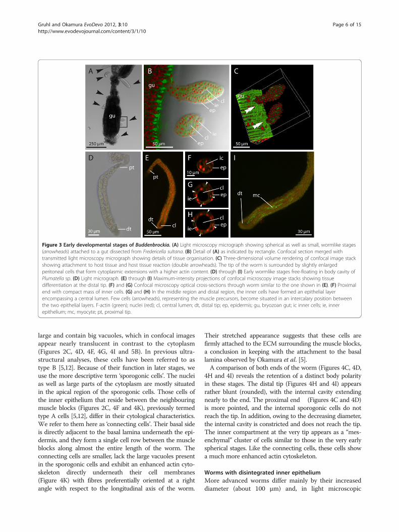
Figure 3 Early developmental stages of Buddenbrockia. (A) Light microscopy micrograph showing spherical as well as small, wormlike stages(arrowheads) attached to a gut dissected from Fredericella sultana. (B) Detail of (A) as indicated by rectangle. Confocal section merged withtransmitted light microscopy micrograph showing details of tissue organisation. (C) Three-dimensional volume rendering of confocal image stackshowing attachment to host tissue and host tissue reaction (double arrowheads). The tip of the worm is surrounded by slightly enlargedperitoneal cells that form cytoplasmic extensions with a higher actin content. (D) through (I) Early wormlike stages free-floating in body cavity ofPlumatella sp. (D) Light micrograph. (E) through (I) Maximum-intensity projections of confocal microscopy image stacks showing tissuedifferentiation at the distal tip. (F) and (G) Confocal microscopy optical cross-sections through worm similar to the one shown in (E). (F) Proximalend with compact mass of inner cells. (G) and (H) In the middle region and distal region, the inner cells have formed an epithelial layerencompassing a central lumen. Few cells (arrowheads), representing the muscle precursors, become situated in an intercalary position betweenthe two epithelial layers. F-actin (green); nuclei (red); cl, central lumen; dt, distal tip; ep, epidermis; gu, bryozoan gut; ic inner cells; ie, innerepithelium; mc, myocyte; pt, proximal tip.
Gruhl and Okamura EvoDevo 2012, 3:10 Page 6 of 15http://www.evodevojournal.com/content/3/1/10
large and contain big vacuoles, which in confocal imagesappear nearly translucent in contrast to the cytoplasm(Figures 2C, 4D, 4F, 4G, 4I and 5B). In previous ultra-structural analyses, these cells have been referred to astype B [5,12]. Because of their function in later stages, weuse the more descriptive term ‘sporogonic cells’. The nucleias well as large parts of the cytoplasm are mostly situatedin the apical region of the sporogonic cells. Those cells ofthe inner epithelium that reside between the neighbouringmuscle blocks (Figures 2C, 4F and 4K), previously termedtype A cells [5,12], differ in their cytological characteristics.We refer to them here as ‘connecting cells’. Their basal sideis directly adjacent to the basal lamina underneath the epi-dermis, and they form a single cell row between the muscleblocks along almost the entire length of the worm. Theconnecting cells are smaller, lack the large vacuoles presentin the sporogonic cells and exhibit an enhanced actin cyto-skeleton directly underneath their cell membranes(Figure 4K) with fibres preferentially oriented at a rightangle with respect to the longitudinal axis of the worm.
Their stretched appearance suggests that these cells arefirmly attached to the ECM surrounding the muscle blocks,a conclusion in keeping with the attachment to the basallamina observed by Okamura et al. [5].A comparison of both ends of the worm (Figures 4C, 4D,
4H and 4I) reveals the retention of a distinct body polarityin these stages. The distal tip (Figures 4H and 4I) appearsrather blunt (rounded), with the internal cavity extendingnearly to the end. The proximal end (Figures 4C and 4D)is more pointed, and the internal sporogonic cells do notreach the tip. In addition, owing to the decreasing diameter,the internal cavity is constricted and does not reach the tip.The inner compartment at the very tip appears as a “mes-enchymal” cluster of cells similar to those in the very earlyspherical stages. Like the connecting cells, these cells showa much more enhanced actin cytoskeleton.
Worms with disintegrated inner epitheliumMore advanced worms differ mainly by their increaseddiameter (about 100 μm) and, in light microscopic

Figure 4 Early stage Buddenbrockia plumatellae worm from Plumatella sp. (A) Whole-mount light microscopy micrograph. (B) Confocalimage of the same specimen, maximum-intensity projection showing body musculature. (C) through (I) Confocal images. (C), (E) and (H)Maximum-intensity projections of whole three-dimensional image stacks. (D), (G) and (I) Optical median horizontal sections. (F) Opticalcross-section. Details of proximal tip (C) and (D) with mesenchymal inner cells (arrowheads), middle-body region (E), (F) and (G) and distal tip(H) and (I). Scale bar as in (H). (J) Horizontal optical section through distal tip showing intact inner epithelium and nuclei in muscle cells.(K) Detail caption of row of connecting cells between neighbouring muscle blocks, horizontal aspect, maximum-intensity projection. F-actin(green); nuclei (red); cc, connecting cell; cl, central lumen; dt, distal tip; ep, epidermis; ie, inner epithelium; mb, muscle block; nu, nucleus; pt,proximal tip; sc, sporogonic cell.
Gruhl and Okamura EvoDevo 2012, 3:10 Page 7 of 15http://www.evodevojournal.com/content/3/1/10
images, by the more granular appearance of their innercontents, which is due to ongoing spore development(see below) (Figure 6A).The muscle blocks have become wider and flatter in
cross-section (Figure 6E). The muscle cells are stillarranged obliquely and in two parallel rows (Figures 6B,6D and 6G) but have elongated to about 50 μm and be-come much thinner. Cross-sections and longitudinal
sections show F-actin to be located mainly underneaththe cell surfaces facing the epidermis and central lumen,but not in regions facing neighbouring muscle cells(Figures 6C, 6E, 6F and 6H).As also shown previously by ultrastructure [25], the inner
epithelium has largely disintegrated and the sporogoniccells are now found floating in the central lumen, wherethey undergo spore formation (Figures 5C to 5F, and 6C,
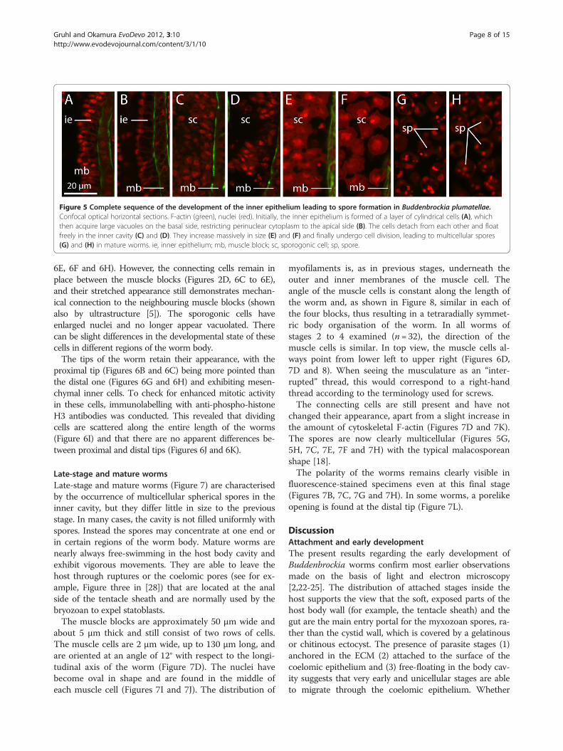
Figure 5 Complete sequence of the development of the inner epithelium leading to spore formation in Buddenbrockia plumatellae.Confocal optical horizontal sections. F-actin (green), nuclei (red). Initially, the inner epithelium is formed of a layer of cylindrical cells (A), whichthen acquire large vacuoles on the basal side, restricting perinuclear cytoplasm to the apical side (B). The cells detach from each other and floatfreely in the inner cavity (C) and (D). They increase massively in size (E) and (F) and finally undergo cell division, leading to multicellular spores(G) and (H) in mature worms. ie, inner epithelium; mb, muscle block; sc, sporogonic cell; sp, spore.
Gruhl and Okamura EvoDevo 2012, 3:10 Page 8 of 15http://www.evodevojournal.com/content/3/1/10
6E, 6F and 6H). However, the connecting cells remain inplace between the muscle blocks (Figures 2D, 6C to 6E),and their stretched appearance still demonstrates mechan-ical connection to the neighbouring muscle blocks (shownalso by ultrastructure [5]). The sporogonic cells haveenlarged nuclei and no longer appear vacuolated. Therecan be slight differences in the developmental state of thesecells in different regions of the worm body.The tips of the worm retain their appearance, with the
proximal tip (Figures 6B and 6C) being more pointed thanthe distal one (Figures 6G and 6H) and exhibiting mesen-chymal inner cells. To check for enhanced mitotic activityin these cells, immunolabelling with anti-phospho-histoneH3 antibodies was conducted. This revealed that dividingcells are scattered along the entire length of the worms(Figure 6I) and that there are no apparent differences be-tween proximal and distal tips (Figures 6J and 6K).
Late-stage and mature wormsLate-stage and mature worms (Figure 7) are characterisedby the occurrence of multicellular spherical spores in theinner cavity, but they differ little in size to the previousstage. In many cases, the cavity is not filled uniformly withspores. Instead the spores may concentrate at one end orin certain regions of the worm body. Mature worms arenearly always free-swimming in the host body cavity andexhibit vigorous movements. They are able to leave thehost through ruptures or the coelomic pores (see for ex-ample, Figure three in [28]) that are located at the analside of the tentacle sheath and are normally used by thebryozoan to expel statoblasts.The muscle blocks are approximately 50 μm wide and
about 5 μm thick and still consist of two rows of cells.The muscle cells are 2 μm wide, up to 130 μm long, andare oriented at an angle of 12° with respect to the longi-tudinal axis of the worm (Figure 7D). The nuclei havebecome oval in shape and are found in the middle ofeach muscle cell (Figures 7I and 7J). The distribution of
myofilaments is, as in previous stages, underneath theouter and inner membranes of the muscle cell. Theangle of the muscle cells is constant along the length ofthe worm and, as shown in Figure 8, similar in each ofthe four blocks, thus resulting in a tetraradially symmet-ric body organisation of the worm. In all worms ofstages 2 to 4 examined (n= 32), the direction of themuscle cells is similar. In top view, the muscle cells al-ways point from lower left to upper right (Figures 6D,7D and 8). When seeing the musculature as an “inter-rupted” thread, this would correspond to a right-handthread according to the terminology used for screws.The connecting cells are still present and have not
changed their appearance, apart from a slight increase inthe amount of cytoskeletal F-actin (Figures 7D and 7K).The spores are now clearly multicellular (Figures 5G,5H, 7C, 7E, 7F and 7H) with the typical malacosporeanshape [18].The polarity of the worms remains clearly visible in
fluorescence-stained specimens even at this final stage(Figures 7B, 7C, 7G and 7H). In some worms, a porelikeopening is found at the distal tip (Figure 7L).
DiscussionAttachment and early developmentThe present results regarding the early development ofBuddenbrockia worms confirm most earlier observationsmade on the basis of light and electron microscopy[2,22-25]. The distribution of attached stages inside thehost supports the view that the soft, exposed parts of thehost body wall (for example, the tentacle sheath) and thegut are the main entry portal for the myxozoan spores, ra-ther than the cystid wall, which is covered by a gelatinousor chitinous ectocyst. The presence of parasite stages (1)anchored in the ECM (2) attached to the surface of thecoelomic epithelium and (3) free-floating in the body cav-ity suggests that very early and unicellular stages are ableto migrate through the coelomic epithelium. Whether

Figure 6 Medium stage Buddenbrockia worms. (A) through (H) Buddenbrockia plumatellae from Plumatella sp. (A) Whole-mount lightmicroscopic micrograph. (B) through (H) Confocal images. (B), (D) and (G) Maximum-intensity projections of whole three-dimensional imagestacks. (C), (F) and (H) Optical median horizontal section. (E) Optical cross-section. Details of the proximal tip (B and (C), middle-body region(D) through (F) and distal tip (G) and (H). Scale bar is the same as in (G). (I) through (K) Buddenbrockia sp. from Fredericella sultana. Labellingwith anti-phospho-histone H3 antibody (pH3) shows distribution of mitosing cells over the full length of the worm, with no apparent differencesbetween the ends. (I) Overview. (J) Proximal tip. (K) Distal tip. F-actin (green); nuclei (red); pH3 immunoreactivity (cyan); cc, connecting cell; dt,distal tip; ep, epidermis; mb, muscle block; mc, myocyte; pt, proximal tip; sc, sporogonic cell.
Gruhl and Okamura EvoDevo 2012, 3:10 Page 9 of 15http://www.evodevojournal.com/content/3/1/10
these develop into attached or free-floating stages may de-pend on their position within the host when growth com-mences. Earlier studies suggested that the onset of wormgrowth may be triggered by warmer temperature or avail-ability of nutrients, since worms were observed to developwhen subject to these conditions in a laboratory meso-cosm [25]. There is strong evidence that such conditionstrigger development of the closely related malacosporeanTetracapsuloides bryosalmonae [29-31].The host tissue reaction may additionally strengthen the
attachment of the worm, as it involves thickening andreinforcement of the cytoskeleton of the peritoneal cellsthat surround the attachment area. As worms of all lengths
were found attached as well as free-floating, we suggest thatdetachment is not a scheduled event during developmentbut occurs haphazardly, possibly facilitated by the increas-ingly vigorous movements of the worms. This hypothesis issupported by a higher proportion of detached older worms.The undifferentiated state and slightly ruptured appearanceof the tissues at the proximal tip of detached worms ren-ders it possible that a few cells could remain in the “wound”after detachment. Such material may therefore result in on-going covert infection and the future growth of anotherworm. Note that covert infections are also likely to beachieved by the persistence of early stages, which we haveobserved in host tissues simultaneously with developing

Figure 7 Late-stage Buddenbrockia plumatellae worm from Plumatella sp. (A) Whole mount, light micrograph. (B) through (H) Confocalimages. (B), (D) and (G) Maximum-intensity projections of whole three-dimensional image stacks. (C), (F) and (H) Optical median horizontalsection. (E) Optical cross section. Details of proximal tip (B) and (C); middle-body region (D), (E) and (F); and distal tip (G) and (H). Scale bar as in(G). (I) Horizontal optical section through muscle block showing distribution of muscle cell nuclei. (J) Detail of (I), maximum-intensity projectionof muscle block. (K) Horizontal optical section through row of connecting cells. (L) Horizontal section through distal tip of mature worm showingporelike opening (arrowhead). F-actin (green); nuclei (red); cc, connecting cell; dt, distal tip; ecm, extracellular matrix; ep, epidermis; mb, muscleblock; mc, myocyte; mr, myocyte row; nu, nucleus; pt, proximal tip; sp, spore.
Gruhl and Okamura EvoDevo 2012, 3:10 Page 10 of 15http://www.evodevojournal.com/content/3/1/10
and mature worms. Such covert infections may promotelong-term persistence in hosts such as occurs in T. bryosal-monae [31,32].We have not found any evidence for the idea that the
worms’ muscle cells could be derived from bryozoanmuscle cells, as suggested by Morris and Adams [24].The latter look rather different, and, in addition, muscledevelopment occurs in the distal end of the worm. Fur-thermore, bryozoan nuclei are considerably larger than
those of Buddenbrockia (see also [25] for ultrastructuraldifferences), making potential chimeras easily detectable.We thus conclude that the musculature is a native Bud-denbrockia feature that is likely to have been retainedfrom a free-living cnidarian ancestor. In addition, ourdata do not support the idea that early multicellularstages form by the accumulation of migratory unicellularstages rather than by mitosis [24]. This would requirethe unicellular stages to move horizontally throughout

Figure 8 Tetraradial symmetry pattern of muscle architecture in mature Buddenbrockia plumatellae. (A) Three-dimensional volumerendering of confocal image stack from midsection of worm. (B) through (E) Maximum-intensity projections of image stack rotated in 90° stepsto show arrangement and angle of muscle fibres in each muscle block as indicated in the inset in (A). F-actin (green), nuclei (red), mb, muscleblock.
Gruhl and Okamura EvoDevo 2012, 3:10 Page 11 of 15http://www.evodevojournal.com/content/3/1/10
the ECM, and a progression from loose clusters of cellsto densely packed tissuelike stages would be expected.Such a progression is not evident in our data.The present data demonstrate that the connecting
cells persist in the mature worm and do not contributeto sporogony, as also shown by ultrastructure [5]. Thepossibility that they serve a mechanical function is sup-ported by their attachment to the surrounding ECM,their stretched appearance and their pronounced actincytoskeleton. However, the possibility of a neuronalfunction, as suggested by Schröder [2], cannot be ruledout completely.
The presence of a polarised primary body axisOur study shows that, during ontogeny, Buddenbrockiaworms acquire a distinct polarity along their primary bodyaxis, which is reflected by directional growth and a gradi-ent of tissue differentiation. The inner cells as well as themusculature at the distal tip of developing worms aremore differentiated than at the proximal tip. This polaritypersists in later stages, and, in some fully mature worms, aporelike opening appears at the distal tip. However, fromthe present data, it cannot be determined whether this is areal pore or an opening as a result of rupture. Althoughinternal cells at the distal pole sometimes differ in theirmorphology, we found no indication that these representspermatids [2]. Anti-phospho-histone H3 staining doesnot give evidence of the presence of a distinct growth zoneat either end of the worm. However, as the proportion ofnuclei labelled with this method is low in comparison to
methods such as bromodeoxyuridine labelling, which inte-grate over longer time periods, the latter might provide amore detailed picture in future studies.Because Buddenbrockia lacks a gastrulalike stage, an
intestinal tract and evidence of nervous structures, it isdifficult to relate this axial polarity to that of other ani-mals. Recent experimental and gene expression data (es-pecially those based on Wnt/β-catenin signalling)demonstrate that the development of polyps from planu-lae and of asexual buds is in accord with early morph-ology-based hypotheses for a homology of the cnidarianoral-aboral axis and the bilaterian anteroposterior axis[33-36]. These data also suggest, although with lowerconfidence, that the cnidarian oral pole may correlatewith the bilaterian posterior pole. Together with findingsthat growth at the posterior pole is most likely an ances-tral character in bilaterian animals [37], this implies thatthe proximal tip of Buddenbrockia may correspond tothe bilaterian posterior pole and thus the cnidarian oralpole. However, in cnidarians, no generalised pattern ofeither anterior or posterior growth has so far beendemonstrated.Another question is when and how polarity is deter-
mined. In cnidarians and bilaterians, the main body axisis either identical to or oriented at a particular angle tothe embryonic animal-vegetal axis. This is achieved, forexample, by gradients of maternally expressed transcrip-tion factors in the egg or the position of the egg with re-spect to the ovarial tissue (reviewed, for example, in[38,39]). However, the unicellular stages found in the

Gruhl and Okamura EvoDevo 2012, 3:10 Page 12 of 15http://www.evodevojournal.com/content/3/1/10
bryozoan tissue are most likely derived from sporo-plasms that came from spores produced in the verte-brate intermediate host [12]. They are amoeboid and donot show any recognisable polarity before mitotic divi-sions leading to early multicellular stages. It is thereforelikely that polarisation is induced by external factors.This might be achieved via the orientation of the earlystages in the bryozoan host tissue, such as with growthdirected away from the basal lamina. Such a scenariocould be tested by detailed comparison with closelyrelated, saclike malacosporeans whose trophic stageslack a distinct body axis, such as the Buddenbrockiaparasite of Cristatella mucedo [28]; Buddenbrockia all-mani in Lophopus crystallinus [40]; or Tetracapsuloidesbryosalmonae [18], which predominantly parasitises Fre-dericella sultana. For the latter species, data on early de-velopment indicate that presaccular stages are situatedon the surface of the peritoneum rather than underneathas in Buddenbrockia [16], rendering the previous explan-ation possible.
Mesodermal musculature and germ layersAs unequivocally indicated by the presence of nuclei inthe muscle cells, the musculature of the Buddenbrockiaworm is formed by independent myocytes and not byepitheliomuscular cells as in most other cnidarians[41,42]. If the epidermis is regarded as ectodermal andthe inner epithelium (although functioning only as re-productive rather than digestive tissue) as endodermal,then the muscle cells are, at least by topological defin-ition [43], mesodermal as they reside in the ECM be-tween the two epithelial layers and are not connected tothe latter. We note that the same argument applies ifadaptation to parasitism has involved an inversion ofectoderm and endoderm as occurs in Polypodium hydri-forme [44]. This may have bearing on questions concern-ing the evolution of metazoan body plans, since thediploblastic vs. triploblastic organisation of the last com-mon ancestor of Cnidaria and Bilateria is highly debated(see, for example, [19-21,45]). Bilaterian mesoderm isusually characterised by (1) topology, (2) germ layer-spe-cific derivation during gastrulation or (3) the presence ofa common set of regulatory genes [45]. The commonlycited example for topological mesoderm in Cnidaria isthe entocodon, a tissue that invaginates during hydro-zoan medusa bud formation and gives rise to an inde-pendent striated subumbrellar musculature in themedusa (summarised in [20]). The musculature in free-living stages of the parasitic cnidarian Polypodium hydri-forme also appears to be topologically mesodermal[46,47].In contrast to the hydrozoan entocodon, the meso-
derm of Buddenbrockia is formed early in ontogeny andmight thus qualify as a bona fide germ layer potentially
homologous to mesoderm in bilaterians. It also developscloser to the inner endodermal layer than to the outerepidermal layer. However, the lack of a distinct cleavagepattern, blastula-like stage and gastrulation-like processhinders further comparisons with cnidarian or bilateriandevelopment. Of course, a mesodermal as opposed to amyoepithelial musculature may have functional advan-tages in a wormlike organism (see for example, [48]),thus promoting a convergent origin of this character inBuddenbrockia and bilaterians. The vermiform parasiticsea anemone Edwardsiella lineata, however, has typicalanthozoan endodermal longitudinal muscles [49]. Geneexpression data from Buddenbrockia should enable fur-ther insights into, for example, tissue homologies andaxial patterning.
Tetraradial symmetry and chiralityThe arrangement of the muscle cells within the muscleblocks demonstrates that Buddenbrockia worms arecharacterised by tetraradial symmetry. Although trad-itionally regarded as radially symmetric, recent evidence,especially from developmental and gene expression stud-ies, suggests that the ancestor of all cnidarians was a bi-laterally symmetrical animal, a pattern still reflected inthe organisation of many recent anthozoans [50-52] (butsee [53]). Tetraradial symmetry must be regarded as hav-ing evolved in the Medusozoa, which form the sis-tergroup to Anthozoa [54]. The corroboration of atetraradial symmetry in Buddenbrockia worms thereforeprovides further support for a medusozoan affinity ofmyxozoans [11].A further interesting finding is the presence of a con-
sistent handedness or chirality (mirror asymmetry) inthe arrangement of the muscle fibres, which align in aright-handed thread in all individuals examined in thisstudy. The coincidence of radial symmetry, or mathem-atically more precisely, rotational symmetry, togetherwith mirror symmetry, may jointly characterise objects,with examples being a regular geometric star or the ma-jority of radially symmetrical animals. However, the twotypes of symmetry are not necessarily linked. Many rota-tionally symmetrical objects are chiral and exhibit rota-tionally repeated elements with no mirror symmetry.Such chiral forms exist in two (dextral vs. sinistral) enan-tiomorphs (see [55] for a review of symmetry patterns).Chirality is a well known phenomenon in many bilater-
ian animals (e.g. molluscs, annelids, pterobranchs, nema-todes, vertebrates) [56], but has so far only rarely beendescribed in non-bilaterians. An interesting non-bilaterian case includes certain conulariids, which are afossil group inferred to have been scyphozoans. Thus, thetetraradially symmetrical skeleton of the conulariidMetaconularia anomala shows torsion along the longitu-dinal axis [57]. M. anomala also exhibits a strong

Gruhl and Okamura EvoDevo 2012, 3:10 Page 13 of 15http://www.evodevojournal.com/content/3/1/10
preference for a sinistral coil [57]. Other nonbilaterianexamples include the siphonophore Bargmannia elon-gata, in which bud formation leads to consistently asym-metrical colony forms [58], and fossils probablybelonging to the ctenophoran lineage and which possessarms that coil preferentially dextrally [59].Fixed chiralities such as the one described here in
Buddenbrockia are in almost all cases heritable [60]. So,is it likely that chirality is adaptive? A similar angle ofthe muscle fibres throughout the body is probablystrongly selected for because of its functional signifi-cance for spiralling movements. However, such move-ment should be achieved regardless of whether the angleis dextral or sinistral. A possible explanation for thedominance of one chiral form might be if concertedmovements of multiple worms within a host minimiseinterference.How is the chirality established in Buddenbrockia worms?
In most bilaterians, where the developmental mechanismleading to chirality is known, symmetry breaking occursafter the establishment of the dorsoventral axis (see, for ex-ample, [61]). However, a deeper underlying mechanism thatestablishes chirality on a subcellular level is generally in-ferred. This has not been clearly identified and may differfrom case to case. The main theories are that cellular chir-ality is a result of (1) molecular chirality of the cilium, (2)cytoskeletal asymmetries leading to a differential distribu-tion of ion channels and/or pumps on one side of a blasto-mere or (3) nonidentical blastomeres produced by differentepigenetically imprinted patterns [62]. The ciliary modelcan clearly be ruled out for Buddenbrockia, as cilia and cen-trosomes are lacking in all myxozoans [12]. As the chiralityis reflected only in the arrangement of the muscle cells, itcould be explained by a directional shift in the cleavageplane in mitoses leading to the muscle cell lineage.
ConclusionsOur study has provided fundamental insights into theevolution of polarity, symmetry and tissue layers in thelower Metazoa. The presence of a polarised primarybody axis represents a unique retention in Myxozoa ofthis ancient feature, present at least in the last commonancestor of Cnidaria and Bilateria if not in the Metazoa.In the absence of clear morphological features, however,clarification of the homologies of the proximal and distaltips with the cnidarian oral or aboral poles will requiregene expression studies. The mechanism by which theprimary axis is established is probably deviant fromother metazoans and could provide insights into howthis character was lost in all other myxozoans.The tetraradial arrangement of musculature supports a
medusozoan affinity for Myxozoa. The chiral patternrevealed in the orientation of muscle fibres has noknown equivalents within Cnidaria, however, and could
thus be an adaptation that allows the spiralling move-ments of the worm. The unique biomechanics ofBuddenbrockia locomotion will be the subject of a forth-coming publication. Finally, the presence of independentmyocytes instead of cnidarian-like epitheliomuscularcells can be interpreted as further support for the pres-ence of mesoderm and thus triploblasty in cnidarians.An alternative explanation is that the topologicallymesodermal muscle represents convergent evolution toa bilaterian bauplan.
AbbreviationsPBS: phosphate-buffered saline..
Competing interestsThe authors declare no competing interests.
AcknowledgementsThe authors thank Tim Wood (Wright State University, Dayton, OH, USA) andHanna Hartikainen (Natural History Museum, London (NHM)) for fruitfuldiscussions and Jukka Jokela (Eawag, Dübendorf, Switzerland), Tim Woodand Hanna Hartikainen for support in sampling and provision of laboratoryspace during our fieldwork abroad, and two reviewers for their constructivecomments on our manuscript. Many thanks to Alex Ball and Lauren Howard(NHM) for general microscopy support, to Thomas Bartolomaeus and BjörnQuast (University of Bonn, Germany) for kindly providing facilities andsupport for histology, and to Georg Mayer (University of Leipzig, Germany)for donating anti-pH3 antibodies. This work was funded by an EU Synthesysgrant (GB-TAF-5378) and a German Academic Exchange Service (DAAD)postdoctoral fellowship (D/09/42855) (to AG) and by the Percy SladenMemorial Fund and the Department of Zoology (NHM).
Authors’ contributionsAG and BO have collected the animals. AG has conducted the microscopy.AG and BO have analysed the data and drafted the manuscript. All authorshave read and approved the final manuscript.
Received: 15 February 2012 Accepted: 17 May 2012Published: 17 May 2012
References1. Schröder O: Buddenbrockia plumatellae, eine neue Mesozoenart aus
Plumatella repens L. und Pl. fungosa Pall. Z Wiss Zool 1910, 96:525–537.2. Schröder O: Zur Kenntnis der Buddenbrockia plumatellae Ol. Schröder. Z
Wiss Zool 1912, 102:79–91.3. Braem F: Beiträge zur Fauna Turkestans aufgrund des von D. D.
Pedaschenko gesammelten Materials. VII. Bryozoen und deren Parasiten.Travaux de la Société Impériale des Naturalists de St. Pétersbourg 1911,42:1–34.
4. Jiménez-Guri E, Okamura B, Holland PWH: Origin and evolution of amyxozoan worm. Int Comp Biol 2007, 47:752–758.
5. Okamura B, Curry A, Wood TS, Canning EU: Ultrastructure ofBuddenbrockia identifies it as a myxozoan and verifies the bilaterianorigin of the Myxozoa. Parasitology 2002, 124:215–223.
6. Monteiro AS, Okamura B, Holland PWH: Orphan worm finds a home:Buddenbrockia is a myxozoan. Mol Biol Evol 2002, 19:968–971.
7. Weill R: L’interpretation des Cnidosporidies et la valeur taxonomique deleur cnidome. Leur cycle comparé à la phase larvaire des Narcomedusescuninides. Trav Stn Zool Wimeraux 1938, 13:727–744.
8. Siddall ME, Martin DS, Bridge D, Desser SS, Cone DK: The demise of aphylum of protists: Phylogeny of Myxozoa and other parasitic Cnidaria. JParasitol 1995, 81:961–967.
9. Holland JW, Okamura B, Hartikainen H, Secombes CJ: A novel minicollagengene links cnidarians and myxozoans. Proc Biol Sci 2011, 278:546–553.
10. Evans NM, Holder MT, Barbeitos MS, Okamura B, Cartwright P: Thephylogenetic position of Myxozoa: exploring conflicting signals inphylogenomic and ribosomal data sets. Mol Biol Evol 2010,27:2733–2746.

Gruhl and Okamura EvoDevo 2012, 3:10 Page 14 of 15http://www.evodevojournal.com/content/3/1/10
11. Jiménez-Guri E, Philippe H, Okamura B, Holland PWH: Buddenbrockia is acnidarian worm. Science 2007, 317:116–118.
12. Canning EU, Okamura B: Biodiversity and evolution of the Myxozoa. AdvParasitol 2004, 56:43–131.
13. Lom J, Dykova I: Myxozoan genera: definition and notes on taxonomy,life-cycle terminology and pathogenic species. Folia Parasitol (Praha) 2006,53:1–36.
14. Grabner DS, El-Matbouli M: Transmission of Tetracapsuloidesbryosalmonae (Myxozoa: Malacosporea) to Fredericella sultana(Bryozoa: Phylactolaemata) by various fish species. Dis Aquat Organ2008, 79:133–139.
15. Anderson CL, Canning EU, Okamura B: Molecular data implicatebryozoans as hosts for PKX (Phylum Myxozoa) and identify a cladeof bryozoan parasites within the Myxozoa. Parasitology 1999,119:555–561.
16. Morris DJ, Adams A: Proliferative, presaccular stages ofTetracapsuloides bryosalmonae (Myxozoa: Malacosporea) within theinvertebrate host Fredericella sultana (Bryozoa: Phylactolaemata). JParasitol 2006, 92:984–989.
17. Grabner DS, El-Matbouli M: Experimental transmission of malacosporeanparasites from bryozoans to common carp (Cyprinus carpio) and minnow(Phoxinus phoxinus). Parasitology 2010, 137:629–639.
18. Canning EU, Curry A, Feist SW, Longshaw M, Okamura B: A newclass and order of myxozoans to accommodate parasites ofbryozoans with ultrastructural observations on Tetracapsulabryosalmonae (PKX organism). J Eukaryot Microbiol 2000, 47:456–468.
19. Burton PM: Insights from diploblasts: the evolution of mesodermand muscle. J Exp Zool B Mol Dev Evol 2008, 310:5–14.
20. Seipel K, Schmid V: Mesodermal anatomies in cnidarian polyps andmedusae. Int J Dev Biol 2006, 50:589–599.
21. Martindale MQ, Pang K, Finnerty JR: Investigating the origins oftriploblasty: “mesodermal” gene expression in a diploblasticanimal, the sea anemone Nematostella vectensis (phylum, Cnidaria;class, Anthozoa). Development 2004, 131:2463–2474.
22. Canning EU, Curry A, Okamura B: Early development of the myxozoanBuddenbrockia plumatellae in the bryozoans Hyalinella punctata andPlumatella fungosa, with comments on taxonomy and systematics of theMyxozoa. Folia Parasitol (Praha) 2008, 45:241–255.
23. McGurk C, Morris DJ, Adams A: Sequential development of Buddenbrockiaplumatellae (Myxozoa: Malacosporea) within Plumatella repens (Bryozoa:Phylactolaemata). Dis Aquat Organ 2006, 73:159–169.
24. Morris DJ, Adams A: Sacculogenesis of Buddenbrockia plumatellae(Myxozoa) within the invertebrate host Plumatella repens (Bryozoa) withcomments on the evolutionary relationships of the Myxozoa. Int JParasitol 2007, 37:1163–1171.
25. Canning EU, Tops S, Curry A, Wood TS, Okamura B: Ecology, developmentand pathogenicity of Buddenbrockia plumatellae Schröder, 1910(Myxozoa, Malacosporea) (syn. Tetracapsula bryozoides) andestablishment of Tetracapsuloides n. gen. for Tetracapsula bryosalmonae. JEukaryot Microbiol 2002, 49:280–295.
26. Tops S, Curry A, Okamura B: Diversity and systematics of the Malacosporea(Myxozoa). Invertebr Biol 2005, 124:285–295.
27. Hirose M, Dick MH, Mawatari SF: Molecular phylogenetic analysis ofphylactolaemate bryozoans based on mitochondrial gene sequences. InBryozoan Studies 2007. Edited by Hageman GS, Key MMJ, Winston JE.Martinsville, VA: Virginia Museum of Natural History; 2008:346.
28. Canning EU, Okamura B, Curry A: Development of a myxozoan parasiteTetracapsula bryozoides gen. n. et sp. n. in Cristatella mucedo (Bryozoa:Phylactolaemata). Folia Parasitol (Praha) 1996, 43:259–261.
29. Tops S, Lockwood W, Okamura B: Temperature-driven proliferation ofTetracapsuloides bryosalmonae in bryozoan portends salmonid declines.Dis Aquat Organ 2006, 70:227–236.
30. Okamura B, Hartikainen H, Schmidt-Posthaus H, Wahli T: Life cyclecomplexity, environmental change and the emerging status of salmonidproliferative kidney disease. Freshw Biol 2011, 56:735–753.
31. Hartikainen H, Okamura B: Castrating parasites and colonial hosts.Parasitology 2012, 139:547–556.
32. Tops S, Hartikainen HL, Okamura B: The effects of infection byTetracapsuloides bryosalmonae (Myxozoa) and temperature onFredericella sultana (Bryozoa). Int J Parasitol 2009, 39:1003–1010.
33. Duffy DJ, Plickert G, Kuenzel T, Tilmann W, Frank U: Wnt signaling promotesoral but suppresses aboral structures in Hydractinia metamorphosis andregeneration. Development 2010, 137:3057–3066.
34. Petersen CP, Reddien PW: Wnt signaling and the polarity of the primarybody axis. Cell 2009, 139:1056–1068.
35. Guder C, Philipp I, Lengfeld T, Watanabe H, Hobmayer B, Holstein TW:The Wnt code: cnidarians signal the way. Oncogene 2006, 25:7450–7460.
36. Lengfeld T, Watanabe H, Simakov O, Lindgens D, Gee L, Law L, Schmidt HA,Ozbek S, Bode H, Holstein TW: Multiple Wnts are involved in Hydraorganizer formation and regeneration. Dev Biol 2009, 330:186–199.
37. Martin BL, Kimelman D: Wnt signaling and the evolution of embryonicposterior development. Curr Biol 2009, 19:R215–R219.
38. Martindale MQ: The evolution of metazoan axial properties. Nat Rev Genet2005, 6:917–927.
39. Nielsen C: Animal Evolution: Interrelationships of the Living Phyla. 3rd edition.Oxford: Oxford University Press; 2012:402.
40. Canning EU, Curry A, Hill SLL, Okamura B: Ultrastructure of Buddenbrockiaallmani n. sp. (Myxozoa, Malacosporea), a parasite of Lophopuscrystallinus (Bryozoa, Phylactolaemata). J Eukaryot Microbiol 2007,54:247–262.
41. Ax P: Das System der Metazoa I. Stuttgart: Gustav Fischer; 1995.42. Schmidt-Rhaesa A: The Evolution of Organ Systems. New York: Oxford
University Press; 2007:363.43. Ruppert EE: Introduction to the aschelminth phyla: a consideration of
mesoderm, body cavities, and cuticle. In Microscopic Anatomy ofInvertebrates: Aschelminthes. Volume 4. Edited by Harrison FW, Ruppert EE.New York: Wiley Liss; 1991:1–17.
44. Raikova EV: Life-Cycle, cytology, and morphology of Polypodiumhydriforme, a coelenterate parasite of the eggs of acipenseriform fishes. JParasitol 1994, 80:1–22.
45. Technau U, Scholz CB: Origin and evolution of endoderm and mesoderm.Int J Dev Biol 2003, 47:531–539.
46. Lipin A: Die Morphologie und Biologie von Polypodium hydriforme Uss.Zoologische Jahrbücher, Abteilung für Anatomie und Ontogenie der Tiere 1911,31:317–426.
47. Raikova EV, Ibragimov AY, Raikova OI: Muscular system of a peculiarparasitic cnidarian Polypodium hydriforme: A phalloidin fluorescencestudy. Tissue Cell 2007, 39:79–87.
48. Rieger MR, Ladurner P: The significance of muscle cells for the origin ofmesoderm in Bilateria. Int Comp Biol 2003, 43:47–54.
49. Reitzel AM, Daly M, Sullivan JC, Finnerty JR: Comparative anatomyand histology of developmental and parasitic stages in the lifecycle of the lined sea anemone Edwardsiella lineata. J Parasitol2009, 95:100–112.
50. Finnerty JR, Pang K, Burton P, Martindale MQ: Origins of bilateralsymmetry: Hox and Dpp expression in a sea anemone. Science2004, 304:1335–1337.
51. Ball EE, de Jong DM, Schierwater B, Shinzato C, Hayward DC, MillerDJ: Implications of cnidarian gene expression patterns for theorigins of bilaterality: is the glass half full or half empty? Int CompBiol 2007, 47:701–711.
52. de Jong DM, Hislop NR, Hayward DC, Reece-Hoyes JS, Pontynen PC,Ball EE, Miller DJ: Components of both major axial patterningsystems of the Bilateria are differentially expressed along theprimary axis of a “radiate” animal, the anthozoan cnidarianAcropora millepora. Dev Biol 2006, 298:632–643.
53. Manuel M: Early evolution of symmetry and polarity in metazoanbody plans. C R Biol 2008, 332:184–209.
54. Collins AG, Schuchert P, Marques AC, Jankowski T, Medina M,Schierwater B: Medusozoan phylogeny and character evolutionclarified by new large and small subunit rDNA data and anassessment of the utility of phylogenetic mixture models. Syst Biol2006, 55:97–115.
55. Savriama Y, Klingenberg CP: Beyond bilateral symmetry: geometricmorphometric methods for any type of symmetry. BMC Evol Biol2011, 11:280.
56. Palmer RA: Animal asymmetry. Curr Biol 2009, 19:R473–R477.57. Sendino C, Zágoršek K, Taylor PD: Asymmetry in an Ordovician
conulariid cnidarian. Lethaia, . doi:10.1111/j.1502-3931.2011.00302.x. inpress.
Gruhl and Okamura EvoDevo 2012, 3:10 Page 15 of 15http://www.evodevojournal.com/content/3/1/10
58. Dunn CW: Complex colony-level organization of the deep-seasiphonophore Bargmannia elongata (Cnidaria, Hydrozoa) isdirectionally asymmetric and arises by the subdivision of pro-buds.Dev Dyn 2005, 234:835–845.
59. Tang F, Bengtson S, Wang Y, Wang X, Yin C: Eoandromeda and theorigin of Ctenophora. Evol Dev 2011, 13:408–414.
60. Palmer RA: Symmetry breaking and the evolution of development.Science 2004, 306:828–833.
61. Grande C: Left-right asymmetries in Spiralia. Integr Comp Biol 2010,50:744–755.
62. Vandenberg LN, Levin M: Far from solved: a perspective on what weknow about early mechanisms of left-right asymmetry. Dev Dyn 2010,239:3131–3146.
doi:10.1186/2041-9139-3-10Cite this article as: Gruhl and Okamura: Development and myogenesisof the vermiform Buddenbrockia (Myxozoa) and implications forcnidarian body plan evolution. EvoDevo 2012 3:10.
Submit your next manuscript to BioMed Centraland take full advantage of:
• Convenient online submission
• Thorough peer review
• No space constraints or color figure charges
• Immediate publication on acceptance
• Inclusion in PubMed, CAS, Scopus and Google Scholar
• Research which is freely available for redistribution
Submit your manuscript at www.biomedcentral.com/submit