DEVELOPMENT AND DISEASE Evidence that Myc activation...
16
INTRODUCTION The epidermis, which forms the outer covering of mammalian skin, is maintained throughout adult life by proliferation of stem cells that reside in specific locations. Stem cell progeny undergo terminal differentiation to form the interfollicular epidermis (IFE), sebaceous glands and hair follicles (Niemann and Watt, 2002; Fuchs and Raghavan, 2002). The stem cell compartment is multi-potential and a given cell has the ability to differentiate along at least 10 distinct lineages (Niemann and Watt, 2002; Panteleyev et al., 2001). There is good evidence that in the epidermis, as in other tissues maintained by stem cells (Watt and Hogan, 2000; Spradling et al., 2001), the choice of differentiation pathway is profoundly influenced by the local microenvironment, or niche (Watt, 2001; Niemann and Watt, 2002). Considerable progress has been made in identifying factors that regulate exit from the epidermal stem cell compartment and differentiation along specific lineages (Watt, 2001; Niemann and Watt, 2002; Fuchs and Raghavan, 2002). Wnt activity controls the choice of differentiation into IFE or hair follicles: high levels of β-catenin promote hair follicle morphogenesis (Gat et al., 1998), whereas inhibition of β-catenin signalling results in conversion of hair follicles into IFE and sebocytes (Huelsken et al., 2001; Merrill et al., 2001; Niemann et al., 2002). Lineage choice within hair follicles is controlled by the interplay of several key signalling pathways and transcription factors, including Notch, Bmp family members and Hox genes (Niemann and Watt, 2002; Fuchs and Raghavan, 2002). The majority of the known regulators of epidermal differentiation belong to well-conserved pathways that are used repeatedly in multiple tissues throughout development. It is thus surprising that activation of Myc (c-Myc) also affects epidermal lineage commitment. Myc is a proto-oncogene that is primarily reported to stimulate proliferation and growth and to induce apoptosis (Pelengaris et al., 2000; Grandori et al., 2000; Eisenman, 2001). Activation of Myc in the epidermal basal layer stimulates exit from the stem cell compartment and differentiation of keratinocytes into IFE and sebocytes at the expense of the hair lineages (Gandarillas and Watt, 1997; Arnold and Watt, 2001; Waikel et al., 2001). Its mode of action in promoting epidermal differentiation is unknown and indeed is counter-intuitive, given that where effects of Myc on differentiation have been reported in other systems Myc acts as a differentiation inhibitor (Pelengaris et al., 2000; Grandori et al., 2001; Eisenman, 2001). 2793 Development 130, 2793-2808 © 2003 The Company of Biologists Ltd doi:10.1242/dev.00462 Activation of Myc (c-Myc) causes epidermal cells to exit the stem cell compartment and differentiate into sebocytes and interfollicular epidermis at the expense of the hair lineages. To investigate how Myc exerts these effects we analysed the transcription of more than 10,000 genes following Myc activation in the basal layer of mouse epidermis for 1 or 4 days. The major classes of induced genes were involved in synthesis and processing of RNA and proteins, in cell proliferation and in differentiation. More than 40% of the downregulated genes encoded cell adhesion and cytoskeleton proteins. Repression of these genes resulted in profound changes in the adhesive and motile behaviour of keratinocytes. Myc activation inhibited cell motility and wound healing, correlating with decreased expression of a large number of extracellular matrix proteins. Cell adhesion and spreading were also impaired, and this correlated with decreased expression of the α6β4 integrin, decreased formation of hemidesmosomes and decreased assembly of the actomyosin cytoskeleton. We propose that Myc stimulates exit from the stem cell compartment by reducing adhesive interactions with the local microenvironment or niche, and that the failure of hair differentiation reflects an inability of keratinocytes to migrate along the outer root sheath to receive hair inductive stimuli. Key words: Myc, Epidermis, Stem cells, Differentiation, Cell adhesion SUMMARY DEVELOPMENT AND DISEASE Evidence that Myc activation depletes the epidermal stem cell compartment by modulating adhesive interactions with the local microenvironment Michaela Frye 1 , Clare Gardner 2 , Elizabeth R. Li 1 , Isabel Arnold 1, * and Fiona M. Watt 1,† 1 Keratinocyte Laboratory, Cancer Research UK, 44 Lincoln’s Inn Fields, London WC2A 3PX, UK 2 Pfizer Global Research and Development, Sandwich CT13 9NJ, UK *Present address: Institute for Genetics, University of Cologne, Cologne D-50674, Germany † Author for correspondence (e-mail: [email protected]) Accepted 4 March 2003
Transcript of DEVELOPMENT AND DISEASE Evidence that Myc activation...

INTRODUCTION
The epidermis, which forms the outer covering of mammalianskin, is maintained throughout adult life by proliferation ofstem cells that reside in specific locations. Stem cell progenyundergo terminal differentiation to form the interfollicularepidermis (IFE), sebaceous glands and hair follicles (Niemannand Watt, 2002; Fuchs and Raghavan, 2002). The stem cellcompartment is multi-potential and a given cell has the abilityto differentiate along at least 10 distinct lineages (Niemann andWatt, 2002; Panteleyev et al., 2001). There is good evidencethat in the epidermis, as in other tissues maintained by stemcells (Watt and Hogan, 2000; Spradling et al., 2001), the choiceof differentiation pathway is profoundly influenced by the localmicroenvironment, or niche (Watt, 2001; Niemann and Watt,2002).
Considerable progress has been made in identifying factorsthat regulate exit from the epidermal stem cell compartment anddifferentiation along specific lineages (Watt, 2001; Niemannand Watt, 2002; Fuchs and Raghavan, 2002). Wnt activitycontrols the choice of differentiation into IFE or hair follicles:high levels of β-catenin promote hair follicle morphogenesis(Gat et al., 1998), whereas inhibition of β-catenin signalling
results in conversion of hair follicles into IFE and sebocytes(Huelsken et al., 2001; Merrill et al., 2001; Niemann et al.,2002). Lineage choice within hair follicles is controlled by theinterplay of several key signalling pathways and transcriptionfactors, including Notch, Bmp family members and Hox genes(Niemann and Watt, 2002; Fuchs and Raghavan, 2002).
The majority of the known regulators of epidermaldifferentiation belong to well-conserved pathways that are usedrepeatedly in multiple tissues throughout development. It isthus surprising that activation of Myc (c-Myc) also affectsepidermal lineage commitment. Myc is a proto-oncogene thatis primarily reported to stimulate proliferation and growth andto induce apoptosis (Pelengaris et al., 2000; Grandori et al.,2000; Eisenman, 2001). Activation of Myc in the epidermalbasal layer stimulates exit from the stem cell compartment anddifferentiation of keratinocytes into IFE and sebocytes at theexpense of the hair lineages (Gandarillas and Watt, 1997;Arnold and Watt, 2001; Waikel et al., 2001). Its mode of actionin promoting epidermal differentiation is unknown and indeedis counter-intuitive, given that where effects of Myc ondifferentiation have been reported in other systems Myc actsas a differentiation inhibitor (Pelengaris et al., 2000; Grandoriet al., 2001; Eisenman, 2001).
2793Development 130, 2793-2808 © 2003 The Company of Biologists Ltddoi:10.1242/dev.00462
Activation of Myc (c-Myc) causes epidermal cells to exit thestem cell compartment and differentiate into sebocytes andinterfollicular epidermis at the expense of the hair lineages.To investigate how Myc exerts these effects we analysed thetranscription of more than 10,000 genes following Mycactivation in the basal layer of mouse epidermis for 1 or 4days. The major classes of induced genes were involved insynthesis and processing of RNA and proteins, in cellproliferation and in differentiation. More than 40% ofthe downregulated genes encoded cell adhesion andcytoskeleton proteins. Repression of these genes resulted inprofound changes in the adhesive and motile behaviour ofkeratinocytes. Myc activation inhibited cell motility andwound healing, correlating with decreased expression of a
large number of extracellular matrix proteins. Celladhesion and spreading were also impaired, and thiscorrelated with decreased expression of the α6β4 integrin,decreased formation of hemidesmosomes and decreasedassembly of the actomyosin cytoskeleton. We propose thatMyc stimulates exit from the stem cell compartmentby reducing adhesive interactions with the localmicroenvironment or niche, and that the failure of hairdifferentiation reflects an inability of keratinocytes tomigrate along the outer root sheath to receive hairinductive stimuli.
Key words: Myc, Epidermis, Stem cells, Differentiation, Celladhesion
SUMMARY
DEVELOPMENT AND DISEASE
Evidence that Myc activation depletes the epidermal stem cell compartment
by modulating adhesive interactions with the local microenvironment
Michaela Frye 1, Clare Gardner 2, Elizabeth R. Li 1, Isabel Arnold 1,* and Fiona M. Watt 1,†
1Keratinocyte Laboratory, Cancer Research UK, 44 Lincoln’s Inn Fields, London WC2A 3PX, UK2Pfizer Global Research and Development, Sandwich CT13 9NJ, UK*Present address: Institute for Genetics, University of Cologne, Cologne D-50674, Germany†Author for correspondence (e-mail: [email protected])
Accepted 4 March 2003

2794
Myc expression is tightly regulated within the epidermis. Ininterfollicular epidermis it is confined to the basal layer, wherethe proliferating keratinocytes, including the stem cells, reside(Hurlin et al., 1995; Gandarillas and Watt, 1997; Bull et al.,2001). It is highly expressed in the stem cells of the hairfollicle, which lie in a region known as the bulge (Bull et al.,2001), and is also expressed in keratinocytes undergoingcommitment to hair shaft and inner root sheath differentiation(Rumio et al., 2000; Bull et al., 2001).
The first evidence that Myc could regulate stem cell fatecame from studies of the proliferative potential of human IFEkeratinocytes in culture (Gandarillas and Watt, 1997). Primarykeratinocytes from neonatal foreskin were infected with aretroviral vector encoding MycER, a fusion protein in whichthe ligand-binding domain (ER) of a mutant estrogen receptor(Danielian et al., 1993) is fused to the carboxy terminus ofMyc. ER lacks intrinsic transactivation activity but responds tothe synthetic steroid 4-hydroxytamoxifen (OHT) (Littlewoodet al., 1995). When OHT is applied to transduced keratinocytesthere is no effect on the proportion of cycling cells andapoptosis is not stimulated. However, within a few days thereis a marked reduction in growth rate and the cells undergoterminal differentiation. Activation of Myc causes cells to exitthe stem cell compartment, divide a small number of times ascommitted progenitors (also known as transit amplifying cells)and then undergo IFE terminal differentiation (Gandarillas andWatt, 1997). In response to Myc keratinocytes can undergoterminal differentiation at any phase of the cell cycle(Gandarillas and Watt, 1997; Gandarillas et al., 2000).
When Myc is targeted to the basal layer of the epidermis intransgenic mice via the keratin 14 promoter proliferation isstimulated, consistent with recruitment of quiescent stem cellsinto cycle (Arnold and Watt, 2001; Waikel et al., 2001). Thenumber of stem cells is reduced, as determined by a reductionin label retaining cells (Waikel et al., 2001). Terminaldifferentiation of keratinocytes into IFE and sebocytes isstimulated at the expense of hair lineage differentiation(Arnold and Watt, 2001). Using mice expressing MycER underthe control of the keratin 14 promoter it has been demonstratedthat a single application of OHT is as efficient as repeateddoses in inducing the phenotype, even though the activation ofMyc is transient (Arnold and Watt, 2001). In contrast, whenMycER is expressed in terminally differentiating keratinocytesvia the involucrin promoter, the phenotype, one ofpreneoplasia, is fully reversible (Pelengaris et al., 1999).
It is unclear which Myc target genes, direct or indirect, couldbe responsible for the observed effects on the epidermal stemcell compartment. Given the role of Myc in other cells, it maybe that the relevant genes are not keratinocyte-specific, but thattheir effects are context-specific. To investigate these issues wehave performed a screen of genes that are regulated by Myc inthe epidermis in vivo.
MATERIALS AND METHODS
Transgenic mice The K14MycER transgenic mice used in this study were from founderline 2184C.1 with 70 copies of the transgene (Arnold and Watt, 2001).MycER was activated by topical application of 4-hydroxytamoxifen(OHT, 1 mg dissolved in 0.2 ml ethanol; 1 mg per mouse per day) to
a shaved area of dorsal skin. In total 18 female age-matched (2 monthold) mice were used for the microarray analysis: nine transgenic andnine control wild-type littermates. RNA was isolated from total skin(epidermis plus dermis).
To analyse wound healing in transgenic and wild-type mice,matched according to age and sex, two full thickness wounds weremade on the dorsal skin with a 3 mm biopsy punch (Stiefel) undergeneral anaesthetic (Halothane-Vet). Three days after wounding themice received daily OHT treatment. At different time points afterwounding the mice were sacrificed and the wounds collected forhistological analysis.
Screen of Affymetrix oligonucleotide arrays and dataanalysisTotal RNA was prepared using Trizol Reagent (Gibco BRL) andpurified using Qiagen columns according to the manufacturers’instructions. Double stranded cDNA was generated from 10 µg totalRNA using the Superscript Choice kit (Life Technologies) with aT7-polyT primer. The cDNA was used to generate biotinylatedcRNA by in vitro transcription using a Bioarray High Yield RNATranscript Labeling Kit (Enzo). Fragmented cRNA (10 µg) washybridised in 100 mM MES, 1 M Na+, 20 mM EDTA, 0.01% Tween20, 0.1 mg/ml herring sperm DNA, 0.5 mg/ml acetylated BSA plus50 pM control oligonucleotide and eukaryotic hybridisation controlsto MGU74A GeneChip Oligonucleotide Arrays (Affymetrix) at45°C for 16 hours. The probe sets represented 10,043 murine genesand ESTs. Arrays were washed using Affymetrix protocols in non-stringent buffer (6×SSPE, 0.01% Tween 20, 0.005% antifoam) at25°C and stringent wash buffer (100 mM MES, 0.1 M Na+, 0.01%Tween) at 50°C and stained with streptavidin phycoerythrin(10 µg/ml), including an antibody amplification step. Arrays werescanned using a laser confocal scanner to generate fluorescenceintensities.
The data were analysed using Microarray Analysis Suite version4.0 (Affymetrix) applying a mask file (MG_U74A.msk) to remove aset of nonfunctional probe sets. Normalized raw data were furtheranalyzed using GeneSpring™ (Silicon Genetics, version 4.1.4) andExcel (Microsoft, version 8.0).
Human keratinocyte culture and retroviral infectionPrimary human keratinocytes were isolated from neonatal foreskin(strain kq) and cultured in the presence of a feeder layer of J2-3T3cells in FAD medium [1 part Ham’s F12 medium, 3 parts Dulbecco’smodified Eagle’s medium (DMEM), 1.8 × 10⁻⁴ M adenine]supplemented with 10% fetal calf serum (FCS) and a cocktail of0.5µg/ml hydrocortisone, 5 µg/ml insulin, 10–10 M cholera toxin and10 ng/ml epidermal growth factor (EGF) as described previously(Gandarillas and Watt, 1997). J2-3T3 cells were cultured in DMEMcontaining 10% donor calf serum.
Keratinocytes were infected with the following retroviral vectors:pBabe puro (empty vector) (Morgenstern and Land, 1990); pBabe-MycER and pBabe106ER (Littlewood et al., 1995). In the 106ERconstruct amino acids 106-143 have been deleted from Myc(Littlewood et al., 1995). Keratinocytes were infected by co-culturewith retroviral producer cells as described previously and used withinone or two passages after infection (Gandarillas and Watt, 1997).Activation of the steroid-inducible constructs was performed byadding 100 nM OHT (Sigma) to the culture medium.
Mouse keratinocyte cultureKeratinocytes were isolated from three-day-old transgenic and wild-type mice and cultured on collagen-coated dishes (Becton Dickinson)in low Ca2+ FAD medium containing 10% chelated FCS and a cocktailof 0.5 µg/ml hydrocortisone, 5 µg/ml insulin, 10–10 M cholera toxinand 10 ng/ml EGF, as described previously (Roper et al., 2001).Keratinocytes were cultured to 80% confluence, incubated for 2 dayswith 100 nM OHT, harvested and analysed by FACS.
M. Frye and others

2795Myc effects on epidermal cell adhesion
Northern blottingTotal RNA was extracted from mouse skin with TRI Reagent®
(Helena BioScience) according to the manufacturers’ instructions.Northern blotting was performed as described by Gandarillas and Watt(Gandarillas and Watt, 1995). The MyBP-H probe was amplified byPCR using the forward primer 5′ ATG ACA GGA AAA GCC ACCTCT G and the reverse primer 5′ CTT CCA GAT GCA CGG TGAGCT C.
Real-time PCRTotal RNA from mouse skin was isolated as described above. RNAquantification was performed by a two-step RT-PCR procedure. Foreach sample, first strand cDNA was prepared using 1-5 µg totalRNA, 100 pmol random nonamers (Sigma) and M-MLV ReverseTranscriptase (Gibco BRL) according to the manufacturers’instructions. PCR amplification was performed using four differentdilutions (1, 1:2, 1:5, and 1:10) of each first strand cDNA plus primerand probe master mix containing 900 nM of each primer and 250 nMTaqMan probe in TaqMan Universal PCR Master Mix (AppliedBiosystems). The following oligonucleotides and TaqMan probes (5′label VIC® and 3′quencher TAMRA, Applied Biosystems) were usedfor the analysis: fibronectin (forward primer 5′GGT TCG GGA AGAGGT TGT GA, reverse primer 5′ TGA GTC ATC TGT AGG CTGGTT CAG, probe 5′ TCG CTG ACA GCG TTG CCC ACA), α6(forward primer 5′TTC CTA CCC CGA CCT TGC T, reverse primer5′ CTG GCC GGG ATC TGA AAA TA, probe 5′ TGG GCT CCCTCT CAG ACT CGG TCA), and adducin (forward primer: 5′ CAGCGG TCT CTG CGA TGA A, reverse primer 5′ GCA ACA TCTCCA AGG GAA AGT G, probe 5′ TGT GGA CTC TTG CCT ATCTCC CCG G). Rodent GAPDH Control Reagent (VIC probe, AppliedBiosystems) was used according to the manufacturers’ instructions.Real-time PCR reactions and analysis were performed with a ABIPrism 7700 Sequence Detection System (Applied Biosystems).Relative quantification of each gene was determined using thestandard curve method. The relative amount of each mRNA wasnormalized to the level of GAPDH in each sample.
In situ hybridisationIn situ hybridisation was performed with a plasmid containing a BSSPcDNA fragment (kindly provided by Peter Angel) as described byMeier et al. (Meier et al., 1999), using 35S-labeled riboprobes.Hybridisation with a β-actin antisense probe served as a positivecontrol. The BSSP sense probe was used as a negative control.
Antibodies and immunostainingAntibodies against the following proteins were used: Myc (9E10)(Gandarillas and Watt, 1997), murine estrogen receptor (HL7)(Arnold and Watt, 2001), keratin 6 (MK6, Covance), keratin 17 (kindgift of P. Coulombe) (McGowan and Coulombe, 1998), cornifin(SQ37C, kindly provided by A. M. Jetten) (Fujimoto et al., 1997),filaggrin (AFIII, Covance), fibronectin (Sigma), fibrillin 1 (Fbn-1,Santa Cruz Biotechnology), laminin (Sigma), E-cadherin (HECD-1,kindly provided by M. Takeichi), desmoplakin (11-5F, kindlyprovided by D. R. Garrod) (Parrish et al., 1987), vinculin (V284,Serotec), α6 integrin (MP4F10) (Anbazhagan et al., 1995) and GoH3(Serotec), β4 integrin (3E1, Gibco BRL), β1 integrin (MB1.2)(Niemann et al., 2002), CD98 (4F2, BD PharMingen), myosinII(Sigma), adducin (ADD1, kind gift of H.-W. Kaiser) (Kaiser et al.,1993) and actin (AC-40, Sigma).
Tissue samples were either fixed overnight in neutral bufferedformalin and embedded in paraffin or else frozen, unfixed, in OCTcompound (Miles) on a frozen isopentane surface (cooled with liquidnitrogen). Sections (5 µm) were used for Hematoxylin and Eosinstaining and immunofluorescence. Frozen sections of skin weresubjected to indirect immunostaining as described previously (Carrollet al., 1995). Paraffin sections were microwaved in antigen retrievalsolution (Bio Genex) for approximately 4 minutes and incubated for
another 15 minutes with the retrieval solution. Tissue sections werefixed with 4% paraformaldehyde for 10 minutes and if necessarytreated for 5 minutes with 0.2% Triton X 100. After blocking thesections with 10% FCS in phosphate buffered saline (PBS) antibodieswere incubated for one hour diluted in 10% FCS in PBS. Secondaryantibodies were conjugated with AlexaFluor 488 or 594 (MolecularProbes).
Immunostaining of human cultured keratinocytes was performed asdescribed above, except when using antibodies against α6 andβ4 integrin subunits. The distribution of those proteins inhemidesmosomes was examined as described by Sterk et al. (Sterk etal., 2000). Stained preparations were viewed and photographed witha Zeiss 510 confocal microscope.
Electron microscopyBack skin from wild-type and transgenic animals that had been treatedwith OHT for 9 days was fixed in 2.5% glutaraldehyde and 4%paraformaldehyde in Sorensen’s buffer (pH 7.4). The tissues wereembedded in araldite resin and 100 nm sections were cut on a Reichertultracut S ultramicrotome. Sections were stained with uranyl acetateand lead citrate and viewed with a JEOL 1010 electron microscope.
Western blottingKeratinocytes were solubilised in RIPA buffer containing proteaseinhibitor cocktail tablets (Roche) and an equal volume of 2% loadingbuffer [4% sodium dodecyl sulfate (SDS), 12% glycerol, 50 mM Tris,2% 2-mercaptoethanol, 0.01% Serva Blue G, 4 M urea, pH 6.8]. Theproteins were resolved by SDS-polyacrylamide gel electrophoresisand transferred to PVDF membranes (NEN). Blots were incubatedovernight at 4°C with primary antibodies. Primary antibodies werevisualized by incubating with anti-mouse or anti-rabbit IgG horse-radish-peroxidase linked antibodies for 1 hour (AmershamPharmacia) and by using the ECL detection kit (AmershamPharmacia).
FACS analysisSingle cell suspensions of cultured primary mouse keratinocytes wereincubated with antibodies against the β1 integrin subunit (MB1.2),CD98 (4F2) or the α6 integrin subunit (GoH3) and subsequently withAlexaFluor 488-conjugated secondary antibody directed against ratIgG. FACS analysis was performed using a Becton-DickinsonFACScan.
Analysis of wound healing in vitroHuman keratinocytes expressing MycER (Kq-MycER), 106ER (Kq-106ER) or the empty retroviral vector (Kq-pBP) were grown to 90%confluence and incubated with 100 nM OHT for 24 hours. Cells weretreated with mitomycin C in culture medium for 2 hours to inhibitproliferation, then washed with PBS. A 1 mm-diameter scrape wasmade across the cultures using a yellow pipette tip (Fisher). The cellswere washed three times with PBS then transferred to completeculture medium. Wound healing was monitored by photography every12 hours.
Spreading assaySpreading assays were performed using Kq-MycER, Kq106ER andKq-pBP. Keratinocytes were cultured in complete FAD medium andtreated with 100 nM OHT for 24 hours. Cells were harvested, washedtwice with PBS and cultured on collagen-coated dishes for 3 hours inFAD medium (without serum and growth factors) or in FAD mediumcontaining 10 ng/ml EGF (Peprotec), 100 ng/ml IGF (Gibco BRL) or5 µg/ml cytochalasin D (Sigma). Cells were washed three times withPBS, fixed with 4% paraformaldehyde and stained with phalloidin(Sigma). Two-hundred randomly selected cells per treatment groupwere examined by microscopy. Photographs were taken using a digitalcamera (Optronics, MagnaFire version 1.0). The spreading area ofeach cell was measured in pixels using NIH image version 1.58. Each

2796
experiment was performed twice. Median values and standard errorswere estimated using Excel.
Motility assayTo analyse the motility of Kq-MycER, Kq-106ER and Kq-pBP, cellswere cultured in complete FAD medium and incubated with OHT for24 hours, then harvested and cultured on collagen- or laminin-coateddishes (Becton Dickinson). The cells were kept humidified at 37°Cin 5% CO2 and videotaped for 48 hours. Frames were taken every4 minutes using Olympus IMT1 or IMT2 inverted microscopesdriven by Broadcast Animation Controllers (BAC 900) and fittedwith monochrome CCD cameras and video recorders (Sony M370CE and PVW-2800P, respectively). Recordings were digitised andthe sequence of all frames was run on a PC. Motility was measuredusing a cell tracking extension (Cancer Research UK) written forIPLab (Signal Analytics Inc.), and speed was calculated using aprogram written in Mathematica by Daniel Zicha (Cancer ResearchUK).
RESULTS
Microarray analysisMicroarray analysis was performed using RNA extracted fromwhole skin of K14MycER transgenic mice, which expresshuman Myc2 fused to the G525R mutant murine estrogenbinding domain (MycER; Fig. 1A) (Arnold and Watt, 2001).The keratin 14 (K14) promoter directs transgene expression toall cells of the basal layer of interfollicular epidermis, theperiphery of the sebaceous glands and along the entire lengthof the outer-root sheath of the hair follicle. It thus targets allthe stem cells and the majority of committed progenitors. Theconstruct is activated via the ER™ domain by topicalapplication of 4-hydroxy-tamoxifen (OHT). Within 4 daysof daily OHT application proliferation within the IFE isstimulated and there is an increase in the number
of differentiated sebocytes and IFEkeratinocytes (Fig. 1B) (Arnold and Watt2001). The microarray experiments wereperformed in triplicate using whole skin ofwild-type and transgenic animals that wereeither untreated (0d/OHT; Fig. 1C) or givendaily applications of OHT for a total of 1 dayor 4 days (1d/OHT; 4d/OHT; Fig. 1C).
Upregulated genes involved in cellproliferation and differentiationTen thousand and forty-three genes on eachmicroarray chip were analysed. When datafrom the three individual experiments werecompared, 137 genes were consistentlyupregulated in 4 days treated transgenicversus 4 days treated wild-type skin. All ofthose genes were increased more thantwofold, and exhibited a minimum absoluteexpression value of 400 (raw data) in 4 daystreated transgenic animals (Table 1). The 137genes were grouped according to theirfunctional roles (Fig. 2A). At least 45% wereinvolved in RNA or protein synthesis andprocessing (27%), proliferation (11%) or thecell cycle (7%). Examples of upregulatedgenes involved in cellular proliferation aregiven in Table 2.
To validate the results obtained from themicroarray analysis (Fig. 3A), sections ofskin from wild-type and transgenic micetreated with OHT for 4 days were labelledwith antibodies to proteins encoded byupregulated genes. Activation of Myc for4 days was sufficient to cause
M. Frye and others
Fig. 1.Generation of microarray data. (A) Schematic representation of the K14MycERexpression cassette (top) and immunofluorescence staining (bottom) of wild-type andtransgenic epidermis with an antibody to the murine estrogen receptor (HL7), showingexpression of transgene in basal layer of epidermis and outer root sheath of hair folliclesafter 4 days treatment with OHT. No signal is observed in skin of wild-type mice.(B) Model (top) for effects of Myc on the epidermis: red arrows indicate thatdifferentiation into interfollicular epidermis and sebocytes is stimulated. Red crossindicates that stem cell renewal is inhibited. Histology (bottom) of skin 4 days after dailytreatment with OHT. Note thickening of interfollicular epidermis (IFE) and enlargedsebaceous glands (SG) in transgenic relative to wild-type control skin. (C) List of the 6treatment groups subjected to microarray analysis. Skin was harvested after 1 day or 4days of daily OHT treatment or was untreated (0d). HF, hair follicle. Scale bars: 10 µm.
Table 1. Breakdown of the number of genes upregulated more than twofold in skin of transgenic and wild-type micetreated for 4 days with OHT
Experiment 1 Experiment 2 Experiment 3 Summary
Total number of genes 10,043 10,043 10,043 10,043Induction (fold) >2.0 >2.0 >2.0 >2.0Number of genes upregulated 255 320 291 137
Data are shown for each independent experiment. Summary column shows the number of genes that were consistently upregulated in all three experiments.

2797Myc effects on epidermal cell adhesion
hyperproliferation of the interfollicular epidermis in transgenicbut not in wild-type mice (Fig. 3B). This was accompanied byincreased expression of the nuclear marker Ki67 (Fig. 3A,B)and of keratins 6 and 17 (Fig. 3A,C) at both the RNA andprotein levels. In OHT treated and untreated wild-typeepidermis keratins 6 and 17 are confined to the hair follicles,whereas in transgenic epidermis each keratin was alsoexpressed in the IFE (Fig. 3C). Therefore, the increase inkeratin 6 and 17 RNA reflects an increase in theproportion of epidermal cells that are expressing eachprotein.
Activation of Myc in K14MycER mice not onlystimulates epidermal proliferation, but also promotesterminal differentiation of IFE keratinocytes andsebocytes (Arnold and Watt, 2001). This was reflected inthe array data, because 6% of the upregulated genesencoded markers of differentiation or genes implicated indifferentiation control (Fig. 2A). Two examples ofupregulated markers of IFE differentiation are filaggrinand SPRP-1 (cornifin) (Fig. 3A). Filaggrin is a componentof keratohyalin granules and SPRP-1 is a precursor of theepidermal cornified envelope. Immunolocalisation of eachprotein confirmed that the increase in expression reflectedan increase in the number of differentiated cell layers (Fig.3C), as reported previously (Arnold and Watt, 2001).
The most highly upregulated gene on the arrays (129-fold in 4 days treated transgenic versus wild-type skin)was the brain and skin serine protease (BSSP) (Fig. 3A).BSSP is predominantly expressed in the sebaceous glandsof the hair follicle and in the distal part of the outer rootsheath, probably corresponding to the bulge (Meier et al.,1999). In situ hybridisation analysis confirmed that BSSPwas highly induced on activation of Myc in transgenic
mice. In untreated transgenic animals BSSP expression wasweak and largely confined to the sebaceous glands (Fig. 3D,tg-0d, arrowheads). In treated transgenics the expression insebaceous glands increased and BSSP-RNA was also highlyexpressed in patches of interfollicular epidermis (Fig. 3D, tg-4d, arrowheads). The increase in BSSP mRNA thus reflectedan increase in the number of cells expressing BSSP.
Table 2. Examples of upregulated genes in skin of transgenic mice treated for 4 days with OHTAccession number Name Induction Description
ProliferationV00835 Metallothionein 1 3.93 Zinc, copper metabolism, acute-phase response, proliferationK02236 Metallothionein 2 3.01 Zinc, copper metabolism, acute-phase response, proliferationX82786 Ki67 6.25 Marker for proliferative cells M13805 Keratin 17 2.03 Marker for hyperproliferative epidermisK02108 Keratin 6 2.96 Marker for hyperproliferative epidermis
Regulation of cell cycleD26090 mCDC46 9.82 DNA replication licensingD26091 mCDC47 2.36 DNA replication licensingX72310 Dp 1 3.38 Sequence-specific binding protein present in DRTF1/E2FM38724 p34 (CDC2) 12.29 G1 phase shortening and stimulating entry into S-PhaseAW061324 p55CDC (CDC20) 3.36 Activator of anaphase-promoting complex (APC)
Regulation of RNAAW061243 eIF2a 2.91 Translational expression: eukaryotic initiation factor AW120719 eIF2b 2.44 Translational expression: eukaryotic initiation factor AW124859 eIF4a 27.27 Translational expression: eukaryotic initiation factor U70733 eIF3 p44 2.93 Translational expression: eukaryotic initiation factor AB012580 eIF3 p66 2.74 Translational expression: eukaryotic initiation factor
Protein synthesis and processingAI849453 ArgRS 2.24 Arginyl-tRNA synthetase AI837853 snRNP D2 2.34 Small nuclear ribonucleoprotein, pre-mRNA bindingM58558 snRNP D1 2.32 Small nuclear ribonucleoproteinAI851198 snRNP A (Nola1) 2.81 Small nuclear ribonucleoproteinX96767 snRNP C (U1) 2.60 Small nuclear ribonucleoproteinU97079 snRNP C (U5) 2.36 Small nuclear ribonucleoproteinAW120557 LSm4 2.65 snRNA-associated SM-like protein 4 X07699 Nucleolin 4.02 Synthesis, packaging and maturation of pre-rRNA
A
B
RNA/Protein Synthesisand Processing
Proliferation
DNA-BindingMetabolismCell Cycle
Differentiation
Cytoskeleton
Signalling
Unknown
Others
27%
11%
8%7%7%6%6%
3%
10%15%
Adhesion
Cytokine/ImmunityCytoskeleton
Metabolism
Differentiation
Signalling
DNA-Binding
ApoptosisUnknown/Others
30%
20%11%7%
4%
5%
5%
4%
15%
Fig. 2. (A) Pie chart of the 137 consistently upregulated genes (Table 1)grouped according to their functional roles. (B) Pie chart of the 81consistently downregulated genes (Table 3) grouped according to theirfunctional roles.

2798
Most downregulated genes are involved in celladhesionWhen the data from the three individual experiments werecompared, 81 genes were consistently downregulated in 4 daystreated transgenic versus wild-type skin (Table 3). All theidentified downregulated genes were decreased more thantwofold, and exhibited a minimum absolute expression valueof 400 (raw data) in 4 days treated wild-type animals. The 81genes were grouped according to their functional roles (Fig.2B). Most (30%) were involved in cellular adhesion (Fig. 2B)
and included genes mediating cell-cell adhesion andcomponents of the extracellular matrix (ECM). A further 11%encoded components of the cytoskeleton or cytoskeletonregulatory factors (Fig. 2B). Examples of downregulated genesin 4 days treated transgenic animals are given in Table 4.
Decreased expression of ECM proteinsIn Fig. 4A the RNA expression profiles of a selection of ECMcomponents in wild-type and transgenic animals are shown. Tovalidate the microarray results skin sections of 4 days treated
M. Frye and others
Fig. 3. (A) RNAexpression profile ofupregulated genes in skinof untreated (0d) andtreated (1d and 4d)transgenic and wild-typemice.(B,C) Immunolabeling of4 days OHT-treated skinwith antibodies to theproteins indicated. Scalebars: 10 µm (B), 5 µm(C). (D) In situhybridisation of BSSPusing radiolabelled RNAantisense probes (BSSP-as), comparing wild-type(wt) and transgenic (tg)skin untreated (0d) orharvested 1 day or 4 days(1d, 4d) after OHTtreatment. A β-actinprobe served as positivecontrol and a BSSP senseprobe (BSSP-s) asnegative control. Thearrowheads indicate theexpression of BSSPmRNA in sebaceousglands in untreatedtransgenic mice (tg-0d)and in the interfollicularepidermis of transgenicmice treated with OHTfor 4 days (tg-4d). Scalebar: 10 µm

2799Myc effects on epidermal cell adhesion
Fig. 4. (A) RNAexpression profile ofdownregulated ECMgenes in skin of untreated(0d) and treated (1d and4d) transgenic and wild-type mice.(B) Immunolabelling of 4days treated skin withantibodies to the proteinsindicated. (C) Westernblotting with anti-fibronectin antibody(upper panel) of wild-type(wt) and transgenic (tg)mice skin untreated (0d)or treated with OHT for 1day or 4 days (1d, 4d).Actin was used as aloading control (lowerpanel). (D) Relative PCRdetermination offibronectin mRNA levelsin skin of wild-type (wt)and transgenic (tg)animals treated with OHTfor 4 days (4d) (P=0.055).(E) Western blotting withanti-fibronectin antibody(upper panel) and anti-actin antibody as control(lower panel). Primaryhuman keratinocytes over-expressing Myc(Kq-MycER) arecompared to controls(Kq-106ER, Kq-pBP).(F) Immunolabelling offibronectin and laminin atthe wound margins of skinfrom wild-type andtransgenic mice that hadbeen wounded and thentreated with OHT for 4days. Skin was harvestedafter the number of daysshown. Arrowheadsindicate the expression offibronectin and laminin atthe basement membranezone. Scale bar: 10 µm.
Table 3. Breakdown of the number of genes downregulated more than twofold in skin of transgenic and wild-type micetreated for 4 days with OHT
Experiment 1 Experiment 2 Experiment 3 Summary
Total number of genes 10,043 10,043 10,043 10,043Repression >2.0 >2.0 >2.0 >2.0Number of genes downregulated 216 293 242 81
Data are shown for each independent experiment. Summary column shows the number of genes that were consistently downregulated in all three experiments.
2800
transgenic and wild-type mice were labelled with antibodies tofibronectin and fibrillin 1. Fibrillin 1 was clearly decreasedin the keratinocytes of transgenic hair follicles (Fig. 4B).Fibronectin is predominantly expressed in the dermis and nodifference between wild-type and transgenic skin could bevisualised by immunofluorescence staining of undamagedskin (Fig. 4B). Downregulation of fibronectin was, however,detectable by Western-blotting protein extracts from the skinof transgenic (tg) and wild-type (wt) mice (Fig. 4C). Treatmentwith OHT for 1 day or 4 days reduced fibronectin levels intransgenic but not wild-type skin (Fig. 4C). Downregulation offibronectin mRNA was validated by using real-time PCR (Fig.4D): the amount of mRNA was 1.7-fold lower in skin fromtransgenic mice compared to wild-type mice after 4 days OHTtreatment.
To analyse whether Myc activation specifically decreasedexpression of fibronectin by keratinocytes, Western blottingwas performed on OHT treated human keratinocytestransduced with a MycER retroviral vector (Kq-MycER) (Fig.4E). As controls, keratinocytes expressing the empty retroviralvector (Kq-pBP) or a form of MycER with a deletion withinthe transactivation domain (Kq-106ER) were also examined(Fig. 4E). The Western blots showed that fibronectin synthesisby keratinocytes was indeed decreased by activation of Myc(Fig. 4E).
Myc activation results in delayed wound healing invivo and in cultureWe performed a range of assays to examine the functionalconsequences of Myc-mediated downregulation of genesinvolved in cell adhesion. Waikel et al. (Waikel et al., 2001)
have previously reported that constitutive expression of Myc inthe basal layer of the epidermis causes impaired wound healing.We therefore investigated whether activation of MycER had anyeffect on wound healing in K14MycER transgenic mice. Threedays after wounding, control mice and transgenic mice receiveddaily OHT treatment for 4 days. The wounds of wild-typeanimals were covered with hyperproliferative epidermis;however, the healing of transgenic wounds was incomplete (Fig.5A). The same results were obtained when OHT treatment wasbegun immediately after wounding (data not shown). By 14days the wounds in transgenic mice had re-epithelialised (datanot shown), indicating that the effect of Myc activation was todelay rather than to completely inhibit wound healing. Thedelay in wound closure did not reflect a defect in proliferationas Ki67 was highly upregulated at the wound margins intransgenic animals (Fig. 5A).
To examine whether the effect of Myc activation on woundclosure correlated with reduced deposition of extracellularmatrix proteins at the basement membrane zone, we stainedsamples of wounded skin with antibodies to fibronectin (acomponent of the provisional extracellular matrix) and laminin(a component of mature basement membrane) (Fig. 4F).Although downregulation of fibronectin was not detectable byimmunofluorescence staining of undamaged skin (Fig. 4B), itwas observed at the basement membrane zone of woundmargins (Fig. 4F, arrowheads). The amount of fibronectin atthe basement membrane zone was decreased at 7, 10 and 14days after wounding OHT-treated transgenic mice. Lamininwas reduced at 7 and 10 days of wound healing, but by 14 dayslaminin levels were comparable to those in 7-day wounds ofwild-type mice (Fig. 4F, arrowheads).
M. Frye and others
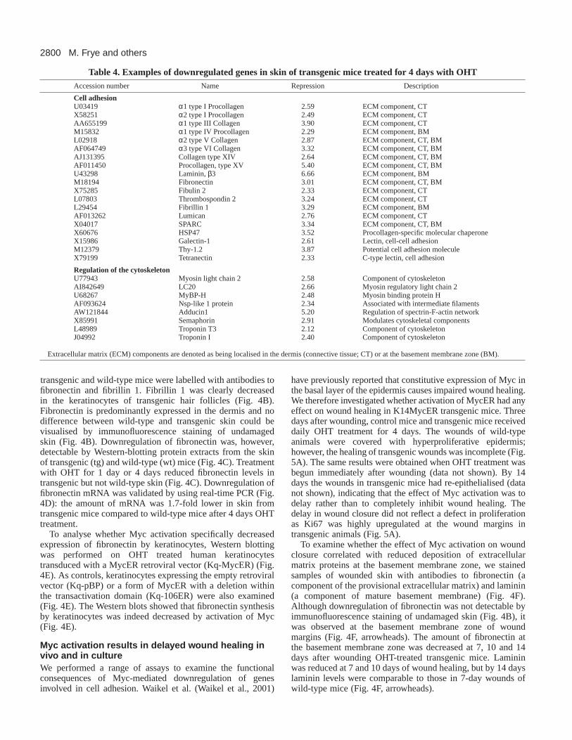
Table 4. Examples of downregulated genes in skin of transgenic mice treated for 4 days with OHTAccession number Name Repression Description
Cell adhesion U03419 α1 type I Procollagen 2.59 ECM component, CTX58251 α2 type I Procollagen 2.49 ECM component, CTAA655199 α1 type III Collagen 3.90 ECM component, CTM15832 α1 type IV Procollagen 2.29 ECM component, BML02918 α2 type V Collagen 2.87 ECM component, CT, BMAF064749 α3 type VI Collagen 3.32 ECM component, CT, BMAJ131395 Collagen type XIV 2.64 ECM component, CT, BMAF011450 Procollagen, type XV 5.40 ECM component, CT, BMU43298 Laminin, β3 6.66 ECM component, BMM18194 Fibronectin 3.01 ECM component, CT, BMX75285 Fibulin 2 2.33 ECM component, CTL07803 Thrombospondin 2 3.24 ECM component, CTL29454 Fibrillin 1 3.29 ECM component, BMAF013262 Lumican 2.76 ECM component, CTX04017 SPARC 3.34 ECM component, CT, BMX60676 HSP47 3.52 Procollagen-specific molecular chaperoneX15986 Galectin-1 2.61 Lectin, cell-cell adhesionM12379 Thy-1.2 3.87 Potential cell adhesion moleculeX79199 Tetranectin 2.33 C-type lectin, cell adhesion
Regulation of the cytoskeletonU77943 Myosin light chain 2 2.58 Component of cytoskeletonAI842649 LC20 2.66 Myosin regulatory light chain 2U68267 MyBP-H 2.48 Myosin binding protein HAF093624 Nsp-like 1 protein 2.34 Associated with intermediate filamentsAW121844 Adducin1 5.20 Regulation of spectrin-F-actin networkX85991 Semaphorin 2.91 Modulates cytoskeletal componentsL48989 Troponin T3 2.12 Component of cytoskeletonJ04992 Troponin I 2.40 Component of cytoskeleton
Extracellular matrix (ECM) components are denoted as being localised in the dermis (connective tissue; CT) or at the basement membrane zone (BM).

2801Myc effects on epidermal cell adhesion
Re-epithelialisation of skin wounds involves acomplex cascade of events that not only requireskeratinocyte migration and proliferation, butalso deposition of a provisional ECM and aninflux of inflammatory cells into the dermis. Toexamine wound healing in the absence of othercell types and independent of Myc effectson proliferation, confluent sheets of humankeratinocytes expressing MycER (Kq-MycER)or the empty retroviral vector (Kq-pBP) werepretreated with OHT for 1 day, then treated withmitomycin C to inhibit proliferation and scrapedwith a pipette tip to create a wound. The widthof the wounds was monitored daily for 3 days.After 24 hours the size of each wound wassignificantly reduced in control cultures, and by48 hours keratinocytes on either side of thewound had made contact (Fig. 5B). In contrast,keratinocytes expressing MycER had not movedinto the wound by 24 hours and by 48 hours thewounds were reduced in width but contactbetween the wound edges had not beenestablished (Fig. 5B). The results presented inFig. 5B are representative of more than fiveindependent experiments.
Motility and spreading are impaired byMyc activationTo examine whether the inhibition of woundhealing by Myc activation reflected decreasedmigration of keratinocytes, motility assays wereperformed using time lapse microscopy. Humankeratinocytes (Kq-MycER, Kq-106ER and Kq-pBP) were pretreated with OHT, plated oncollagen- or laminin-coated dishes and filmedfor 36 hours. Fig. 6A shows the results of asingle experiment that is representative of fourindependent experiments on collagen. The pathtaken by individual cells was determined bymarking the start (green spot) and end co-ordinates (red spot). The average path length ofKq-MycER cells (n=24) on collagen was decreased comparedto controls (Kq-106ER, n=24; Kq-pBP, n=23). The averagespeed of all cells in Fig. 6A is shown in Fig. 6B: there was nosignificant difference in the motility of cells expressing 106ERor the empty vector, but the speed of cells expressing Myc wasdecreased three fold. A similar reduction in motility wasobserved when Myc was activated in keratinocytes plated onlaminin (data not shown).
The effect of Myc on spreading of human keratinocytes wasalso determined (Fig. 6C,D). Kq-MycER, Kq-106ER and Kq-pBP were treated with OHT for 1 day, plated on collagen andincubated in serum-free medium (FAD–; Fig. 6C) or mediumcontaining IGF, EGF or cytochalasin D for 3 hours. Fig. 6Dshows the median spreading area of Kq-MycER, Kq-106ERand Kq-pBP (n=200) from two independent experiments.Cytochalasin D was added as a negative control, because whenactin polymerisation is inhibited keratinocytes attach but do notspread. IGF was added to stimulate cell spreading and EGF toinduce cell contraction (Haase et al., 2003). Kq-MycER cellsshowed a decreased ability to spread in serum-free medium and
in the presence of IGF or EGF. Keratinocytes over-expressing106ER spread to a greater extent than Kq-pBP in FAD or FADplus EGF (Fig. 6D), consistent with the observation that thismutant can act as a dominant negative form of Myc under someconditions (Gandarillas and Watt, 1997).
Effects of Myc on cell-cell and cell-ECM receptorsMyc over-expressing keratinocytes were less motile and spreadto a lower extent than controls, even in the presence ofexogenous extracellular matrix proteins. This suggested thatMyc might affect expression of integrin ECM receptors.Indeed, activation of Myc results in reduced β1 integrinexpression by human keratinocytes in vitro (Gandarillas andWatt, 1997) and in transgenic mice that constitutively expressMyc via the K14 promoter (Waikel et al., 2001). However,several of the integrin receptor genes expressed bykeratinocytes, in particular α2, α5 and β1, were notrepresented on the Affymetrix arrays; β4 RNA levels wereunchanged; and α6, though 1.5-fold downregulated, had a rawexpression value of less than 200, placing the result within the
Fig. 5.Myc activation delays epidermal wound healing in vivo and in vitro. (A) Threedays after wounding wild-type and transgenic mice received daily OHT treatment for4 days. Wounds of wild-type mice had completely re-epithelialised, but wounds oftransgenic mice remained open. Arrowheads indicate wound edges. Ki67 labelling ofwound margins is shown in right-hand panels. Scale bar: 100 µm. (B) Wounds weremade in sheets of cultured human keratinocytes (0h) and monitored after 24 or 48hours. Myc over-expressing keratinocytes (Kq-MycER) were compared to controls(Kq-pBP). Cells were stained with phalloidin. Scale bar: 10 µm.

2802
low confidence level category. To establish whether α6 mRNAwas indeed downregulated by Myc, we performed real-timePCR analysis of RNA extracted from whole skin of transgenicmice (Fig. 7C). α6 mRNA was twofold downregulated intransgenic mice after 4 days treatment with OHT compared tountreated transgenic animals (Fig. 7C). We therefore usedFACS, immunofluorescence staining and electron microscopyto investigate whether expression of cell-ECM and cell-celladhesion receptors was altered upon Myc activation.
When cultured human keratinocytes were stained withantibodies to E-cadherin and desmoplakin there was nodifference between cells expressing activated Myc andcontrols, suggesting that Myc did not affect the assembly ofadherens and desmosomal junctions (Fig. 7A). Vinculinstaining demonstrated that focal adhesion formation was alsonormal (Fig. 7A). There was no significant decrease in surfaceβ1 integrins in K14MycER transgenic keratinocytes, althoughthe β1-associated protein CD98 (Fenczik et al., 1997) wasupregulated on the cell surface as well as at the RNA level (5.0fold on the arrays) (Fig. 7B). The only other integrin-associatedprotein to be upregulated was the tetraspan NAG-2 (3.9 fold;data not shown) (Tachibana et al., 1997).
Activation of Myc in transgenic mouse keratinocytes led toa marked reduction in cell surface expression of the α6β4
integrin (Fig. 7B). In cultured human keratinocytes Mycactivation resulted in reduced localisation of α6β4 in theimmature hemidesmosomes that are assembled in culture (Fig.7A). Reduced assembly of these junctions was confirmed byimmunolabelling for plectin (data not shown). We concludethat of the major types of cell-cell and cell-ECM adhesivejunctions, only hemidesmosome formation was affected byMyc activation in cultured keratinocytes.
We next examined α6 expression and hemidesmosomeformation in intact skin. Immunolabelling the skin oftransgenic mice after 4 days treatment with OHT confirmed adecrease in α6 levels at the basement membrane zone (Fig.8A,B). We then performed electron microscopy on the skin oftransgenic and wild-type mice treated for 9 days with OHT(Fig. 8C-F). The number of hemidesmosomes in the epidermisof transgenic animals was greatly reduced compared to wild-type mice (Fig. 8E,F) and those hemidesmosomes that werepresent were smaller than in wild-type epidermis (Fig. 8C,D,arrowheads).
Cytoskeleton and cytoskeleton regulatory factorsare downregulatedGiven that 11% of the downregulated genes on the microarrayswere components of the cytoskeleton or cytoskeleton
M. Frye and others
Fig. 6.Effect of Mycactivation on keratinocytemotility and spreading. Mycover-expressing keratinocytes(Kq-MycER) were comparedwith keratinocytes expressingthe empty retroviral vector(Kq-pBP) or the mutatedMyc construct (Kq-106ER).(A) Movements of individualcells plated on collagen areshown. Green spots: start co-ordinates; red spots: end co-ordinates. (B) Average speedsof Kq-MycER (n=24), Kq-106ER (n=24) and Kq-pBP(n=23) on collagen. (C) Cellspreading in serum-freemedium (FAD-). Cells werestained with phalloidin. Scalebar: 10 µm. (D) Median ofcells spreading in serum-freemedium (FAD) or mediumsupplemented with EGF, IGFor cytochalasin D (cyto. D).

2803Myc effects on epidermal cell adhesion
regulatory factors (Fig. 2B), we asked whether they mightcontribute to the low motility and impaired spreading of Mycover-expressing keratinocytes. Examples of some of thedownregulated genes are given in Fig. 9A. These includecomponents of the cytoskeleton such as myosin II andtroponin, and cytoskeleton regulatory factors such as LC20,MyBP-H and adducin (for explanation, see Table 4).
Downregulation of MyBP-H mRNA was confirmed byNorthern blotting (Fig. 9B). Compared to wild-type mice anduntreated transgenic mice MyBP-H mRNA was downregulatedin the skin of transgenic animals after 1 day and 4 daystreatment with OHT (Fig. 9B). Decreased expression ofmyosin II was validated by immunolabelling of the skin ofnon-transgenic and transgenic mice (Fig. 9C). The cellulardistribution of myosin II changed from concentration at theplasma membrane to a weak cytoplasmic localisation (Fig.9C).
Adducin is a membrane skeletal protein that binds to F-actin,modulates the assembly and disassembly of the spectrin-F-actin network (Gardner and Bennett, 1987), and is thought toplay a crucial role in cell motility (Fukata et al., 1999).Adducin was localised at the plasma membranes of all cells inthe epidermis in wild-type mice, but was no longer detectablein keratinocytes in the basal layer of OHT-treated transgenicepidermis (Fig. 9C, arrowheads). Real-time PCR confirmed a1.8-fold downregulation of adducin mRNA in transgenic skincompared to wild-type skin treated for 4 days with OHT (Fig.9F). We also measured adducin protein levels by western
blotting (Fig. 9G). There was a 3.3-fold decrease of adducinprotein in Kq-MycER cells after 2 days treatment with OHT,whereas adducin levels were not altered upon OHT treatmentof Kq-pBP (Fig. 9G). Adducin protein was 1.7-fold lower inuntreated Kq-MycER cells compared to Kq-pBP, suggestingthat when expressed at high levels in cultured cells the MycERconstruct is somewhat ‘leaky’ (see also Gandarillas and Watt,1997). Consistent with the in vivo observations (Fig. 9C),immunofluorescence demonstrated that the level of adducinprotein at cell-cell borders was decreased upon activation ofMyc in Kq-MycER cells compared to Kq-pBP (Fig. 9E).
To analyse how activated Myc might influence thecytoskeleton we examined the distribution of actin and myosinin cultured human keratinocytes (Fig. 9D). In Kq-MycER cellstreated with OHT the number and size of the leading membranelamellae were reduced compared to the controls (Kq-pBP) (Fig.9D, arrowheads). This correlated with reduced polymerisationof actin and myosin at the cell periphery (Fig. 9D).
DISCUSSION
Gene expression profiling has been used before to identifypotential Myc target genes. Myc null and wild-type fibroblastshave been compared (Guo et al., 2000; O’Connell et al., 2003)and fibroblasts transduced with a MycER construct have beenexamined 9 hours after OHT treatment (Coller et al., 2000).Other investigators have focused on the oncogenic properties
Fig. 7.Myc activation didnot disrupt cell-celladhesion or focaladhesions, but did decreasehemidesmosomeformation. (A) Culturedhuman keratinocytes (Kq-MycER, Kq-106ER, Kq-pBP) were stained withantibodies to the proteinsshown. Nuclei are stainedfor Myc using 9E10antibody. (B) Flowcytometry of primarykeratinocytes isolated fromtransgenic or wild-typemice 2 days after OHTtreatment using antibodiesagainst the β1 and α6integrin subunits andCD98. (C) Real-time PCRdetermination of α6integrin subunit mRNAlevels in transgenic (tg)mice untreated (0d) ortreated with OHT for 4days (4d) (P<0.01).

2804
of Myc, identifying Myc targets in neuroblastoma and Burkitt’slymphoma (Boon et al., 2001; Schuhmacher et al., 2001;Schuldiner and Benvenisty, 2001). Our study is the first toexamine Myc regulated genes in vivo, and to examine geneexpression in non-malignant epithelial cells.
In considering how gene expression is modulated by Myc,direct versus indirect regulation is an important issue(Eisenman, 2001). Direct targets are those genes whoseexpression is altered by direct Myc binding (Eisenman, 2001).Max-Myc-TRRAP coactivator complexes bind E-boxes,generating acetylation of histone H4 in the vicinity of thebinding site and stimulating induction of target gene expression(Eisenman, 2001). Most Myc target genes identified usingcultured cells are involved in growth and metabolism,consistent with the function of Myc in regulating the growthrate (i.e. size and mass) of cells (Eisenman, 2001). Genesupregulated in response to Myc are involved in ribosomebiogenesis, energy and nucleotide metabolism and translational
regulation (Coller et al., 2000; Guo et al., 2000; Boon et al.,2001; Schuhmacher et al., 2001).
The major classes of genes that were upregulated inK14MycER mouse skin were those well documented to beregulated by Myc, including genes that are known to be directtargets. This is the case for the eukaryotic initiation factors(eIF) (Table 2), mRNA cap-binding proteins rate limiting forprotein synthesis (Rosenwald et al., 1993, Coller et al., 2000;Jones et al., 1996). Activation of Myc induces proteinsynthesis by upregulating ribosomal proteins in fibroblasts(Guo et al., 2000), in Drosophila (Johnston et al., 1999),human B cells (Iritani and Eisenman, 1999) and mice(Schuhmacher et al., 1999). Ribosomal proteins were alsoinduced in K14MycER skin, including the direct Myc target,nucleolin (Greasley et al., 2000). Myc plays an essential rolein regulating entry into S-phase by shortening G1; severalproteins upregulated in MycER mouse skin stimulate entryinto S-phase (Table 2), including mCDC47 (MCM7), which
has an E-box binding site for Mycin its promoter (Suzuki et al.,1998). Another well documentedMyc target gene is Ornithinedecarboxylase (ODC) (Bello-Fernandez et al., 1993). AlthoughODC has been previouslydescribed to be upregulated inK14MycER transgenic mice(Arnold and Watt, 2001), themRNA was not regulated on themicroarray chips; a possibleexplanation for this is a highbackground value with the controlmismatch probe.
Although our screen was able todetect direct Myc target genes,other identified genes wereundoubtedly indirectly regulated.The most obvious categories weregenes that were expressed intransgene-negative cells. Theseinclude the differentiation markersexpressed in the suprabasalepidermal layers (Fig. 3C) anddermal (connective tissue)-specificECM genes (Table 4). As discussedbelow, the induction ofdifferentiation genes is mostprobably a secondary consequenceof the Myc-mediated effects on celladhesion. The mechanism by whichdermal gene expression is alteredremains to be investigated;however, one possibility is that it isvia the effects of Myc on thecytokines expressed by basalkeratinocytes (see Fig. 2B).
The induction of cell cycle anddifferentiation marker genesreflects the stimulation ofproliferation and differentiation byMycER that we have reported
M. Frye and others
Fig. 8.Decrease in α6 integrin levels and hemidesmosomes by activation of Myc.(A,B) Immunolabelling of the epidermis with antibodies to α6 of (A) wild-type mice or (B)transgenic mice after 4 days treatment with OHT. (C-F) Electron microscopy of the basementmembrane zone of skin from transgenic mice (D,F) and wild-type mice (C,E) treated for 9 dayswith OHT. Arrows in C, D and E indicate hemidesmosomes. Arrows in F indicate absence ofhemidesmosomes at the basement membrane. Scale bars: 5 µm (B), 200 nm (D), 1 µm (F). BM,basement membrane; IFE, interfollicular epidermis; HF, hair follicle.

2805Myc effects on epidermal cell adhesion
previously (Arnold and Watt, 2001). However, it is the genesthat were repressed by Myc that provide a clue as to themechanism by which Myc exerts its surprising differentiation-promoting effects on the epidermis. Myc-mediated repressionof gene expression is incompletely understood. It has beenreported to involve direct binding by Myc-Max complexes toINR elements; alternatively, it may depend on interaction ofMyc with positively acting transcription factors (reviewed inOrian and Eisenman, 2001). Myc can also form a complex withthe transcription factor Miz-1 and thereby inhibit Miz-1-mediated transcriptional activation (e.g. Staller et al., 2001).There is also evidence for repression of E-box-dependent
transcription via Myc recruitment of a transcriptionalcorepressor complex (Satou et al., 2001).
The major classes of genes that were downregulated in thetransgenics were those involved in cell adhesion and thecytoskeleton. There is already evidence from several studiesthat Myc regulates cell adhesion. Cells transformed byderegulated expression of Myc or N-Myc are characterised byreduced expression of cell adhesion molecules, including arange of integrin subunits (Inghirami et al., 1990; Judware andCulp, 1995; Barr et al., 1998; Fujimoto et al., 2001) and Myccan act directly on certain integrin promoters (Barr et al., 1998;López-Rodríguez et al., 2000). Fibronectin, alpha-1 type 3
Fig. 9. (A) RNA expressionprofile of downregulatedcytoskeleton components andcytoskeleton regulatory factorsin untreated (0d) and treated(1d and 4d) wild-type andtransgenic mice. (B) Northernblot for MyBP-H of RNA fromskin of wild-type (wt) andtransgenic (tg) mice, untreated(0d) or treated for 1 day (1d)or 4 days (4d) with OHT.(C) Immunolabelling withantibodies to adducin andmyosin II of skin from wild-type and transgenic animalsafter 4 days OHT treatment.Scale bar: 10 µm. (D) Culturedhuman keratinocytes(Kq-MycER and Kq-pBP)were stained with antibodies toactin (red; left panels) andmyosin II (green; right panels).Cells shown are at theperiphery of clones or at theedges of wounds made inconfluent sheets. Scale bar:5 µm. Nuclei are stained forMyc using 9E10 antibody.(E-G) Downregulation ofadducin upon activation ofMyc visualised byimmunofluorescence (E), real-time PCR (F), and Westernblotting (G). (E,G) Adducinprotein expression in Mycexpressing humankeratinocytes (Kq-MycER) isdecreased compared tocontrols (Kq-pBP). Scale bar:10 µm. (F) Relative RNAexpression is reduced in skinof transgenic (tg) animalstreated with OHT for 4 days(4d) compared to 4 daystreated wild-type (wt) anduntreated (0d) transgenic mice(P<0.05).

2806
collagen and tropomyosin alpha chain genes are downregulatedwithin 9 hours of MycER activation in fibroblasts (Coller et al.,2000) and there is evidence that Myc suppresses collagengenes by interference with NF-1 (Yang et al., 1991; Yang etal., 1993), as proposed for the PDGF receptor promoter (Osteret al., 2000). It thus seems probable that at least some of thegenes downregulated by Myc are direct targets.
The categories of adhesion molecules that were repressed byMyc in the skin included ECM proteins, regulators of thecytoskeleton and membrane proteins such as the α6 integrinsubunit. These changes had profound effects on the behaviourof keratinocytes, whether assayed in vivo or in vitro. Woundhealing in vivo was impaired, as reported previously (Waikelet al., 2001). The motility of keratinocytes in culture wasseverely reduced, both within cell sheets (Fig. 5B) and at thelevel of single cells (Fig. 6A,B). The spreading of keratinocyteswas decreased (Fig. 6C,D) and the ability of the cells toform lamellipodia was compromised (Fig. 9D), reflectingdownregulation of proteins that control the assembly andcontractility of the actin cytoskeleton (Table 4). This was alsoreflected by the downregulation of adducin, a protein that playsa crucial role in cell motility (Fig. 9E-G) (Fukata et al., 1999).In vivo, there was a substantial reduction in the numberof hemidesmosomes and those hemidesmosomes that werepresent were reduced in size (Fig. 8C-F).
Based on the effects of Myc on keratinocyte adhesion and
motility we can now propose a model for the phenotype ofK14MycER mouse epidermis. The model is that reduced cell-ECM adhesion stimulates exit from the stem cell compartment,whereas reduced motility determines that IFE and sebocytedifferentiation are promoted at the expense of the hair lineages(Fig. 10). There is good evidence that epidermal stem cells aremore adhesive to ECM than their differentiating daughters;this is true both for human (Jones et al., 1995) and mouse(Bickenbach and Chism, 1998) keratinocytes. Furthermore,reduced ECM adhesion is known to promote differentiation:both human and mouse keratinocytes undergo terminaldifferentiation when placed in suspension (reviewed by Watt,2001) and in human keratinocytes reduced expression ofβ1 integrins in vitro stimulates exit from the stem cellcompartment (Zhu et al., 1999). High expression of the α6β4integrin is thought to be a marker of stem cells in mouseepidermis (Tani et al., 2000) and α6β4 was markedlydownregulated by MycER. It therefore seems probable thatMyc-induced repression of adhesion stimulates epidermal stemcells to differentiate.
The stem cells that give rise to mature sebocytes andsuprabasal IFE keratinocytes lie directly underneath theirdifferentiated progeny (Fig. 10A). In contrast, differentiationalong the hair lineages involves migration of keratinocytes tothe base of the hair follicle, where contact with the specialisedmesenchymal cells of the dermal papilla provides the necessaryhair inductive stimuli (Fig. 10A) (Niemann and Watt, 2002).The inhibition of keratinocyte motility by Myc may thereforeexplain why differentiation along the hair lineages is notstimulated; instead the cells that exit the stem cell compartmentremain in the upper follicle and are exposed to an environmentthat is conducive to sebocyte differentiation (Fig. 10A,B). It isinteresting that BSSP was the most highly induced gene on thearrays, because it is predominantly expressed in the sebaceousglands of hair follicles of nude mice (Meier et al., 1999). Thepatchy expression of BSSP in IFE in response to Mycactivation could potentially reflect the presence of scatteredsebocytes in IFE of MycER mice (Arnold and Watt, 2001).
The control of stem cell fate in many, if not all, tissues andorganisms involves reciprocal interactions between stem cellsand their local microenvironment or niche (Watt and Hogan,2001; Spradling et al., 2001). The expression and functions ofMyc could provide an example of such reciprocity, becausecell-ECM adhesion can regulate Myc. In epithelial cells Myclevels decrease in suspended cells, correlating with cell cyclearrest or differentiation (Gandarillas and Watt, 1995; Benaudand Dickson, 2001a). Conversely, adhesion of epithelial cellsto fibronectin or collagen induces Myc expression in a
M. Frye and others
Interfollicular epidermis (IFE)
Dermal Papilla
BulgeInner Root Sheath
SebaceousGland
Outer RootSheath
Stem cell
stem cells
interfoll icularepider mis
hair folli cle
seb aceousgland
c-Myc (transient)
TA cells
detachmentfrom BM
detachmentfrom BMmigration
along ORS
Hair Follicle
stem cells
interfoll icularepider mis
hair folli cle
seb aceousgland
c-Myc (sustained)
TA cells
detachmentfrom BM
detachmentfrom BMmigration
along ORS
XWild-type Transgenic
A
B
X
Fig. 10.Model for mechanism by which Mycstimulates exit from the stem cell compartmentand differentiation into IFE and sebocytes.(A) Location of stem cell populations, showingthat stem cells for IFE and sebocytes are in closeproximity to their differentiated daughters,whereas the progeny of bulge stem cells migrateto the dermal papilla before differentiating alongthe hair lineages. (B) Comparison of the role ofMyc activation in wild-type and K14MycERepidermis.

2807Myc effects on epidermal cell adhesion
concentration-dependent fashion (Benaud and Dickson,2001b). Pathways implicated in the induction of Myc, namelyc-Src, Erk1/2 MAPK and Ras/Akt (Barone and Courtneidge,1995; Sears et al., 2000; Benaud and Dickson, 2001b) areactivated by integrin ligation (Frame et al., 2002; Howe et al.,2002).
In conclusion, we have presented evidence that the effectsof Myc on epidermal differentiation are a consequence of itseffects on cell adhesion and motility. This would explain whyMyc promotes differentiation in keratinocytes (Gandarillas andWatt, 1997; Arnold and Watt, 2001; Waikel et al., 2001) yetsuppresses differentiation in non-epithelial cells (Eisenman,2001). Because inhibition of cell-ECM adhesion promotesapoptosis of certain cell types (Frisch and Screaton, 2001), therepression of cell adhesion genes could also play a role in Myc-induced apoptosis of some cells. Thus, generic effects of Mycon gene expression have context-specific outcomes in terms ofcell behaviour.
We are grateful to everyone who provided us with advice orreagents, and to Laurence Levy and Frank Burslem for encouragingus to carry out the project. We thank Richard Poulsom, DeborahAubyn and Ken Blight for technical support. M.F. is supported by afellowship from the DFG, FR 1575/1-1. F.M.W. was a recipient of theCE.R.I.E.S. Research Award. I.A. was supported by an EU MarieCurie Fellowship and an EMBO Long Term Fellowship.
REFERENCES
Anbazhagan, R., Bartkova, J., Stamp, G., Pignatelli, M., Gusterson, B. andBartek, J. (1995). Expression of integrin subunits in the human infant breastcorrelates with morphogenesis and differentiation. J. Pathol. 176, 227-232.
Arnold, I. and Watt, F. M. (2001). c-Myc activation in transgenic mouseepidermis results in mobilization of stem cells and differentiation of theirprogeny. Curr. Biol. 11, 558-568.
Barone, M. V. and Courtneidge, S. A.(1995). Myc but not Fos rescue ofPDGF signalling block caused by kinase-inactive Src. Nature378, 509-512.
Barr, L. F., Campbell, S. E., Bochner, B. S. and Dang, C. V.(1998).Association of the decreased expression of alpha3beta1 integrin with thealtered cell, environmental interactions and enhanced soft agar cloningability of c-myc-overexpressing small cell lung cancer cells. Cancer Res.58, 5537-5545.
Bello-Fernandez, C., Packham, G. and Cleveland, J. L.(1993). Theornithine decarboxylase gene is a transcriptional target of c-Myc. Proc. Natl.Acad. Sci. USA90, 7804-7808.
Benaud, C. M. and Dickson, R. B.(2001a). Adhesion-regulated G1 cell cyclearrest in epithelial cells requires the downregulation of c-Myc. Oncogene20, 4554-4567.
Benaud, C. M. and Dickson, R. B.(2001b). Regulation of the expression ofc-Myc by beta1 integrins in epithelial cells. Oncogene20, 759-768.
Bickenbach, J. R. and Chism, E.(1998). Selection and extended growth ofmurine epidermal stem cells in culture. Exp. Cell Res.244, 184-195.
Boon, K., Caron, H. N., van Asperen, R., Valentijn, L., Hermus, M. C.,van Sluis, P., Roobeek, I., Weis, I., Voute, P. A., Schwab, M. et al. (2001).N-myc enhances the expression of a large set of genes functioning inribosome biogenesis and protein synthesis. EMBO J.20, 1383-1393.
Bull, J. J., Muller-Rover, S., Patel, S. V., Chronnell, C. M., McKay, I. A.and Philpott, M. P. (2001). Contrasting localization of c-Myc with otherMyc superfamily transcription factors in the human hair follicle and duringthe hair growth cycle. J. Invest. Dermatol. 116, 617-622.
Carroll, J. M., Romero, M. R. and Watt, F. M. (1995). Suprabasal integrinexpression in the epidermis of transgenic mice results in developmentaldefects and a phenotype resembling psoriasis. Cell 83, 957-968.
Coller, H. A., Grandori, C., Tamayo, P., Colbert, T., Lander, E. S.,Eisenman, R. N. and Golub, T. R.(2000). Expression analysis witholigonucleotide microarrays reveals that MYC regulates genes involved ingrowth, cell cycle, signaling, and adhesion. Proc. Natl. Acad. Sci. USA97,3260-3265.
Danielian, P. S., White, R., Hoare, S. A., Fawell, S. E. and Parker, M. G.(1993). Identification of residues in the estrogen receptor that conferdifferential sensitivity to estrogen and hydroxytamoxifen. Mol. Endocrinol.7, 232-240.
Eisenman, R. N.(2001). Deconstructing myc. Genes Dev. 15, 2023-2030. Fenczik, C. A., Sethi, T., Ramos, J. W., Hughes, P. E. and Ginsberg, M. H.
(1997). Complementation of dominant suppression implicates CD98 inintegrin activation. Nature390, 81-85.
Frame, M. C., Fincham, V. J., Carragher, N. O. and Wyke, J. A.(2002).v-Src’s hold over actin and cell adhesions. Nat. Rev. Mol. Cell Biol. 3, 233-245.
Frisch, S. M. and Screaton, R. A.(2001). Anoikis mechanisms. Curr. Opin.Cell Biol. 13, 555-562.
Fuchs, E. and Raghavan, S. (2002). Getting under the skin of epidermalmorphogenesis.Nat. Rev. Genet.3, 199-209.
Fujimoto, H., Tanaka, Y., Liu, Z. J., Yagita, H., Okumura, K., Kosugi, A.,Morinobu, A., Umehara, H., Yamamura, H. and Minami, Y. (2001).Down-regulation of alpha6 integrin, an anti-oncogene product, by functionalcooperation of H-Ras and c-Myc. Genes Cells. 6, 337-343.
Fujimoto, W., Nakanishi, G., Arata, J. and Jetten, A. M. (1997).Differential expression of human cornifin alpha and beta in squamousdifferentiating epithelial tissues and several skin lesions. J. Invest. Dermatol.108, 200-204.
Fukata, Y., Oshiro, N., Kinoshita, N., Kawano, Y., Matsuoka, Y., Bennett,V., Matsuura, Y. and Kaibuchi, K. (1999). Phosphorylation of adducin byRho-kinase plays a crucial role in cell motility. J. Cell Biol.145, 347-361.
Gandarillas, A. and Watt, F. M. (1995). Changes in expression of membersof the fos and jun families and myc network during terminal differentiationof human keratinocytes. Oncogene11, 1403-1407.
Gandarillas, A. and Watt, F. M. (1997). c-Myc promotes differentiation ofhuman epidermal stem cells. Genes Dev. 11, 2869-2882.
Gandarillas, A., Davies, D. and Blanchard, J. M.(2000). Normal and c-Myc-promoted human keratinocyte differentiation both occur via a novelcell cycle involving cellular growth and endoreplication. Oncogene19,3278-3289.
Gardner, K. and Bennett, V. (1987). Modulation of spectrin-actin assemblyby erythrocyte adducin. Nature328, 359-362.
Gat, U., DasGupta, R., Degenstein, L. and Fuchs, E.(1998). De novo hairfollicle morphogenesis and hair tumors in mice expressing a truncated beta-catenin in skin. Cell 95, 605-614.
Grandori, C., Cowley, S. M., James, L. P. and Eisenman, R. N.(2000). TheMyc/Max/Mad network and the transcriptional control of cell behavior.Annu. Rev. Cell Dev. Biol. 16, 653-699.
Greasley, P. J., Bonnard, C. and Amati, B.(2000). Myc induces the nucleolinand BN51 genes: possible implications in ribosome biogenesis. NucleicAcids Res. 28, 446-453.
Guo, Q. M., Malek, R. L., Kim, S., Chiao, C., He, M., Ruffy, M., Sanka,K., Lee, N. H., Dang, C. V. and Liu, E. T.(2000). Identification of c-mycresponsive genes using rat cDNA microarray. Cancer Res. 60, 5922-5928.
Haase, I., Evans, R., Pofahl, R. and Watt, F. M. (2003). Regulation ofkeratinocyte shape, migration and wound epithelialisation by IGF-1 andEGF-dependent signalling pathways. J. Cell Sci. (in press).
Howe, A. K., Aplin, A. E. and Juliano, R. L.(2002). Anchorage-dependentERK signaling – mechanisms and consequences. Curr. Opin. Genet. Dev.12, 30-35.
Huelsken, J., Vogel, R., Erdmann, B., Cotsarelis, G. and Birchmeier, W.(2001). beta-Catenin controls hair follicle morphogenesis and stem celldifferentiation in the skin. Cell. 105, 533-545.
Hurlin, P. J., Foley, K. P., Ayer, D. E., Eisenman, R. N., Hanahan, D. andArbeit, J. M. (1995). Regulation of Myc and Mad during epidermaldifferentiation and HPV-associated tumorigenesis. Oncogene11, 2487-2501.
Inghirami, G., Grignani, F., Sternas, L., Lombardi, L., Knowles, D. M.and Dalla-Favera, R. (1990). Down-regulation of LFA-1 adhesionreceptors by C-myc oncogene in human B lymphoblastoid cells. Science250, 682-686.
Iritani, B. M. and Eisenman, R. N.(1999). c-Myc enhances protein synthesisand cell size during B lymphocyte development. Proc. Natl. Acad. Sci. USA96, 13180-13185.
Johnston, L. A., Prober, D. A., Edgar, B. A., Eisenman, R. N. and Gallant,P. (1999). Drosophila myc regulates cellular growth during development.Cell 98, 779-790.
Jones, P. H., Harper, S. and Watt, F. M.(1995). Stem cell patterning andfate in human epidermis. Cell 80, 83-93.

2808
Jones, R. M., Branda, J., Johnston, K. A., Polymenis, M., Gadd, M.,Rustgi, A., Callanan, L. and Schmidt, E. V.(1996). An essential E box inthe promoter of the gene encoding the mRNA cap-binding protein(eukaryotic initiation factor 4E) is a target for activation by c-myc. Mol. CellBiol. 16, 4754-4764.
Judware, R. and Culp, L. A. (1995). Over-expression of transfected N-myconcogene in human SKNSH neuroblastoma cells down-regulates expressionof beta 1 integrin subunit. Oncogene11, 2599-2607.
Kaiser, H. W., Ness, W., O’Keefe, E., Balcerkiewicz, A. and Kreysel, H. W.(1993). Localization of adducin in epidermis. J. Invest. Dermatol.101, 783-788.
Littlewood, T. D., Hancock, D. C., Danielian, P. S., Parker, M. G. and Evan,G. I. (1995). A modified oestrogen receptor ligand-binding domain as animproved switch for the regulation of heterologous proteins. Nucleic AcidsRes.23, 1686-1690.
López-Rodríguez, C., Delgado, M. D., Puig-Kröger, A., Nueda, A.,Muñoz, E., León, J., Bernabéu, C. and Corbi, A. L.(2000). c-Mycinhibits CD11a and CD11c leukocyte integrin promoters. Eur. J. Immunol.30, 2465-2471.
McGowan, K. M. and Coulombe, P. A. (1998). Onset of keratin 17expression coincides with the definition of major epithelial lineages duringskin development. J. Cell Biol. 143, 469-486.
Meier, N., Dear, T. N. and Boehm, T.(1999). A novel serine proteaseoverexpressed in the hair follicles of nude mice. Biochem. Biophys. Res.Commun. 258, 374-378.
Merrill, B. J., Gat, U., DasGupta, R. and Fuchs, E.(2001). Tcf3 and Lef1regulate lineage differentiation of multipotent stem cells in skin. Genes Dev.15, 1688-1705.
Morgenstern, J. P. and Land, H.(1990). A series of mammalian expressionvectors and characterisation of their expression of a reporter gene in stablyand transiently transfected cells. Nucleic Acids Res. 18, 1068.
Niemann, C., Owens, D. M., Hulsken, J., Birchmeier, W. and Watt, F. M.(2002). Expression of DeltaNLef1 in mouse epidermis results indifferentiation of hair follicles into squamous epidermal cysts and formationof skin tumours. Development129, 95-109.
Niemann, C. and Watt, F. M.(2002). Designer skin: lineage commitment inpostnatal epidermis. Trends Cell Biol.12, 185-192.
O’Connell, B. C., Cheung, A. F., Simkevich, C. P., Tam, W., Ren, X.,Mateyak, M. K. and Sedivy, J. M.(2003). A large scale genetic analysisof c-Myc-regulated gene expression patterns. J. Biol. Chem.2781, 12563-12573.
Orian, A. and Eisenman, R. N.(2001). TGF-beta flips the Myc switch. Sci.STKE88, PE1.
Oster, S. K., Marhin, W. W., Asker, C., Facchini, L. M., Dion, P. A., Funa,K., Post, M., Sedivy, J. M. and Penn, L. Z.(2000). Myc is an essentialnegative regulator of platelet-derived growth factor beta receptor expression.Mol. Cell. Biol. 20, 6768-6778.
Panteleyev, A. A., Jahoda, C. A. and Christiano, A. M.(2001). Hair folliclepredetermination. J. Cell Sci.114, 3419-3431.
Parrish, E. P., Steart, P. V., Garrod, D. R. and Weller, R. O.(1987).Antidesmosomal monoclonal antibody in the diagnosis of intracranialtumours. J. Pathol. 153, 265-273.
Pelengaris, S., Littlewood, T., Khan, M., Elia, G. and Evan, G.(1999).Reversible activation of c-Myc in skin: induction of a complex neoplasticphenotype by a single oncogenic lesion. Mol. Cell 3, 565-577.
Pelengaris, S., Rudolph, B. and Littlewood, T.(2000). Action of Myc in vivo– proliferation and apoptosis. Curr. Opin. Genet. Dev. 10, 100-105.
Roper, E., Weinberg, W., Watt, F. M. and Land, H. (2001). p19ARF-independent induction of p53 and cell cycle arrest by Raf in murinekeratinocytes. EMBO Rep. 2, 145-150.
Rosenwald, I. B., Rhoads, D. B., Callanan, L. D., Isselbacher, K. J. and
Schmidt, E. V. (1993). Increased expression of eukaryotic translationinitiation factors eIF-4E and eIF-2 alpha in response to growth induction byc-myc. Proc. Natl. Acad. Sci. USA.90, 6175-6178.
Rumio, C., Donetti, E., Imberti, A., Barajon, I., Prosperi, E., Brivio, M.F., Boselli, A., Lavezzari, E., Veraldi, S., Bignotto, M. et al. (2000). c-Myc expression in human anagen hair follicles. Br. J. Dermatol. 142, 1092-1099.
Satou, A., Taira, T., Iguchi-Ariga, S. M. and Ariga, H. (2001). A noveltransrepression pathway of c-Myc. Recruitment of a transcriptionalcorepressor complex to c-Myc by MM-1, a c-Myc-binding protein. J. Biol.Chem. 276, 46562-46567.
Schuhmacher, M., Staege, M. S., Pajic, A., Polack, A., Weidle, U. H.,Bornkamm, G. W., Eick, D. and Kohlhuber, F. (1999). Control ofcell growth by c-Myc in the absence of cell division. Curr. Biol.9, 1255-1258.
Schuhmacher, M., Kohlhuber, F., Holzel, M., Kaiser, C., Burtscher, H.,Jarsch, M., Bornkamm, G. W., Laux, G., Polack, A., Weidle, U. H. etal. (2001). The transcriptional program of a human B cell line in responseto Myc. Nucleic Acids Res. 29, 397-406.
Schuldiner, O. and Benvenisty, N.(2001). A DNA microarray screen forgenes involved in c-MYCand N-MYConcogenesis in human tumors.Oncogene20, 4984-4994.
Sears, R., Nuckolls, F., Haura, E., Taya, Y., Tamai, K. and Nevins, J. R.(2000). Multiple Ras-dependent phosphorylation pathways regulate Mycprotein stability. Genes Dev.14, 2501-2514.
Spradling, A., Drummond-Barbosa, D. and Kai, T.(2001). Stem cells findtheir niche. Nature414, 98-104.
Staller, P., Peukert, K., Kiermaier, A., Seoane, J., Lukas, J., Karsunky, H.,Moroy, T., Bartek, J., Massague, J., Hanel, F. et al. (2001). Repressionof p15INK4b expression by Myc through association with Miz-1. Nat. CellBiol. 3, 392-399.
Sterk, L. M., Geuijen, C. A., Oomen, L. C., Calafat, J., Janssen, H. andSonnenberg, A.(2000). The tetraspan molecule CD151, a novel constituentof hemidesmosomes, associates with the integrin α6β4 and may regulate thespatial organization of hemidesmosomes. J. Cell Biol.149, 969-982.
Suzuki, S., Adachi, A., Hiraiwa, A., Ohashi, M., Ishibashi, M. and Kiyono,T. (1998). Cloning and characterization of human MCM7 promoter. Gene216, 85-91.
Tachibana, I., Bodorova, J., Berditchevski, F., Zutter, M. M. and Hemler,M. E. (1997). NAG-2, a novel transmembrane-4 superfamily (TM4SF)protein that complexes with integrins and other TM4SF proteins. J. Biol.Chem.272, 29181-29189.
Tani, H., Morris, R. J. and Kaur, P. (2000). Enrichment for murinekeratinocyte stem cells based on cell surface phenotype. Proc. Natl. Acad.Sci. USA97, 10960-10965.
Waikel, R. L., Kawachi, Y., Waikel, P. A., Wang, X. J. and Roop, D. R.(2001). Deregulated expression of c-Myc depletes epidermal stem cells. Nat.Genet. 28, 165-168.
Watt, F. M. and Hogan, B. L.(2000). Out of Eden: stem cells and their niches.Science287, 1427-1430.
Watt, F. M. (2001). Stem cell fate and patterning in mammalian epidermis.Curr. Opin. Genet. Dev. 11, 410-417.
Yang, B. S., Geddes, T. J., Pogulis, R. J., de Crombrugghe, B. and Freytag,S. O. (1991). Transcriptional suppression of cellular gene expression by c-Myc. Mol. Cell. Biol. 11, 2291-2295.
Yang, B. S., Gilbert, J. D. and Freytag, S. O.(1993). Overexpression of Mycsuppresses CCAAT transcription factor/nuclear factor 1-dependentpromoters in vivo. Mol. Cell. Biol.13, 3093-3102.
Zhu, A. J., Haase, I. and Watt, F. M.(1999). Signaling via beta1 integrinsand mitogen-activated protein kinase determines human epidermal stem cellfate in vitro. Proc. Natl. Acad. Sci. USA96, 6728-6733.
M. Frye and others