DEVELOPMENT AND DISEASE Axial skeletal defects caused by...
12
INTRODUCTION Notch signalling is an evolutionarily conserved mechanism used by metazoans to control the specification of cell fates through local interactions between cells (Artavanis-Tsakonas et al., 1999). As ligand and receptor are membrane associated, signalling is triggered by direct interaction of adjacent cells. In general, the Notch receptor is widely distributed within a cell population, while the ligand is restricted to a subset of cells (Fleming et al., 1990; Heitzler and Simpson, 1991; Vassin et al., 1987; Wharton et al., 1985). While several proteins participate in transmitting and regulating Notch signalling, a group of elements are defined as the core of this signalling pathway: in Drosophila, Delta and Serrate are Notch ligands, the transcription factor Suppressor of Hairless [Su(H)] is the major downstream effector (Bailey and Posakony, 1995; Lecourtois and Schweisguth, 1995), and genes of the Enhancer of Split [E(Spl)] locus (also transcription factors) are the primary targets of the pathway (Egan et al., 1998; Greenwald, 1998). Mammalian homologues have been identified for each of these core components and include Notch1, Notch2, Notch3 and Notch4 (Lardelli et al., 1994; Uyttendaele et al., 1996; Weinmaster et al., 1991; Weinmaster et al., 1992); Delta-like1 (Dll1), Dll3 and Dll4 (Bettenhausen et al., 1995; Dunwoodie et al., 1997; Shutter et al., 2000); Serrate homologues Jag1 and Jag2 (Lindsell et al., 1995; Shawber et al., 1996); Su(H) homologue RBPjK (Furukawa et al., 1992; Schweisguth and Posakony, 1992) and Hairy and Enhancer of Split homologues Hes1, Hes5 (Sasai et al., 1992; Takebayashi et al., 1995), Hey1 and Hey2 (also known as HRT/Hesr) (Kokubo et al., 1999; Leimeister et al., 1999; Nakagawa et al., 1999). The Notch signalling pathway is deployed in three types of processes: lateral inhibition, lineage decisions and boundary formation (Bray, 1998). In vertebrates, somite segmentation relies on boundary formation in rostral presomitic mesoderm, coincident with expression of genes associated with Notch signalling (del Barco Barrantes et al., 1999). Accordingly, boundary formation with respect to somitogenesis commands 1795 Development 129, 1795-1806 (2002) Printed in Great Britain © The Company of Biologists Limited 2002 DEV14507 A loss-of-function mutation in the mouse delta-like3 (Dll3) gene has been generated following gene targeting, and results in severe axial skeletal defects. These defects, which consist of highly disorganised vertebrae and costal defects, are similar to those associated with the Dll3-dependent pudgy mutant in mouse and with spondylocostal dysplasia (MIM 277300) in humans. This study demonstrates that Dll3 neo and Dll3 pu are functionally equivalent alleles with respect to the skeletal dysplasia, and we suggest that the three human DLL3 mutations associated with spondylocostal dysplasia are also functionally equivalent to the Dll3 neo null allele. Our phenotypic analysis of Dll3 neo /Dll3 neo mutants shows that the developmental origins of the skeletal defects lie in delayed and irregular somite formation, which results in the perturbation of anteroposterior somite polarity. As the expression of Lfng, Hes1, Hes5 and Hey1 is disrupted in the presomitic mesoderm, we suggest that the somitic aberrations are founded in the disruption of the segmentation clock that intrinsically oscillates within presomitic mesoderm. Key words: Notch signalling, Somite, Spondylocostal dysplasia, Pudgy, Dll3, Mouse SUMMARY DEVELOPMENT AND DISEASE Axial skeletal defects caused by mutation in the spondylocostal dysplasia/pudgy gene Dll3 are associated with disruption of the segmentation clock within the presomitic mesoderm Sally L. Dunwoodie 1,2, *, Melanie Clements 1 , Duncan B. Sparrow 2 , Xin Sa 3 , Ronald A. Conlon 3 and Rosa S. P. Beddington 1 1 Division of Mammalian Development, National Institute for Medical Research, The Ridgeway, Mill Hill, London NW7 1AA, UK 2 Developmental Biology Unit, The Victor Chang Cardiac Research Institute, 384 Victoria Street, Darlinghurst, NSW 2010, Australia 3 Department of Genetics, Case Western Reserve University, and University Hospitals Cleveland, 10900 Euclid Avenue, Cleveland, OH 44106-4955, USA *Author for correspondence (e-mail: [email protected]) Accepted 14 December 2001 This article is dedicated to Rosa Beddington (March 23, 1956 to May 18, 2001) an extraordinary embryologist and a great friend
Transcript of DEVELOPMENT AND DISEASE Axial skeletal defects caused by...

INTRODUCTION
Notch signalling is an evolutionarily conserved mechanismused by metazoans to control the specification of cell fatesthrough local interactions between cells (Artavanis-Tsakonaset al., 1999). As ligand and receptor are membrane associated,signalling is triggered by direct interaction of adjacent cells. Ingeneral, the Notch receptor is widely distributed within a cellpopulation, while the ligand is restricted to a subset of cells(Fleming et al., 1990; Heitzler and Simpson, 1991; Vassin etal., 1987; Wharton et al., 1985). While several proteinsparticipate in transmitting and regulating Notch signalling, agroup of elements are defined as the core of this signallingpathway: in Drosophila, Delta and Serrate are Notch ligands,the transcription factor Suppressor of Hairless [Su(H)] is themajor downstream effector (Bailey and Posakony, 1995;Lecourtois and Schweisguth, 1995), and genes of the Enhancerof Split [E(Spl)] locus (also transcription factors) are theprimary targets of the pathway (Egan et al., 1998; Greenwald,
1998). Mammalian homologues have been identified for eachof these core components and include Notch1, Notch2, Notch3and Notch4 (Lardelli et al., 1994; Uyttendaele et al., 1996;Weinmaster et al., 1991; Weinmaster et al., 1992); Delta-like1(Dll1), Dll3 and Dll4 (Bettenhausen et al., 1995; Dunwoodieet al., 1997; Shutter et al., 2000); Serrate homologues Jag1andJag2 (Lindsell et al., 1995; Shawber et al., 1996); Su(H)homologue RBPjK (Furukawa et al., 1992; Schweisguth andPosakony, 1992) and Hairy and Enhancer of Split homologuesHes1,Hes5(Sasai et al., 1992; Takebayashi et al., 1995), Hey1and Hey2 (also known as HRT/Hesr) (Kokubo et al., 1999;Leimeister et al., 1999; Nakagawa et al., 1999).
The Notch signalling pathway is deployed in three types ofprocesses: lateral inhibition, lineage decisions and boundaryformation (Bray, 1998). In vertebrates, somite segmentationrelies on boundary formation in rostral presomitic mesoderm,coincident with expression of genes associated with Notchsignalling (del Barco Barrantes et al., 1999). Accordingly,boundary formation with respect to somitogenesis commands
1795Development 129, 1795-1806 (2002)Printed in Great Britain © The Company of Biologists Limited 2002DEV14507
A loss-of-function mutation in the mouse delta-like3 (Dll3)gene has been generated following gene targeting, and resultsin severe axial skeletal defects. These defects, which consistof highly disorganised vertebrae and costal defects, aresimilar to those associated with the Dll3-dependent pudgymutant in mouse and with spondylocostal dysplasia (MIM277300) in humans. This study demonstrates that Dll3neoandDll3pu are functionally equivalent alleles with respect to theskeletal dysplasia, and we suggest that the three humanDLL3 mutations associated with spondylocostal dysplasiaare also functionally equivalent to the Dll3neonull allele. Ourphenotypic analysis of Dll3neo/Dll3neomutants shows that the
developmental origins of the skeletal defects lie in delayedand irregular somite formation, which results in theperturbation of anteroposterior somite polarity. As theexpression of Lfng, Hes1, Hes5and Hey1is disrupted in thepresomitic mesoderm, we suggest that the somiticaberrations are founded in the disruption of thesegmentation clock that intrinsically oscillates withinpresomitic mesoderm.
Key words: Notch signalling, Somite, Spondylocostal dysplasia,Pudgy, Dll3, Mouse
SUMMARY
DEVELOPMENT AND DISEASE
Axial skeletal defects caused by mutation in the spondylocostal
dysplasia/pudgy gene Dll3 are associated with disruption of the segmentation
clock within the presomitic mesoderm
Sally L. Dunwoodie 1,2,*, Melanie Clements 1, Duncan B. Sparrow 2, Xin Sa3, Ronald A. Conlon 3
and Rosa S. P. Beddington 1
1Division of Mammalian Development, National Institute for Medical Research, The Ridgeway, Mill Hill, London NW7 1AA, UK2Developmental Biology Unit, The Victor Chang Cardiac Research Institute, 384 Victoria Street, Darlinghurst, NSW 2010, Australia3Department of Genetics, Case Western Reserve University, and University Hospitals Cleveland, 10900 Euclid Avenue, Cleveland,OH 44106-4955, USA*Author for correspondence (e-mail: [email protected])
Accepted 14 December 2001
This article is dedicated to Rosa Beddington (March 23, 1956 to May 18, 2001) an extraordinary embryologist and a great friend

1796
considerable interest because, in mouse, core Notch signallingcomponents (Notch1, Dll1, Dll3 and RBPjK) and signallingmodifiers [lunatic fringe (Lfng) and presenilin 1] are requiredfor normal somite formation and anterior-posterior somitepolarity (Conlon et al., 1995; Evrard et al., 1998; Hrabe deAngelis et al., 1997; Kusumi et al., 1998; Oka et al., 1995;Swiatek et al., 1994; Wong et al., 1997; Zhang and Gridley,1998). In zebrafish, a mutation in deltaD is responsible for theafter eightmutant (which makes only the first eight somites),demonstrating that Notch signalling is also required in thisspecies (Holley et al., 2000).
In presomitic mesoderm, Notch signalling activity is notrestricted to boundary formation, but also appears to be requiredat earlier (albeit interrelated) stages during the development ofpresomitic mesoderm (Pourquie, 2000). Presomitic mesodermacquires a prepattern that distinguishes rostral presomiticmesoderm from caudal, and rostrally this culminates insegmentation with anteroposterior polarity being established ina single presomite unit. The periodicity with which thisprepattern develops is postulated to require a ‘segmentationclock’ that oscillates in accordance with the formation of eachnew somite (Cooke, 1998; Cooke and Zeeman, 1976). Geneshave been identified in chick (hairy-1), mouse (Lfng,Hes1,Hes7and Hey2) and in zebrafish (her1, deltaC anddeltaD) thatproduce transcripts that are seen to pass in a caudal to rostraldirection (Aulehla and Johnson, 1999; Forsberg et al., 1998;Jiang et al., 2000, Bessho et al., 2001; Jouve et al., 2000;Leimeister et al., 2000; Leimeister et al., 1999; McGrew et al.,1998; Palmeirim et al., 1997). It is likely that Notch signallingis associated with the ‘segmentation clock’ because these genesare allied with Notch signalling: Fringein Drosophila actsupstream of the pathway by modifying the response of Notch toligand binding; deltaC anddeltaD are ligands of Notch; andHairy and Enhancer of Splithomologues (chairy-1, Hes1,Hes7and Hey2) are likely or proven downstream target genes of Notchsignalling (Bessho et al., 2001; de la Pompa et al., 1997; delBarco Barrantes et al., 1999; Fleming et al., 1997; Holley et al.,2000; Jouve et al., 2000; Klein and Arias, 1998; Leimeister etal., 2000; Leimeister et al., 1999; Ohtsuka et al., 1999; Panin etal., 1997). This oscillatory pattern of gene expression consists ofrostral and caudal expression components within the presomiticmesoderm. Characteristically, the rostral domain is condensedand corresponds to a half-somite segment, while the caudaldomain is broader and moves rostrally from the caudalpresomitic mesoderm. In cases where the core components ofNotch signalling have been targeted, null mutant embryos showdisrupted oscillatory gene expression within the presomiticmesoderm. The most severe effects are seen in Dll1 and RBPjΚmutants, while milder expression perturbations have beenreported for Notch1and the Dll3pu (pudgy) mutant allele (delBarco Barrantes et al., 1999; Jouve et al., 2000; Kusumi et al.,1998; Leimeister et al., 2000). This suggests that Notchsignalling is required to propagate and maintain oscillation ofthe segmentation clock, probably through a feedback mechanismsimilar to that identified in Drosophilaand nematode (de Celisand Bray, 1997; Huppert et al., 1997; Kimble and Simpson,1997). However, studies in zebrafish by Jiang and colleaguespropose that Notch signalling is not required to establishoscillation within the presomitic mesoderm but rather to keepthe oscillations of neighbouring cells synchronised (Jiang et al.,2000).
To understand how presomitic mesoderm prepatterning andsegmentation culminates in somite formation, the role of corecomponents of Notch signalling needs to be examined. Inmouse, three Notch ligands are expressed in presomiticmesoderm. While Dll1and jagged 1 (Jag1) expression iscoincident in the posterior half of the forming somite, Dll3isexpressed in the anterior half (del Barco Barrantes et al.,1999; Dunwoodie et al., 1997; Mitsiadis et al., 1997; Zhangand Gridley, 1998), leading to the juxtaposition of Dll1/Jag1co-expressing cells with Dll3-expressing cells across aforming somite boundary and within a forming somite.Genetic analysis reveals no somitic or vertebral defect inmouse Jag1 null mutants; however, butterfly vertebrae dooccur in Alagille Syndrome in which JAG1is mutated(Krantz et al., 1997). By contrast, in Dll1mutants the basicmetameric unit within paraxial mesoderm is maintained albeitwith a loss of anteroposterior polarity (del Barco Barrantes etal., 1999; Hrabe de Angelis et al., 1997; Xue et al., 1999). Inthe case of Dll3, pudgy mice have a highly disorganisedvertebrocostal skeleton with delayed somite formation(Gruneberg, 1961; Kusumi et al., 1998). In humans,spondylocostal dysplasia (SCD) is characterised by similarvertebrocostal defects, and where SCD follows a recessivemode of inheritance, mutations have been reported in theDLL3 gene (Bulman et al., 2000).
We report the phenotypic analysis of a loss-of-functionmutation in mouse Dll3and demonstrate that this mutationaffects the axial skeleton and components of the peripheralnervous system. The skeletal defects are severe and similar tothose observed in cases of DLL3-dependent SCD in humansand Dll3/pudgy mice. In addition we show that the two mouseDll3 mutant alleles, Dll3neo and Dll3pu, are functionallyequivalent with respect to the skeletal defects. We use the nullDll3neo allele to show that the skeletal defects originate inaberrant somite formation, which are probably due to analtered ‘segmentation clock’ in presomitic mesoderm.
MATERIALS AND METHODS
Targeting vector and generation of chimaerasThe Dll3 genomic clone was isolated from a 129sv library(Stratagene). Genomic DNA (2.5 kb and 3.4 kb) was cloned eitherside of PGK1-neomycin (Fig. 1A). This vector was linearised withXhoI and electroporated into CGR8 embryonic stem (ES) cells asdescribed (Harrison et al., 1995). After double selection with G418and gancyclovir, 800 ES cell clones were picked, expanded and frozenaccording to standard methods (Hogan et al., 1994). Homologousrecombinants were identified following BamHI restriction andhybridisation with sequences located 5′ (Fig. 1A) and 3′ external tothe recombination sites. Four targeted clones were identified andchimaeric males representing three clones were mated with C57BL6females to establish F0 heterozygotes. These were crossed to C57BL6mice and their progeny were intercrossed for phenotypic analysis.Results were pooled from the three distinct targeted Dll3/Dll3neolinesas individuals were phenotypically identical.
Genotyping Dll3 , Dll3Neo and Dll3pu allelesGenotyping was performed by PCR or Southern blot (Fig. 1B,C)(Hogan et al., 1994). PCR primers used to distinguish between Dll3and Dll3neo were D3F (5′-tatgcaagactccatcattgagcc-3′), D3R (5′-ccaatggaggagccttatccag-3′) and PGK1 (5′-atgctccagactgccttggg-3′).The Dll3pu allele was identified according to Kusumi et al. (1998).
S. L. Dunwoodie and others

1797Delta3 is required for normal somitogenesis
Histology, in situ hybridisation andimmunohistochemistry For histology, embryos were fixed in Bouin’s fixative, dehydrated,embedded in paraffin wax, sectioned and stained with Hematoxylin-Eosin as described (Kaufman, 1992). Whole-mount RNA in situhybridisation was performed as described (Harrison et al., 1995).Probes for the following genes were used: Dll3 (Dunwoodie et al.,1997), Uncx4.1(Mansouri et al., 1997), Cer1 (Biben et al., 1998),Hes1, Hes5(Akazawa et al., 1992; Sasai et al., 1992), Lfng (Johnstonet al., 1997) and Mesp2(Saga et al., 1997). pSPORT1-beta-spectrin2(6412-8172bp) was linearised with SalI and antisense RNA generatedusing SP6 RNA polymerase. Skeletal preparations were performed at14.5 dpc according to Jegalian and De Robertis (Jegalian and DeRobertis, 1992). Whole-mount immunohistochemistry with anti-neurofilament monoclonal antibody 2H3 (Developmental StudiesHybridoma Bank) was performed according to Mark et al. (Mark etal., 1993).
RESULTS
Targeted disruption of the Dll3 gene and generationof null mutant mice To engineer a Dll3null mutation a targeting vector wasconstructed deleting 5.4 kb of genomic sequence (Fig. 1A)including amino acids G135-S556 containing the DSL (Notchbinding domain), all EGF repeats and the transmembranedomain (Dunwoodie et al., 1997; Kusumi et al., 1998). Miceheterozygous for the targeted allele (Dll3neo) appearednormal. Dll3 wild-type and targeted (Dll3neo) alleles weredistinguished by Southern blot or multiplex PCR analysis (Fig.1B,C). Heterozygous (Dll3/Dll3neo) intercrosses resulted in thebirth of homozygous (Dll3neo/Dll3neo) null mice. Genotypic
analysis at post birthday (PBD) ten showed a deviation fromthe expected Mendelian ratio with 87% fewer Dll3neo/Dll3neo
mutants present (Table 1). Further analysis indicated thatDll3neo/Dll3neomutants were dying between birth and PBD10,as the genotype showed no deviation from the expected ratioduring the prenatal period and at birth.
Skeletal defects in homozygous mutantsDll3neo/Dll3neomutants were easily identified because they had
Fig. 1.Generation of Dll3null mutantmice. (A) Mouse Dll3locus, targetingvector and targeted allele, exons areboxed with coding exons in black. Theneomycin resistance gene and theHerpes simplex virus thymidine kinasegene transcribed by the PGK1 promoterare shown (PGK-Neo, PGKtk). Thegenomic probe that identifies differentHindIII fragment sizes for the wild type(14 kb) and targeted (10.5 kb) alleles isshown. PCR primers are defined byblack arrowheads 1 (D3F) and 2 (D3R),and PGK-1. (B) Southern blot analysisof genomic DNA from wild-type(Dll3/Dll3), heterozygous(Dll3/Dll3neo) and homozygous null(Dll3neo/Dll3neo) mice. (C) PCRgenotyping of embryos and mice fromheterozygous matings. Primers D3F,D3R and PGK1 amplified 340 bp and450 bp representing the Dll3andDll3neoalleles, respectively.
Table 1. Genotypes of mice resulting from heterozygousintercrosses
Genotype
Stage Dll3/Dll3 Dll3/Dll3 neo Dll3neo/Dll3neo P
Postnatal 366 544 84 0.000Birth 58 120 36 0.021Pre-natal 307 575 282 0.53718.5 dpc 2 6 5 0.48117.5 dpc 2 3 2 0.98215.5 dpc 5 9 4 0.94614.5 dpc 8 15 9 0.91113.5 dpc 15 21 9 0.40612.5 dpc 16 37 16 0.83411.5 dpc 41 38 21 0.00110.5 dpc 103 163 88 0.1759.5 dpc 73 172 60 0.0478.5 dpc 46 95 56 0.5327.5 dpc 11 43 26 0.048
The genotype analysis combines data from three independent clones. Micegrouped into the postnatal category were between day 10 and day 20 at thetime of tail biopsy. Ratios of genotypes were tested for goodness of fit toexpected Mendelian segregation (1:2:1) by χ2 analysis, calculated with twodegrees of freedom. dpc, days post coitum.

1798
a shortened body (40% reduced) and a short tail (Fig. 2A). Thisdefect was completely penetrant and was apparent inpreskeletal cartilage in embryos at 14.5 dpc (Fig. 2B). Skeletaldisorganisation extended from the most rostral vertebra(cervical 1) along the length of the vertebral column. Thevertebral arches were highly disorganised with ribs sometimesfused or absent (compare Fig. 2C with 2D). Shortening of the
body was probably due to fewer vertebrae, and single vertebrashowed more than one centre of ossification. In addition, theshort tail in Dll3neo/Dll3neo individuals was due to the absenceof approximately 20 coccygeal vertebrae.
Histological analysis at 13.5 dpc demonstrated irregularitiesin the peripheral nervous system (Fig. 3). In Dll3/Dll3neo
embryos, the cartilage primordia of the vertebrae wereregularly spaced like the dorsal root ganglia (Fig. 3A-C), whilethese were disorganised in Dll3neo/Dll3neo embryos (Fig. 3D-F). The cartilage primordium of the bassioccipital boneappeared normal, with disorganisation apparent from therostralmost vertebra (cervical 1) and extending along the entirelength of the vertebral column (Fig. 2 and data not shown).
Neural crest cells arise without periodicity along the lengthof the neural tube; those that migrate ventrally condense toform ganglia (Larsen, 1997; Tosney, 1978; Weston, 1963).Similarly, axons of motoneurones that pass through a ventralroot leave the neural tube along a broad front but they toocondense to form discrete units. These ganglia and axons arelocated periodically along the length of the trunk despite thefact that they arise without periodicity from the neural tube.Periodicity is generated as the passage of neural crest andaxons is restricted so that they migrate only through theanterior of the sclerotome (Stern and Keynes, 1987). Thisbehaviour is not autonomous to the neural crest cells andaxons, but rather is enforced by the sclerotome (BronnerFraser, 1986; Rickmann et al., 1985; Teillet et al., 1987). Anti-neurofilament antibody confirmed the regular periodicity withwhich the spinal nerves and ganglia form in Dll3/Dll3neo
embryos (Fig. 4A,B,F). Conversely, Dll3neo/Dll3neo embryosexhibited either lost or irregular periodic arrangement ofganglia and axons (Fig. 4C-E). In addition, the neural tube wasoften ‘kinked’ in Dll3neo/Dll3neo embryos (compare Fig. 4Fwith 4G).
Somitogenesis is abnormal in Dll3neo/Dll3neo
embryosEpithelial somites form from mesenchymal presomiticmesoderm in a rostrocaudal manner such that cells at therostralmost part of the presomitic mesoderm will be the nextto undergo a mesenchymal to epithelial transition to form asomite. Accordingly, cells at the caudal aspect of thepresomitic mesoderm have only recently been recruited fromthe primitive streak (or tail bud) and so will form a somite onlyonce they are located at the rostralmost position of thepresomitic mesoderm. Epithelial somites were formed inDll3neo/Dll3neo embryos; however, somite formation wasdelayed and the degree of condensation was reduced (Fig. 5).Using morphological landmarks and Mesp2gene expression(Saga et al., 1997), the site of somite boundary formation wasclearly identified in Dll3/Dll3neo embryos (Fig. 5A,B). InDll3neo/Dll3neo embryos, the paraxial mesoderm was notorganised into epithelial somites immediately rostral to thissite (Fig. 5C-F). The extent of mesenchyme was inconstant,suggesting that the delay in somite formation was variablebetween embryos. The expression of Mesp2 where theboundary should form in the Dll3neo/Dll3neo mutantsdemonstrates that this site is defined at the molecular level,despite the fact that a morphological transition was absent andthat Mesp2expression is independent of Dll3 function. Next,we examined whether somitogenesis was delayed from the
S. L. Dunwoodie and others
Fig. 2.Dll3neo/Dll3neomutants have a truncated body axis andskeletal dysplasia. (A) Dll3neo/Dll3neomutants have a shortened bodyand tail compared with Dll3/Dll3neomice. (B) Lateral view of AlcianBlue-stained embryos (14.5 dpc). The positions of vertebrae: cervical(c1 and c2), thoracic (t1), lumbar (l1), sacral (s1) and coccygeal(co1) are indicated. (C,D) Dorsal view of developing skeleton.(C) Dll3/Dll3neoembryo from left in (B). (D)Dll3neo/Dll3neoembryofrom right in B. Red dots indicate centrum corresponding to theposition of t1, white dots indicate centrum of thoracic vertebrae.Note that in Dll3neo/Dll3neoembryos, ossification centres lie two andthree in a row instead of lying in column as seen in Dll3/Dll3neo
embryo (C). Scale bar: 1.35 mm in B; 675 µm in C.

1799Delta3 is required for normal somitogenesis
Fig. 3.Skeletal dysplasia in Dll3neo/Dll3neomutants leadsto disorganisation of the peripheral nervous system.Parasagittal sections of 13.5 dpc Dll3/Dll3neo(A-C) andDll3neo/Dll3neo(D-F) embryos. (A) Dorsal root ganglia andcartilage primordia of vertebrae are evenly spaced along theaxis. (B) Enlargement of thoracic region shown in A.(C) The cartilage primordium of the bassioccipital bone andthe first two cervical vertebrae are clearly identifiable as isthe rostralmost dorsal root ganglion which lies caudal tocervical vertebra 2. Note the even spacing of spinal nerves(dots) in (B,C). (D) Dorsal root ganglia are irregular in sizeand shape and are fused. This is evident in the thoracicregion (E), where the arrangement of vertebrae and ribheads is also highly disorganised. Fused dorsal root gangliaare also evident in the cervical region (F) where thecartilage primordia of cervical vertebrae 1 and 2 are fused.Note the uneven distribution of spinal nerves (dots) in(E,F). Dorsal root ganglion (g), vertebra (v), head of rib(r), cervical vertebra (c), thoracic vertebra (t), basioccipitalbone (b). Scale bar: 1.8 mm in A,D; 680 µm in B,C,E,F.
Fig. 4.Elements of the peripheralnervous system are disorganised inDll3neo/Dll3neomutants. Whole-mountimmunohistochemistry with an anti-neurofilament antibody of Dll3/Dll3neo
embryos (A,B,F) and Dll3neo/Dll3neo
mutant embryos (C-E,G) at 10.5-11.5dpc. Lateral view (A-E) and dorsalview (F,G). (A) Dorsal root ganglia(drg), spinal nerve (sn) and sensorychain ganglia (scg) are evenly spaced.(B) The region between the fore andhind limbs of (A) is marked with a line(anterior towards the top). Lines dorsaland ventral to the somites markindividual somitic segments and showthat ventral spinal axons passexclusively through the anterior of thesomite segment. (C) dsg, sn and scg areunevenly spaced. (D) The regionbetween the fore and hind limbs of (C)is marked with a line (anterior towardsthe top). Lines mark out individualsomitic segments and show that thespinal axons pass through the anterior,posterior or central part of the somitesegment. (E)Dll3neo/Dll3neomutantembryo (11.5 dpc). The disarray ofspinal axons and scg is more severethan in (C). (F) Dorsal view of A shows a straight neural tube, while in G the same view of E indicates that the neural tube is ‘kinked’(anterior towards the top). Scale bar: 730 µm in A,C,E; 150 µm in B,D; 365 µm in F,G.
1800
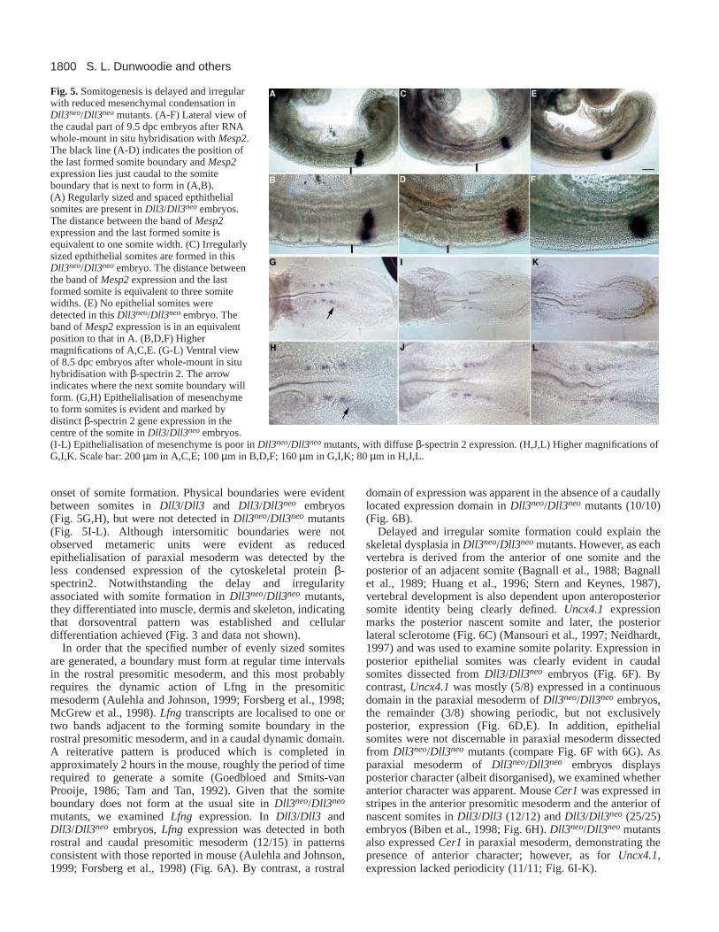
onset of somite formation. Physical boundaries were evidentbetween somites in Dll3/Dll3 and Dll3/Dll3neo embryos(Fig. 5G,H), but were not detected in Dll3neo/Dll3neo mutants(Fig. 5I-L). Although intersomitic boundaries were notobserved metameric units were evident as reducedepithelialisation of paraxial mesoderm was detected by theless condensed expression of the cytoskeletal protein β-spectrin2. Notwithstanding the delay and irregularityassociated with somite formation in Dll3neo/Dll3neo mutants,they differentiated into muscle, dermis and skeleton, indicatingthat dorsoventral pattern was established and cellulardifferentiation achieved (Fig. 3 and data not shown).
In order that the specified number of evenly sized somitesare generated, a boundary must form at regular time intervalsin the rostral presomitic mesoderm, and this most probablyrequires the dynamic action of Lfng in the presomiticmesoderm (Aulehla and Johnson, 1999; Forsberg et al., 1998;McGrew et al., 1998). Lfng transcripts are localised to one ortwo bands adjacent to the forming somite boundary in therostral presomitic mesoderm, and in a caudal dynamic domain.A reiterative pattern is produced which is completed inapproximately 2 hours in the mouse, roughly the period of timerequired to generate a somite (Goedbloed and Smits-vanProoije, 1986; Tam and Tan, 1992). Given that the somiteboundary does not form at the usual site in Dll3neo/Dll3neo
mutants, we examined Lfngexpression. In Dll3/Dll3 andDll3/Dll3neo embryos, Lfng expression was detected in bothrostral and caudal presomitic mesoderm (12/15) in patternsconsistent with those reported in mouse (Aulehla and Johnson,1999; Forsberg et al., 1998) (Fig. 6A). By contrast, a rostral
domain of expression was apparent in the absence of a caudallylocated expression domain in Dll3neo/Dll3neo mutants (10/10)(Fig. 6B).
Delayed and irregular somite formation could explain theskeletal dysplasia in Dll3neo/Dll3neomutants. However, as eachvertebra is derived from the anterior of one somite and theposterior of an adjacent somite (Bagnall et al., 1988; Bagnallet al., 1989; Huang et al., 1996; Stern and Keynes, 1987),vertebral development is also dependent upon anteroposteriorsomite identity being clearly defined. Uncx4.1expressionmarks the posterior nascent somite and later, the posteriorlateral sclerotome (Fig. 6C) (Mansouri et al., 1997; Neidhardt,1997) and was used to examine somite polarity. Expression inposterior epithelial somites was clearly evident in caudalsomites dissected from Dll3/Dll3neo embryos (Fig. 6F). Bycontrast, Uncx4.1 was mostly (5/8) expressed in a continuousdomain in the paraxial mesoderm of Dll3neo/Dll3neo embryos,the remainder (3/8) showing periodic, but not exclusivelyposterior, expression (Fig. 6D,E). In addition, epithelialsomites were not discernable in paraxial mesoderm dissectedfrom Dll3neo/Dll3neo mutants (compare Fig. 6F with 6G). Asparaxial mesoderm of Dll3neo/Dll3neo embryos displaysposterior character (albeit disorganised), we examined whetheranterior character was apparent. Mouse Cer1 was expressed instripes in the anterior presomitic mesoderm and the anterior ofnascent somites in Dll3/Dll3(12/12) and Dll3/Dll3neo (25/25)embryos (Biben et al., 1998; Fig. 6H). Dll3neo/Dll3neomutantsalso expressed Cer1in paraxial mesoderm, demonstrating thepresence of anterior character; however, as for Uncx4.1,expression lacked periodicity (11/11; Fig. 6I-K).
S. L. Dunwoodie and others
Fig. 5.Somitogenesis is delayed and irregularwith reduced mesenchymal condensation inDll3neo/Dll3neomutants. (A-F) Lateral view ofthe caudal part of 9.5 dpc embryos after RNAwhole-mount in situ hybridisation with Mesp2.The black line (A-D) indicates the position ofthe last formed somite boundary and Mesp2expression lies just caudal to the somiteboundary that is next to form in (A,B).(A) Regularly sized and spaced epthithelialsomites are present in Dll3/Dll3neoembryos.The distance between the band of Mesp2expression and the last formed somite isequivalent to one somite width. (C) Irregularlysized epthithelial somites are formed in thisDll3neo/Dll3neoembryo. The distance betweenthe band of Mesp2expression and the lastformed somite is equivalent to three somitewidths. (E) No epithelial somites weredetected in this Dll3neo/Dll3neoembryo. Theband of Mesp2expression is in an equivalentposition to that in A. (B,D,F) Highermagnifications of A,C,E. (G-L) Ventral viewof 8.5 dpc embryos after whole-mount in situhybridisation with β-spectrin 2. The arrowindicates where the next somite boundary willform. (G,H) Epithelialisation of mesenchymeto form somites is evident and marked bydistinct β-spectrin 2 gene expression in thecentre of the somite in Dll3/Dll3neoembryos.(I-L) Epithelialisation of mesenchyme is poor in Dll3neo/Dll3neomutants, with diffuse β-spectrin 2 expression. (H,J,L) Higher magnifications ofG,I,K. Scale bar: 200 µm in A,C,E; 100 µm in B,D,F; 160 µm in G,I,K; 80 µm in H,J,L.

1801Delta3 is required for normal somitogenesis
Defining downstream effectors of Dll3-mediatedNotch signallingAs Hes5, Hes1and Hey1have been identified as genesresponsive to Notch signalling, their expression was examinedin Dll3neo/Dll3neo embryos. Hes5 is normally expressed as aband in rostral presomitic mesoderm in the posterior half of theforming somite (de la Pompa et al., 1997; del Barco Barranteset al., 1999; Takebayashi et al., 1995). Analysis of geneexpression at 10.5 dpc revealed four distinct patterns of
expression in presomitic mesoderm of Dll3/Dll3andDll3/Dll3neo embryos. These patterns of expression indicatethat Hes5, like closely related Hes1 andHes7, is dynamicallyexpressed in the presomitic mesoderm (Fig. 7A-D) (Bessho etal., 2001; Jouve et al., 2000). By contrast, at 10.5 dpc, Hes5expression was not detected in presomitic mesoderm in eight
Fig. 6.The ‘segmentation clock’ and anteroposterior identity aredisrupted in trunk paraxial mesoderm of Dll3neo/Dll3neomutantembryos. (A-E) Lateral views. (A) At 9.5 dpc, Lfng expression isdetected in two domains in the presomitic mesoderm of thisDll3/Dll3neoembryo: rostrally, as one or two bands (the anteriormostis just caudal to the forming somite boundary); and caudallyextending to the primitive streak. (B)Lfngexpression is neverdetected caudally in the presomitc mesoderm of Dll3neo/Dll3neo
embryos and rostral expression was diffuse. Uncx4.1expressionshows clear periodicity in Dll3/Dll3neoembryos at 9.5 dpc (C).(F) Expression is clearly restricted to the posterior of epithelialsomites dissected from (C). (D,E) Uncx4.1expression is reduced inDll3neo/Dll3neomutants. Periodic expression is partially retained insome embryos (D) or lost (E). (G) Trunk paraxial mesodermdissected from D; expression appears periodic but is not restricted tothe posterior somite (note the lack of epithelial structure). Dorsalview showing Cer1expression in the tail region at 10.5 dpc ofDll3/Dll3neo(H) and Dll3neo/Dll3neoembryos (I-K). (H) Cer1isexpressed anteriorly in the presumptive somite and the nascentsomite. Arrows mark the site of the most recently formed somiteboundary and vertical bars show expression in the anterior of thenascent somite. (I-K) Cer1is expressed in a broad domain (verticalbar) as the bands of expression are missing. Scale bar: 125 µm inA,B; 300 µm in C-E; 60 µm in F,G; 120 µm (H-K).
Fig. 7.Hes5,Hes1and Hey1expression is altered in Dll3neo/Dll3neo
mutants. Gene expression determined by whole-mount RNA in situhybridisation. Dorsal view of tail region from 10.5 dpc embryos,caudal boundary of most recently formed somite marked with anarrow. (A-F) Hes5expression; (A-D) four distinct patterns of Hes5expression identified in Dll3/Dll3 and Dll3/Dll3neoembryos. A tightband of Hes5expression is marked with a horizontal line; broadercaudal domains of expression are marked with a vertical line. Hes5isalso strongly expressed in the neural tube. (E,F)Hes5is not detectedin the presomitic mesoderm of Dll3neo/Dll3neomutants, despite beingstrongly detected in the neural tube. (G-I) Three distinct patterns ofHes1expression are detected in the presomitic mesoderm ofDll3/Dll3 and Dll3/Dll3neoembryos. Vertical lines indicatebands/domains of expression. Hes1expression is also detected in thecaudal somite. (J,K) Only a single, relatively narrow band of Hes1expression is detected in the presomitic mesoderm ofDll3neo/Dll3neo
mutant, this is located rostrally. Tail regions showing Hes5(A-D) andHes1(G-I) expression are arranged in a hypothetical progression,according to the oscillatory expression of Hes1(Jouve et al., 2000).Hey1expression is clearly detected in the caudal somite in Dll3/Dll3and Dll3/Dll3neoembryos (L,M) but not Dll3neo/Dll3neomutants (N).Dynamic Hey1expression is represented in (L,M) with a broad bandof expression in the presomitic mesoderm (vertical line, L) thatcondenses (vertical line, M) in Dll3/Dll3and Dll3/Dll3neoembryos.Only a single narrow band of expression (vertical line) is detected inDll3neo/Dll3neomutants (N). Scale bar: 200 µm in A-F; 250 µm inG-K.

1802
out of nine Dll3neo/Dll3neomutant embryos (Fig. 7E,F). In oneembryo, a single faint band was detected in rostral presomiticmesoderm (data not shown). Similarly, at 9.5 dpc, Hes5expression was not detected in presomitic mesoderm in six outof six Dll3neo/Dll3neomutant embryos (data not shown). Hes1is expressed in the caudal half of nascent somites anddynamically in presomitic mesoderm (Jouve et al., 2000). Inthe presomitic mesoderm of Dll3/Dll3and Dll3/Dll3neo
embryos, dynamic expression is detected as a broad caudaldomain that appears to narrow as it moves rostrally to form atight band coincident with somite formation (Fig. 7G-I).Rostral expression is evident alone or in combination with thiscaudal domain of expression, depending upon the stage of thecycle. At 10.5 dpc Dll3/Dll3 embryos exhibited either rostralalone (3/10) or rostral and caudal domains (7/10) of Hes1expression. Similarly Hes1expression was detected as a singlerostral domain (6/11) or with rostral and caudal domains (5/11)in Dll3/Dll3neo. This pattern of expression was not evident inDll3neo/Dll3neomutant embryos because, in ten out of ten, onlya single narrow band of Hes1expression was detected in therostral presomitic mesoderm (Fig. 7J,K). In addition, no Hes1expression was detected in the somites where normally it isdetected caudally (compare Fig. 7G-I with 7J,K). Hey1 isexpressed in the caudal half of the most recently formed somiteand in a band in the rostral presomitic mesoderm whichnarrows as a somite forms (Kokubo et al., 1999; Leimeister etal., 1999). Hey1 expression in wild-type (Dll3/Dll3) andheterozygous (Dll3/Dll3neo) embryos reflects this pattern ofexpression with 10/24 embryos the same as Fig. 7L and 14/24the same as Fig. 7M. By contrast, in 18 out of 18Dll3neo/Dll3neo mutant embryos, Hey1expression appearedstatic because only a single band of expression was detected inthe rostral presomitic mesoderm (Fig. 7N). In addition,expression normally present in the caudal somites was notdetectable in most mutants (compare Fig. 7L,M with 7N). Insummary, these data demonstrate that Dll3is required for thenormal expression of Hes5, Hes1 andHey1 in presomiticmesoderm.
Dll3neo and Dll3pu are equivalent alleles with respectthe generation of axial skeletal defectsPostnatal analysis of pudgy mice demonstrated that the Dll3pu
allele resulted in truncation of the body, misaligned vertebrae,rib fusions and a short tail in homozygous individuals(Gruneberg, 1961; Kusumi et al., 1998). We extended thisanalysis and observed at 14.5 dpc that defects in thepreskeleton of Dll3pu/Dll3pu were very similar to those ofDll3neo/Dll3neo embryos. However, slight differences wereevident such that the dysplasia of Dll3pu/Dll3pu embryos wasless severe and the tail was longer. Genetic analysis revealedthat the Dll3neoand Dll3pu alleles were equivalent with respectto the skeletal defects as preskeletons in Dll3neo/Dll3pu,Dll3neo/Dll3neo and Dll3pu/Dll3pu were similar (Fig. 8).
DISCUSSION
The mutant Dll3 alleles; Dll3neo, Dll3pu and DLL3-SCD are functionally equivalent with respect toskeletal dysplasiaUncertainty has surrounded the mouse Dll3pu allele because itis unclear whether this allele is null. Although a four basedeletion is predicted to generate a stop codon in exon 3 (N-terminal to the DSL that lies in exon 4) (Kusumi et al., 1998),it is possible that splicing around the deletion occurs and somefunctional Dll3 protein is produced. As we have been unableto generate anti-Dll3 antibodies this scenario has remaineduntested; however, our genetic complementation studiesindicate that this is unlikely to be the case because Dll3neoandDll3pu are equivalent alleles with respect to skeletal dysplasia(Fig. 8). We observed that Dll3neo/Dll3neo individuals areslightly more severely affected than Dll3pu/Dll3pu, but this islikely to be due to differences in mouse strain (Dll3neo/Dll3neo
embryos are 129Ola/C57BL6, whereas the Dll3pu/Dll3pu
embryos are C3H/He/C57BL6). In humans, sequence analysishas defined three SCD-associated DLL3mutations. SD1contains a five base insertion, SD2 a two base deletion and SD3a missense mutation in EGF repeat number 5 (Bulman et al.,2000). The effect of the missense mutation on protein functionis unknown; however, SD1 and SD2 mutations generatetruncated proteins that are not membrane tethered but couldinteract with Notch because the DSL (Notch-binding region)is present either in full (SD2) or in part (SD1). This raises thepossibility that these mutants do not represent null alleles,
S. L. Dunwoodie and others
Fig. 8. Dll3neoand Dll3pu areequivalent alleles with respect toskeletal dysplasia. Lateral views ofAlcian Blue-stained 14.5 dpcembryos. (A)Dll3neo/Dll3neo,(B) Dll3neo/Dll3pu,(C) Dll3pu/Dll3pu. The skeletalprimordia appear to be in greaterdisarray and there are fewercoccygeal vertebrae in A than inC. Scale bar: 1.25 mm.

1803Delta3 is required for normal somitogenesis
because soluble DLL3 forms could interact with Notch andeither activate the receptor without being tethered to aneighbouring cell or prevent another ligand from bindingNotch. However, as the Dll3neo null mutation has very similarphenotypic effects on the development of the axial skeleton,we suggest that each of the human SCD alleles are likely torepresent null mutations.
The developmental origin of skeletal defectsassociated with SCD lie in the disruption of thesegmentation clock within the presomitic mesodermGeneration of the Dll3neo mutant mouse lines has allowed usto examine the developmental origins of the skeletal defectspresented in SCD. The core SCD phenotype is characterisedby multiple hemi-vertebrae with rib fusions and deletions. Thedevelopmental origins of this phenotype reside in aberrantsomite formation – a defect that appears grounded in the lossof the oscillatory mechanism that drives the regular periodicitywith which somites are formed. The molecular analysis ofDll3neo/Dll3neoembryos identifies genes associated with Notchsignalling, whose normal expression in the presomiticmesoderm is dependent upon Dll3 function. These includeLfng, Hes5,Hes1and Hey1, and therefore these are candidategenes responsible for cases of SCD that show no link toDLL3/19q13.
Are Dll1 and Dll3 distinct ligands of Notch inparaxial mesoderm?Both Dll1 and Dll3 are required for normal somite formationand correct specification of anteroposterior polarity within thepresomitic mesoderm (Gruneberg, 1961; Hrabe de Angelis etal., 1997; Kusumi et al., 1998) (Figs 5, 6). As Dll1 and Dll3are both ligands of Notch, what evidence is there that they aredistinct ligands that elicit different downstream responses?This study shows that markers of anterior (Cer1) and posterior(Uncx4.1) somite identity are expressed at normal levels in theabsence of Dll3, but that the periodic expression of Uncx4.1and Cer1, which is characteristic of anteroposterior polarity,is lost (Fig. 6 and summarised in Fig. 9). By contrast,anteroposterior identity is lost in Dll1mutants, as Uncx4.1isnot detected, while Cer1(and EphA4another marker ofanterior) are severely downregulated (del Barco Barrantes etal., 1999) (Fig. 9). In addition, we present evidence to suggestthat Dll1 and Dll3 elicit distinct responses from genesassociated with Notch signalling. For example, a loss-of-function mutation in Dll1results in severely downregulated(and largely undetected) expression of Lfng,Hes5,Hes1,Hey1,Mesp1 and Mesp2 in presomitic mesoderm (del BarcoBarrantes et al., 1999; Jouve et al., 2000; Kokubo et al., 1999)(Fig. 9). By contrast, with the exception of Hes5, theexpression of each of these genes is readily detected inpresomitic mesoderm of Dll3null mutants (Figs 5-7, 9; Mesp1was not examined). That Dll1 and Dll3 may be distinct isfurther supported by the fact that Dll3 is a highly divergentDelta homologue (Dunwoodie et al., 1997) and has only 18%identity to the Notch binding DSL of Dll1, compared with the51% identity between Dll4 and Dll1. It is, however, possiblethat when Dll1and Dll3 mutants are compared that some ofthe observed differences in gene expression do not indicatediscrete functions for these ligands but rather reflect thepossibility that Dll1and Dll3 perform the same function and
affect the expression of specific genes to different extents. AsDll1 and Dll3 are differentially expressed in presumptive andnascent somites, this issue could best be addressed by placingthe Dll1 cDNA under the regulatory control of Dll3 or viceversa using a cDNA ‘knock-in’ approach.
Oscillatory gene expression in the presomiticmesodermGenes expressed in an oscillatory manner in the presomiticmesoderm are likely to hold the key to our understanding ofhow exactly Notch signalling controls somitogenesis. We showfor the first time that Hes5exhibits a number of distinctpatterns of expression in the presomitic mesoderm. Thissuggests that Hes5, like Hes1 and Hes7, is expressed under thecontrol of oscillatory stimuli. The regulatory parameters thatcontrol the dynamic expression of these ‘clock’ genes inpresomitic mesoderm are unknown. We present data to indicatethat the rostral and caudal expression components of thesegenes are differentially controlled by Dll3 and Dll1.Expression of Lfngis severely downregulated (del BarcoBarrantes et al., 1999) and Hes1is not detected (Jouve et al.,2000) in the presomitic mesoderm of Dll1 null mutants, but inDll3neonull mutants, only the caudal expression component islost with the rostral band clearly evident (Figs 6, 7, 9). Thesedifferences could simply be due to different levels of geneexpression; however, this does not appear to be the casebecause the rostral and caudal expression domains of Hes1in
Fig. 9.A comparison of gene expression in paraxial mesoderm ofnormal and Delta mutants. Black and grey areas representlocalisation of transcript and the wavy line represents dynamic geneexpression. The most recently formed somite is SI, the formingsomite is S0, and the block of presomitic mesoderm cells of onesomite length (caudal to S0) is S-I according to (Dale and Pourquie,2000; Ordahl, 1993). Gene expression was determined by RNA insitu hybridisation. This study examined expression in wild-type andDll3 mutant embryos. Wild-type expression patterns were inaccordance with those previously reported: Mesp2 (Saga et al.,1996), Cer1(Biben et al., 1998),Uncx4.1 (Mansouri et al., 1997;Neidhardt, 1997),Lfng (Forsberg et al., 1998; Johnston et al., 1997),Hes1 (Jouve et al., 2000), Dll1 (Bettenhausen et al., 1995;Dunwoodie et al., 1997) and Dll3 (Dunwoodie et al., 1997). ForHes5, we identified four distinct patterns of expression in presomiticmesoderm (Fig. 7). Gene expression in Dll1 mutants is based onprevious reports (del Barco Barrantes et al., 1999; Jouve et al., 2000).The levels of expression of Dll1in Dll3 mutant embryos was lowwith diffuse boundaries and is indicated by grey shading.

1804
the presomitic mesoderm normally occur at equal levels and sothe loss of just the caudal domain in Dll3 mutants cannot beexplained by an overall reduction in expression levels. In thecase of Lfng, even though the rostral domain is normallyexpressed at levels higher than that seen caudally, rostralexpression in Dll3mutants was readily detectable and nocaudal expression was ever observed, even under extensiveperiods of staining. This suggests that the rostral and caudalcomponents that drive expression of oscillatory genes such asHes1and Lfngin the presomitic mesoderm are independentlycontrolled and that both components require Dll1, but only thecaudal component requires Dll3.
A comparison of Dll3 and deltaD mutantsThe mutant phenotype of Dll3 resembles that of deltaD (aftereight) in zebrafish at a number of levels. First, in both mutantssomite formation occurs in the first instance with what appearsto be the correct periodicity. This is followed by delayed somiteformation in Dll3 mutants, and lack of somite formation indeltaD mutants. However, even though metamerism wasapparent in Dll3mutants, borders between somites were notevident and condensation of paraxial mesoderm into somiteswas reduced compared with wild type (Fig. 5). Thatsomitogenesis is not completely normal is supported by the factthat the vertebra caudal to and including cervical 1 (which iscomprised of the anterior part of the fifth formed somite) wasnot properly formed. Second, marker gene expression indicatesthat paraxial mesoderm in Dll3and deltaDmutants has bothanterior (Dll3 – Cer1andMesp2;deltaD– mesp-a,EphA4,fgf8anddeltaD) and posterior (Dll3– Uncx4.1andCited1;deltaD– ephrin-B2 and MyoD) character (Fig. 6, data not shown)(Durbin et al., 2000). Third, although paraxial mesoderm hasanterior and posterior identity in both mutants, like cells arenot grouped and spaced periodically (Fig. 6) (Durbin et al.,2000). Finally, both mutants show disrupted expression ofgenes expressed in a cyclical manner in the presomiticmesoderm. In Dll3mutants, Lfng,Hes1and Hes5expressionis disrupted, while in deltaD,her1expression is disrupted. Asmutant expression of Lfng, Hes1and her1 consists of whatappears to be a static band in the rostral presomitic mesodermin the absence of caudal expression, there is potentially acommon mechanism that is responsible for the oscillatory geneexpression in presomitic mesoderm (Fig. 7) (Holley et al.,2000).
We thank A. Stewart and E. Grigorieva for excellent technicalassistance. Clones were provided by Y. Saga (Mesp2), R. Johnston(Lunatic Fringe), B. Hermann (Uncx4.1), R. Harvey (Cer-1), M.Gessler (Hey1) and C. Lobe (Hes1, Hes5). This work was funded byNHMRC project grant #142006 (SLD), the National ScienceFoundation and the March of Dimes (RAC) and Core MRCprogramme support (RSPB).
REFERENCES
Akazawa, C., Sasai, Y., Nakanishi, S. and Kageyama, R.(1992). Molecularcharacterization of a rat negative regulator with a basic helix-loop-helixstructure predominantly expressed in the developing nervous system.J. Biol.Chem. 267, 21879-21885.
Artavanis-Tsakonas, S., Rand, M. D. and Lake, R. J.(1999). Notchsignaling: cell fate control and signal integration in development.Science284, 770-776.
Aulehla, A. and Johnson, R. L.(1999). Dynamic expression of lunatic fringesuggests a link between notch signaling and an autonomous cellularoscillator driving somite segmentation.Dev. Biol. 207, 49-61.
Bagnall, K. M., Higgins, S. J. and Sanders, E. J.(1988). The contributionmade by a single somite to the vertebral column: experimental evidence insupport of resegmentation using the chick- quail chimaera model.Development103, 69-85.
Bagnall, K. M., Higgins, S. J. and Sanders, E. J.(1989). The contributionmade by cells from a single somite to tissues within a body segment andassessment of their integration with similar cells from adjacent segments.Development107, 931-943.
Bailey, A. M. and Posakony, J. W.(1995). Suppressor of hairless directlyactivates transcription of enhancer of split complex genes in response toNotch receptor activity.Genes Dev. 9, 2609-2622.
Bessho, Y., Miyoshi, G., Sakata, R. and Kageyama, R.(2001). Hes7: abHLH-type repressor gene regulated by Notch and expressed in thepresomitic mesoderm.Genes Cells6, 175-185.
Bettenhausen, B., Hrabe de Angelis, M., Simon, D., Guenet, J. L. andGossler, A. (1995). Transient and restricted expression during mouseembryogenesis of Dll1, a murine gene closely related to Drosophila Delta.Development121, 2407-2418.
Biben, C., Stanley, E., Fabri, L., Kotecha, S., Rhinn, M., Drinkwater, C.,Lah, M., Wang, C. C., Nash, A., Hilton, D. et al. (1998). Murine cerberushomologue mCer-1: a candidate anterior patterning molecule.Dev. Biol.194, 135-151.
Bray, S. (1998). Notch signalling in Drosophila: three ways to use a pathway.Semin. Cell Dev. Biol. 9, 591-597.
Bronner Fraser, M. (1986). Analysis of the early stages of trunk neural crestmigration in avian embryos using monoclonal antibody HNK-1.Dev. Biol.155, 44-55.
Bulman, M. P., Kusumi, K., Frayling, T. M., McKeown, C., Garrett, C.,Lander, E. S., Krumlauf, R., Hattersley, A. T., Ellard, S. and Turnpenny,P. D. (2000). Mutations in the human delta homologue, DLL3, cause axialskeletal defects in spondylocostal dysostosis.Nat. Genet. 24, 438-441.
Conlon, R. A., Reaume, A. G. and Rossant, J.(1995). Notch1 is requiredfor the coordinate segmentation of somites.Development121, 1533-1545.
Cooke, J. and Zeeman, E. C.(1976). A clock and wavefront model for controlof the number of repeated structures during animal morphogenesis.J. Theor.Biol. 58, 455-476.
Cooke, J. (1998). A gene that resuscitates a theory–somitogenesis and amolecular oscillator.Trends Genet. 14, 85-88.
Dale, K. J. and Pourquie, O.(2000). A clock-work somite.BioEssays22, 72-83.
de Celis, J. F. and Bray, S.(1997). Feed-back mechanisms affecting Notchactivation at the dorsoventral boundary in the Drosophila wing.Development124, 3241-3251.
de la Pompa, J. L., Wakeham, A., Correia, K. M., Samper, E., Brown, S.,Aguilera, R. J., Nakano, T., Honjo, T., Mak, T. W., Rossant, J. et al.(1997). Conservation of the Notch signalling pathway in mammalianneurogenesis.Development124, 1139-1148.
del Barco Barrantes, I., Elia, A. J., Wunsch, K., De Angelis, M. H., Mak,T. W., Rossant, J., Conlon, R. A., Gossler, A. and de la Pompa, J. L.(1999). Interaction between Notch signalling and Lunatic fringe duringsomite boundary formation in the mouse.Curr. Biol. 9, 470-480.
Dunwoodie, S. L., Henrique, D., Harrison, S. M. and Beddington, R. S.(1997). Mouse Dll3: a novel divergent Delta gene which may complementthe function of other Delta homologues during early pattern formation inthe mouse embryo.Development124, 3065-3076.
Durbin, L., Sordino, P., Barrios, A., Gering, M., Thisse, C., Thisse, B.,Brennan, C., Green, A., Wilson, S. and Holder, N. (2000).Anteroposterior patterning is required within segments for somite boundaryformation in developing zebrafish.Development127, 1703-1713.
Egan, S. E., St-Pierre, B. and Leow, C. C.(1998). Notch receptors, partnersand regulators: from conserved domains to powerful functions.Curr. Top.Microbiol. Immunol. 228, 273-324.
Evrard, Y. A., Lun, Y., Aulehla, A., Gan, L. and Johnson, R. L.(1998).lunatic fringe is an essential mediator of somite segmentation and patterning.Nature394, 377-381.
Fleming, R. J., Scottgale, T. N., Diederich, R. J. and Artavanis-Tsakonas,S. (1990). The gene Serrate encodes a putative EGF-like transmembraneprotein essential for proper ectodermal development in Drosophilamelanogaster.Genes Dev. 4, 2188-2201.
Fleming, R. J., Gu, Y. and Hukriede, N. A. (1997). Serrate-mediatedactivation of Notch is specifically blocked by the product of the gene fringe
S. L. Dunwoodie and others

1805Delta3 is required for normal somitogenesis
in the dorsal compartment of the Drosophila wing imaginal disc.Development124, 2973-2981.
Forsberg, H., Crozet, F. and Brown, N. A.(1998). Waves of mouse Lunaticfringe expression, in four-hour cycles at two-hour intervals, precede somiteboundary formation.Curr. Biol. 8, 1027-1030.
Furukawa, T., Maruyama, S., Kawaichi, M. and Honjo, T. (1992). TheDrosophila homolog of the immunoglobulin recombination signal- bindingprotein regulates peripheral nervous system development.Cell 69, 1191-1197.
Goedbloed, J. F. and Smits-van Prooije, A. E.(1986). Quantitative analysisof the temporal pattern of somite formation in the mouse and rat. A simpleand accurate method for age determination.Acta Anat. 125, 76-82.
Greenwald, I. (1998). LIN-12/Notch signaling: lessons from worms and flies.Genes Dev. 12, 1751-1762.
Gruneberg, H. (1961). Genetical studies on the skeleton of the mouse XXIXPudgy.Genetic Res. 2, 384-393.
Harrison, S. M., Dunwoodie, S. L., Arkell, R. M., Lehrach, H. andBeddington, R. S.(1995). Isolation of novel tissue-specific genes fromcDNA libraries representing the individual tissue constituents of thegastrulating mouse embryo.Development121, 2479-2489.
Heitzler, P. and Simpson, P.(1991). The choice of cell fate in the epidermisof Drosophila.Cell 64, 1083-1092.
Hogan, B., Beddington, R., Costantini, F. and Lacy, E.(1994).Manipulating the Mouse Embryo. A Laboratory Manual. New York: ColdSpring Harbor Laboratory Press.
Holley, S. A., Geisler, R. and Nusslein-Volhard, C.(2000). Control of her1expression during zebrafish somitogenesis by a delta- dependent oscillatorand an independent wave-front activity.Genes Dev. 14, 1678-1690.
Hrabe de Angelis, M., McIntyre, J., 2nd and Gossler, A.(1997).Maintenance of somite borders in mice requires the Delta homologue DII1.Nature386, 717-721.
Huang, R., Zhi, Q., Neubuser, A., Muller, T. S., Brand-Saberi, B., Christ,B. and Wilting, J. (1996). Function of somite and somitocoele cells in theformation of the vertebral motion segment in avian embryos.Acta Anat. 155,231-241.
Huppert, S. S., Jacobsen, T. L. and Muskavitch, M. A.(1997). Feedbackregulation is central to Delta-Notch signalling required for Drosophila wingvein morphogenesis.Development124, 3283-3291.
Jegalian, B. G. and De Robertis, E. M.(1992). Homeotic transformations inthe mouse induced by overexpression of a human Hox3.3 transgene.Cell71, 901-910.
Jiang, Y.-J., Aerne, B. L., Smithers, L., Haddon, C., Ish-Horowicz, D. andLewis, J. (2000). Notch signalling and the synchronization of the somitesegmentation clock.Nature408, 475-479.
Johnston, S. H., Rauskolb, C., Wilson, R., Prabhakaran, B., Irvine, K. D.and Vogt, T. F. (1997). A family of mammalian Fringe genes implicated inboundary determination and the Notch pathway.Development124, 2245-2254.
Jouve, C., Palmeirim, I., Henrique, D., Beckers, J., Gossler, A., Ish-Horowicz, D. and Pourquie, O.(2000). Notch signalling is required forcyclic expression of the hairy-like gene HES1 in the presomitic mesoderm.Development127, 1421-1429.
Kaufman, M. H. (1992). The Atlas of Mouse Development. London:Academic Press.
Kimble, J. and Simpson, P.(1997). The LIN-12/Notch signaling pathway andits regulation.Annu. Rev. Cell Dev. Biol. 13, 333-361.
Klein, T. and Arias, A. M. (1998). Interactions among Delta, Serrate andFringe modulate Notch activity during Drosophila wing development.Development125, 2951-2962.
Kokubo, H., Lun, Y. and Johnson, R. L. (1999). Identification andexpression of a novel family of bHLH cDNAs related to Drosophila hairyand enhancer of split.Biochem. Biophys. Res. Commun. 260, 459-465.
Krantz, I. D., Piccoli, D. A. and Spinner, N. B.(1997). Alagille syndrome.J. Med. Genet. 34, 152-157.
Kusumi, K., Sun, E. S., Kerrebrock, A. W., Bronson, R. T., Chi, D. C.,Bulotsky, M. S., Spencer, J. B., Birren, B. W., Frankel, W. N. andLander, E. S.(1998). The mouse pudgy mutation disrupts Delta homologueDll3 and initiation of early somite boundaries.Nat. Genet. 19, 274-278.
Lardelli, M., Dahlstrand, J. and Lendahl, U. (1994). The novel Notchhomologue mouse Notch 3 lacks specific epidermal growth factor-repeatsand is expressed in proliferating neuroepithelium.Mech. Dev. 46, 123-136.
Larsen, W. J. (1997). Development of the peripheral nervous system. InHuman Embryology. pp. 107-125. New York: Churchill Livingstone.
Lecourtois, M. and Schweisguth, F.(1995). The neurogenic suppressor of
hairless DNA-binding protein mediates the transcriptional activation of theenhancer of split complex genes triggered by Notch signaling.Genes Dev.9, 2598-2608.
Leimeister, C., Externbrink, A., Klamt, B. and Gessler, M. (1999). Heygenes: a novel subfamily of hairy- and Enhancer of split related genesspecifically expressed during mouse embryogenesis.Mech. Dev. 85, 173-177.
Leimeister, C., Dale, K., Fischer, A., Klamt, B., Hrabe de Angelis, M.,Radtke, F., McGrew, M. J., Pourquie, O. and Gessler, M.(2000).Oscillating expression of c-hey2 in the presomitic mesoderm suggests thatthe segmentation clock may use combinatorial signaling through multipleinteracting bHLH factors.Dev. Biol. 227, 91-103.
Lindsell, C. E., Shawber, C. J., Boulter, J. and Weinmaster, G.(1995).Jagged: a mammalian ligand that activates Notch1.Cell 80, 909-917.
Mansouri, A., Yokota, Y., Wehr, R., Copeland, N. G., Jenkins, N. A. andGruss, P. (1997). Paired-related murine homeobox gene expressed in thedeveloping sclerotome, kidney, and nervous system.Dev. Dyn. 210, 53-65.
Mark, M., Lufkin, T., Vonesch, J. L., Ruberte, E., Olivo, J. C., Dolle, P.,Gorry, P., Lumsden, A. and Chambon, P.(1993). Two rhombomeres arealtered in Hoxa-1 mutant mice.Development119, 319-338.
McGrew, M. J., Dale, J. K., Fraboulet, S. and Pourquie, O.(1998). Thelunatic fringe gene is a target of the molecular clock linked to somitesegmentation in avian embryos.Curr. Biol. 8, 979-982.
Mitsiadis, T. A., Henrique, D., Thesleff, I. and Lendahl, U.(1997). MouseSerrate-1 (Jagged-1): expression in the developing tooth is regulated byepithelial-mesenchymal interactions and fibroblast growth factor-4.Development124, 1473-1483.
Nakagawa, O., Nakagawa, M., Richardson, J. A., Olson, E. N. andSrivastava, D.(1999). HRT1, HRT2, and HRT3: a new subclass of bHLHtranscription factors marking specific cardiac, somitic, and pharyngeal archsegments.Dev. Biol. 216, 72-84.
Neidhardt, L. M., Kispert, A. and Herrmann, B. G. (1997). A mouse geneof the paired-related homeobox class expressed in the caudal somitecompartment and in the developing vertebral column, kidney and nervoussystem.Dev. Genes Evol. 207, 330-339.
Ohtsuka, T., Ishibashi, M., Gradwohl, G., Nakanishi, S., Guillemot, F. andKageyama, R.(1999). Hes1 and Hes5 as notch effectors in mammalianneuronal differentiation.EMBO J. 18, 2196-2207.
Oka, C., Nakano, T., Wakeham, A., de la Pompa, J. L., Mori, C., Sakai,T., Okazaki, S., Kawaichi, M., Shiota, K., Mak, T. W. and Honjo, T.(1995). Disruption of the mouse RBP-J kappa gene results in earlyembryonic death.Development121, 3291-3301.
Ordahl, C. (1993). In Myogenic Lineages within the Developing Somite(ed.M. Bernfield), pp. 165-176. New York: John Wiley and Sons.
Palmeirim, I., Henrique, D., Ish-Horowicz, D. and Pourquie, O.(1997).Avian hairy gene expression identifies a molecular clock linked to vertebratesegmentation and somitogenesis.Cell 91, 639-648.
Panin, V. M., Papayannopoulos, V., Wilson, R. and Irvine, K. D.(1997).Fringe modulates Notch-ligand interactions.Nature387, 908-912.
Pourquie, O.(2000). Vertebrate segmentation: is cycling the rule? Curr. Opin.Cell Biol. 12, 747-751.
Rickmann, M., Fawcett, J. W. and Keynes, R. J.(1985). The migration ofneural crest cells and the growth of motor axons through the rostral half ofthe chick somite.J. Embryol. Exp. Morphol. 90, 437-455.
Saga, Y., Hata, N., Kobayashi, S., Magnuson, T., Seldin, M. F. and Taketo,M. M. (1996). MesP1: a novel basic helix-loop-helix protein expressed inthe nascent mesodermal cells during mouse gastrulation.Development122,2769-2778.
Saga, Y., Hata, N., Koseki, H. and Taketo, M. M.(1997). Mesp2: a novelmouse gene expressed in the presegmented mesoderm and essential forsegmentation initiation.Genes Dev. 11, 1827-1839.
Sasai, Y., Kageyama, R., Tagawa, Y., Shigemoto, R. and Nakanishi, S.(1992). Two mammalian helix-loop-helix factors structurally related toDrosophila hairy and Enhancer of split.Genes Dev. 6, 2620-2634.
Schweisguth, F. and Posakony, J. W.(1992). Suppressor of Hairless, theDrosophila homolog of the mouse recombination signal-binding proteingene, controls sensory organ cell fates.Cell 69, 1199-1212.
Shawber, C., Boulter, J., Lindsell, C. E. and Weinmaster, G.(1996).Jagged2: a serrate-like gene expressed during rat embryogenesis.Dev. Biol.180, 370-376.
Shutter, J. R., Scully, S., Fan, W., Richards, W. G., Kitajewski, J.,Deblandre, G. A., Kintner, C. R. and Stark, K. L. (2000). Dll4, a novelNotch ligand expressed in arterial endothelium.Genes Dev. 14, 1313-1318.
Stern, C. D. and Keynes, R. J.(1987). Interactions between somite cells: the

1806
formation and maintenance of segment boundaries in the chick embryo.Development99, 261-272.
Swiatek, P. J., Lindsell, C. E., del Amo, F. F., Weinmaster, G. and Gridley,T. (1994). Notch1 is essential for postimplantation development in mice.Genes Dev. 8, 707-719.
Takebayashi, K., Akazawa, C., Nakanishi, S. and Kageyama, R.(1995).Structure and promoter analysis of the gene encoding the mouse helix- loop-helix factor HES-5. Identification of the neural precursor cell- specificpromoter element.J. Biol. Chem. 270, 1342-1349.
Tam, P. P. and Tan, S. S.(1992). The somitogenetic potential of cells in theprimitive streak and the tail bud of the organogenesis-stage mouse embryo.Development115, 703-715.
Teillet, M. A., Kalcheim, C. and Le Douarin, N. M. (1987). Formation ofthe dorsal root ganglia in the avian embryo: segmental origin and migratorybehavior of neural crest progenitor cells.Dev. Biol. 120, 329-347.
Tosney, K. W. (1978). The early migration of neural crest cells in the trunkregion of the avian embryo: An electron microscopic study.Dev. Biol. 62,317-333.
Uyttendaele, H., Marazzi, G., Wu, G., Yan, Q., Sassoon, D. and Kitajewski,J. (1996). Notch4/int-3, a mammary proto-oncogene, is an endothelial cell-specific mammalian Notch gene.Development122, 2251-2259.
Vassin, H., Bremer, K. A., Knust, E. and Campos-Ortega, J. A.(1987). Theneurogenic gene Deltaof Drosophila melanogasteris expressed in
neurogenic territories and encodes a putative transmembrane protein withEGF-like repeats.EMBO J. 6, 3431-3440.
Weinmaster, G., Roberts, V. J. and Lemke, G.(1991). A homolog ofDrosophila Notch expressed during mammalian development.Development113, 199-205.
Weinmaster, G., Roberts, V. J. and Lemke, G.(1992). Notch2: a secondmammalian Notch gene.Development116, 931-941.
Weston, J. A. (1963). An radioautographic analysis of the migration andlocalization of trunk neural crest cells in the chick.Dev. Biol. 6, 279-310.
Wharton, K. A., Johansen, K. M., Xu, T. and Artavanis-Tsakonas, S.(1985). Nucleotide sequence from the neurogenic locus notch implies a geneproduct that shares homology with proteins containing EGF-like repeats.Cell 43, 567-581.
Wong, P. C., Zheng, H., Chen, H., Becher, M. W., Sirinathsinghji, D. J.,Trumbauer, M. E., Chen, H. Y., Price, D. L., Van der Ploeg, L. H. andSisodia, S. S.(1997). Presenilin 1 is required for Notch1 and DII1expression in the paraxial mesoderm.Nature387, 288-292.
Xue, Y., Gao, X., Lindsell, C. E., Norton, C. R., Chang, B., Hicks, C.,Gendron-Maguire, M., Rand, E. B., Weinmaster, G. and Gridley, T.(1999). Embryonic lethality and vascular defects in mice lacking the Notchligand Jagged1.Hum. Mol. Genet. 8, 723-730.
Zhang, N. and Gridley, T. (1998). Defects in somite formation in lunaticfringe-deficient mice.Nature394, 374-377.
S. L. Dunwoodie and others