Development and characterisation of a decellularised ... · electrospinning PCL fibres. This...
11
SPECIAL ISSUE: ESB 2014 Biocompatibility Studies Development and characterisation of a decellularised bovine osteochondral biomaterial for cartilage repair Hazel L. Fermor 1 • Serena L. Russell 2 • Sophie Williams 2 • John Fisher 2 • Eileen Ingham 1 Received: 26 July 2014 / Accepted: 19 March 2015 / Published online: 21 April 2015 Ó The Author(s) 2015. This article is published with open access at Springerlink.com Abstract It is proposed that an acellular natural osteo- chondral scaffold will provide a successful repair material for the early intervention treatment of cartilage lesions, to prevent or slow the progression of cartilage deterioration to osteoarthritis. Here, we investigated the efficacy of meth- ods for the decellularisation of bovine osteochondral plugs. The plugs were subject to four freeze/thaw cycles followed by two cycles of washes in hypotonic solution and low concentration (0.1 % w/v) sodium dodecyl sulphate with protease inhibitors. Plugs were treated with nuclease (DNase and RNase) treatment followed by sterilization in peracetic acid. Full tissue decellularisation was achieved as confirmed by histological analysis and DNA quantification, however the resultant acellular matrix had reduced gly- cosaminoglycan content which led to an increased percent deformation of cartilage. Furthermore, the acellular scaf- fold was not reproducibly biocompatible. Additional ter- minal washes were included in the process to improve biocompatibility, however, this led to visible structural damage to the cartilage. This damage was found to be minimised by reducing the cut edge to cartilage area ratio through decellularisation of larger cuts of osteochondral tissue. 1 Introduction Osteoarthritis (OA) is the progressive degeneration of nat- ural joint tissues including articular cartilage, bone and supporting ligaments which results in pain and loss of motion for sufferers [1]. OA is the most common disorder affecting joints [2], in the UK an estimated 8.75 million people aged 45 and over sought treatment for the disease [3]. The causes of OA are multifactorial and not fully understood. One known cause of OA is the development of initial cartilage lesions, often as a result of joint trauma [4]. These lesions are unable to heal as the tissue is avascular, so progressively deteriorate over time with normal joint loading and activity [5]. Current surgical interventions to repair initial cartilage lesions, such as marrow stimulation techniques [6], au- tologous mosaicplasty [7], autologous chondrocyte implan- tation [8] and matrix-induced chondrocyte implantation [9] have been reported to have variable clinical outcomes. Many treatments do not produce a hyaline-like cartilage repair, leading to uncertain long term prognosis. Due to the limitation of current interventions, the repair of cartilage lesions using tissue engineered approaches is being explored. Synthetic biomaterials such as polycapro- lactone (PCL) [10] and polylactic acid [11] are easily manufactured with precise material properties; however achieving satisfactory biological integration is often a challenge. Biological materials such as fibrin [12] and gelatin [13] are biodegradable and biocompatible, however there remain concerns over scaffold integration. Due to the complex biological and biochemical structure of natural articular cartilage the tissue exhibits extraordi- nary biomechanical and frictional properties [14]; this is difficult to recapitulate using conventional biomaterial approaches, although advances are being made. Recently, anisotropic cartilage biomaterials have been produced by & Hazel L. Fermor [email protected] 1 Faculty of Biological Sciences, University of Leeds, Leeds, UK 2 School of Mechanical Engineering, University of Leeds, Leeds, UK 123 J Mater Sci: Mater Med (2015) 26:186 DOI 10.1007/s10856-015-5517-0
Transcript of Development and characterisation of a decellularised ... · electrospinning PCL fibres. This...

SPECIAL ISSUE: ESB 2014 Biocompatibility Studies
Development and characterisation of a decellularised bovineosteochondral biomaterial for cartilage repair
Hazel L. Fermor1 • Serena L. Russell2 • Sophie Williams2 • John Fisher2 •
Eileen Ingham1
Received: 26 July 2014 / Accepted: 19 March 2015 / Published online: 21 April 2015
� The Author(s) 2015. This article is published with open access at Springerlink.com
Abstract It is proposed that an acellular natural osteo-
chondral scaffold will provide a successful repair material
for the early intervention treatment of cartilage lesions, to
prevent or slow the progression of cartilage deterioration to
osteoarthritis. Here, we investigated the efficacy of meth-
ods for the decellularisation of bovine osteochondral plugs.
The plugs were subject to four freeze/thaw cycles followed
by two cycles of washes in hypotonic solution and low
concentration (0.1 % w/v) sodium dodecyl sulphate with
protease inhibitors. Plugs were treated with nuclease
(DNase and RNase) treatment followed by sterilization in
peracetic acid. Full tissue decellularisation was achieved as
confirmed by histological analysis and DNA quantification,
however the resultant acellular matrix had reduced gly-
cosaminoglycan content which led to an increased percent
deformation of cartilage. Furthermore, the acellular scaf-
fold was not reproducibly biocompatible. Additional ter-
minal washes were included in the process to improve
biocompatibility, however, this led to visible structural
damage to the cartilage. This damage was found to be
minimised by reducing the cut edge to cartilage area ratio
through decellularisation of larger cuts of osteochondral
tissue.
1 Introduction
Osteoarthritis (OA) is the progressive degeneration of nat-
ural joint tissues including articular cartilage, bone and
supporting ligamentswhich results in pain and loss ofmotion
for sufferers [1]. OA is the most common disorder affecting
joints [2], in theUKan estimated 8.75million people aged 45
and over sought treatment for the disease [3]. The causes of
OA are multifactorial and not fully understood. One known
cause of OA is the development of initial cartilage lesions,
often as a result of joint trauma [4]. These lesions are unable
to heal as the tissue is avascular, so progressively deteriorate
over time with normal joint loading and activity [5].
Current surgical interventions to repair initial cartilage
lesions, such as marrow stimulation techniques [6], au-
tologous mosaicplasty [7], autologous chondrocyte implan-
tation [8] and matrix-induced chondrocyte implantation [9]
have been reported to have variable clinical outcomes.Many
treatments do not produce a hyaline-like cartilage repair,
leading to uncertain long term prognosis.
Due to the limitation of current interventions, the repair
of cartilage lesions using tissue engineered approaches is
being explored. Synthetic biomaterials such as polycapro-
lactone (PCL) [10] and polylactic acid [11] are easily
manufactured with precise material properties; however
achieving satisfactory biological integration is often a
challenge. Biological materials such as fibrin [12] and
gelatin [13] are biodegradable and biocompatible, however
there remain concerns over scaffold integration.
Due to the complex biological and biochemical structure
of natural articular cartilage the tissue exhibits extraordi-
nary biomechanical and frictional properties [14]; this is
difficult to recapitulate using conventional biomaterial
approaches, although advances are being made. Recently,
anisotropic cartilage biomaterials have been produced by
& Hazel L. Fermor
1 Faculty of Biological Sciences, University of Leeds, Leeds,
UK
2 School of Mechanical Engineering, University of Leeds,
Leeds, UK
123
J Mater Sci: Mater Med (2015) 26:186
DOI 10.1007/s10856-015-5517-0
electrospinning PCL fibres. This nanofabrication tech-
nology enabled tangential alignment of fibres at the surface
and random orientation in the rest of the material, increased
fibre diameter was included in the base of the material to
mimic natural cartilage zonal microstructure showing
promising in vitro results [10].
An alternative approach is decellularisation of natural
tissues. Decellularisation of natural tissues has been shown
to produce extracellular matrix (ECM) scaffolds with the
same structure and function as the original tissues whilst
removing immunogenic cells [15, 16]. This approach has
led to the clinical translation of acellular allogeneic and
xenogeneic tissues for use in cardiovascular [17] and
connective tissue [18, 19] applications. This approach has
also been investigated to develop acellular cartilage and
osteochondral scaffolds for use in cartilage lesion repair
[20–23]. It is proposed that an acellular osteochondral
scaffold will have superior biological and biomechanical
characteristics and will show improved integration com-
pared to other tissue engineered cartilage repair materials,
due to the presence of the subchondral bone. Kheir et al.
[21] presented initial data on the decellularisation of por-
cine osteochondral tissues from 4–6 month old pigs using
low concentration sodium dodecyl sulphate (SDS) and
protease inhibitors [15, 16]. However, mature bovine tissue
may be a more appropriate source material [24]. Here, we
present data on the development of an improved method to
decellularise mature bovine osteochondral plugs (clinically
relevant in mosaicplasty-like procedures). The resultant
acellular scaffold was analysed using biological, bio-
chemical and biomechanical methods and the biocom-
patibility of the material was determined.
2 Materials and methods
2.1 Tissue preparation and decellularisation
of osteochondral plugs
Osteochondral plugs (9 mm diameter, 12 mm thickness)
were extracted from the medial patello-femoral groove of
18 month old bovine knee joints using bespoke corers and
a hand held electric drill. The osteochondral plugs were
either retained as native (n = 5 from 5 cows) or decellu-
larised using one of five iterative methods based on the
processes described by Booth et al. [15], Stapleton et al.
[16] and Kheir et al. [21].
2.1.1 Process (1)
Osteochondral plugs (n = 5 from five cows) were frozen at
-20 �C followed by thawing at 42 �C, this process was
repeated once more then a further two times with the plugs
submerged in a hypotonic solution (10 mM tris–HCl, pH
8.0; Sigma plus 10 KIU ml-1 aprotinin; Nordic Pharma).
Thawed plugs were washed for 3 9 10 min in phosphate
buffered saline (PBS; Oxoid) and washed in hypotonic so-
lution for 18 h, followed by 24 h in hypotonic solution
containing SDS (0.1 % (w/v); Sigma). Plugs were then
washed 3 9 10 min in PBS before being incubated in nu-
clease solution (50 mM Tris solution, pH 7.5, with 10 mM
magnesium chloride, 50 U ml-1 DNAase and 1 U ml-1
RNAase; Sigma). Following a further 3 9 10 min washes
in PBS plugs were sterilised using peracetic acid (PAA;
0.1 % v/v in PBS; Sigma). Finally plugs were washed
3 9 10 min in PBS and stored at -20 �C until analysed.
All incubations were carried out at 42 �C with the exception
of the nuclease treatment which was performed at 37 �Cand PAA sterilisation which was performed at 27 �C. Allwashes were performed with 125 ml solution with agitation
on an orbital shaker. The washes following PAA sterilisa-
tion were performed aseptically in a Class II safety cabinet.
2.1.2 Process (2)
This method differed from process (1) only in that there
was an extended terminal 36 h wash in PBS added to the
end of the process (n = 5 plugs from three cows).
2.1.3 Process (3)
This method was the same as process (2) with the addition
of use of a dental water flosser (Water Pik, Inc.) to
physically remove the bone marrow from the osteochon-
dral plugs with a fine jet of PBS (400 ml per plug). This
was performed following the first freeze/thaw cycle (n = 5
plugs from three cows).
2.1.4 Process (4)
This method was the same as process (3) with the addition
of an incubation of the plugs PBS at 42 �C for 18 h fol-
lowing the first freeze/thaw cycle prior to the use of the
water flosser (n = 5 plugs from three cows).
2.1.5 Process (5)
This method was the same as process (4) with an additional
24 h wash in hypotonic solution and 24 h wash in SDS in
hypotonic solution after the original SDS wash (n = 5
plugs from three cows).
2.1.6 Extended washes
PBS washes (4 9 24 h) were added at the end of process
(5) using n = 5 plugs from three cows.
186 Page 2 of 11 J Mater Sci: Mater Med (2015) 26:186
123

2.2 Histological analysis
Osteochondral plugs (n = 5 native, n = 5 decellularised
using process 1; n = 3 decellularised using processes 2–5)
were fixed for 48 h in 10 % (v/v) neutral buffered formalin
(NBF; Bios Europe Ltd), before being decalcified in
12.5 % (w/v) EDTA (pH 7: Fisher Scientific) for 4 weeks
or until soft enough to be cut with a scalpel. Plugs were
bisected before being dehydrated and paraffin wax and
embedded using an automated process (Lecia TP 1020,
Lecia Microsystems). Sections of 6 lm thickness were cut
through the cartilage surface into the bone, encompassing
the different cartilage zones and subchondral bone. Hae-
matoxylin and eosin (H&E; Bios Europe Ltd) staining was
used to assess tissue histoarchitecture. DAPI (Sigma)
staining was used to visualise cell nuclei. Safranin O and
Fast Green (Sigma) staining was used to visualise gly-
cosaminoglycan (GAG) distribution.
2.3 Biochemical analysis
2.3.1 DNA quantification
The Qiagen DNeasy blood and tissue kit was used to digest
cartilage and extract and purify tissue DNA according to
the manufacturers’ instructions (Qiagen). Cartilage (n = 5
native, n = 5 decellularised using process 1; n = 3 de-
cellularised using processes 2–5) was cut away from the
subchondral bone and digested using proteinase K (Qia-
gen). Purified DNA was quantified using a nanodrop nano
spectrophotometer (Nanodrop). DNA content is expressed
in ng mg-1 of wet weight of tissue.
2.3.2 Sulphated sugar quantification
Lyophilised cartilage (n = 5 native, n = 5 decellularised
using process 1; n = 3 decellularised using processes
2–5) was digested using papain solution (5 ml, 50 U ml-1
papain, Sigma, in PBS at pH 6 with 5 mM L-cysteine
hydrochloride, Sigma, and 5 mM EDTA) for 48 h at
60 �C. GAG content was quantified following a method
adapted from Farndale et al. [25]. Briefly, standard con-
centrations of chondroitin B sulphate (Sigma) were pro-
duced, 1,9-dimethylmethylene blue (Sigma) solution
(250 ll) was added to each standard and sample (40 ll)in a clear flat bottomed 96-well plate, the plate was
agitated for 2 min before the absorbance was read on a
spectrophotometer (Multiscan Spectrum, Thermo Lab
Systems) at 525 nm. Interpolation of sample absorbance
from the standard curve gave the GAG concentration of
each digest which was then expressed in lg mg-1 of dry
weight of tissue.
2.3.3 Collagen quantification
Lyophilised cartilage (n = 5 native, n = 5 decellularised
using process 1; n = 3 decellularised using processes
2–5) was hydrolysed in 6 M hydrochloric acid (HCl,
VWR) for 16 h at 80 �C followed by neutralisation with
6 M sodium hydroxide (NaOH: Fisher). Hydroxyproline
content was quantified following a method adapted from
Edwards & O’Brien [26]. Briefly, standard concentrations
of trans-4-hydroxy-L-proline (Sigma) were produced,
Chloramine-T oxidation solution (100 ll; Sigma) was
added to each standard and sample (50 ll) in a clear flat
bottomed 96-well plate, the plate was agitated for 5 min.
Ehrlich’s reagent (100 ll; Sigma) was then added and the
plate was incubated in a water bath at 60 �C for 45 min
before the absorbance was read at 570 nm on a spec-
trophotometer. Interpolation of sample absorbance from
the standard curve gave the hydroxyproline concentration
of each sample. This was then expressed as lg mg-1 dry
weight of tissue.
2.4 Compressive testing using an indenter
The methods used have been previously described by
Pawaskar et al. [27] and Taylor et al. [28] to assess the
biomechanical properties of cartilage attached to bone.
Briefly, osteochondral plugs (n = 5 native, n = 5 decellu-
larised using process 1; n = 3 decellularised using processes
2–5) were compressed in a purpose built indentation rig using
a 3 mmdiameter, hemispherical, stainless steel indenter under
a load of 0.8 N. Plugs were submerged in PBS during testing
to maintain cartilage hydration. The deformation of cartilage
was measured at a sampling frequency of 5 Hz over 1 h, after
which all samples had reached equilibrium. Following com-
pression, plugs were fully rehydrated in PBS before cartilage
thickness was measured. A needle indenter was used to
penetrate the cartilage, lowering at a rate of 4.5 mm min-1;
the resistance tomotionwasmeasured using a 500 N load cell
(Instron 3365). An increase in load was recorded when the
needle first contacted the cartilage surface and a second in-
creasewhen entering the bone, the distance between these two
changes in load was taken as the cartilage thickness. Defor-
mation of cartilage was normalised to thickness to give per-
centage deformation for each osteochondral plug.
2.5 Biocompatibility testing
2.5.1 Contact cytotoxicity
Babyhamster kidney cells (BHK,HealthProtection,England)
were cultured in BHK culture medium (GMEM; Sigma, 5 %
(v/v) foetal bovine serum (FBS),SeraLab, 10 %(v/v) tryptone
phosphate broth, 2 mM L-glutamine, 100 U ml-1 penicillin
J Mater Sci: Mater Med (2015) 26:186 Page 3 of 11 186
123
and 100 mg ml-1 streptomycin, Sigma) at 37 �C in 5 % (v/v)
CO2 in air. 3T3 murine fibroblasts (European Collection of
Cell Cultures) were cultured in 3T3 culturemedium (DMEM;
Sigma, 10 % (v/v) FBS, 2 mM L-glutamine, 100 U ml-1
penicillin and 100 mg ml-1 streptomycin) at 37 �C in 5 % (v/
v) CO2 in air. Decellularised osteochondral tissue samples
(n = 3, 1 mm 9 3 mm 9 5 mm)were attached to the centre
of 6-well culture plates using 3 M Steri-strips (Medisave).
Steri-strips and cyanoacrylate were applied to separate wells
in triplicate to act as negative and positive controls respec-
tively. BHK or 3T3 cells were seeded into each well at the
appropriate cell density to achieve confluence under usual
culture conditions after 48 h. Following culture with tissue
samples and controls for 48 h at 37 �C in 5 % (v/v) CO2 in air
cells were examined using phase contrast microscopy to vi-
sualise changes in cell morphology and confluence.
2.5.2 SDS quantification
In order to determine the levels of SDS remaining in the
osteochondral tissues following processing, decellularisation
(process 5) was performed using SDS in hypotonic solution
which had been spiked with 14C radiolabelled SDS
(0.001 lCi ml-1; Hartmann Analytical). Macerated decel-
lularised cartilage or bone (40 mg) were added in triplicate
to a flat bottomed 96 well OptiPlateTM (Perkin Elmer) and
MicroScintTM-20 (160 ll; Perkin Elmer) was added to each
well. Counts per minute were measured using Top countTM
(Perkin Elmer), with each well being counted for 20 min. A
standard curve of counts per minute was produced from
known concentrations of SDS, from which the concentration
of SDS in the test tissue could be interpolated.
2.5.3 SDS cytotoxicity
To determine the concentration at which SDS exhibited
toxic effects upon mammalian cells, BHK and 3T3 cells
were cultured in triplicate in 96-well culture plates for
24 h; culture medium was aspirated and replaced with
BHK or 3T3 medium containing a range of known con-
centrations of SDS. BHK or 3T3 culture medium and 40 %
(v/v) dimethyl sulphoxide (DMSO; Sigma) were used as
negative and positive controls for cell toxicity respectively.
Cells were cultured for a further 24 h at 37 �C in 5 % (v/v)
CO2 in air and the ATP content measured using the
ATPLite-M� assay kit (Perkin Elmer) according to the
manufacturers’ instructions to quantify cell viability.
2.6 Statistical analysis
Numerical data was analysed using Microsoft Excel (ver-
sion 2010, Microsoft) and is presented as the mean (n = 5
or n = 3) ±95 % confidence levels (CL). One way
analysis of variance (ANOVA) was performed and indi-
vidual differences between group means were identified by
calculating the minimum significant difference at P = 0.05
using the T-method, or T’-method when comparing groups
of unequal sample size [29].
3 Results
Initial attempts to decellularise bovine osteochondral tissues
using process (1) were not successful and this lead to nu-
merous iterations of the decellularisation process which are
documented below. Images of sections of native bovine
osteochondral tissue and tissue following the various de-
cellularisation processes stained with H&E, DAPI and
Safranin O are presented in Fig. 1. The total DNA and GAG
content of native bovine cartilage and the cartilage following
the various decellularisation processes are presented in
Figs. 2 and 3. The data for the percentage deformation of the
native bovine osteochondral tissues and tissues following the
various decellularisation processes are presented in Fig. 4.
3.1 Process (1)
Following application of decellularisation process (1) stain-
ing of treated tissue sections with H&E and DAPI revealed
that there were still a number of cells present in the cartilage
and bone (Fig. 1b, h). The general structure of the tissue was
maintained, however marrow was still present in the bone.
There was no significant reduction in the total DNA content
of the cartilage compared to native tissue (Fig. 2). The GAG
content (222.1 ± 77.1 lg mg-1) of cartilage following de-
cellularisation process (1) was not significantly different
from native tissue (261.1 ± 33.3 lg mg-1; Fig. 3); however
a reduction was observed in sections of the superficial and
middle zones compared to native cartilage when stained with
Safranin O (Fig. 1n). The percentage deformation
(37.5 ? 12.0/-11.2 %) of cartilage decellularised using
process (1) was not significantly different from native tissues
(30.4 ? 7.6/-7.1 %; Fig. 4).
3.2 Process (2)
Following the introduction of an extended PBS wash at the
end of decellularisation Process (1), sections of osteochon-
dral plugs decellularised using Process (2) showed a much
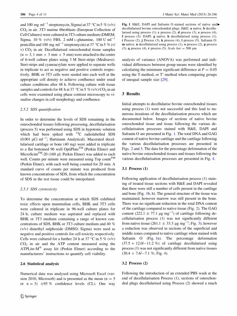
cFig. 1 H&E, DAPI and Safranin O stained sections of native and
decellularised bovine osteochondral plugs. H&E: a native, b decellu-
larised using process (1), c process (2), d process (3), e process (4),
f process (5). DAPI: g native, h decellularised using process (1),
i Process (2), j Process (3), k process (4), l process (5). Safranin O:
m native, n decellularised using process (1), o process (2), p process
(3), q process (4), r process (5). Scale bar = 500 lm
186 Page 4 of 11 J Mater Sci: Mater Med (2015) 26:186
123

J Mater Sci: Mater Med (2015) 26:186 Page 5 of 11 186
123

greater reduction in cell content. Nucleated cells were,
however, still visible in the deep/calcified cartilage regions
and in the subchondral bone (Fig. 1c, i). Total DNA content
(16.2 ± 3.1 ng mg-1) showed a significant reduction com-
pared to native bovine tissue (119.0 ± 35.9 ng mg-1;
Fig. 2). There was no significant difference in the GAG
content of the treated cartilage (193.4 ± 79.9) compared to
native cartilage (Fig. 3), however histologically a loss of
GAGs throughout the cartilage tissue was seen (Fig. 1o). No
significant difference was observed in the percentage de-
formation (32.2 ? 15.2/-13.5 %) of the cartilage compared
to native tissue (Fig. 4).
3.3 Process (3)
To increase decellularisation solution access to the bone
and deep cartilage layers a water flosser was used to
remove the bone marrow from within the trabecular spaces.
Visibly, little marrow was removed following application
of the water jet. Histologically, no damage to the cartilage
or bone was seen, however cells were still visible in the
tissue within denser areas of bone (Fig. 1d, j). The total
DNA content of cartilage (2.2 ± 7.4 ng mg-1) was sig-
nificantly reduced compared to native cartilage (Fig. 2).
There was no significant difference in the quantified GAG
content of the treated cartilage compared to native cartilage
(Fig. 3) or tissue biomechanics (Fig. 4), however histo-
logical analysis showed reduced GAG content throughout
the tissue compared to native cartilage (Fig. 1p).
3.4 Process (4)
The bovine osteochondral plugs were agitated in PBS for
18 h at 42 �C before treatment with the water flosser; this
loosened the bone marrow and greatly improved ease and
extent of bone marrow removal. A few cell nuclei however
remained in the tissue at the cartilage-bone interface fol-
lowing treatment using process (4) Fig. 1e, k). The amount
of DNA remaining in the cartilage (11.0 ± 3.9 ng mg-1)
was again significantly reduced (Fig. 2). There was, how-
ever, a significant reduction in cartilage GAG content
(141.0 ± 100.6 lg mg-1) compared to native tissue
(Fig. 3) which was also seen histologically (Fig. 1q),
although this did not have any effect on the percentage
deformation (26.8 ± 0.3 %) of the tissue (Fig. 4).
3.5 Process (5)
Application of process (5) to the bovine osteochondral
plugs showed that two cycles of treatment with hypotonic
solution and SDS were required to successfully remove all
histological evidence of cell nuclei from the osteochondral
Fig. 2 Total DNA content of native and decellularised cartilage.
Data is shown as the mean (native and decellularised by process (1)
n = 5, others n = 3) ±95 % confidence limits. * indicates significant
difference compared to native tissue P\ 0.05 (ANOVA)
Fig. 3 GAG content of native and decellularised bovine cartilage.
Data is shown as the mean (native and decellularised by process (1)
n = 5, others n = 3) ±95 % confidence limits. *indicates significant
difference compared to native tissue P\ 0.05 (ANOVA)
Fig. 4 Percent deformation of native and decellularised cartilage.
Data was subject to arcsine transformation prior to calculation of the
95 % confidence limits and analysis of variance. Data is shown as the
back transformed mean (native and decellularised using process (1)
n = 5, others n = 3) ±95 % confidence limits. * indicates sig-
nificantly different compared to native tissue P\ 0.05 (ANOVA)
186 Page 6 of 11 J Mater Sci: Mater Med (2015) 26:186
123

tissues (Fig. 1f, l). DNA content per wet weight of tissue
(7.0 ± 5.9 ng mg-1) was significantly reduced compared to
native tissue (Fig. 2). The DNA content of cartilage decel-
lularised using process (5) was 39.7 ± 34.1 ng mg-1 per
cartilage dry weight. Quantification of the GAG content
however revealed that almost all of the GAGs had been
removed from the cartilage following decellularisation using
process (5) (Fig. 3) with no Safranin O staining observed in
the processed cartilage (Fig. 1r). This had an effect on the
tissue biomechanics and the percentage deformation was
increased to 55.9 % (?20.1/-21.2 %) compared to native
tissue (Fig. 4). Cartilage decellularised using process (5)
also showed a significant increase in hydroxyproline content
(146.3 ± 65.1 lg mg-1) compared to native bovine carti-
lage (72.3 ± 26.5 lg mg-1) which had not been evident for
cartilage decellularised using processes (1–4).
Since the decellurisation process (5) appeared to be suc-
cessful, it was necessary to determine whether the acellular
tissue was biocompatible with cells. Culture of tissues
(n = 3) decellularised using process (5) showed that all three
samples of decellularised cartilage were compatible with
BHK and 3T3 cells. Samples of decellularised bone (n = 3)
were also compatible with 3T3 cells, however 1/3 samples of
decellularised bone was cytotoxic to BHK cells (Fig. 5), with
a zone of inhibited cell growth observable around the tissue.
Hence, the concentration of residual SDS in decellularised
tissue was determined and this was found to be 364 ± 43
ng mg-1 per cartilagewetweight and 374 ± 187 ng mg-1 in
bone. It was therefore relevant to determine the toxicity of
varying levels of SDS to the two cell types. Culture of BHK
cells with SDS showed a significant decrease in cell viability
at SDS concentrations of 50 lg ml-1 and above whereas 3T3
cells only showed significantly reduced viability in the pres-
ence of SDS concentrations above 250 lg ml-1.
3.6 Extended washes
To improve biocompatibility by removing residual SDS a
further 4 days of PBS washes were included at the end of
decellularisation process (5). The addition of the extra
wash step had a detrimental effect on the cartilage ECM,
which became visibly damaged between the 2 and 3 day
time point (Fig. 6), shrinking away from the bone or be-
coming completely detached. The remaining cartilage was
contracted and softened having a gelatinous appearance
and a roughened, dull surface.
4 Discussion
The aims of this study were to investigate the utility of
tissue decellularisation processes for the generation of
acellular bovine osteochondral plugs of potential use in
osteochondral lesion repair. The objectives were to develop
a process which removed whole cells and nuclear material
from the ECM scaffold to eliminate any adverse im-
munogenic effects of xenogeneic cellular antigens. Since
the major structural macromolecules of the ECM, such as
collagens, are relatively conserved throughout higher
mammalian species, these matrix proteins should not evoke
an immune response [30].
A protocol for decellularisation was developed, based on
previous work by Booth et al. [19] and developments of the
original protocol for porcine meniscus and osteochondral
tissues [16, 21] which had shown that increased incubation
temperatures were applied to overcome issues of the dense
matrix and high GAG concentrations of cartilaginous tis-
sues to improve the diffusion of decellularisation solutions.
In a further change to the original process, EDTA which
was used in the decellularisation process for soft tissues
[19] as a metalloproteinase inhibitor was removed from the
process to avoid decalcification of the bony component of
the osteochondral plugs.
In the final iteration of the decellularisation process
described here, histological analysis of bovine osteo-
chondral plugs subject to process (5) showed removal of
whole cells and large cellular debris, and the total DNA
content of the cartilage was significantly reduced to
39 ng mg-1 per cartilage dry weight, below the recom-
mended levels of 50 ng mg-1 double stranded DNA for
tissue acellularity as established by Crapo et al. [31]. To
achieve this level of decellularisation of the mature bovine
osteochondral plugs, the various iterations of the process
(processes (1–4)) showed that it was necessary to initially
loosen and remove the bone marrow prior to subjecting
the tissue to decellularisation solutions. This was achieved
by incubation in PBS at 42 �C with agitation for 18 h and
then use of a water flosser. It is therorised that removal of
the bone marrow enabled improved diffusion of decellu-
larisation solutions through the bone and up to the sub-
chondral plate. Four cycles of freeze/thaw were required
(two in hypotonic solution) to open up the ECM through
formation of ice crystals to improve diffusion of solutions
through the tissue. Two cycles of hypotonic buffer and
low concentration SDS washes in series were required to
remove the cells from the calcified cartilage and the
subchondral bone plate, as these areas were most dense.
This number of SDS cycles was fewer than required in the
protocol developed by Khier et al. [21] for porcine os-
teochondral tissues which may have been due to the fact
that the process used by Kheir et al. did not address re-
moval of bone marrow or the smaller volume of tissue
being decellularised, or indeed both. Extensive washing in
PBS following nuclease treatment and PAA sterilisation
was required to remove nucleic acids from the tissue
(process 2 compared to process 3).
J Mater Sci: Mater Med (2015) 26:186 Page 7 of 11 186
123

186 Page 8 of 11 J Mater Sci: Mater Med (2015) 26:186
123

Although decellularisation was achieved, there were
detrimental effects on the ECM. An almost complete loss of
GAGs was observed in the cartilage decellularised using
process (5) which had an adverse effect on the mechanical
properties of the tissue resulting in a significant increase in
cartilage deformation from 30 % in native tissue to 56 % in
the decellularised tissue. Previous studies have found that
decellularisation using SDS has led to removal of GAGs from
tissue ECM [15, 16, 21]. Ionic interruption of link protein by
SDS leading to disaggregation of aggrecan and hyaluronan,
thus increased GAG mobility, has been suggested as a po-
tential mechanism of GAG loss from tissues [21].
The DNA content of the bony section of the osteo-
chondral tissue was not quantified; hence the residual DNA
content of the bone is currently unknown. However, there
was no evidence of cellular material in the bone as judged
by histology and DAPI staining revealed no evidence of
cell nuclei. Retention of some DNA fragments in the bone
was not thought to be problematic since DNA alone will
not elicit an immune response [32]. Residual DNA is
known to result in calcification of decellularised tissues
following implantation [33], however this would not pose
an issue in mineralised tissue such as bone.
For acellular scaffolds to be used in patients, it is
essential that they are not toxic or harmful. SDS, a deter-
gent included in the decellularisation process to solubilise
cellular membranes has been shown to have cytotoxic ef-
fects when not thoroughly removed from decellularised
tissues [34]. SDS is able to impart a negative charge on
proteins, which may inhibit cell attachment and prolif-
eration which may affect cell survival [35]. Quantification
of the residual SDS in the decellularised tissues clearly
showed that the majority of the SDS was removed during
wash steps, and that the decellularised bone and cartilage
contained \37.5 ng mg-1 SDS per wet weight (circa
0.0375 % (w/w)), following decellularisation using process
(5). The in vitro contact cytotoxicity tests carried out on
these decellularised tissues showed inconclusive results.
The acellular cartilage was biocompatible with both BHK
and 3T3 cells. The acellular bone was biocompatible with
3T3 cells, however 1/3 acellular bone samples showed
cytotoxicity with BHK cells. Further investigation revealed
that the 3T3 cells were less sensitive to the cytotoxic ef-
fects of SDS in culture (sensitive to 250 lg ml-1 or
0.025 % (w/v)), than BHK cells (sensitive to 50 lg ml-1 or
0.005 % (w/v)), perhaps explaining the difference in cel-
lular response to the acellular bone. It is likely that due to
binding of the SDS to the 3D structure of the tissue, not all
of the residual SDS in the tissue was available to interact
with the cells in culture, thus although the levels of SDS
remaining in both the cartilage and bone tissues were toxic
the effects on the cells in culture were minimal.
Nevertheless, since the residual levels of SDS in the
tissues was relatively high, further modifications to process
(5) were investigated which involved increasing the num-
ber and duration of PBS washes at the end of the process to
improve SDS wash out. When additional PBS washes were
included, tissue samples began to show signs of damage.
The cartilage contracted in from the bone and in some
cases became detached. Schwarz et al. [22] produced an
acellular scaffold from nasal cartilage. It was of interest to
note, that although not recognised by the authors as dam-
aged, the histology images of the decellularised cartilage
showed similar morphology to the damaged cartilage found
in the present study, they also identified a large reduction in
GAG content.
Although the overall objective of this study, which was
to develop a protocol for the successful decellularisation of
bovine osteochondral plugs, was not achieved, this study
bFig. 5 Contact cytotoxicity of decellularised bovine osteochondral
tissues when cultured for 48 h with BHK and 3T3 cells. BHK cells:
a cells only, b cyanoacrylate positive control, c steri-strip negative
control, d–f decellularised bovine bone (n = 3), g–i decellularisedbovine cartilage (n = 3). 3T3 cells: j cells only, k cyanoacrylate
positive control, l steri-strip negative control, m–o decellularised
bovine bone (n = 3), p–r decellularised bovine cartilage (n = 3)
Fig. 6 Macroscopic observation of osteochondral plugs decellu-
larised using process (5) and process (5) with extended washes. Top
panel shows smooth shiny cartilage following decellularisation with
process (5). Bottom panel shows the dull, mottled cartilage surface
(left) and the cartilage contracted in from the bone following
decellularisation with process (5) with extended PBS washes
J Mater Sci: Mater Med (2015) 26:186 Page 9 of 11 186
123

has led to important knowledge regarding the difficulties of
producing acellular osteochondral tissues. It is hypothesised
that this is due to the unique structure of the cartilage ECM.
The fibres of the collagen II network in cartilage have a
characteristic orientation [36] and are under tension, pro-
viding the tensile strength of the tissue. Proteoglycans are
dispersed throughout the collagen network and bind water
in the tissue, providing the compressive strength of the
tissue [37]. Preparing osteochondral tissues for decellu-
larisation by cutting plugs would have severed the collagen
fibres and resulted in loss of tension, particularly the fibres
orientated parallel to the surface in the cartilage superficial
zone. With the loss of tension, the collagen network would
have had a reduced ability to counter the repulsive forces of
negatively charged proteoglycans being held in close
proximity to one another, resulting in an increase in porosity
allowing proteoglycans to be washed out (GAGs removed),
water to enter the tissue and the collagen fibres to therefore
clump together in the remaining ECM.
In order to test the hypothesis that decellularisation of
cuts of bovine osteochondral tissues (with a reduced ratio of
cut edges to cartilage volume) would result in a tissue with
less damage, a preliminary study was undertaken where by
bovine osteochondral cuts of 20 mm 9 40 mm 9 12 mm
thickness (n = 3) from the femoral groove were decellu-
larised using process (5) plus 11 9 24 h extended PBS
washes. Following decellularisation the cartilage did not
detach from the bone or appear damaged macroscopically or
microscopically, with the exception of the cut edges. It was
therefore demonstrated that reducing the ratio of cut edges to
cartilage area greatly improved cartilage stability during the
decellularisation processes. However, the tissue was not
completely decellularised. Research will now focus on de-
veloping a process for complete decellularisation of bovine
osteochondral cuts, utilising the knowledge gained from this
study, with a view to harvesting acellular osteochondral pins
of clinically relevant size from the undamaged central re-
gions of these decellularised large osteochondral constructs.
Decellularisation of cartilage for therapeutic applica-
tions has been approached in a number of ways. One ap-
proach is the homogenisation of cartilage tissue following
lyophilisation and grinding [38] or shattering into frag-
ments and centrifuging to produce microfilaments [39]
prior to decellularisation. This produced a material that
provided bioactive cues for cell growth, however lacked
any 3D architecture essential for tissue biomechanical
function. Decellularisation of intact 3D cartilage tissues has
been investigated [20, 22] however both resultant scaffolds
had inferior biochemical composition and thus biome-
chanical properties. Furthermore, the lack of subchondral
bone in these cartilage scaffolds would limit the integration
of implanted grafts. The decellularisation of immature
porcine osteochondral tissues has been demonstrated [21],
however the resultant scaffold again had reduced GAG
content and therefore inferior mechanical properties. The
current study investigated the decellularisation of a mature
bovine osteochondral tissue. The method presented here is
currently the most effective known for decellularisation of
intact bovine osteochondral tissues, with no visible cell
nuclei present and a residual DNA content of\50 ng mg-1
in the decellularised cartilage matrix. This protocol in-
cluded the novel use of a water flossing technique to re-
move bone marrow, which enabled more complete
decellularisation. Fewer cycles of SDS were required to
fully remove cells compared to previously reported
methodologies [21]. However, loss of GAG and reduced
biomechanical properties was observed, as reported for
other protocols [20–23].
5 Conclusion
This study has increased knowledge and understanding of
the effects of decellularisation processes on osteochondral
tissues which will form the basis for future development of
a bioactive, acellular, natural tissue engineered repair ma-
terial for osteochondral lesions to prevent or delay the
onset of OA.
Acknowledgments This work was funded by EPSRC and partially
through WELMEC, a Centre of Excellence in Medical Engineering
funded by the Wellcome Trust and EPSRC, under Grant Number WT
088908/Z/09/Z. J Fisher is an NIHR senior investigator. Additionally
the principle investigators J Fisher and E Ingham are supported in part
by the NIHR LMBRU Leeds Musculoskeletal Biomedical Research
Unit and the ERC. E Ingham and J Fisher are academic founders of
Tissue Regenix and are shareholders and advisers to Tissue Regenix
Group PLC.
Open Access This article is distributed under the terms of the
Creative Commons Attribution 4.0 International License (http://
creativecommons.org/licenses/by/4.0/), which permits unrestricted
use, distribution, and reproduction in any medium, provided you give
appropriate credit to the original author(s) and the source, provide a
link to the Creative Commons license, and indicate if changes were
made.
References
1. McGonagle D, Tan AL, Carey J, Benjamin M. The anatomical
basis for a novel classification of osteoarthritis and allied disor-
ders. J Anat. 2010;216(3):279–91.
2. Gabriel SE, Michaud K. Epidemiological studies in incidence,
prevalence, mortality and comorbidity of the rheumatic diseases.
Arthritis Res Ther. 2009;11:229–45.
3. Osteoarthritis in general practice: data and perspectives. arthritis
research UK. 2013. http://www.arthritisresearchuk.org/arthritis-
information/data-and-statistics/*/media/EFAEFCE432734F3AA5
FB1C64329E02D1.ashx. Accessed 20 Jul 2014.
4. Ding C, Garnero P, Cicuttini F, Scott F, Cooley H, Jones G. Knee
cartilage defects: association with early radiographic
186 Page 10 of 11 J Mater Sci: Mater Med (2015) 26:186
123

osteoarthritis, decreased cartilage volume, increased joint surface
area and type II collagen breakdown. Osteoarthritis cartilage.
2005;13(3):198–205.
5. Chaing H, Jiang CC. Repair of articular cartilage defects: review
and perspectives. J Formos Med Assoc. 2009;108:87–101.
6. Bae DK, Song SJ, Yoon KH, Heo DB, Kim TJ. Survival analysis
of microfracture in the osteoarthritic knee—Minimum 10-year
follow-up. Arthroscopy. 2013;29(2):244–50.
7. Solheim E, Hegna J, Øyen J, Harlem T, Strand T. Results at 10 to
14 years after osteochondral autografting (mosaicplasty) in ar-
ticular cartilage defects in the knee. Knee. 2013;20(4):287–90.
8. Moradi B, Schonit E, Nierhoff C, Hagmann S, Oberle D, Got-
terbarm T, Schmitt H, Zeifang F. First-generation autologous
chondrocyte implantation in patients with cartilage defects of the
knee: 7 to 14 years’ clinical and magnetic resonance imaging
follow-up evaluation. Arthroscopy. 2012;28(12):1851–61.
9. Bauer S, Khan RJK, Ebert JR, Robertson WB, Breidahl W, Ack-
land TR, Wood DJ. Knee joint preservation with combined neu-
tralising high tibial osteotomy (HTO) and matrix-induced
autologous chondrocyte implantation (MACI) in younger patients
with medial knee osteoarthritis: a case series with prospective
clinical and MRI follow-up over 5 years. Knee. 2012;19(4):431–9.
10. McCullen SD, Autefage H, Callanan A, Gentleman E, Stevens
MM. Anisotropic fibrous scaffolds for articular cartilage regen-
eration. Tissue Eng Part A. 2012;18(19–20):2073–83.
11. Oshima Y, Harwood FL, Coutts RD, Kubo T, Amiel D. Variation
of mesenchymal cells in polylactic acid scaffold in an osteo-
chondral repair model. Tissue Eng Part C Methods. 2009;15(4):
595–604.
12. Ahmed TA, Giulivi A, Griffith M, Hincke M. Fibrin glues in
combination with mesenchymal stem cells to develop a tissue-
engineered cartilage substitute. Tissue Eng Part A. 2010;17(3–4):
323–35.
13. Shin H, Olsen BD, Khademhosseini A. The mechanical proper-
ties and cytotoxicity of cell-laden double-network hydrogels
based on photocrosslinkable gelatin and gellan gum biomacro-
molecules. Biomaterial. 2012;33(11):3143–52.
14. Mow VC, Gu WY, Chen FH. Structure and function of articular
cartilage and meniscus. In: Mow VC, Huiskes R, editors. Basic
orthopaedic biomechanics and mechano-biology. Philadelphia:
Lippincott Williams & Wilkins; 2005. p. 181–258.
15. Booth C, Korossis SA, Wilcox HE, Watterson KG, Kearney JN,
Fisher J, Ingham E. Tissue engineering of cardiac valve pros-
theses I: development and histological characterization of an
acellular porcine scaffold. J Heart Valve Dis. 2002;11(4):457–62.
16. Stapleton TW, Ingram J, Katta J, Knight R, Korossis S, Fisher J,
Ingham E. Development and characterization of an acellular
porcine medial meniscus for use in tissue engineering. Tissue Eng
Part A. 2008;14(4):505–18.
17. da Costa FD, Costa AC, Prestes R, Domanski AC, Balbi EM,
Ferreira AD, Lopes SV. The early and midterm function of de-
cellularized aortic valve allografts. Ann Thorac Surg. 2010;90(6):
1854–60.
18. Hogg P, Rooney P, Ingham E, Kearney JN. Development of a
decellularised dermis. Cell Tissue Bank. 2013;14(3):465–74.
19. Greaves NS, Benatar B, Baguneid M, Bayat A. Single-stage ap-
plication of a novel decellularized dermis for treatment-resistant
lower limb ulcers: positive outcomes assessed by SIAscopy, laser
perfusion, and 3D imaging, with sequential timed histological
analysis. Wound Repair Regen. 2013;21(6):813–22.
20. Elder BD, Eleswarapu SV, Athanasiou KA. Extraction techniques
for the decellularization of tissue engineered articular cartilage
constructs. Biomaterials. 2009;30(22):3749.
21. Kheir E, Stapleton T, Shaw D, Jin Z, Fisher J, Ingham E.
Development and characterization of an acellular porcine
cartilage bone matrix for use in tissue engineering. J Biomed
Mater Res A. 2011;99(2):283–94.
22. Schwarz S, Koerber L, Elsaesser AF, Goldberg-Bockhorn E,
Seitz AM, Durselen L, Ignatius A, Walther P, Breiter R, Rotter N.
Decellularized cartilage matrix as a novel biomatrix for cartilage
tissue-engineering applications. Tissue Eng Part A. 2012;18:
2195–209.
23. Benders KE, Weeren P, Badylak SF, Saris DB, Dhert WJ, Malda
J. Extracellular matrix scaffolds for cartilage and bone regen-
eration. Trends Biotechnol. 2013;31(3):169–76.
24. Fermor HL, McLure SWD, TaylorSD, Russell SL, Williams S,
Fisher J, Ingham E. Biological, biochemical and biomechanical
characterisation of articular cartilage from the porcine, bovine
and ovine hip and knee. Bio Med Mater Eng. In press.
25. Farndale RW, Sayers CA, Barrett AJ. A direct spectrophoto-
metric microassay for sulphated glycosaminoglycans in cartilage
structures. Connect Tissue Res. 1982;9:247–8.
26. Edwards CA, O’Brien JR. Modified assay for determination of
hydroxyproline in a tissue hydrolyzate. Clin Chim Acta.
1980;104:161–7.
27. Pawaskar SS, Fisher J, Jin Z. Robust and general method for
determining surface fluid flow boundary conditions in articular
cartilage contact mechanics modelling. J Biomech Eng. 2010;
132:1–8.
28. Taylor SD, Tsiridis E, Ingham E, Jin Z, Fisher J, Williams S.
Comparison of human and animal femoral head chondral prop-
erties and geometries. Proc Inst Mech Eng H. 2012;226:55–62.
29. Sokal RR, Rohlf FJ. Biometry: the principals and practice of
statistics in biological research. 3rd ed. New York: W.H. Freeman
and company; 1995.
30. Bayrak A, Pruger P, Stock UA, Seifert M. Absence of immune
responses with xenogeneic collagen and elastin. Tissue Eng Part
A. 2013;19(13–14):1592–600.
31. Crapo PM, Gilbert TW, Badylak SF. An overview of tissue and
whole organ decellularization processes. Biomaterials. 2011;
32(12):3233–43.
32. Knight R, Ingham E. Allogeneic cells and tissues. In: Akay M,
editor. Wiley encyclopedia of biomedical engineering. Boston:
Wiley; 2006.
33. Schoen FJ, Levy RJ. Calcification of tissue heart valve substi-
tutes: progress toward understanding and prevention. Ann Thorac
Surg. 2005;79(3):1072–80.
34. Rieder E, Kasimir MT, Silberhumer G, Seebacher G, Wolner E,
Simon P, Weigel G. Decellularization protocols of porcine heart
valves differ importantly in efficiency of cell removal and sus-
ceptibility of the matrix to recellularization with human vascular
cells. J Thorac Cardiovasc Surg. 2004;127(2):399–405.
35. Seddon AM, Curnow P, Booth PJ. Membrane proteins, lipids and
detergents: not just a soap opera. Biochim Biophys Acta.
2004;1666(1):105–17.
36. Jeffery AK, Blunn GW, Archer CW, Bently G. Three-dimen-
sional collagen architecture in bovine articular cartilage. J Bone
Joint Surg Br. 1991;73:795–801.
37. Roughley PJ. The structure and function of cartilage proteogly-
cans. Eur Cell Mater. 2006;12:92–101.
38. Yang Z, Shi Y, Wei X, He J, Yang S, Dickson G, Tang J, Xiang J,
Song C, Li G. Fabrication and repair of cartilage defects with a
novel acellular cartilage matrix scaffold. Tissue Eng Part C.
2010;16(5):865–76.
39. Yang Q, Peng J, Guo Q, Huang J, Zhang L, Yao J, Yang F, Wang
S, Xu W, Wang A, Lu S. A cartilage ECM-derived 3-D porous
acellular matrix scaffold for in vivo cartilage tissue engineering
with PKH26-labeled chondrogenic bone marrow-derived mes-
enchymal stem cells. Biomaterials. 2008;29(15):2378–87.
J Mater Sci: Mater Med (2015) 26:186 Page 11 of 11 186
123