Developed by the Federation of American Societies for
16
Developed by the Federation of American Societies for Experimental Biology (FASEB) to educate the general public about the benefits of fundamental biomedical research. INSIDE this issue Building Electronic Bridges to Bionics: The Basic Science of Neural Prosthetics Introduction to Neural Prosthetics 1 Electrifying Experiments 1 Journey into Inner Space 2 An Electronic Bridge to Hearing 4 From Redundancy to Artificial Retinas 6 Battling Blindness 7 Creating the Bionic Eye 8 Closing the Gap for Amputees 10
Transcript of Developed by the Federation of American Societies for

Developed by the Federation of American Societies for Experimental Biology (FASEB) to educate the general public about the benefits of fundamental biomedical research.
INSIDEthis issueBBuilding Electronic Bridges to Bionics:
The Basic Science of Neural Prosthetics
Introduction to Neural Prosthetics
1Electrifying Experiments
1Journey into Inner Space
2An Electronic Bridge to Hearing
4From Redundancy to Artificial Retinas
6Battling Blindness
7Creating the Bionic Eye
8Closing the Gap for Amputees
10
AcknowledgmentsBuilding Electronic Bridges toBionics: The Basic Science ofNeural Prosthetics
Author, Margie Patlak
Scientific Advisor, William Craelius, Ph.D., Rutgers,
The State University of New Jersey
Scientific Reviewer, Shu Chien, M.D., Ph.D.,
University of California, San Diego
BREAKTHROUGHS IN BIOSCIENCE COMMITTEE
Fred R. Naider, Ph.D., Chair, College of Staten Island, CUNY
James E. Barrett, Ph.D., Drexel University Collegeof Medicine
Marnie Halpern, Ph.D., Carnegie Institution of Washington
Tony T. Hugli, Ph.D., Torrey Pines Institute for
Molecular Studies
Richard G. Lynch, M.D., University of Iowa College
of Medicine
Mary Lou King, Ph.D., University of Miami School
of Medicine
Loraine Oman-Ganes, M.D., FRCP(C), CCMG, FACMG,
RBC Insurance, Toronto, Canada
BREAKTHROUGHS IN BIOSCIENCE PRODUCTION STAFF
Science Policy Committee Chair: Avrum Gotlieb, M.D., CM,
FRCPC, University of Toronto
Managing Editor: Carrie D. Wolinetz, Ph.D., Director of
Scientific Affairs and Public Relations, FASEB Office of
Public Affairs
Production Staff: Jennifer Pumphrey, Communications
Assistant, FASEB Office of Public Affairs
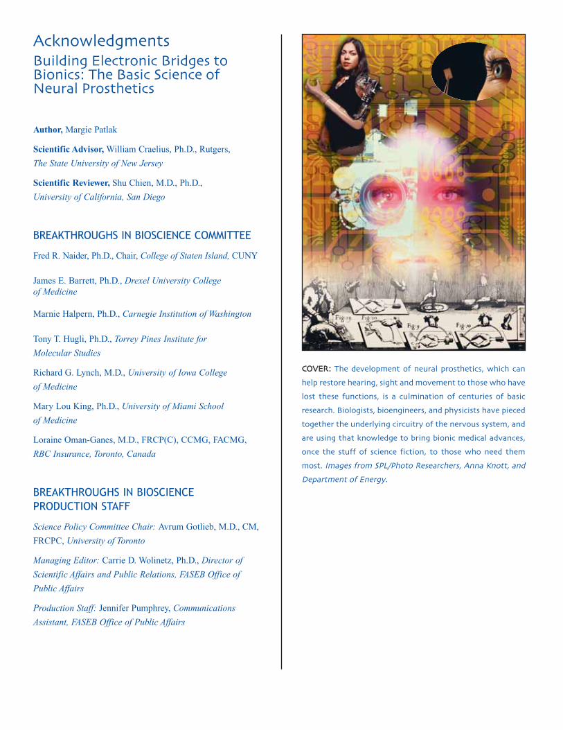
CCOOVVEERR:: The development of neural prosthetics� which can
help restore hearing� sight and movement to those who have
lost these functions� is a culmination of centuries of basic
research� Biologists� bioengineers� and physicists have pieced
together the underlying circuitry of the nervous system� and
are using that knowledge to bring bionic medical advances�
once the stuff of science fiction� to those who need them
most� Images from SPL/Photo Researchers� Anna Knott� andDepartment of Energy�

NEURAL PROSTHETICS
Breakthroughs in Bioscience 1Breakthroughs in Bioscience 1
Building Electronic Bridges to Bionics: The Basic Science of Neural Prosthetics Margie Patlak
At first sight Rachel looks likea typical teenager—she spendsendless hours on her cell phonetalking to her friends, and enjoysrap music and the television showAmerican Idol. It is only aftertalking to her mother aboutRachel’s medical history, thatthe amazement sets in. Despiteappearances to the contrary,Rachel is deaf.
Born with a birth defect thatdestroyed a portion of both of herinner ears, Rachel cannot hearanything without her cochlearimplant. This electronic deviceenables most sounds enteringRachel’s ear to complete theneural pathway to Rachel’s brainwhere they are perceived asvoices, birdsongs or a panoplyof other auditory features thatthe hearing take for granted.Surgically inserted when shewas just a year old, the cochlearimplant has enabled Rachel tolead a normal life free of thedisability of deafness.
Rachel is one of a growingnumber of people with a missingfunction that has been restoredwith a neural prosthetic device.The oldest and most widely usedof these electrical, and oftencomputerized, devices is thecochlear implant, which has pro-vided hearing to thousands ofcongenitally deaf people in this
country. Recently, the use of thecochlear implant is expanding tothe elderly, who frequently suffermajor hearing loss. More cuttingedge are artificial retinas, whichare helping dozens of blindpeople see, and “smart” artificialarms and legs that amputees canmaneuver by thoughts alone, andthat feel more like real limbs.
Such pioneering devices arebrought to us by the collaborativeefforts of a myriad of scientistsfrom a diverse range of fieldsincluding biomedical engineering,physics, biology, neuroscience,and computer sciences. Theseresearchers, whose curiosity ledthem to explore frog legs dancingduring thunderstorms, a snail-shaped organ in the inner ear, andhow various eye cells react tolight, have fostered an understand-ing of how to “talk” to the nervoussystem. That understandingcombined with the miniaturizationof electronics and enhancedcomputer processing has enabledprosthetic devices that often canbridge the gap in nerve signalingthat is caused by disease or injury.
Electrifying ExperimentsToday’s neural prosthetics can
trace their origin, in part, to a pairof frog legs that caused quite alaboratory sensation in Mozart’stime (c.18th century). The labbelonged to the Italian anatomy
professor and physicist LuigiGalvani, who was dissecting afrog at a table. Also on the tablewas a wheel that generated staticelectricity for Galvani’s physicsexperiments. Just as Galvani puta scalpel to the sciatic nerve,which connects to the musclesin the frogs’ legs, his assistanthappened to discharge a sparkof electricity from the wheel.Galvani noticed that when thespark was released, the legs of hisdissected frog jerked. Apparentlythe static electricity released intothe air was picked up by themetal scalpel and passed to thenerve (Figure 1).
This led Galvani to conductother experiments, including onein which the static electricity ofa thunderstorm prompted froglegs on a rooftop to dance, andanother one in which touchingthe exposed nerve to a frog legmuscle was enough to cause themuscle to twitch. The results ofthese experiments led Galvani toconclude that there was “animalelectricity” stored in the nervesof all living creatures, and literallysparked the notion that nervesuse electrical energy to triggermuscle movement. (Galvani’sexperiments also apparentlyinspired Mary Shelley, who readabout them shortly before writingher famous novel Frankenstein, inwhich electricity is used to bringto life Dr. Frankenstein’s monster[Figure 2].)

Breakthroughs in Bioscience 2
In the 19th century, the inventionof an amplifier of electric currentcalled the galvanometer (namedafter Galvani), enabled severalbasic science researchers toeavesdrop on the electrical chatterof muscles and nerves. Theexperiments these Europeanphysicists did on frogs revealedthat an electrical current appliedto the nerve can briefly reverse
the charge emitted by that sectionof the nerve. This flip-flop incharge from positive to negativequickly spreads down the lengthof the nerve and to the musclewhere it causes the muscle tocontract. In other words, byexploring the innate electricalactivity of tissues and how thatchanges in response to electricalstimulation, these researchers hadcollectively stumbled upon theelectrical nerve impulse, which isthe basic signaling mechanismthat nerves use to communicatewith tissues and organs (Figure 3).
Later, studies in the 1920’srevealed that the electrical activityruns on a two-way street—it canpass not just from the motornerve to the muscle, but in thereverse direction from the muscleto a sensory nerve. The brainuses motor nerves to tell themuscle to contract or relax.But the muscle can talk back to
Breakthroughs in Bioscience 2
the brain via sensory nerves thatconvey the amount of tension inthe muscle.
One particularly telling experi-ment was that of the Britishphysiologist (and later NobelPrize winner) Edgar DouglasAdrian. He had suspended a frogleg from a brass hook and wassurprised by the spike of irregularelectrical activity coming fromnerves in the leg. He first thoughtthat this was due to faulty equip-ment used to record the electricalactivity and frustratingly expectedthat he would have to spendmonths rebuilding it. But then henoticed that when the frog legwas placed on a glass plate, theirregular electrical activity stoppedonly to come back when the legwas suspended again. That’swhen it dawned on him that theelectrical activity was actually anelectrical signal being passedfrom muscle to nerve to tell thenerve (and ultimately the brain inan intact animal) that the musclein the frog leg was beingstretched. (Such stretching didn’toccur when the leg was merelylaying on the plate.)
These “electrifying” findingsopened up a whole new way ofunderstanding how the bodyworks, and led investigatorsto electrically explore how acacophony of sounds that passthrough our ears become musicto our minds.
Journey into Inner SpaceMeanwhile, by the middle of
the 19th century, anatomistshad mapped out the inner ear.
Figure � – Frankenstein’s monster:Galvani’s experiments showing thatelectrical impulses are carried throughnerves to trigger muscle movementhelped inspire Mary Shelley’s novel�Frankenstein� later popularized in film�
Figure � – Galvani’s frog experiment: Illustration showing the various experimentsconducted by Italian physicist Luigi Galvani in ���� on frog muscle and electricity� The result was that the muscles in the frog’s leg twitched� indicating the presence ofelectricity� This engraving was taken from Galvani’s De viribus electricitatus in motumusculari commentarius� ����� Credit: SPL/Photo Researchers� Inc�

Breakthroughs in Bioscience 3
Their dissections revealed thatin addition to a thin membrane,called the ear drum, and some ofthe tiniest bones in the body, theinner ear had a strange snail-shapedstructure called the cochlea. Thecochlea was filled with fluid andcarpeted with thousands of tinyhairs. Because these hair cellsconnected with the auditory nervethat travelled from the ear to thebrain (Figure 4), it was assumedthat the cochlea played a crucialrole in hearing. But there was noexperimental evidence to confirmthis. Also, ear dissections didnot answer a key puzzle—howthe cochlea could transformthe multitude of sounds thatentered the ear into distincttones (frequencies) that thebrain uses to tell the differencebetween a vowel and a consonant,or between various notes in themusical scale. Fortunately, insightin this regard was provided in1928 by Georg von Bekesy, aphysicist who had an ingeniousway of seeing sound.
While working for the HungarianPost Office on how to improvetelephone communications, vonBekesy turned to the inner ear
for guidance. In order to besttransform the human voice intomeaningful signals, he firstwanted to know how the humanear processes sound. So hedissected the inner ears of animalsand human cadavers such that thecochlea remained intact andfunctioning—not an easy featgiven that the cochlea is encasedin the bones of the skull. He thensprinkled light-sensitive silverflakes on the hair cell lining andused strobe photography to mark
how the hair cells moved inresponse to various tones. Hisbasic research revealed that notall hair cells were alike—thoseclose to the base of the cochleamoved more in response to high-pitched tones, whereas hair cellsat the tip of the cochlea weremore swayed by low-pitchedtones. For this research vonBekesy later won the NobelPrize (Figure 5).
A year after von Bekesy photo-graphically saw the cochlea’s
FFigure � – Nerve Impulse:Electrical impulses travelthrough motor neurons�or nerve cells� to signalmuscle contraction� Asdescribed in later figures�electrical signals can alsobe carried from tissues back through sensoryneurons to the brain tocommunicate the amountof tension in the muscle�Figure designed by Corporate Press�
Figure – Anatomy of the ear: The discovery that the cochlea was critical for hearingled scientists to explore the way in which this snail shaped organ helped translatesound waves to nerve impulses� This� in turn� led to the breakthrough finding thatelectrical signals could be used to stimulate the auditory nerve� and ultimately resulted in the development of cochlear implants� Figure designed by Corporate Press�
➔ ➔ ➔ ➔
Breakthroughs in Bioscience 4Breakthroughs in Bioscience 4Breakthroughs in Bioscience 4Breakthroughs in Bioscience 4Breakthroughs in Bioscience 4Breakthroughs in Bioscience 4
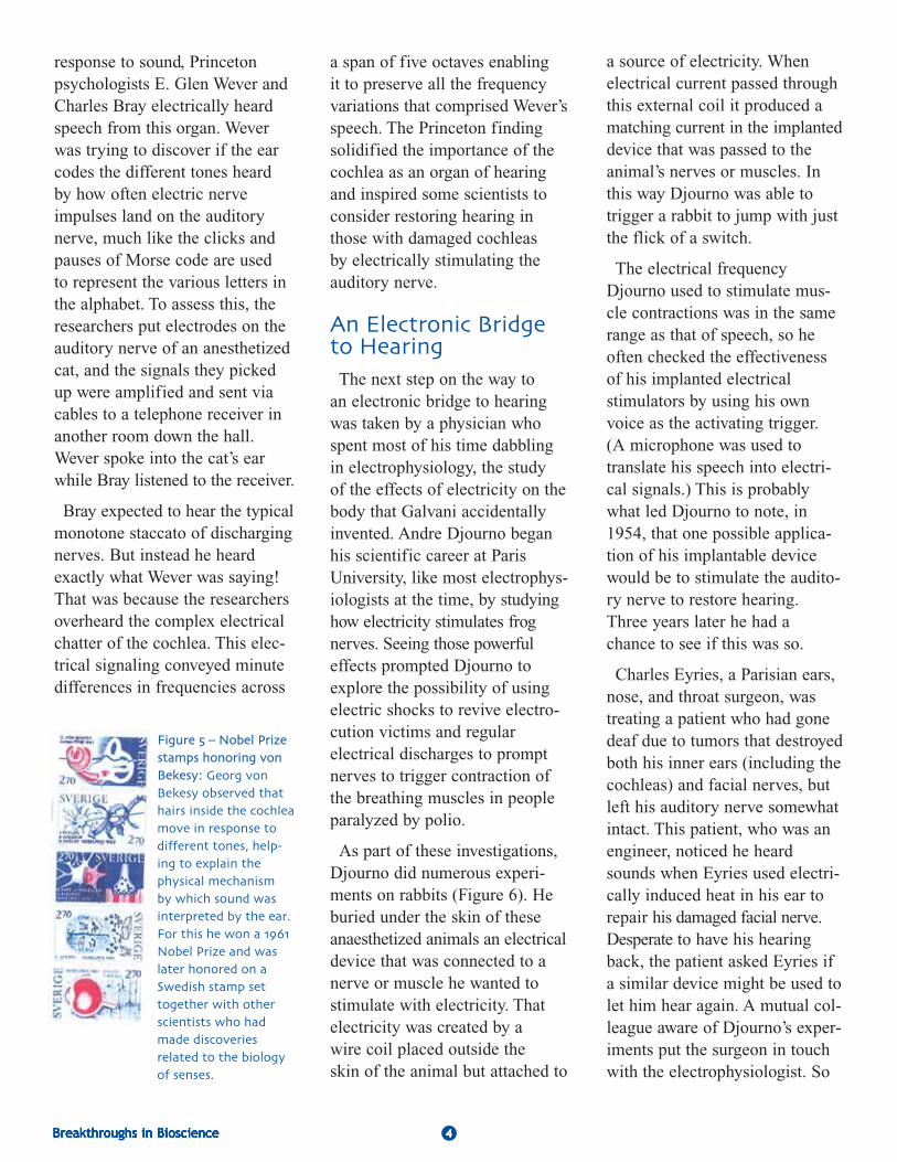
response to sound, Princetonpsychologists E. Glen Wever andCharles Bray electrically heardspeech from this organ. Weverwas trying to discover if the earcodes the different tones heardby how often electric nerveimpulses land on the auditorynerve, much like the clicks andpauses of Morse code are usedto represent the various letters inthe alphabet. To assess this, theresearchers put electrodes on theauditory nerve of an anesthetizedcat, and the signals they pickedup were amplified and sent viacables to a telephone receiver inanother room down the hall.Wever spoke into the cat’s earwhile Bray listened to the receiver.
Bray expected to hear the typicalmonotone staccato of dischargingnerves. But instead he heardexactly what Wever was saying!That was because the researchersoverheard the complex electricalchatter of the cochlea. This elec-trical signaling conveyed minutedifferences in frequencies across
a span of five octaves enablingit to preserve all the frequencyvariations that comprised Wever’sspeech. The Princeton findingsolidified the importance of thecochlea as an organ of hearingand inspired some scientists toconsider restoring hearing inthose with damaged cochleasby electrically stimulating theauditory nerve.
An Electronic Bridgeto HearingThe next step on the way to
an electronic bridge to hearingwas taken by a physician whospent most of his time dabblingin electrophysiology, the studyof the effects of electricity on thebody that Galvani accidentallyinvented. Andre Djourno beganhis scientific career at ParisUniversity, like most electrophys-iologists at the time, by studyinghow electricity stimulates frognerves. Seeing those powerfuleffects prompted Djourno toexplore the possibility of usingelectric shocks to revive electro-cution victims and regularelectrical discharges to promptnerves to trigger contraction ofthe breathing muscles in peopleparalyzed by polio.
As part of these investigations,Djourno did numerous experi-ments on rabbits (Figure 6). Heburied under the skin of theseanaesthetized animals an electricaldevice that was connected to anerve or muscle he wanted tostimulate with electricity. Thatelectricity was created by awire coil placed outside theskin of the animal but attached to
Figure � – Nobel Prizestamps honoring vonBekesy: Georg vonBekesy observed thathairs inside the cochleamove in response to different tones� helping to explain thephysical mechanism by which sound wasinterpreted by the ear�For this he won a ����Nobel Prize and waslater honored on aSwedish stamp settogether with otherscientists who hadmade discoveries related to the biologyof senses�
a source of electricity. Whenelectrical current passed throughthis external coil it produced amatching current in the implanteddevice that was passed to theanimal’s nerves or muscles. Inthis way Djourno was able totrigger a rabbit to jump with justthe flick of a switch.
The electrical frequencyDjourno used to stimulate mus-cle contractions was in the samerange as that of speech, so heoften checked the effectivenessof his implanted electricalstimulators by using his ownvoice as the activating trigger.(A microphone was used totranslate his speech into electri-cal signals.) This is probablywhat led Djourno to note, in1954, that one possible applica-tion of his implantable devicewould be to stimulate the audito-ry nerve to restore hearing.Three years later he had achance to see if this was so.
Charles Eyries, a Parisian ears,nose, and throat surgeon, wastreating a patient who had gonedeaf due to tumors that destroyedboth his inner ears (including thecochleas) and facial nerves, butleft his auditory nerve somewhatintact. This patient, who was anengineer, noticed he heardsounds when Eyries used electri-cally induced heat in his ear torepair his damaged facial nerve.Desperate to have his hearingback, the patient asked Eyries ifa similar device might be used tolet him hear again. A mutual col-league aware of Djourno’s exper-iments put the surgeon in touchwith the electrophysiologist. So

Breakthroughs in Bioscience 5Breakthroughs in Bioscience 5Breakthroughs in Bioscience 5Breakthroughs in Bioscience 5Breakthroughs in Bioscience 5Breakthroughs in Bioscience 5
Figure � – Animal models: Animal models�including rabbits� cats� and frogs� were vital to discoveries that led to the development ofneural prosthetics such as the cochlear implant�Laboratory animals still play an important role in biomedical research� allowing researchers tostudy how cells and tissues interact inside thebody or how disease affects living systems�Credit: Getty Images�
when Eyries’ deaf patient under-went surgery to try to relievefacial paralysis, he implantedDjourno’s electrical stimulatorjust above the patient’s ear andthreaded a wire from it to theauditory nerve. The external coilwas placed nearby on thepatient’s head.
Much to the patient’s delight,he experienced some hearingfollowing his surgery. Afteryears of silence, he could onceagain hear doors opening andclosing, and make out a fewwords spoken to him. But hishearing couldn’t distinguish thedifferent frequencies of vowelsand consonants so that he couldunderstand conversations.
Encouraged by his initialresults, Djourno tried to improvehis hearing device, but when hewas denied a grant to supportthis work, he turned to otherlines of research. However, hisfindings inspired researchersaround the world to take up thetorch, including an Australiangroup, and three separate groupsin California, whose work wasmainly funded by the NationalInstitutes of Health. This
governmental support enabledthem to conduct basic researchon how exactly the cochleatransmits speech sounds to thebrain, as well as to do animaland clinical studies on the safetyand effectiveness of the cochlearimplants they devised to restorehearing. By the early 1970’s,the first cochlear implant madeits debut.
All contemporary cochlearimplants work by having micro-phones that pick up sound,computer chips that code thatsound by frequency, and a seriesof electrodes that electronicallyconvey frequency information tothe auditory nerve (Figure 7).Sound is filtered into differentfrequency channels in theseimplants because of the work ofBekesy and others that revealedthat this is what happens in anormal cochlea via the hair cells.The first cochlear implant onlyhad one electrode and channel.But studies revealed that speechperception greatly improved withthe multichannel devices that sur-faced shortly thereafter.
Today more than 23,000 adultsand 15,000 children in the United
States have cochlear implants,including a Miss America and aradio talk show host, according tothe National Institute on Deafnessand Other CommunicationDisorders (NIDCD, one of theNational Institutes of Health).Success of these implants variesdepending on how long patientswere deaf prior to the implant,how intact their nerves are intheir inner ear, whether they hadsome hearing before they wentdeaf, and how successful theirpost-implant training was.Cochlear implants often enabledeaf or severely hearing impairedpeople to understand speechwithout the aid of lip reading,and to speak clearly with theright volume. Some peoplewith the implants can also enjoymusic, birdsongs, and hear manyother environmental soundsbesides speech.
In deaf children, the deviceswork best the younger the ageat implant. About 2 or 3 out ofevery 1,000 children in theUnited States are born deafor hard-of-hearing, and morelose their hearing later duringchildhood, according to NIDCD.

Breakthroughs in Bioscience 6Breakthroughs in Bioscience 6Breakthroughs in Bioscience 6Breakthroughs in Bioscience 6Breakthroughs in Bioscience 6Breakthroughs in Bioscience 6Breakthroughs in Bioscience 6Breakthroughs in Bioscience 6
When such congenital hearingloss is not effectively treatedearly, it can lead to permanentproblems with language develop-ment and linked learning andsocial difficulties. The Instituterecommends screening newbornswithin the first month of life forhearing loss because childrenbegin learning speech andlanguage in the first 6 monthsof life. Most children whoreceive the cochlear implantand training before age 5 or 6are able to speak and understandspeech as well as their normalhearing peers, and those whoreceive the implant as infants ortoddlers tend to develop languageat the same pace as those withnormal hearing.
Some experts in hearing weresurprised by the success of the
cochlear implants. How could asmall number of electrodes mimicthe sensitivity to sounds of the15,000 hair cells seen in the earsof those who can hear adequately?That challenging question sentbasic researchers scurrying backto the lab to try to find answers.What they discovered not onlyfurthered understanding of howwe hear, but made the prospectsof giving sight to the blind anattainable goal.
From Redundancy toArtificial RetinasIt turns out that when it comes
to hearing, only a fraction ofthe information on the frequencyof sounds sent to the brain isneeded to understand speech.Recent studies indicate thatthe normal cochlear divides
frequencies into the equivalentof 28 channels before sendingthem to the auditory nerve,but we can hear with far fewerchannels. Researchers at ArizonaState University used processorsto divide and compress thefrequency bands in speechlistened to by people with normalhearing. (The processing of thespeech mimicked what is doneby a cochlear implant.) Theirstudies reveal that as few as 8channels are needed to discernspeech adequately. Presumably,a lot of the complex soundinformation sent to the brain isredundant, experts now think,so one doesn’t have to captureit completely to provideadequate hearing.
Part of the success of cochlearimplants is also due to the amazingability of the brain to see the bigpicture from sketchy information.For example, consider thosepersonalized license plates that,due to the limited number ofcharacters they can have, omitvowels yet are still understandableto the person reading them duringa traffic jam. We see “LVMYDG”yet realize it stands for “Love mydog.” Our brain is able to fill inthe missing letters so we canread the license plate. Similarly,our brain can fill in missingfrequencies so we can hear whatpeople are saying to us.
Those of us with years ofexperience listening to peopletalk are better able to fill in themissing sounds. This may explainwhy people who have been deaffor a short time do better withcochlear implants than those with
Figure � – Diagram of cochlear implant: A microphone placed above the ear convertssounds to electrical signals� which it sends to a speech processor worn behind the earor attached to a belt� The speech processor is a computer chip that divides up andencodes the electrical signals by frequency� which our brains interpret as pitch� Thisfrequency data and the power needed to activate the cochlea implant are sent� oftenvia radio waves� to a receiverstimulator implanted near the ear� This device decodesthe sound information back into electrical impulses� with each of the frequencygroups (channels) sent to eight or more different electrodes placed in the cochlea�These electrodes convey the signals to various portions of the auditory nerve� whichthen carries them to the brain (the ultimate processor) where they are perceived asspeech and environmental sounds� Image designed by Corporate Press�

Breakthroughs in Bioscience 7Breakthroughs in Bioscience 7Breakthroughs in Bioscience 7
long-term deafness. But withproper training, many peoplewho have been deaf for most oftheir lives can learn how to makesense of what they are hearingwith their cochlear implants.Studies show that people with theimplants do better at discerningspeech over time as they gainmore experience with them. Thisshows the remarkable plasticityof the brain that refutes the notionthat the brain is permanentlyhard-wired.
However, studies in cats revealthat there does appear to be acritical window of time duringwhich the part of the brain thatprocesses sounds and fosterslanguage skills develops mostreadily. These studies havemapped the sensitivity of variousregions of the brain to varioussound frequencies and found thatwhen it comes to hearing, its allabout location—high frequencypitches activate different minutesections of the brain than lowtones. When kittens are deafenedshortly after birth, their brainsnever develop this sound special-ization unless their hearing isrestored within a short periodof time. Yet when these deafenedkittens are given cochlearimplants, the sound-processingregions of their brains appear todevelop normally. This probablyexplains why cochlear implantsare often so successful in youngchildren, and has importantclinical implications now thatuniversal screening of newbornsmakes it possible to detectdeafness by one month of age.
Battling BlindnessEncouraged by the success of
cochlear implants and the findingson redundancy of nerve signalingand brain plasticity, researchersbegan to tackle in earnest thepossibility of creating electroniceye prosthetics for people blindedby various diseases. Their effortswere not a literal stab in thedark, but hinged on more than acentury’s worth of basic researchon the eye. This research hadsolved the intriguing puzzle ofhow light entering our eyes istransformed into images inour brain.
In early studies, anatomistsdiscovered and drew in painstakingdetail all of the eye’s cellularcomponents and the nervepathway from the eye to thebrain. But the functions of theseeye components remained hiddenuntil biochemists and electro-physiologists in the 20th centuryshowed how the various cells inthe eye responded chemically orelectrically to light or darkness.Their research revealed that thekingpin of sight is the retina, thelight-sensitive inner lining at theback of the eye. It is there thatlight encodes the scene before usinto an image that is sent to thebrain, much like film encodes animage captured by a camera.
The retina has a layer of cellscalled rods and cones that containspecial pigments. When lightstrikes these pigments, it triggersa chemical reaction that createselectrical signals. These signalsare passed to a layer of nervecells called ganglion cells, which
convey them to the optic nerve.The nerve travels from the eyeto the brain, where the electricalsignals are transformed intorecognizable images (Figure 8).Throughout this visual pathway,the spatial configuration of all theelements seen is maintained. Inother words, an American flag willbe encoded in both the retina andthe brain with stars on the upperleft and stripes beside and below it.Because the retina plays such akey role in sight, damage to it cancause blindness. More than onemillion people in the UnitedStates are blind due to diseasesof the retina that destroyed theirrods and cones, according to theNational Eye Institute (anotherone of the National Institutes ofHealth). There are no effectivetreatments for these disorders,which take a tremendous toll onpatients. They need significant aidto carry on their daily tasks, letalone adjust to the psychologicalburden of no longer being able tohave an independent lifestyle. Witha growing aging population, thenumber of people affected byblinding diseases may triple by2025, some experts predict,creating a future pandemic ofvision loss. (Macular degenerationis a blinding vascular disorder inthe eye that damages the retinaand affects more than one-third ofpeople 75 years of age and over.)
Until recently, it was assumedthat destruction of the rods andcones would cause the ganglioncells to deteriorate as well. Thisfrequently happens to tissues nolonger fed signals from nerves.Eager to see if that was the case
Breakthroughs in Bioscience 7Breakthroughs in Bioscience 7Breakthroughs in Bioscience 7Breakthroughs in Bioscience 7

Breakthroughs in Bioscience 8Breakthroughs in Bioscience 8Breakthroughs in Bioscience 8
was ophthalmologist and bioengi-neer Mark Humayun. Inspired byhis grandmother, whose blindnessdeveloped during his teenageyears and contributed to herdemise, Humayun’s doctoralresearch was a blueprint for adevice that could act as a signalingrescue operation in eyes withdamaged rods and cones. Thesuccess of that device hinged onhaving enough ganglion cells inthe retina still working.
In the early 1990s, Humayun andhis colleagues dissected the eyesof people with retinal diseasesthat were donated after death. Hewas thrilled to discover that evenpeople with severe disease stillhad more than one-quarter oftheir ganglion cells and thosewith milder disease had most ofthese cells intact. But did thoseremaining ganglion cells stillwork? To test this, Humayun gavevolunteers blinded by retinal
disease a local anesthetic and thenput tiny electrode probes in theireyes. These individuals all hadmajor loss of their rods and cones.But when Humayun electricallystimulated different parts of theirretina with the probe, the patientsreported seeing streaks or dots oflight. When two probes stimulatedthe retina at two different sites,patients reported seeing twodifferent light bursts with thesame position in space as theelectrodes were on their retinas.This suggested that evenly spacedelectrical stimulation of theremaining retina cells couldpreserve the spatial configurationof retinal signaling that is soimportant for sight.
Creating the Bionic EyeBut creating an implantable
array of electrodes in the retinawas a daunting task. Thinner thanthe tip of a pen and with the
strength of a wet Kleenex, theretina could be easily torn by aninflexible device, or damaged bythe heat released by the numerouselectrodes needed to activatesufficient nerve cells. (More thana million nerve cells comprisethe optic nerve compared to the30,000 cells that make up theauditory nerve.) To top it off, anydevice implanted in the eye has tosurvive the corrosive effectsof a salty world and deliverinformation to the brain quickly,so a person doesn’t realize theyare seeing a curb a minute afterthey trip over it.
To meet these bioengineeringchallenges, Humayan is collabo-rating with several engineers,materials scientists, ophthalmolo-gists, neuroscientists and otherexperts funded by the Departmentof Energy, the National ScienceFoundation and the National Eye
Breakthroughs in Bioscience 8
Figure – Anatomy of the eye: Light entering the eye passes through to the retina� where specialized vision cells� called rods andcones� will be stimulated to set off a chain of reactions converting light to electrical impulses� These impulses are conveyed via ganglion cells� a type of nerve cell� to the optic nerve� which ultimately transmits them to the brain� where visual input is translated�Understanding how this circuitry works helped scientists create an artificial retina to restore some sight in those suffering from retinal damage� Image designed by Corporate Press�
CORNEA
PUPIL
LENS
IRIS
RETINA
OPTICALNERVE

Breakthroughs in Bioscience 9Breakthroughs in Bioscience 9Breakthroughs in Bioscience 9Breakthroughs in Bioscience 9Breakthroughs in Bioscience 9Breakthroughs in Bioscience 9Breakthroughs in Bioscience 9
Institute. The first artificialretina these researchers created isshowing promise in tests on blindpeople. This device consists ofa tiny camera and computerchip mounted on sunglasses thatcapture, code, and convert whatis “seen,” into electrical signalsthat are relayed to the ganglioncells via an electrode-studdedcomputer chip. This chip isimplanted in between the retinaand the jelly-like inner portion ofthe eyeball (Figure 9). The minoramount of heat given off by thechip is carried away by the fluidin the inner eye so it doesn’t buildup and harm the retina.
After extensive testing of theimplant in dogs and rabbits,Humayan finally implanted thefirst model of this artificial retinain 6 blind volunteers in 2002.Humayan didn’t expect muchfrom his pilot retinal implantmodel, which after all only hasonly 16 electrodes that are a farcry from the million nerve cellsin the optical nerve. But, in whathe has described as a definingmoment in his life, testing of thepatients revealed that they wereable to distinguish a cup froma plate or a knife and detectmovement of objects across ascreen. This crude vision is help-ing some of these patients bettermaneuver in their environment—they can avoid a tree branchblocking their way because itappears to them as a bright whiteline, and they can discern a tableor a door.
Such surprisingly good resultswere unexpected and attributedto the remarkable ability of
the brain to learn and fill inthe blanks when given sketchysignals. And to the patientvolunteers, some of whom hadbeen blind for 50 years, thosesketchy signals were immenselyrewarding. One patient wasthrilled when he could see theshadow of his 18 year-old son,whom he hadn’t seen since theboy was 5 years old. None ofthese volunteers have experiencedsurgical complications or damageto their eyes caused by the implantso far. The devices were notintended to last indefinitely, butin one patient it has continued tooperate for more than 5 years.
By taking advantage of therecent advances in microelectronics,which enable more electrodes andprocessing power to be packedinto smaller spaces, (see side bar)the Humayan group created anewer version of its artificial retina.This model has 60 electrodesand is currently being tested in alarger number of volunteers withthe expectation that it will improvetheir unaided mobility. They havealso developed an artificial retinawith 200 electrodes that has manyimprovements over the previoustwo models. Thanks to innovativetechnology developed by collabo-rators at Lawrence Livermore
Figure � – Artificial retina: The camera and microprocessor substitute� in part� for themissing lightsensitive cells in the retina� The camera videotapes the patient’s surroundings and sends a digital black and white version of the scene to the microprocessor� Thiscomputer chip compresses and simplifies the data it receives� and converts them intoelectrical signals� These signals are sent to the receiver and its attached wires that connect to the retinal implant� which is about an inch long and tacked to the retina� Therethe signals are passed on to the ganglion cells and the optic nerve� which carries them to the brain� A wireless battery pack� worn on a belt� powers the device� Image courtesyof U�S� Department of Energy Artificial Retina Project� artificialretina�energy�gov
Breakthroughs in Bioscience 10
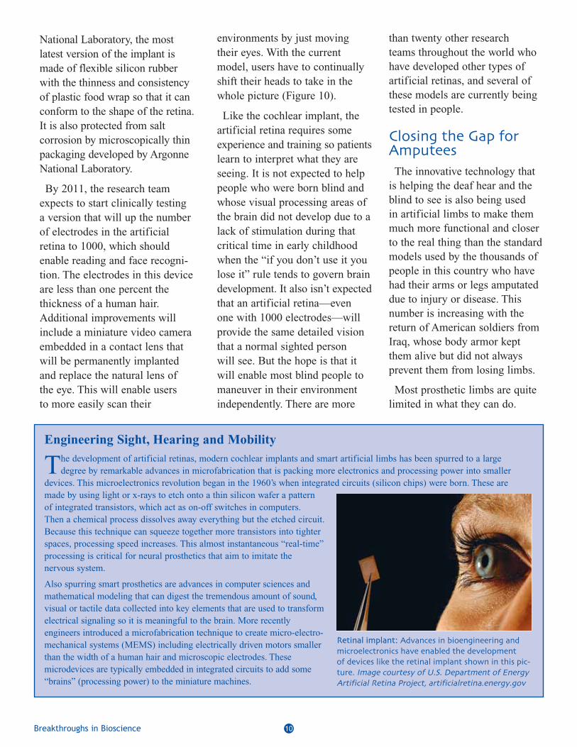
National Laboratory, the mostlatest version of the implant ismade of flexible silicon rubberwith the thinness and consistencyof plastic food wrap so that it canconform to the shape of the retina.It is also protected from saltcorrosion by microscopically thinpackaging developed by ArgonneNational Laboratory.
By 2011, the research teamexpects to start clinically testinga version that will up the numberof electrodes in the artificialretina to 1000, which shouldenable reading and face recogni-tion. The electrodes in this deviceare less than one percent thethickness of a human hair.Additional improvements willinclude a miniature video cameraembedded in a contact lens thatwill be permanently implantedand replace the natural lens ofthe eye. This will enable usersto more easily scan their
environments by just movingtheir eyes. With the currentmodel, users have to continuallyshift their heads to take in thewhole picture (Figure 10).
Like the cochlear implant, theartificial retina requires someexperience and training so patientslearn to interpret what they areseeing. It is not expected to helppeople who were born blind andwhose visual processing areas ofthe brain did not develop due to alack of stimulation during thatcritical time in early childhoodwhen the “if you don’t use it youlose it” rule tends to govern braindevelopment. It also isn’t expectedthat an artificial retina—evenone with 1000 electrodes—willprovide the same detailed visionthat a normal sighted personwill see. But the hope is that itwill enable most blind people tomaneuver in their environmentindependently. There are more
than twenty other researchteams throughout the world whohave developed other types ofartificial retinas, and several ofthese models are currently beingtested in people.
Closing the Gap forAmputeesThe innovative technology that
is helping the deaf hear and theblind to see is also being usedin artificial limbs to make themmuch more functional and closerto the real thing than the standardmodels used by the thousands ofpeople in this country who havehad their arms or legs amputateddue to injury or disease. Thisnumber is increasing with thereturn of American soldiers fromIraq, whose body armor keptthem alive but did not alwaysprevent them from losing limbs.
Most prosthetic limbs are quitelimited in what they can do.
Engineering Sight, Hearing and Mobility
The development of artificial retinas, modern cochlear implants and smart artificial limbs has been spurred to a largedegree by remarkable advances in microfabrication that is packing more electronics and processing power into smaller
devices. This microelectronics revolution began in the 1960’s when integrated circuits (silicon chips) were born. These aremade by using light or x-rays to etch onto a thin silicon wafer a patternof integrated transistors, which act as on-off switches in computers.Then a chemical process dissolves away everything but the etched circuit.Because this technique can squeeze together more transistors into tighterspaces, processing speed increases. This almost instantaneous “real-time”processing is critical for neural prosthetics that aim to imitate thenervous system.
Also spurring smart prosthetics are advances in computer sciences andmathematical modeling that can digest the tremendous amount of sound,visual or tactile data collected into key elements that are used to transformelectrical signaling so it is meaningful to the brain. More recentlyengineers introduced a microfabrication technique to create micro-electro-mechanical systems (MEMS) including electrically driven motors smallerthan the width of a human hair and microscopic electrodes. Thesemicrodevices are typically embedded in integrated circuits to add some“brains” (processing power) to the miniature machines.
Retinal implant: Advances in bioengineering andmicroelectronics have enabled the development of devices like the retinal implant shown in this picture� Image courtesy of U�S� Department of EnergyArtificial Retina Project� artificialretina�energy�gov

Breakthroughs in Bioscience 11Breakthroughs in Bioscience 11Breakthroughs in Bioscience 11
Artificial arms, for example, usea system of pulleys and cables orelectronic motors to enable themto bend at the elbow or pick upitems with a hook that opens andcloses. These prosthetics cannotrestore the full range of motionand capabilities of a real arm—they may let you pick up a fork,but they don’t let you feed your-self with it. They also require alot of concentration to use—youhave to not only manipulate thedevice with your other arm ornearby muscles, but you have tocontinually watch the prostheticin action so it can pick up anObject or carry out someother function.
This is no surprise when youconsider what our nervous systemdoes every time we drink a cupof coffee. Our brain sends electricsignals that travel to the motornerves of our arm and hand.There they simultaneously triggerour hand and arm muscles tocontract or extend so we cangrasp the cup. At the same time,thousands of nerve cell sensorsin our skin, muscles and jointsof our hand and arm signal tosensory nerves and then the brainso we can feel the cup in ourhand, gauge if we are grasping ittight enough or lifting it withenough force, as well as let usknow where our arm/hand is inspace. This information is quickly
sent back to the brain which thenuses it to guide our arm and handmotor nerves so we can lift thecup and precisely position it toour lips. Such a simple actionrequires our nervous system toinstantly coordinate millions ofelectrical nerve impulses with aprecision that we take for granted,and at which bioengineers marvel(Figure 11A).
When someone loses a limbor finger, the portion of thebrain and motor nerves that oncegoverned its movements continuesto send signals to the missingbody part, despite the fact thatthese signals never reach theirtarget. Even the sensory nervesonce connected to the amputatedlimb or finger will send signalsback to the brain if they arestimulated any place along theirpathway. For example, if asensory nerve for touch that oncewas connected to the thumb ofa missing hand is electricallystimulated at the arm level, theperson will report feeling thatsomething just brushed againsthis or her missing thumb. Thislingering nerve signaling is whyabout three-quarters of amputeesreport that they still feel theirmissing limbs and have the urgeto move them (Figure 11B). Forexample, when a motorcycleaccident severed the left armof Claudia Mitchell, she didn’teven realize it was missing andcouldn’t understand why it wasn’tworking when she tried to use itto push herself up off the ground.
Fortunately, bioengineers arehelping Mitchell and a handfulof others by connecting the still
Breakthroughs in Bioscience 11
Figure �� –What you see with an artificial retina: These images of a hand (A) and head (B) approximate what patients with retinal devices see� Increasing the number of electrodes (moving from � to ) will result in enhanced visual perception and higher resolution vision� Scientists work with patients who receive artificial retinas for aboutone month after surgery to help them interpret what they see� Deaf individuals withcochlear implants also need such guidance to understand sounds they are hearing for the first time� Technology is also helping to improve processing of signals from retinalimplants: the Artificial Vision Support System (AVSS)� devised and developed by Dr�Wolfgang Fink and associate Mark Tarbell at the California Institute of Technology� performs realtime image processing/enhancement of the videostream provided by the external camera to improve on the limited vision afforded by these implants for the benefit of the blind subjects� Image courtesy of Dr� Wolfgang Fink and Mark Tarbell�Visual and Autonomous Exploration Systems Research Laboratory� California Institute of Technology

Breakthroughs in Bioscience 12
signaling nerves in their stumps
to artificial mechanical arms
fitted with pressure sensors. To
do this, researchers funded by the
Department of Defense and the
National Institute of Neurological
Disorders and Stroke attach
superfine electrode arrays to the
residual nerves of the missing arm
to complete the electrical signaling
communication loop from the
brain to an arm prosthetic and
back in volunteer amputees.
Some of these electrodes connect
with motor nerves and send their
electrical signals to a computer
that directs motors in an attached
artificial arm to move the
prosthetic appropriately. Other
electrodes connect with the
sensory nerve cells to convey
Figure �� – Nerve signal transmission in a healthy arm (A)� amputee (B)� and neuralprosthetic arm ((C)� Images designed by Michael Linkinhoker� Link Studios� LLC�

The Breakthroughs in Bioscience
series is a collection of illustrated
articles that explain recent
developments in basic biomed
ical research and how they are
important to society� Electronic
versions of the articles are avail
able in html and pdf format at
the Breakthroughs in Bioscience
website at:
opa�faseb�org
Breakthroughs in Bioscience 13
the signals sent from silicon-based touch pressure sensorson the prosthetic arm. Thesesensory signals are also sent tothe computer and then the brain(Figure 11C). The end result isthat amputees feel that theartificial arms are more a partof their bodies because they cancontrol them intuitively, and theycan sense what the arm prostheticsare touching and where they arein space.
For example, as soon as Mitchellthinks about opening her left hand,her artificial hand opens. She hasbeen effectively able to use it to
peel a banana, and cut a steak(Figure 12). Other amputeesfitted with the new artificial armscan pull credit cards from theirback pockets, or with their eyesclosed have their “fake” armmimic the position of their realarm. Researchers expect toeventually produce a prostheticarm model with a plastic sleevethat not only looks and feels likeskin, but has the feelings of skinas well, thanks to numeroussensors scattered throughout it.Bioengineers are also pursuingsimilar smart prosthetic legs.These are bound to benefit many
of the nearly two million peoplein the United States livingwithout a limb.
Galvani could not have imaginedhis basic research on nervesignaling in frogs would eventuallyblossom into artificial eyes,cochleas, and arms for people.But none of these smart prostheticswould have been possible if basicresearchers from such diversedisciplines as physics, physiology,ophthalmology, computer sciences,mathematics and engineeringhadn’t explored how we hear andsee and how the nervous systemworks. Collectively these scientistsfostered the development of bio-engineered devices that can closecrucial gaps in nerve signaling sothat the deaf can hear, the blindcan see, and amputees can haveartificial limbs that feel and actlike their missing limbs.
FFigure �� – Claudia Mitchell:After losing her arm in amotorcycle accident� ClaudiaMitchell became the firstwoman to be outfitted with abionic arm developed by theRehabilitation Institute ofChicago� The neural prostheticoperates by detecting themovements of muscles in herchest that have been rewiredto the stumps of nerves thatonce went to her amputatedarm� Credit: Anna KnottPhotography�
BiographiesMargie Patlak writes about biomedical research and health from the Philadelphia region.She has written for Discover, Glamour, Physician’s Weekly, Consumer Reports on Health, TheWashington Post, Los Angeles Times, Dallas Morning News and numerous other publications.She also writes frequently for the National Institutes of Health and the National Academy ofSciences. This is her seventh article in the Breakthroughs in Bioscience series.
William Craelius, Ph.D., is a Professor in the Department of BiomedicalEngineering at Rutgers, The State University of New Jersey, where he also heads up theBiomechanics and Rehabilitation Engineering Laboratory. Dr. Craelius’ research interestsinclude human motor control, bioinstrumentation, biofeedback and rehabilitation technology,ion channels in cell membranes, and cell-substrate interaction. He is the internationally rec-ognized inventor of the Dextra artificial hand, a breakthrough technology that first allowedamputees to use existing neural pathways to control mechanical movement, and founder ofthe company Nia-Crae. Dr. Craelius’ honors include the Mary Switzer Fellowship, DiscoverTechnology Award, and induction into the American Institute for Medical and BiomedicalEngineering College of Fellows.

For reprints or other information:Federation of American Societies for Experimental Biology
Office of Public Affairs9650 Rockville Pike
Bethesda, MD 20814-3998http://opa.faseb.org
Published2008