DETECTION OF LEGIONELLA spp. AND OTHER PATHOGENS IN …
106
DETECTION OF LEGIONELLA spp. AND OTHER PATHOGENS IN WATER SYSTEMS OF NURSING HOMES AND SPA POOLS Tanyaporn Kullaket A Thesis Submitted in Partial Fulfillment of the Requirements for the Degree of Master of Science in Microbiology Suranaree University of Technology Academic Year 2017
Transcript of DETECTION OF LEGIONELLA spp. AND OTHER PATHOGENS IN …

DETECTION OF LEGIONELLA spp. AND OTHER
PATHOGENS IN WATER SYSTEMS OF NURSING
HOMES AND SPA POOLS
Tanyaporn Kullaket
A Thesis Submitted in Partial Fulfillment of the Requirements for the
Degree of Master of Science in Microbiology
Suranaree University of Technology
Academic Year 2017

การตรวจหาเช้ือ Legionella spp. และจุลนิทรีย์ก่อโรค
ในระบบน า้ใช้ของสถานพยาบาลพกัฟ้ืนและสระน า้สปา
นางสาวธันยาภรณ์ กลุเกษ
วทิยานิพนธ์นีเ้ป็นส่วนหน่ึงของการศึกษาตามหลกัสูตรปริญญาวทิยาศาสตรมหาบัณฑิต
สาขาวชิาจุลชีววทิยา
มหาวทิยาลัยเทคโนโลยสุีรนารี
ปีการศึกษา 2560


ACKNOWLEDGEMENTS
I am exceptionally grateful to my supervisor, Associate Professor Dr.Tassanee
Saovana for her valuable advice, encouragement, and kindness. I am also appreciated
for her supporting throughout of my study.
I would like to thank and appreciated to all my thesis committee, Associate
Professor Dr. Griangsak Eumkeb and Dr. Nawarat Nantapong for their comments and
suggestions.
I would like to thank laboratory scientist of the Center for Scientific and
Technology Equipment, Suranaree University of Technology for their helping
throughout the work.
I would like to thank all managers and staffs both of nursing homes and spas for
the good reception and accommodate the sampling collection throughout the
processing.
I would like to thank SUT outstanding academic performance scholarships for
financial and laboratory facility support.
Finally, I thank my friend at SUT for their help throughout the work and my
beloved family for good advice, chance to study and supported me throughout this
thesis.
Tanyaporn Kullaket

CONTENTS
Page
ABSTRACT IN THAI ................................................................................................... I
ABSTRACT IN ENGLISH ......................................................................................... II
ACKNOWLEDGEMENTS ........................................................................................ IV
CONTENTS ................................................................................................................. V
LIST OF TABLES ...................................................................................................... IX
LIST OF FIGURES ..................................................................................................... X
LIST OF ABBREVIATIONS ..................................................................................... XI
CHAPTER
I INTRODUCTION ........................................................................................... 1
1.1 Background/Problems ................................................................................. 1
II LITERATURE REVIEW .............................................................................. 4
2.1 Legionella species ....................................................................................... 4
2.2 Legionella ecology ...................................................................................... 7
2.3 Pathogenesis ................................................................................................ 8
2.4 Legionellosis ............................................................................................... 9
2.5 Amplification factors ................................................................................ 12
2.5.1 Protozoa associations ...................................................................... 12
2.5.2 Biofilm associations ........................................................................ 14
2.5.3 Algal associations ........................................................................... 15

VI
CONTENTS (Continued)
Page
2.6 Distribution of the Legionella spp. to humans .......................................... 16
2.7 Distribution of the Legionella spp. in Thailand ........................................ 18
2.8 Methods for Legionella detection ............................................................. 20
2.8.1 Cultural method ............................................................................... 20
2.8.2 Non-cultural methods ...................................................................... 22
2.9 Legionella disinfection methods ............................................................... 23
2.10 Microbiological evaluation of water sample quality .............................. 24
2.11. Research objectives ................................................................................ 24
2.12. Research hypothesis ............................................................................... 25
III MATERAIALS AND METHODS .............................................................. 26
3.1 Preparation of the standard bacteria, chemicals and reagents ................... 26
3.2 Instrumentation ......................................................................................... 26
3.3 Samples collection and processing ........................................................... 27
3.3.1 Shower heads and faucets ............................................................... 27
3.3.2 Spa pools ......................................................................................... 27
3.3.3 Samples processing ......................................................................... 27
3.4 Microbiological analysis ........................................................................... 28
3.4.1 Detection of Legionella species ...................................................... 28
3.4.2 Isolation and quantitation of total heterotrophic plate count .......... 29
3.4.3 Isolation of gram-negative bacteria ................................................. 29
VII
CONTENTS (Continued)
Page
3.4.4 Isolation of Staphylococcus spp. ..................................................... 30
3.4.5 Isolation of Coliform and E. coli bacteria ....................................... 30
3.4.6 Repeated cultivation after decontamination of Legionella spp.
at positive sites ............................................................................... 32
3.4.7 The decontamination of Legionella spp .......................................... 32
3.5 Relationships between water parameters
and the prevalence of Legionella pneumophila ....................................... 33
IV RESULTS AND DISCUSSIONS ................................................................. 34
4.1 Survey and collection of samples ............................................................. 34
4.2 Detection of Legionella species ................................................................ 35
4.3 Detection of other microorganisms ........................................................... 43
4.4 Microbiological quality of water samples ................................................. 55
4.5 Physical analysis of samples ..................................................................... 56
4.6 Relationships between water parameter
and prevalence of Legionella pneumophila ............................................. 58
4.7 Repeated cultivation after decontamination of Legionella spp.
at the positive sites ................................................................................... 59
VI CONCLUSION ............................................................................................. 64
REFERENCES ........................................................................................................... 68
APPENDICES ............................................................................................................ 77

VIII
CONTENTS (Continued)
Page
APPENDIX A MICROBIOLOGICAL MEDIA ............................................ 78
APPENDIX B CHEMICAL REAGENTS ..................................................... 82
APPENDIX C STANDARD OF TAP WATER RECOMMENDED
BY METROPOLIAN WATERWORKS AUTHORITY
(BASED ON WHO GUIDANCE 2011) ............................... .84
APPENDIX D BIOCHEMICAL TEST OF BACTERIA .............................. .87
CURRICULUM VITAE ............................................................................................. 90

LIST OF TABLES
Table Page
1 Legionella species with standing in nomenclature ........................................... 5
2 Symptoms associated with Legionellosis ....................................................... 10
3 Protozoan species found to harbour intracellular Legionella spp .................... 13
4 Source of aerosol transmission of Legionella spp .......................................... 16
5 Biochemical test of gram-negative bacteria .................................................... 31
6 Amount and duration for elimination the pathogens ...................................... 33
7 Source of samples for detection of Legionella spp. ......................................... 34
8 The density and interpretation level of the contaminated water samples
from GVPC agar .............................................................................................. 37
9 The relative risk assessment of Legionella pneumophila positive samples
of nursing homes ............................................................................................. 39
10 The relative risk assessment of Legionella pneumophila positive samples
of spa pools ..................................................................................................... 40
11 The microorganisms in the water samples of nursing homes ......................... 44
12 The microorganisms in the water samples of spa pools ................................. 46
13 Physical parameters of water samples collected from nursing homes ............ 56
14 Physical parameters of water samples from spa pools .................................... 57
15 Statistical analysis of Pearson’s correlation analysis between water quality
parameters (temperature and pH) and the outbreak of Legionella spp ........... 58

X
LIST OF TABLES (Continued)
Table Page
16 Legionella spp. and other microorganisms detected from water samples
before and after elimination in positive sample sites of nursing homes ......... 60
17 Legionella spp. and other microorganisms detected from water samples
before and after elimination in positive sample sites of spa pools .................. 61
LIST OF FIGURES
Figure Page
1 Legionella pneumophila life cycle in amoebae ................................................ 9
2 Legionella colony grows on GVPC agar ........................................................ 42
3 Gram stain of Legionella spp. under compound light microscope ................. 42
4 Gram stain of Citobacter spp. under compound light microscope ................. 52
5 Gram stain of Pseudomonas spp. under compound light microscope ............ 52
6 Gram stain of Staphylococcus spp. under compound light microscope ......... 53
7 Gram stain of Acinetobacter spp. under compound light microscope ............ 53
8 Gram stain of Enterobacter spp. under compound light microscope ............. 54
9 Gram stain of E. coli spp. under compound light microscope ........................ 54
LIST OF ABBREVIATIONS
% Percent
BCYE Buffered Charcoal Yeast Extract Agar
°C Degree Celsius
CFU/ml Colony Forming Units per milliliter
DFA Direct fluorescence antibody
DNA Deoxyribonucleic acid
ELISA Enzyme-linked immunosorbent assay
et al. et alia (and other)
(m, た) g (milli, micro) Gramme
(た) g/l (micro) Gramme per litre
GVPC Glycine Vancomycin Polymyxin B and Cycloheximide
Agar
IFA Indirect fluorescent antibody
(m, た)l (milli, micro) Litre
M Molar
ml/l Millilitre per litre
min Minute
MWA Metropolitan Waterworks Authority, Thailand
PCR Polymerase chain reaction
sp., spp. Species (singular, plural)

XIII
LIST OF ABBREVIATIONS (Continued)
rRNA Ribosomal ribonucleic acid
WHO World Health Organization
CHAPTER I
INTRODUCTION
1.1 Background/Problems
The water distribution system in many buildings such as hospital, nursery,
nursing home or even swimming pool and spa are very important because it may be a
source of infections, especially respiratory infections that are caused by the inhalation
of bacteria contaminated by water aerosols. The contaminated aerosols that are
generated by respiratory equipments, including humidifiers and nebulizers have been
reported that they can transmit airborne pathogens into the respiratory tract of patients
(Woo, Goetz and Yu, 1992). One of bacterial pathogens that cause respiratory disease
is Legionellae. They are gram-negative and non-spore-forming bacteria. These
bacteria are short rod-shaped cells and are described as coccobacillary (Rodgers,
Macrae and Lewis, 1978) . The representative species of the genus is Legionella
pneumophila that can cause the Legionellosis (Percival and Williams, 2014).
Legionellae are commonly found in natural water environments (e.g. , rivers, lakes,
lagoon and reservoirs) and human-made water systems (e. g. , cooling tower, water
heater tanks, fountains and spa pools) . The water distribution system that is not
appropriately managed can act as the source of major outbreaks of Legionellosis
(Moore and Walker, 2014) . People at risk are the elderly, smokers and the
immunosuppressed patients.

2
The Legionellosis is divided into two distinct clinical entities, Pontiac fever is
a self-limited flu-like illness and has a high rate of infection of about 95% and
Legionnaires’ disease which is a severe multisystem disease involving pneumonia
with about 5%, rate of infection but symptoms are more severe than Pontiac fever and
may lead to death (Fields, Benson and Besser, 2002) . In the United Kingdom,
Legionnaires’ disease caused by L. pneumophila, is rare but serious disease. Between
2009 to 2011, there were 934 confirmed cases in England and Wales, 355 ( 38%)
affected persons occurred diseases while they were travelling abroad ( Moore and
Walker, 2014) . In the water system of nursing home such as one in Iran, Legionella
were found 18.2% from 77 samples ( Ahmadinejad, Shakibaie, Shams and Khalili,
2011) . In year 1990, nursing home in Slovenia found 15 Legionella infected cases
from 234 patients ( Skaza, Beskovnik, Storman, Kese and Ursic, 2012) . The water
systems of hot spring, spa, swimming pool or public baths in Taiwan, 20 Legionella
cases were found from 72 samples, representing 27. 8% from all samples
( Huang et al. , 2010) . In Thailand, between 1984 to 2002, there were 17 patients
reported to be infected with Legionella. Fourteen patients were infected by L.
pneumophila, two patients were infected by Legionella spp. and another one was
infected by L. jordanis. Legionella spp. have been isolated from human-made water
systems and environmental samples in several regions of Thailand ( Bovornkitti,
2010) . During 2006 to 2007 Legionella occurred in travellers in Phuket province.
Total 5 confirmed cases and 1 presumptive case were detected among all
Scandinavians staying at the hotel in Phuket province. The risk factors of infection
were showers in the hotels which had Legionella and people aged more than 45 years
old had increased risk for Legionella spp. infection (Buathong et al., 2013).
3
Other microorganisms that may be found in the water systems and cause
problems to human are gram-negative bacteria that are commonly found in soil, water
and natural environments and may be found in the hospitals causing nosocomial
infections. Most frequently reported microorganisms are Enterobacteriaceae,
Pseudomonas aeruginosa, Staphylococcus aureus, coagulase-negative staphylococci
and fungi which include Flavobacterium, Alcaligenes and Acinetobacter (Vincent et
al., 1995). They cause many diseases and may be the causes of death.
From the above data, it is necessary to study the incident of Legionella spp.
and other microorganisms in water systems of nursing homes and spa pools for more
information. These results will stimulate the staffs to aware since it may affect anyone
who concerns with the water distribution systems.
The purpose of this work was to study the prevalence of legionellae and other
bacterial pathogens in water systems of nursing homes and spa pools and the result
would make nursing homes and spa staffs to concern the possible outbreak of
Legionellosis and other bacterial pathogens.

CHAPTER II
LITERATURE REVIEW
2.1 Legionella species
Legionella is the gram-negative bacteria which is short rod shape
(approximately 0.3–0.9 mm wide and 1–3 mm long) and non-spore-forming.
Legionella is the single genus of the family Legionellaceae. It comprises at least 50
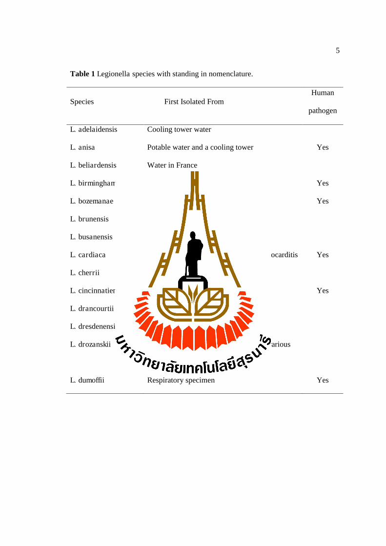
species and is subdivided into 70 distinct serogroups (Table 1). Legionella
pneumophila (serogroup 1) is the most common and be the major genus that causes
the disease. Legionella is the aerobic and fastidious bacteria which its nature will not
grow on traditional bacteriological media but it requires an enriched medium
supplemented with L-cysteine and ferric salts. The optimal growth temperature for
Legionella is 35 °C. Legionella is catalase-positive and unable to reduce nitrate. This
bacterium also does not utilize carbohydrates by either oxidation or fermentation
(Percival and Williams, 2014).
5
Table 1 Legionella species with standing in nomenclature.
Species First Isolated From Human
pathogen
L. adelaidensis
L. anisa
L. beliardensis
L. birminghamensis
L. bozemanae
L. brunensis
L. busanensis
L. cardiaca
L. cherrii
L. cincinnatiensis
L. drancourtii
L. dresdenensis
L. drozanskii
L. dumoffii
Cooling tower water
Potable water and a cooling tower
Water in France
Cardiac transplant recipient
Respiratory specimen
Cooling tower water
Cooling tower water in Korea
Isolated from a case of native valve endocarditis
Water, thermally altered
Pneumonia patient
Water in UK
River wate
Isolated via amoebal enrichment from various
sources in the UK
Respiratory specimen
Yes
Yes
Yes
Yes
Yes
Yes

6
Table 1 (Continued) Legionella species with standing in nomenclature.
Species First Isolated From Human
pathogen
L. erythra
L. fairfieldensis
L. fallonii
L. pneumophila
L. quateirensis
L. quinlivanii
L. rowbothamii
L. rubrilucens
L. sainthelensi
L. santicrucis
L. shakespearei
L. spiritensis
L. steelei
Water, cooling tower
Cooling tower water in Australia
Isolated via amoebal enrichment from various
sources in the UK
Pneumonia patient
Water, shower in bathroom
Water
Isolated via amoebal enrichment from various
sources in the UK
Tap water
Water near Mt. St. Helens
Tap water
Water, cooling tower
Water, lake
Human respiratory specimen
Yes
Yes
Yes
Yes

7
Table 1 (Continued) Legionella species with standing in nomenclature.
Species First Isolated From Human
pathogen
L. steigerwaltii
L. taurinensis
L. tucsonensis
L. tunisiensis
L. wadsworthii
L. waltersii
L. worsleiensis
L. yabuuchiae
Tap water
Water in Italy
Human, renal transplant recipient
Environmental water
Pneumonia patient
Water in Australia
Return flow of cooling tower water
Soil contaminated with industrial wastes in
Japan
Yes
Yes
Yes
Source : (Nazarian, De Jesus and Musser, 2015).
2.2 Legionella ecology
Legionella pneumophila is found in the natural aquatic environment and this
bacterium is capable to survive in the extreme ranges of the environmental conditions
(Fliermans et al., 1981). The natural reservoirs of Legionella are freshwater systems
such as rivers, lakes or thermal waters. Apart from their natural habitat, Legionella
bacteria is also able to colonize in the man-made water systems such as air cooling
towers, conditioning systems, hot water systems, vegetable misters, whirlpools and
dental-unit water lines (Guyard and Low, 2011). Although, Legionella can be found
in water ranging from cold to very hot, its multiplication is restrictive to temperature
between 25-42 °C with an optimal growth at 35 °C (Fields, 2008) and does not

8
multiply at temperature below 20 °C. The Legionella can survive as intracellular
parasites of protozoa, amoebae, ciliated or slime moulds, when the temperature of
aquatic environments changes, it can shift the balance between protozoa and bacteria,
resulting in rapid multiplication of Legionellae, which is the etiology of the human
disease.
Some outbreaks of Legionellosis associated with construction, and can be
transmitted to humans via soil or containing microorganism by not washing hands
after gardening. However, L. pneumophila does not survive in dry environments and
the outbreaks are more likely the result of massive descalement of plumbing systems
due to changes in water pressure during construction (Fields, Benson and Besser,
2002).
2.3 Pathogenesis
Legionella pneumophila serogroup 1 is the most virulent Legionella species
and the most common cause of disease. The infection of Legionella is commonly
found through inhalation of contaminated aerosols produced by water systems such as
cooling towers, showers and faucets. Other modes of transmission of Legionella are
respiratory tract manipulations. Person to person transmission has not been reported
both of Pontiac fever and Legionnaires’ disease (Guyard and Low, 2011). The
Legionella can be found naturally in freshwater and acts as a parasite of amoebae. If
inhaled into the lung, Legionella can replicate within the alveolar macrophages
(Swanson and Hammer, 2000).

9
Figure 1 Legionella pneumophila life cycle in amoebae
(Swanson and Hammer, 2000).
The pathology of Legionellosis is similar to all Legionella spp. There are
heavy inflammatory infiltrations including neutrophils and macrophages, abscess
formation, necrosis, inflammation of small blood vessels (Lau and Ashbolt, 2009) and
other clinical symptoms such as pneumonia.
2.4 Legionellosis
Legionellosis is the disease caused by Legionella. Over 90% of cases of
Legionellosis are caused by L. pneumophila and other species include L. longbeachae,
L. feeleii, L. micdadei and L. anisa which are the causative agents of a less severe
infection known as Pontiac fever. The Legionellosis is divided into two distinct
clinical entities, Legionnaires’ disease, a severe multisystem disease involving
pneumonia and Pontiac fever, a self-limited flu-like illness (Fields, Benson and
Besser, 2002). The incubation period for Legionnaires’ disease is typically 2–14 days,
10
with the infection lasting weeks to months. In Pontiac fever, symptoms include fever,
chills, myalgia and headache. The incubation period for Pontiac fever is 5–66 hours
and symptoms last for 2–7 days (Percival and Williams, 2014). The
Legionnaires’disease presents with a broad spectrum of illness, ranging from a mild
cough and low-grade fever to stupor, respiratory failure, and multiorgan failure. In the
early illness, patients have nonspecific symptoms including fever, malaise, myalgias,
anorexia, and headache (Table 2). The temperature often exceeds 40 °C (Stout and
Yu, 1997).
Table 2 Symptoms associated with Legionellosis.
Legionnaires’ disease
Mild cough to a raidly fatal pneumonia. Death occurs through progressive pneumonia
with respiratory failure and/or shock, acute kidney and multi-organ failure
Incubation period: 2-10 days (up to 16 days in recent outbreaks)
- Fever
- Headache
- Loss of appetite
- Malaise
- Lethargy
In some cases:
- Diarrhea
- Muscle pain
- Confusion
11
Table 2 (Continued) Symptoms associated with Legionellosis.
- Initial mild cough
- Phlegm (up to 50% of patients)
- Blood-streaked phlegm or hemoptysis (1/3 of the patients)
Pontiac fever
Acute self-limiting influenza-like illness lasting 2-5 days
Incubation period: few to 48 h.
- Fever
- Chills
- Headache
- Malaise
- Myalgia
- Not fatal
Source: (Percival and Williams, 2014)
The Legionnaires’disease can generate multilobar in the lungs, with focal or
lobar consolidation presenting as either red or grey hepatization. Acute renal failure,
shock, disseminated intravascular coagulation, coma, respiratory insufficiency and
circulatory collapse are the major factors associated with death (Percival and
Williams, 2014).
There are many risk factors that cause Legionellosis such as the people aged
50 years old or over, smoking or having smoked heavily in the past, drinking alcohol
heavily, including people who have an underlying medical conditions, such as
diabetes, kidney disease or a pre-existing lung condition and having a weak immune
system for example, people with AIDS or cancer.
( ) .

12
For the treatment of Pontiac fever, treatment does not use antibiotics because
it is a self-limited illness and recovery usually occurs within 1 week. The
Legionnaires disease is treated with antibiotics, the two most potent classes of
antibiotics are the macrolides (azithromycin) and the quinolones (ciprofloxacin,
levofloxacin, moxifloxacin, gemifloxacin, trovofloxacin). Other agents that have been
shown to be effective include tetracycline, doxycycline, minocycline, trimethoprim
and sulfamethoxazole. Macrolides (azithromycin) is the drug of choice for children
with suspected or confirmed Legionnaires’disease and quinolones (levofloxacin,
moxifloxacin) are recommended for adults with severe disease. Both of antibiotics are
highly effective and have few side-effects more than other drugs, so, they become to
be antilegionella drugs in healthy and immunocompromised individuals. The
recommended duration of therapy is 5-14 days if azithromycin is used. For the
patients with severe disease or immunocompromised patients should be 2-3 weeks.
(Phin et al., 2014)
2.5 Amplification factor
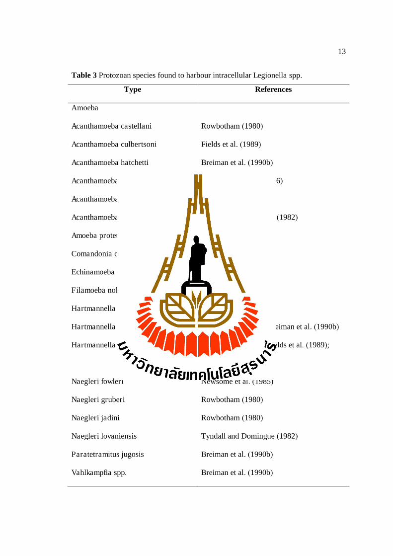
2.5.1 Protozoa associations
Legionella can alive intracellular protozoan parasites (Kwaik et al., 1998) and
the protected environment provided by the protozoan envelope reduces its
susceptibility to disinfection and other harmful conditions. Legionella is residing
within at least 20 species of amoebae, two species of ciliated protozoa and one species
of slime mould (Table 3).
13
Table 3 Protozoan species found to harbour intracellular Legionella spp.
Type References
Amoeba
Acanthamoeba castellani
Acanthamoeba culbertsoni
Acanthamoeba hatchetti
Acanthamoeba polyphaga
Acanthamoeba palestinensis
Acanthamoeba royreba
Amoeba proteus strain x D
Comandonia operculata
Echinamoeba exudans
Filamoeba nolandi
Hartmannella spp.
Hartmannella cantabrigiensis
Hartmannella vermiformis
Naegleri fowleri
Naegleri gruberi
Naegleri jadini
Naegleri lovaniensis
Paratetramitus jugosis
Vahlkampfia spp.
Rowbotham (1980)
Fields et al. (1989)
Breiman et al. (1990b)
Rowbotham (1980, 1986)
Rowbotham (1986)
Tyndall and Domingue (1982)
Park et al. (2004)
Breiman et al. (1990b)
Fields et al. (1989)
Breiman et al. (1990b)
Fields et al. (1989)
Rowbotham (1986); Breiman et al. (1990b)
Rowbotham (1986); Fields et al. (1989);
Breiman et al. (1990b)
Newsome et al. (1985)
Rowbotham (1980)
Rowbotham (1980)
Tyndall and Domingue (1982)
Breiman et al. (1990b)
Breiman et al. (1990b)

14
Table 3 (Continued) Protozoan species found to harbour intracellular Legionella spp.
Type References
Vahlkampfia jugosa
Vahlkampfia ustiana
Ciliate
Tetrahymena pyriformis
Tetrahymena thermophile
Slime Mould
Dictyostelium discoideum
Rowbotham (1986)
Breiman et al. (1990b)
Fields et al. (1984)
Kikuhara et al. (1994)
Hagele et al. (2000)
Source: (Lau and Ashbolt, 2009)
2.5.2 Biofilm associations
Biofilms are defined as complex microbial communities featured by cells that
are attached to a substratum and to each other by process of a matrix of self-produced
extracellular polymeric substances (EPS) (Declerck, 2010). Biofilm formation can
occur worldwide in natural and artificial environments, and on a range of different
surfaces. Microorganisms, including L. pneumophila, form biofilms as a mechanism
to withstand adverse conditions, such as low nutrients or temperature extremes.
Surface adherence commonly occurs by process of an extracellular polysaccharide
substance (EPS) secreted by the cells. This substance (the glycocalyx, or slime) is a
hydrated polyanionic polysaccharide matrix produced by polymerases affixed to the
lipopolysaccharide component of the cell wall (Bartram, 2007).
15
There are five recognized stages in the development of biofilm as follow:
1) Initial reversible attachment of free swimming microorganisms to surface
2) Permanent chemical attachment, single layer, bugs begin making slime
3) Early vertical development
4) Multiple towers with channels between maturing biofilm
5) Mature biofilm with seeding/dispersal of more free swimming
microorganisms
The biofilms not only provide a source of nutrients for Legionella but also
protect them from the antibiotics and other biocides. Biofilm prevention is an
important role to control the proliferation of Legionella and is considered to be vital to
control the Legionellosis.
2.5.3 Algal associations
Algae are the most abundant biofilm forming organisms on earth. On the
surface water, algae can be both in planktonic form and biofilms. In biofilms, they
may contain species which form toxins such as microcystin and represent a serious
threat to human health (Wingender and Flemming, 2011). The Legionella have
symbiotic relationship with some algae and Cyanobacteria, which may involve
phosphorus metabolism and photosynthesis on the surface. Additionally, algal
photosynthetic activity provides oxygen that can be used in aerobic respiration, which
in turn produces CO2, which may be available for algal photosynthesis. This
mutualistic association between algae, Cyanobacteria and Legionella may occur in
natural planktonic communities. The communities of cyanobacteria are Fischerella
sp., Phornidium sp. and Oscillatoria sp. (Tison, Pope, Cherry and Fliermans, 1980).
Recently, a highly sensitive amperometric immunosensor for microcystin detection in

16
algae and their biofilms has been reported. However, drinking water distribution
systems and installations, algae do not occur due to lack of light (Wingender and
Flemming, 2011). From the above data, the relationship of algae, cyanobacteria and
Legionella may play an important role in the colonization and dispersal of Legionella
in water systems.
2.6 Distribution of the Legionella spp. to humans
The transmission mode of Legionellosis is inhalation of Legionella organisms
by contaminated water aerosols or occasionally via direct inoculation of Legionella
into the wound. The transmission can occur from hospital potable hot water sources,
potentially via shower aerosols (Hanrahan et al., 1987). The source of aerosol
transmission of Legionella commonly found in cooling tower, shower, respiratory
therapy device, swimming pool and fountain et cetera, which are shown in Table 4.
Table 4 Source of aerosol transmission of Legionella spp.
Source/reservoir Likely mode of transmission
Taps
Tap water
Direct wound contact—use of contaminated water
to bathe patients
Showers
Hot water supply
Baths
Re-circulating hot water
Inhalation of aerosols generated by shower nozzle
Inhalation of aerosols generated by all-day-
running-hot-water bath

17
Table 4 (Continue) Source of aerosol transmission of Legionella spp.
Source/reservoir Likely mode of transmission
Water supply
Respiratory equipment
Re-usable oxygen humidifier
Aspiration of contaminated water during delivery
(birthing pool)
Inadequate cleaning/disinfection; inhalation of
contaminated aerosols
Nebulizer
Room humidifier
Water features
Decorative fountain
Malfunction of water distillation system; inhalation
of contaminated aerosols
Use of contaminated tap water to fill reservoir;
inhalation of contaminated [cold mist] aerosol
Stagnation of water during maintenance; inhalation
of contaminated aerosol
Source: (Moore and Walker, 2014)
From the previous studies, In Italy (2014), they surveyed ten healthcare
facilities to provide more information on the distribution of Legionella spp. by
collected samples from air and water. They found 78.6% of L.pneumophila serogroup
6 (Lpn sg 6), 9.5% of Lpn sg 9, 5.5% of Lpn sg 1, 5.5% of Lpn sg 7 and 0.8% of Lpn
sg 1and 12. These results showed that Lpn sg 6 was the serogroup, mostly found in
water samples (Montagna et al., 2016).
In Canada, they collected 101 spa water samples and identified quantification
of Legionella spp. by real-time PCR method compare with conventional culture

18
method. They found 13.86% (14 from 101) by culture method and 41.58% (42 from
101) by real-time PCR method. These two methods had low correlation. (Guillemet et
al., 2010).
In Taiwan, they studied about distribution of Legionella in hot tub, spa and
swimming pool by collected samples from 91 sites. They found Legionella in 21 sites
(23%) and the most frequently detected was L.pneumophila. Moreover, they found
Legionella in water temperature ranging from 22-50 °C and pH parameter found in
range 5.0 to 9.0 (Hsu et al., 2006).
2.7 Distributions of Legionella spp. in Thailand
The Legionella spp. could be detected firstly in Thailand in 1984 and have
been isolated in several regions of Thailand. The total number of cases during 1984-
2002 were 17 patients and the most of Legionella species that caused the disease was
Legionella pneumophila (Bovornkitti, 2010) .
Tishyadhigama et al. (1995) had surveyed for the contamination of
Legionella in the environmental sources and cooling towers in several regions of
Thailand. They found 57% of 94 cooling towers and 21.8% of 78 other environmental
sources. The Legionella pneumophila serogroup 1 was the most of organisms
predominating both in the cooling towers and other environmental sources.
Lertkhanawanichakul et al. (2004) had investigated Legionella spp. from the
environments at Walailuk University in Nakronsrithammaraj provice. The samples
were collected from the natural environmental air and man-made aquatic
environments, including biofilm of potables. They found Legionella spp., 2 of 76
water samples (2.6%) from the environmental sources and 3 of 62 air samples (3.2%)
but could not found Legionella spp. in the 30 biofilm samples. In addition, another

19
microorganisms (i.e. Acinetobacter, Pseudomonas, Staphylococcus and mold) were
found in many samples. The exposure to high dose of microorganisms can lead to be
the nosocomial infections in the immunocompromised patients.
Paveenkittiporn, Dejsirilert and Kalambaheti (2012) surveyed for Legionella
organisms during 2003–2007 from various water resources from 33 provinces in
Thailand. The samples were collected from cooling towers, storage tanks, chiller
systems, hot springs, tap water, ponds, drinking-water containers and showers. The
Legionella were firstly confirmed as Legionella species and identified as L.
pneumophila based on PCR. The 256 isolates were confirmed as Legionella species.
Among, 206 isolates (80.47%) were belonged to L. pneumophila and 50 isolates
(19.53%) were identified as non-pneumophila when the samples were detected by
DNA tree analysis.
Phares et al. (2007) studied the Legionella surveillance in 3489 patients with
clinically-defined pneumonia in Sa Kaeo, the rural province in Thailand for 1 year.
The samples were collected from sera, nasopharyngeal swabs, and urines for
immunologic and molecular tests. Incidence of pneumonia was reported as a range
from the lower limit to upper limit. The results showed that the incidence of
pneumonia requiring hospitalization that was caused by Legionella longbeachae were
5–29 cases per 100,000 pneumonia patient population and no case of Legionella
pneumophila pneumonia was observed. Other pathogenic microorganisms such as
Mycoplasma pneumoniae and Chlamydia pneumoniae were frequently associated
with severe pneumonia in Sa Kaeo too. But there were few patients who received
antibiotics before collecting specimens, thus, these might cover atypical pathogens.
20
Buathong et al. (2013) had investigated Legionnaires’ disease outbreak among
EU travelers and hotel staffs in Phuket during 2006-2007. The information of each
hotel guest was provided by home country officials for enquiring any symptoms after
staying at the Phuket hotels. The water samples were collected from rooms and
cooling towers in the hotel for Legionella cultures. Hotel staffs were tested for the
Legionella pneumophila antibody by indirect fluorescent antibody (IFA) technique to
identify the risk factors among hotel workers. The result showed that 5 confirmed
cases (0.78%) and 1 presumptive case (0.16%) of Legionnaires’ disease were traced
from 645 Scandinavians staying at the hotels in Phuket. Among 118 hotel staffs, 78
cases (66.10%) had positive titer. The risk factors of Legionella infection were
showers in the hotels which had Legionella and people aged more than 45 years old
were group of increased risk for Legionella spp. infection.
In 2016, the Regional Medical Sciences Center 11/1, Phuket has been
investigating the outbreak of Legionella spp. in Phuket, Phang-nga and Krabi
provinces. The most common sources were water from showers, spas and faucets.
They collected 1,508 water samples and found 116 samples positive for Legionella
spp. but the amount of bacteria was not high enough to cause the disease in human.
(Karnchanapimai et al., 2016)
2.8 Methods for Legionella detection
2.8.1 Cultural method
The Legionella detection method often uses the culture method which is the
gold standard for the identification of Legionella spp. The first solid medium that is
Mueller-Hinton agar supplemented with 1% IsoVitaleX and 1% hemoglobin (MHIH )
21
(Fields, Benson and Besser, 2002). Then L-cysteine hydrochloride can replace the
IsoVitaleX reagent, and soluble ferric pyrophosphate can replace hemoglobin
(Cordes et al., 1981). Later, starch is replaced with charcoal to detoxify the medium
and the amino acid source is changed to be yeast extract, so a result is charcoal yeast
extract agar (Feeley et al., 1979). The medium has been improved several times, until
resulting in the medium currently used, buffered charcoal-yeast extract (BCYE) agar
enriched with g - ketoglutarate (Edelstein, 1982). Legionella can be isolated from
environmental water, water systems and specimens, including blood, lung tissue, lung
biopsy specimens, respiratory secretions and stool. The antibiotic-containing media
which perform better than the others for growing the stock strains and the clinical
specimens contained with cefamandole, polymyxin B, anisomycin, organic buffer and
g-ketoglutarate (Edelstein, 1981). For the water samples, BCYE agar containing
glycine, vancomycin, polymyxin B and cycloheximide (GVPC) is a selective medium
and suitable for Legionellae growing. The glycine, vancomycin and polymyxin B
inhibit most non-target bacterial species, both gram-positive and gram-negative,
including common contaminants such as Enterococci, Coliform, and Pseudomonas
spp, while cycloheximide suppresses the growth of yeasts and moulds. These plates
are incubated at 35 °C in a humid 2-5% CO2 environment and examined after 4, 8 and
14 days of incubation (Leoni and Legnani, 2001).
The Legionella spp. generally produce small, blue-gray colonies, slow
growing and have ground – glass appearance when examine with dissecting
microscope. The suspected colonies are subcultured on BCYE agar, with and without
cysteine. The Legionella can grow on BCYE with cysteine, but not grow on the
BCYE without cysteine. The Legionella will be confirmed with biochemical test

22
(Hippurate hydrolysis) (Leoni and Legnani, 2001). The positive reaction performs a
purple, a very light purple will be designated as weakly positive and shades of gray or
a very light yellow will be reported as negative for hippurate hydrolysis.
2.8.2 Non-cultural methods
The several non-cultural methods have been developed to detect Legionella in
environmental samples because the cultural method must wait for several days for
growing Legionella. The non-cultural methods offer the potential of increased
sensitivity and have a specificity more than the cultural method. However, the non-
cultural methods have the disadvantage since they cannot provide the information
regarding the viability of Legionella. The several non-cultural methods include, direct
fluorescent antibody (DFA) staining, serological diagnosis (IFA and ELISA), urine
antigen detection and detection of Legionella nucleic acid by polymerase chain
reaction (PCR).
For the clinical and environmental samples, PCR has been successfully used to
detect Legionella DNA and it is the rapid test for diagnosis of Legionellosis. There
are several techniques available using rRNA (ribosomal RNA): 5S rRNA, 16S rRNA
and mip gene (macrophage infectivity potentiator) used as target for PCR. Inoue,
Takama, Yoshizaki and Agata (2015) had detected Legionella species in water
samples and cooling tower water samples by using a combination of conventional
plate culture, quantitative polymerase chain reaction (qPCR) and qPCR combined
with ethidium monoazide treatment (EMA-qPCR) methods. The results showed that,
EMA treatment decreased the number of Legionella-positive bath water samples
detected by qPCR. In contrast, EMA treatment had no effect on cooling tower water
23
samples. So, EMA-qPCR is a useful method for the rapid detection of viable
Legionella spp. from cooling tower water samples.
2.9 Legionella disinfection methods
The Legionella bacteria can cause Legionnaires’ disease and Pontiac fever.
This bacteria is commonly found in the natural water environment and water
distribution systems. The water distribution systems have been reported that they are
the sources of bacterial infections (Moore and Walker, 2014). So, the water systems
need to get rid of bacteria. There are many disinfection methods involving thermal
and chemical methods. For the disinfection of drinking water, chemical methods
using disinfectants have been the most widely used (Kim, Anderson, Mueller, Gaines
and Kendall, 2002).
Chemical methods
Chlorine is an oxidizing agent that efficiently uses as a disinfectant for
controlling pathogens in domestic drinking water. The shock hyperchlorination is
used to inactivate Legionella. Shock hyperchlorination is used by pulse injection of
chlorine in water to achieve concentration of chlorine 20-50 ppm though out the
system. After that water is drained and the system is mixed with water, the residual
chlorine will return to normal concentration (0.5-1 ppm) (Lin, Stout, Yu and Vidic,
1998). When the shock hyperchlorination kills the Legionella bacteria in the water,
then biofilm reduces dramatically. The performance of chlorine is more effective at
higher temperature and higher pH.
Thermal methods
The thermal methods start with flushing all water outlets, faucets, and shower
heads more than 30 min at >60 °C (140 °F) at distal outlets. At this temperature,

24
Legionella colonized in these sites are killed (Kim, Anderson, Mueller, Gaines and
Kendall, 2002).
2.10 Microbiological evaluation of water sample quality
The microbiological parameters of water samples are compared with the
standard of tap water recommended by Metropolitan Waterworks Authority, Thailand
(based on WHO guideline 2011). The WHO’s guideline has recommended the
limitation of the water quality in microbiological parameters that tap water must not
have any E.coli in 100 ml of water sample.
2.11 Research objectives
1. To detect Legionella spp. and other bacterial pathogens in water systems of
nursing homes and spa pools in Bangkok and Nakhon Ratchasima provinces.
2. To prevent infections of Legionella and other bacterial pathogens in the
elders in nursing homes and visitors who came to the swimming pools and spa, if
microorganisms were found more than the accepted standards, these results were
informed to the related persons to get rid of these microorganisms. After treatments,
the samples at the infected sites were investigated again in order to eliminate the
source of infections.
3. To determine the relationships between water parameters (temperature and
pH value) and the prevalence of Legionella spp.
25
2.12 Research hypothesis
The detection of Legionella and other bacterial pathogens would be found in
water systems of nursing homes and spa pools. After the suggestion and
decontamination of Legionella, the samples sites that contaminated would be
decreased.
CHAPTER III
MATERIALS AND METHODS
3.1 Preparation of the Legionella pneumophila bacteria, chemicals
and reagents
Legionella pneumophila serogroup 1 ATCC 33152 were obtained from The
Center of Scientific and Technological Equipment, Suranaree University of
Technology. These Legionella pneumophila bacteria was used to be positive control.
All chemicals and reagents used in this work were the laboratory grades or analytical
grades, purchased from Himedia, Sigma-Aldrich and Amresco.
3.2 Instrumentation
Instruments for the detection of Legionella spp. in water samples from nursing
homes and spa pools were located in the Instrument Building of the Center for
Scientific and Technology Equipment, Suranaree University of Technology, Nakhon
Ratchasima province, Thailand
27
3.3 Samples collection and processing
Water samples were collected from the nursing homes and spa pools in
Nakhon Ratchasima and Bangkok province, Thailand. Sixty samples were collected
for detection of Legionella spp. and other bacterial pathogens, including viable
heterotrophic bacteria, gram - negative bacteria, Staphylococcus spp. and Coliform
that could cause the diseases. The water sample sites were shower heads, faucets and
spa pools that could generate aerosol to the possibly exposed persons.
3.3.1 Shower heads and faucets
Water and biofilm samples from shower heads and sink faucets were collected
by modified method of Cordes et al. (1981). The water samples were collected
approximately 500 ml in the steriled containers.
3.3.2 Spa pools
Water samples from spa pools were collected approximately 500 ml in the
sterile containers and stored samples at room temperature during transporting to the
laboratory.
3.3.3 Samples processing
Each sample of water was collected in a sterile container which had 1 ml of a
10 mg/ml solution of sodium thiosulfate (Na2S2O3) to neutralize residual
disinfectants. The water temperature and pH value were determined immediately after
collection (Nostro, Checchi, Ducci and Pesavento, 2011). The water was carried in the
insulated containers at room temperature to the laboratory and processed within 24 h.
The water samples were concentrated by filtration through 0.22 たm pore size cellulose
acetate membrane filters (Millipore S.p.A., Milan, Italy) (Nostro, Checchi, Ducci and

28
Pesavento, 2011) and the membrane filters were cut into small pieces with aseptic
technique, then put into a sterile tube that containing 1.5 ml sterile distilled water and
vortexed for 30 seconds to remove bacterial cells from the membrane filters.
3.4 Microbiological analysis
3.4.1 Detection of Legionella species
The 1.5 ml of acid solution (HCl - KCl solution pH 2.2) were added to the
concentrated water samples (from 3.3.3) for 5 minutes, then pipetted 1 ml to another
tube that already contained 9 ml of sterile distilled water. The treatment water samples
were tested by spread plate technique at undiluted and 10-1 dilution, 0.1 ml of each
sample was placed in duplicate on Buffered Charcoal Yeast Extract (BCYE) agar and
BCYE agar + glycine, vancomycin, polymyxin B and cycloheximide ( GVPC)
because no one medium will be optimal for the recovery of Legionella from every
environmental site; so different selective media with various antibiotic combination in
a BCYE were necessary. These plates were incubated at 37 °C in the humid chamber
for 3-4 days. If there were Legionella bacteria, the blue-gray bacterial colonies would
presence when using stereo microscope and ground – glass appearance when using
dissecting microscope. The suspect colonies were cultured on BCYE and BCYE
without L-cysteine for testing the requirement of cysteine by streak plate technique
and incubated at 35 °C for 4 days. Legionella spp. were grown on BCYE but were not
grown on BCYE without L-cysteine. L. pneumophila serogroup 1 ATCC 33152 were
used as positive control. The biochemical tests were used to identifiy L. pneumophila
from other legionellae by hippurate hydrolysis reaction (Hebert, 1981).

29
The suspect colony was selected from BCYE and emulsified in microcentrifuge tube
containing 0.4 ml of 1% sodium hippurate. The suspension was placed in an incubator
at 37 °C. After 18 to 20 h of incubation, 0.2 ml of the ninhydrin solution was added to
each microcentrifuge tube. The contents were mixed by shaking and returned to the
incubator for 10 min, then observed the color development within 20 minutes; all
shades of purple will be read as a positive reaction, a very light purple was designated
as weakly positive, and shades of gray or a very light yellow were reported as
negative for hippurate hydrolysis. The number of typical colonies of Legionella spp.
and L. pneumophila were counted, and reported as colony forming units per ml
(CFU/ml).
3.4.2 Isolation and quantitation of total heterotrophic plate count
The determinations of heterotrophic bacteria were analyzed by 10-fold
dilution series of the concentrate water sample. The 0.1 ml of concentrated water
samples were cultured duplicate on plate count agar (PCA) with spread plate
technique. All plates were incubated at 35 °C for 24 – 48 h (Reasoner, 2004). The
number of colonies were counted and reported as colony forming unit per ml
(CFU/ml).
3.4.3 Isolation of gram - negative bacteria
The gram – negative bacteria were cultured by spreading 0.1 ml of
concentrated water samples on the Mac Conkey agar in duplicate. All plates were
incubated at 35 °C for 24 h. The colonies of gram – negative were identified by
morphology and biochemical tests (gram stain, oxidase test, catalase test, motility
indole lysine test, OF-glucose test, simmons citrate agar and triple sugar iron agar,
30
which showed in Appendix D). The gram – negative bacteria were reported in genus
by evaluated from Table 5.
3.4.4 Isolation of Staphylococcus spp.
The isolation of Staphylococcus spp. was analyzed by spread 0.1 ml of
concentrated water samples on the selective medium, Manitol salt agar in duplicate.
All plates were incubated at 35 °C for 24 h. The Staphylococcus aureus produced
yellow colonies with yellow zones, there used for the selective isolation of
presumptive pathogenic Staphylococcus species. The colonies of Staphylococcus spp.
were confirmed by morphology and biochemical tests (gram stain and catalase test as
shown in the Appendix D).
3.4.5 Isolation of Coliform and E.coli bacteria
The determination of Coliform bacteria was analyzed by inoculate sample
water to lactose broth and incubated at 35 °C for 24-48 h. The positive tubes had a
turbidity and produced gas within durham tube. The isolation of E.coli was analyzed
by inoculated the solution in the positive tube to EC medium, streaked plate on eosin
methylene blue agar (EMB), confirmed with urea test, gram stain and catalase test
(Appendix D),then evaluated from Table 5.

32
31
Table 5 Biochemical test of gram-negative bacteria.
+ = Positive - = Negative Y = yellow R = red
v = variable (some strains positive, others strains negative)
d = result different in different species or strain
No Organism Lactose Oxidase Catalase Motility Indole Urease Triple sugar iron Simmon
citrate Butt Slant Gas H2S
1 E.coli + - + + + - Y Y + - - 2 Klebsella + - + - + + Y Y + - + 3 Enterobacter spp. + - + + - - Y Y + - + 4 Citrobacter + - + + - d Y Y/R + d + 5 Salmonella Typhi - - + + - - Y R - + -
6 Salmonella Parayphi-A
- - + + - - Y R + - -
7 S.typhi marium and other
- - + + - - Y R d + d
8 Shigella spp. - - + - d - Y R - d - 9 Proteus - - + + v + Y R + + d 10 Pseudomonas spp. - + + + - d R R - - + 11 Vibrio cholerae - + + + + - Y Y - - d 12 Paraheamolyticus - + + + + - Y Y - - d 13 Serratia mercescus - - + d - d Y R - - +
14 Yersina enterocolitire
- - + + d + Y R - - -
15 Providencia - - + + + - Y R - - +
32
3.4.6 Repeated cultivation after decontamination of Legionella spp. at
positive sites
The positive sites of Legionella spp. were reported to the nursing home and
spa managers. The elimination of pathogens were done according to the
recommended method of Bureau of food and water sanitation, Department of Health,
Ministry of Public Health (Table 6). One month after elimination of pathogens, the
repeated samples were collected and cultured again to prove that the tentative
pathogenic microorganisms were destroyed completely.
3.4.7 The decontamination of Legionella spp.
The water disinfection recommended by Bureau of food and water sanitation,
Department of Health, Ministry of Public Health.
Chemical methods
Chlorine powder is a white powder or white scales. The chlorine has to
dissolve in the water and use the supernatant for disinfection.
- Prepare water into the glass or bucket, put the chlorine powder and mix with
the water thoroughly until dissolve.
- Leave it until the undissolved powder precipitate.
- Add the supernatant into the jar or tank. Mix well. The amount and duration
for elimination the pathogens was shown in Table 6.
Caution
- Keep out of reach of children. Store in a dry place and away from sunlight.
- Do not touch chlorine by hand.
- Do not eat directly.
33
Table 6 Amount and duration for elimination the pathogens.
Concentration
of chlorine
Amount of
chlorine powder Water Duration Category of food
50 ppm Half teaspoon 20 liter 30 min. Vegetable, fruit
100 ppm A teaspoon 20 liter 30 min. Seafood
A teaspoon 20 liter 2 min. Container
A teaspoon 20 liter Cleaning Building
2 ppm A teaspoon 50 bucket 30 min. Drinking water-
water consumption
1/8 teaspoon 8 bucket 30 min. Drinking water-
water consumption
3.5 Relationships between water parameters and the prevalence of
Legionella pneumophila
The microbiological analysis was extended to the other informations by
investigated the relationships between Legionella pneumophila and other water
quality parameters (temperature and pH). The relationships were statistical analyzed
by linear regression analysis (Leoni et al., 2005).
CHAPTER IV
RESULTS AND DISCUSSION
4.1 Survey and collection of samples
The water samples for the detection of Legionella spp. were collected from
nursing homes and spa pools that could generate the aerosols with suspect of
Legionellae contamination in droplets to the exposal persons. A total of 60 water
samples sites were collected from showerheads, faucets and water tanks. The source
of water samples were shown in Table 7.
Table 7 Source of samples for detection of Legionella spp.
Source No. of samples
Nursing homes
Bangkok
- Showerheads
Nakhon Ratchasima
- Faucets
- Showerheads
- Water tanks
6
2
16
6
35
Table 7 (Continued) Source of samples for detection of Legionella spp.
4.2 Detection of Legionella species
Thirty water samples collected from nursing homes and other 30 water
samples from spa pools were examined for the detection of Legionella spp. by spread
plate technique on BCYE agar and GVPC agar (BCYE agar with glycine,
vancomycin, polymixin B and cycloheximide). Colonies of Legionella spp. on BCYE
and GVPC appeared to be blue-gray with slightly convex, circular and total with a
ground glass appearance (Figure 2). The suspected colonies were subsequently
stained with gram stain and the result indicated that they were gram-negative, thin
bacilli (Figure 3). Then, they were confirmed by sub–culturing on BCYE agar
supplemented with and without L-cysteine. Legionella spp. are able to grow only on
BCYE supplemented with L-cysteine. L. pneumophila was then distinguished from
other Legionella spp. by hippurate hydrolysis reaction.
Source No. of samples
Spa pools
Nakhon Ratchasima
- Faucets
30
Total 60
36
4.2.1 Nursing homes
A total of 30 water samples collected from nursing homes were examined for
the Legionellae. The results demonstrated 5 positive out of 30 samples (16.67%) and
were confirmed as L.pneumophila by hipurate hydrolysis reaction. These positive
samples were collected from showers and water tanks; 2 samples from showers and 3
samples from water tanks. The mean value of L.pneumophila cell density was 13.20
CFU/100ml on GVPC agar, whilst no colony was seen on BCYE agar.
4.2.2 Spa pools
Thirty water samples collected from spa pools were screened for the
Legionellae and detected for L.pneumophila by hipurate hydrolysis reaction. Thirteen
samples (43.33%) were confirmed as L.pneumophila. These confirmed positive
samples were collected from faucets of spa pools. The mean values of L.pneumophila
density were 94.50 CFU/100ml on BCYE agar with cysteine and 435.92 CFU/100ml
on GVPC agar, respectively. The L.pneumophila could grow well on GVPC agar
which was the suitable agar medium for the growth of L.pneumophila because the
antibiotics were added (Vancomycin, Polymyxin B and Cycloheximide) to inhibit
gram-positive, fungal and yeast that could interrupt the growth of L.pneumophila.
The densities of the contaminated samples at the sampling sites were
sumarized in Table 8.
37
Table 8 The density and interpretation level of the contaminated water samples from
GVPC agar.
Samples Density of L.pneumophila
(CFU/100ml)
Nursing homes
Showerhead N5 - Nursing homes No.3
Showerhead N13 - Nursing homes No.3
Water tank N27 - Nursing homes No.8
Water tank N28 - Nursing homes No.8
Water tank N29 - Nursing homes No.8
Spa pools
Faucet S3 – spa pools No.3
Faucet S4 – spa pools No.4
Faucet S5 – spa pools No.4
Faucet S6 – spa pools No.4
Faucet S13 – spa pools No.6
Faucet S14 – spa pools No.6
Faucet S24 – spa pools No.7
Faucet S25 – spa pools No.7
Faucet S26 – spa pools No.7
Faucet S27 – spa pools No.7
24
6
12
12
12
30
54
6
12
42
492
759
2,469
126
528
38
Table 8 (Continued) The density and interpretation level of the contaminated water
samples.
Samples Density of L.pneumophila
(CFU/100ml)
Spa pools
Faucet S28 – spa pools No.8
Faucet S29 – spa pools No.8
Faucet S30 – spa pools No.8
72
21
1,056
The relative risk assessments of hazard levels of Legionella pneumophila
The relative risk assessments of hazard levels of Legionella pneumophila were
categorized by Miller and Kenepp (1993) according to the density of L.pneumophila
in water samples of cooling towers associated with outbreaks of Legionnaires’disease.
The hazard levels of L.pneumophila were counted in CFU/100 ml. Our results of
L.pneumophila densities in positive samples were categorized for the risk assessment
and shown in Table 9 and Table 10.
39
Table 9 The relative risk assessment of Legionella pneumophila positive samples of
nursing homes.
Density of Legionella
pneumophila (CFU/100ml) Risk category a
Amount of contaminated
water samples
>100,000
10,000-99,999
1,000-9,999
100-999
<100
Very high
High
Moderate
Low
Very low
-
-
-
-
5
a The relative risk assessment according to Miller and Kenepp (1993)
This table showed that the densities of L.pneumophila in the all positive
samples of nursing homes were in the very low category (<100CFU/100ml) and none
were high or very high.

40
Table 10 The relative risk assessment of Legionella pneumophila positive samples of
spa pools.
Density of Legionella
pneumophila (CFU/100ml) Risk category a
Amount of
contaminated water
samples
>100,000
10,000-99,999
1,000-9,999
100-999
<100
Very high
High
Moderate
Low
Very low
-
-
2
4
7
a The relative risk assessment according to Miller and Kenepp (1993)
This table showed that the densities of L.pneumophila in the positive samples
of spa pools were mostly in the very low category (<100CFU/100ml), 2 of them were
in the moderate risk category, 4 of them were low and none were high or very high.
41
The results showed that 18 of 60 total samples were positive (30%). The
number of positive samples for Legionella spp. in this study was similar to other
studies that were carried out in Thailand. The prevalence of Legionella spp. in water
systems of hotels and resorts in the North-Eastern of Thailand was 24 from 75
(32.0%) hotels and resorts (A. Mahayotha, 2016). During 2003-2007, the prevalence
of Legionella spp. in various water resources from 33 provinces in Thailand were
investigated and 256 Legionella strains were isolated, among these, 206 isolates
(80%) were belonged to L. pneumophila and 50 isolates were identified as non-
pneumophila by DNA tree analysis. (Paveenkittiporn, 2012). In 2004, Borella et al.
studied Legionella infection risk from domestic hot water and found that 22.6%
(33/146) were Legionella spp. and 38.4% (56/146) were Pseudomonas spp.

42
Figure 2 Legionella spp. colonies grow on GVPC agar.
The characteristic of Legionella colonies were the blue-gray bacterial colonies,
glistening, convex, and circular with an entire edge.
Figure 3 Gram stain of Legionella spp. under compound light microscope
(1000x magnification).

43
4.3 Detection of other microorganisms
This research collected 60 samples in total from nursing homes and spa pools.
All samples were cultivated to screen for Legionella spp. and other microorganisms
including total heterotrophic bacteria, gram-negative bacteria, Staphylococcus spp.
and total Coliform bacteria.
The densities of heterotrophic bacteria contaminated in water samples
collected from nursing homes and spa pools are illustrated in Table 8. The average
mean of heterotrophic bacteria contained in water samples collected from nursing
homes was 1.05×10³ CFU/ml with ranged from 1.20 to 1.62×10⁴ CFU/ml. For spa
pools, the average mean was 8.61×10² CFU/ml with ranged from 6.00 to 1.22×10⁴
CFU/ml.
Not only heterotrophic bacteria but there were other microorganisms
contaminated in water samples. These microorganisms were found in water samples
from both nursing homes and spa pools; Coliform 16.67% ( 10 from 60 samples) ,
E.coli 5% (3 from 60 samples) , Staphylococcus spp. 16.67% (10 from 60 samples)
and some gram-negative bacteria including Pseudomonas spp. (35.00%),
Enterobacter spp. (8.33%), Citrobacter spp. (5.00%) and Acinetobacter spp. (8.33%).
The various microorganisms in each sample site were demonstrated in Table 11 and
Table 12.

44
44
Table 11 The microorganisms in the water samples of nursing homes.
Source Total plate count
(CFU/ml)
Legionella spp.
(CFU/100 ml) Gram-negative Staphylococcus spp. Coliform E.coli
N1
N2
N3
N4
N5
N6
N7
N8
N9
N10
N11
N12
N13
N14
32.20
39.50
6.40
6.48×10²
4.26×10³
1.96×10²
1.48×10²
11.70
1.16×10³
4.16×10²
4.64×10³
1.76×10²
37.90
38.30
-
-
-
-
24.00
-
-
-
-
-
-
-
6.00
-
E.coli, Enterobacter spp.
-
-
Pseudomonas spp.
Pseudomonas spp.
E.coli, Acinetobacter spp.
Enterobacter spp.,
Acinetobacter spp.
Enterobacter spp.
Acinetobacter spp.,
Pseudomonas spp.
Pseudomonas spp.
Pseudomonas spp.
-
-
Acinetobacter spp.
-
-
-
-
-
-
-
-
-
-
-
-
-
-
-
-
-
-
-
-
-
-
-
-
-
-
-
-
-
-
45
45
Table 11 (Continued) The microorganisms in the water samples of nursing homes.
Source Total plate count
(CFU/ml)
Legionella spp.
(CFU/100 ml) Gram-negative Staphylococcus spp. Coliform E.coli
N15
N16
N17
N18
N19
N20
N21
N22
N23
N24
N25
N26
N27
N28
N29
N30
37.50
53.80
4.11×10²
1.62×10⁴
2.53×10²
2.72×10³
1.20
26.50
2.40
1.40
22.00
1.80
7.70
18.20
2.30
1.50
-
-
-
-
-
-
-
-
-
-
-
-
12.00
12.00
12.00
-
-
Pseudomonas spp.
-
-
Pseudomonas spp.
-
-
-
-
Acinetobacter spp.
-
Pseudomonas spp.
E.coli
Enterobacter spp.
Pseudomonas spp.
Pseudomonas spp.
-
-
-
-
-
-
-
-
-
-
-
-
-
-
-
-
-
-
-
-
-
-
-
-
-
-
-
-
-
-
-
-
-
-
-
-
-
-
-
-
-
-
46
Table 12 The microorganisms in the water samples of spa pools.
Source Total plate count
(CFU/ml)
Legionella spp.
(CFU/100 ml) Gram-negative Staphylococcus spp. Coliform E.coli
Spa pools
S1
S2
S3
S4
S5
S6
S7
S8
S9
S10
S11
S12
S13
S14
22.60
1.93×10²
1.29×10²
2.94×10²
39.10
28.50
13.60
1.22×10⁴
2.98×10²
1.84×10³
32.70
40.80
2.00×10²
1.50×10²
-
-
30.00
54.00
6.00
12.00
-
-
-
-
-
-
42.00
4.92×10²
Pseudomonas spp.
-
Citrobacter spp.
Pseudomonas spp.
-
-
Pseudomonas spp.
Pseudomonas spp.
Pseudomonas spp.
Pseudomonas spp.
Pseudomonas spp.
-
-
-
-
-
-
-
-
-
-
-
-
-
-
-
-
-
-
-
-
-
-
-
-
-
-
-
-
-
-
-
-
-
-
-
-
-
-
-
-
-
-
-
46
47
Table 12 (Continued) The microorganisms in the water samples of spa pools.
Source Total plate count
(CFU/ml)
Legionella spp.
(CFU/100 ml) Gram-negative Staphylococcus spp. Coliform E.coli
S15
S16
S17
S18
S19
S20
S21
S22
S23
S24
S25
S26
S27
52.40
44.20
1.09×10²
5.70×10³
3.92×10²
2.23×10²
5.40×10²
31.60
6.00
53.00
2.18×10²
1.84×10²
2.82×10²
-
-
-
-
-
-
-
-
-
7.59×10²
2.47×10³
1.26×10²
5.28×10²
-
-
-
-
-
-
-
Pseudomonas spp.
Pseudomonas spp.
Citrobacter spp.
-
-
-
-
-
-
-
-
-
-
-
-
-
-
-
-
-
-
-
-
-
-
-
-
-
-
-
-
-
-
-
-
-
-
-
-
-
-
-
-
-
-
-
47
48
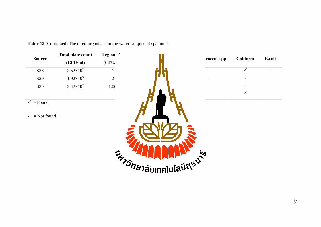
Table 12 (Continued) The microorganisms in the water samples of spa pools.
= Found
- = Not found
Source Total plate count
(CFU/ml)
Legionella spp.
(CFU/100 ml) Gram-negative Staphylococcus spp. Coliform E.coli
S28
S29
S30
2.52×10²
1.92×10³
3.42×10²
72.00
21.00
1.06×10³
Citrobacter spp.
Pseudomonas spp.
Pseudomonas spp.
Enterobacter spp.
-
-
-
-
-
-
-
-
48
49
Regarding to Table 11 and Table 12, the total plate count or heterotrophic
plate counts (HPC) is a method used to measure variety of bacteria that are common
in water for quality assessment of drinking water in storage tanks and in water
distribution systems. Hetero-trophic plate counts are not the indicators of pathogenic
conditions but some of them such as Pseudomonas spp. is the opportunistic pathogens
that can cause some infections in skin and lung and also the other, Aeromonas spp.
cause gastroenteritis (Amanidaz, Zafarzadeh and Mahvi, 2015). Therefore, if
heterotrophic plate counts are high then the risk are increase too. The National
primary drinking water regulations of Environmental Protection Agency, United
States of America recommended that heterotrophic plate counts should no more than
500 CFU/ml for safety water systems. Moreover, the Coliform bacteria were found in
water samples collected from nursing homes and spa pools. The Coliform bacteria
included Citrobacter spp., Enterobacter spp., Hafnia spp., Klebsiella spp. and E.coli.
They were found in intestines of human and warm-blooded animals, so they were
used as indicator for fecal contamination. The Enterobacter, Acinetobacter,
Citrobacter and E.coli were found to be 8.33%, 8.33%, 5% and 5%, respectively. The
Enterobacter, Citrobacter and E.coli could cause many diseases including septicemia,
pneumonia, meningitis and urinary tract infections. While the Acinetobacter could
cause a variety of diseases, ranging from pneumonia to serious blood or wound
infections. Therefore, the Queensland health swimming and spa pool water quality
and operational guidelines 2004 recommended the microbiological criteria that
thermo tolerant (fecal) Coliform or E.coli should not be detected in 100ml and also
Pseudomonas aeroginosa too, for reduce the risk contamination and potential for

50
illness. The Pseudomonas spp. were found to be the most common gram-negative
bacteria contaminated in water samples because Pseudomonas spp. were mostly
resistant to antibiotics and secreted extracellular enzyme, toxin and had ability to
develop biofilm on many surfaces, so the infections of Pseudomonas spp. might be
difficult to eradicate. They were found 35% (21 from 60 samples) and most of them
were found in showerheads and faucets from nursing homes.
These microorganisms found in this present study were similar to other
studies. The study of water system in ICU wards, hospitals in Tehran of Iran was
indicated that the Legionella pneumophila, Pseudomonas aeruginosa and
Acinetobacter were found 9.6%, 11.4% and 1.8%, respectively. The Legionella
pneumophila, Pseudomonas aeruginosa, and Acinetobacter baumanii could survive in
water released from their biofilm into the water stream. These posed a high risk of
infection to people. Legionellosis and other nosocomial waterborne infections were
occurred by the microorganisms presented and amplified in water reservoir,
associated with water biofilms, and the transmission of bacteria (aerosolization,
ingestion, and contact) (Yaslianifard, 2012).
In this study, the number of spa pools that found Legionella spp. was higher
than nursing homes since the water systems of spa pools were high temperature which
was an ideal temperature for Legionella spp. growing. Moreover, the usability of
water systems in spa pools might not be opened every day, thus, bacteria at the
faucets might accumulate and grow while the water systems of the nursing homes
normally were opened every day. Therefore, the spa pools had chances to find
Legionella spp. more than the nursing homes.
51
In this study, Staphylococcus spp. were found 16.67% (10 from 60 samples).
Comparing to Lechevallier and Seidler (1980), they found S.aureus 6.25% (20 from
320 samples) in rural drinking water. The Staphylococcus spp. is a gram-positive
bacteria that can cause a variety of diseases in human such as skin abscesses, pustules,
septicemia, enterocolitis, osteomylitis, and pneumonitis and is an agent of food
poisoning because they can produced endotoxin into the food that cause vomiting and
diarrhea (Lechevallier and Seidler, 1980).
52
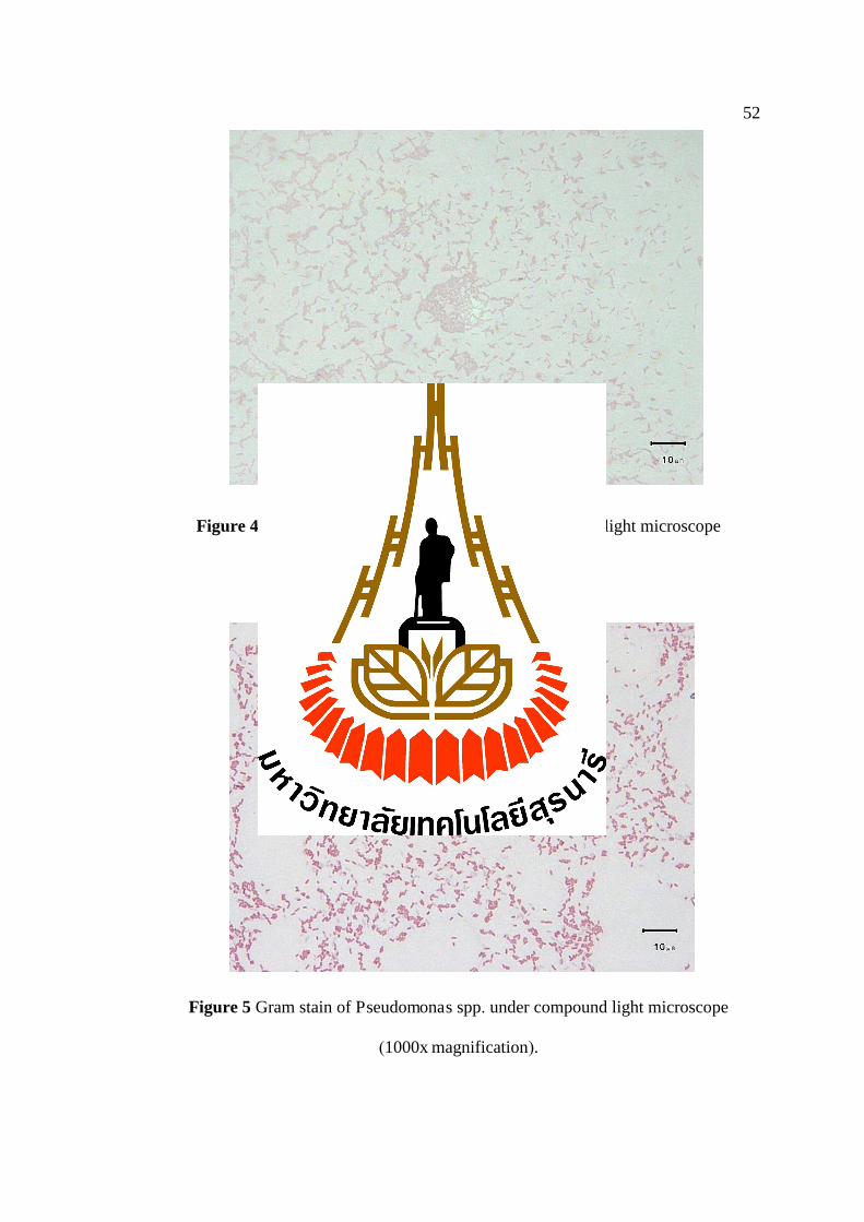
Figure 4 Gram stain of Citrobacter spp. under compound light microscope
(1000x magnification).
Figure 5 Gram stain of Pseudomonas spp. under compound light microscope
(1000x magnification).
53
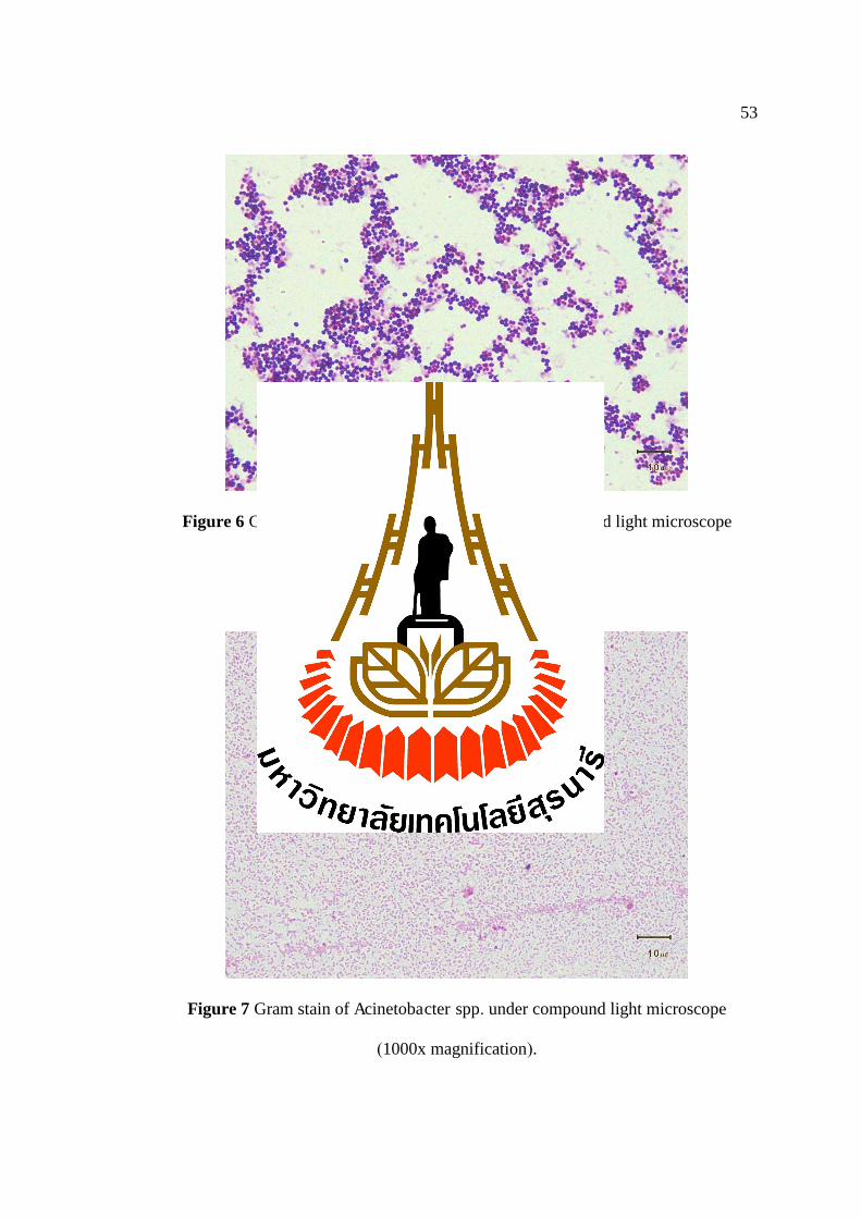
Figure 6 Gram stain of Staphylococcus spp. under compound light microscope
(1000x magnification).
Figure 7 Gram stain of Acinetobacter spp. under compound light microscope
(1000x magnification).
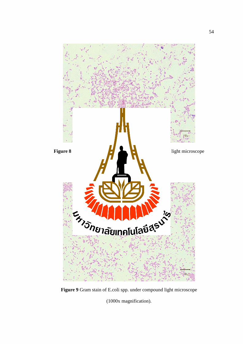
54
Figure 8 Gram stain of Enterobacter spp. under compound light microscope
(1000x magnification).
Figure 9 Gram stain of E.coli spp. under compound light microscope
(1000x magnification).

55
4.4 Microbiological quality of water samples
The microbiological parameters of tap water used in this research were based
on the guideline recommended by Metropolitan Waterworks Authority, Thailand
(based on WHO’s guideline 2011) (Appendix C). The WHO’s guideline
recommended that microbiological parameters of the good water quality of tap water
must not have any E.coli in 100 ml of water sample and no contamination of
Legionella spp. recommended by National primary drinking water regulations of
Environmental Protection Agency, United States of America for safety water in
nursing homes and spa pools.
The results showed that 3 from 60 tap water samples were E.coli
contaminations and 18 from 60 tap water samples contaminated with Legionella spp.
Thus, the water quality of these particular samples did not meet the criteria of safety
water according to the WHO’s guideline. However, the water samples that
contaminated E.coli, we were reported the results of water analysis and provided the
suggestion about elimination of contaminated samples. While, the water samples that
contaminated of Legionella spp., we were reported and elimination of contaminated
samples then recollected the water samples for analysis to prove that the water
samples were not contaminated of the Legionella spp.

56
4.5 Physical analysis of samples
The water samples from nursing homes and spa pools were evaluated for pH
and temperature. The results of the water parameters were concluded in Table 13 and
Table 14.
Table 13 Physical parameters of water samples collected from nursing homes (N=30).
Parameters Source Mean±SEM Median Range (min-max)
pH
Temperature (°C)
Faucets
Showerheads
Water tanks
Faucets
Showerheads
Water tanks
7.36±0.29
7.30±0.07
7.36±0.26
28.25±1.75
29.14±0.53
25.08±1.19
7.36
7.37
7.54
28.25
29.50
24.50
7.07-7.64
6.71-7.75
6.43-8.09
26.50-30.00
25.00-35.00
22.50-30.00
The average pH values of water samples from faucets, showerheads and water
tanks of nursing homes were 7.36±0.29, 7.30±0.07and 7.36±0.26, respectively. The
pH of water was in normal range which was 6.5 to 8.5, recommended by Metropolitan
Waterworks Authority, Thailand (based on WHO’s guideline 2011). For water
temperature, the average value of water samples from faucets, showerheads and water
tanks of nursing homes were 28.25±1.75 °C, 29.14±0.53 °C and 25.08±1.19 °C,
respectively and the optimal water temperature was 35 °C (Tison, 1980).
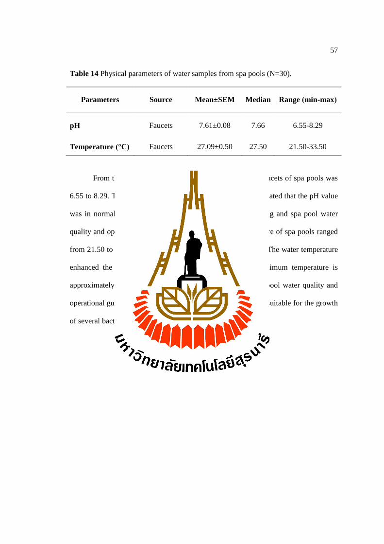
57
Table 14 Physical parameters of water samples from spa pools (N=30).
Parameters Source Mean±SEM Median Range (min-max)
pH
Temperature (°C)
Faucets
Faucets
7.61±0.08
27.09±0.50
7.66
27.50
6.55-8.29
21.50-33.50
From the study, the pH range of water samples from faucets of spa pools was
6.55 to 8.29. The average value was 7.61±0.08. The result indicated that the pH value
was in normal range 7. 2 to 7. 8. (Queensland health swimming and spa pool water
quality and operational guidelines, 2004). The water temperature of spa pools ranged
from 21.50 to 33.50 °C with the average value 27.09±0.50 °C.The water temperature
enhanced the bacterial growth at exceeding 26 °C. The optimum temperature is
approximately 38 °C. (Queensland health swimming and spa pool water quality and
operational guidelines, 2004). These temperature and pH were suitable for the growth
of several bacteria.

58
4.6 Relationships between water parameters and the prevalence of
Legionella pneumophila
The relationships between outbreak of Legionella and physical parameters and
bacteriological parameters of all water samples were examined by Pearson’ s
correlation analysis (IBM SPSS Statistics version 23).
Table 15 Statistical analysis of Pearson’s correlation analysis between water quality
parameters (temperature and pH) and the outbreak of Legionella spp.
Parameter Source r Sig. p
Nursing homes
pH
Temperature
Spa pools
pH
Temperature
Faucets
Showerheads
Water tanks
Faucets
Showerheads
Water tanks
Faucets
Faucets
-
-0.399
0.790
-
-0.103
-0.597
0.331
0.111
-
0.066
0.061
-
0.647
0.211
0.074
0.561
0.05
0.05
0.05
0.05
0.05
0.05
0.05
0.05

59
The results from statistical analysis of the relationship between water
parameters (pH and temperature) and the prevalence of Legionella spp. showed that
no statistical significant correlation in both nursing homes and also in the spa pools
because Sig.>0.05 and r value (Pearson correlation) were very low. The correlation
coefficient ranged between −1 to 1 if the value was zero indicated that there was no
correlation between the two variables. So, these results were suggested that the
relationship between water parameters (pH and temperature) and prevalence of
Legionella spp. had no statistical significant correlation in nursing homes and also in
the spa pools.
4.7 Repeated cultivation after decontamination of Legionella spp.
at the positive sites
The positive sites of Legionella spp. were reported to the nursing homes and
spa managers for decontamination of microorganism’s contaminants according to the
recommendation of Bureau of food and water sanitation, Department of Health,
Ministry of Public Health. After one month of decontamination, water samples were
recollected and cultured for microorganism’s contamination again to prove that
whether the Legionella was destroyed completely or not.
After decontamination, Legionella was mostly eliminated except in some
samples of spa pools, Legionella still remained but the density were decreased. The
other microorganisms (heterotrophic bacteria and gram-negative bacteria) still remain
in those sites. Heterotrophic bacteria were decreased in some samples of nursing
homes and spa pools. The Staphylococcus spp. was not found. These results were
shown in Table 16 and 17.

60
60
Table 16 Legionella spp. and other microorganisms detected from water samples before and after elimination in positive sample sites of
nursing homes.
Sample no.
Density of Legionella spp.
(CFU/100ml)
Total plate count
(CFU/ml) Other microorganisms
before after before after before after
Showerhead N5
Showerhead N13
Water tank N27
Water tank N28
Water tank N29
24
6
12
12
12
0
0
0
0
0
4,260
37.90
7.70
18.20
2.30
46.20
2.86
11.90
6.10
6.80
Pseudomonas spp.
Not found
E.coli
Enterobacter spp.
Pseudomonas spp.
Pseudomonas spp.
Not found
E.coli
Enterobacter spp.
Pseudomonas spp.
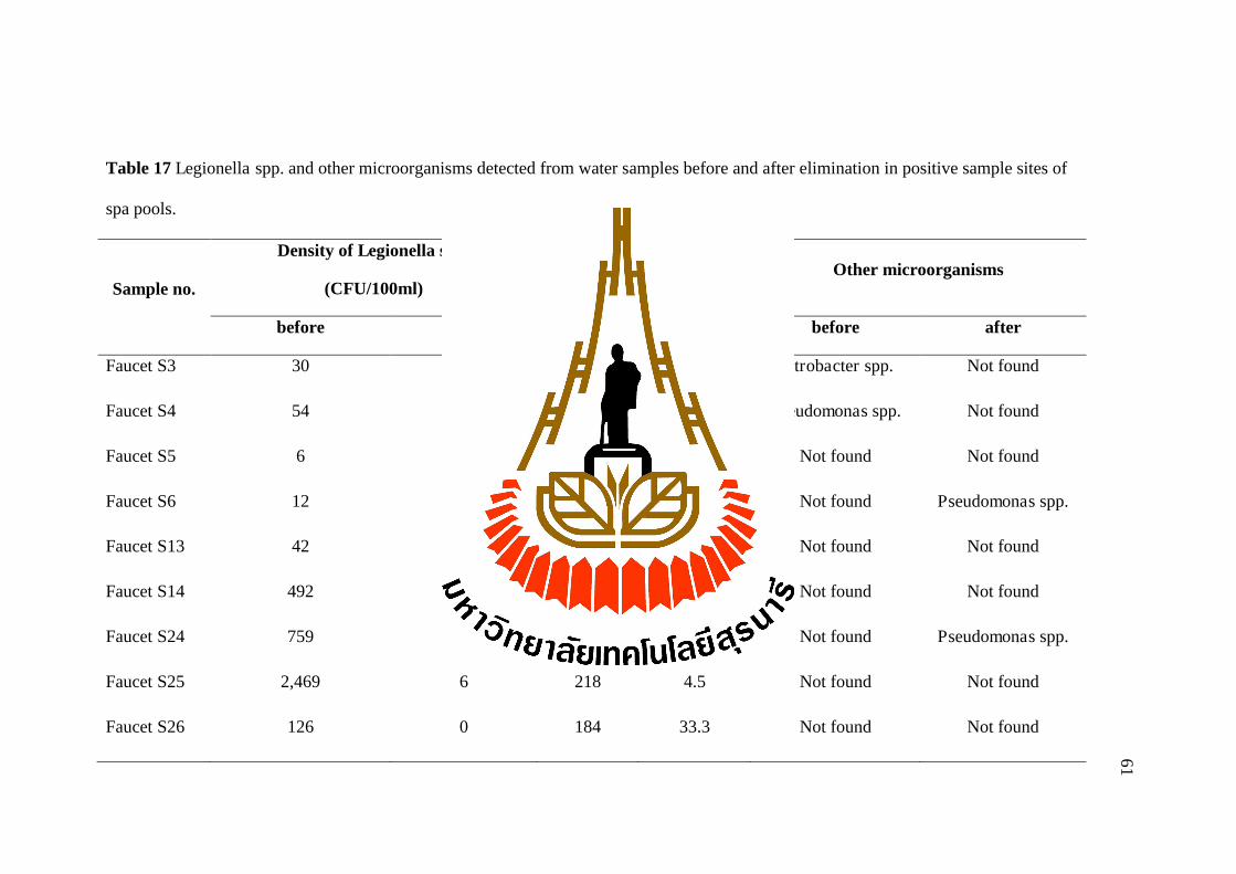
61
61
Table 17 Legionella spp. and other microorganisms detected from water samples before and after elimination in positive sample sites of
spa pools.
Sample no.
Density of Legionella spp.
(CFU/100ml)
Total plate count
(CFU/ml) Other microorganisms
before after before after before after
Faucet S3
Faucet S4
Faucet S5
Faucet S6
Faucet S13
Faucet S14
Faucet S24
Faucet S25
Faucet S26
30
54
6
12
42
492
759
2,469
126
1,311
204
30
24
0
0
6
6
0
129
294
39.1
28.5
200
150
53
218
184
465
37.62
33.81
200
10.6
44.64
9.6
4.5
33.3
Citrobacter spp.
Pseudomonas spp.
Not found
Not found
Not found
Not found
Not found
Not found
Not found
Not found
Not found
Not found
Pseudomonas spp.
Not found
Not found
Pseudomonas spp.
Not found
Not found
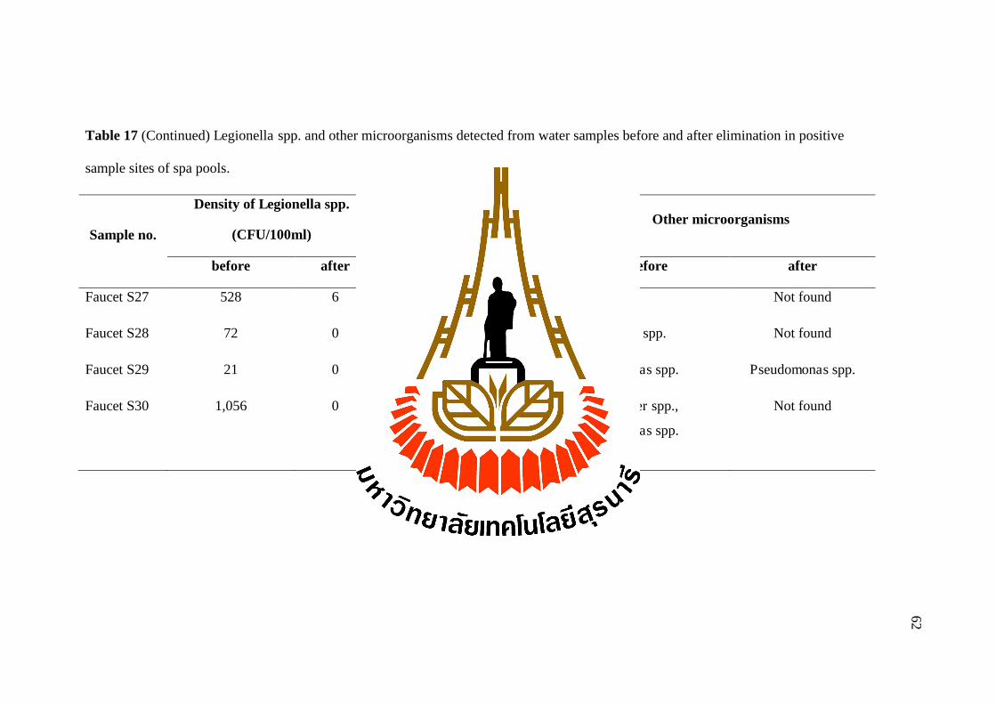
62
62
Table 17 (Continued) Legionella spp. and other microorganisms detected from water samples before and after elimination in positive
sample sites of spa pools.
Sample no.
Density of Legionella spp.
(CFU/100ml)
Total plate count
(CFU/ml) Other microorganisms
before after before after before after
Faucet S27
Faucet S28
Faucet S29
Faucet S30
528
72
21
1,056
6
0
0
0
282
252
1,920
342
428
220
199.04
262
Not found
Citrobacter spp.
Pseudomonas spp.
Enterobacter spp.,
Pseudomonas spp.
Not found
Not found
Pseudomonas spp.
Not found

63
Regarding to the water quality of water samples collected from nursing homes,
the positive sites may be risk to the elder person that stayed there. However, the
people age over 60 years included smokers have more risk than the other people
because they have low immunity and the Legionella infection was found in male more
than female. (Elverdal et al., 2013) The L.pneumophila positive sites (showerhead and
water tank) were reported to manager of nursing homes for elimination of the
microorganisms and they well-cooperated. After elimination, the water samples were
recollected and cultivated to screen for microorganisms contamination. The total
bacteria counts of microorganisms at the positive sites were decreased in some water
samples and absence of Legionella. In the spa pools, the positive sites may be risk to
the customers and spa pool keepers. The route of infection can transmit by inhalation
of bacteria contaminated as aerosol forms. The L.pneumophila positive sites (faucet
from spa pools) were reported and advised the spa managers about the risk of
infection and how to decontaminate the microorganisms. After decontamination, the
water samples were collected again and repeated the cultivation. The total bacteria
counts of microorganisms and Legionella mostly were decreased. For other
Legionella sample sites that were remain positive, after repeat decontamination again,
the water samples will be recollected and cultivated later.
Therefore, the findings of this study should be concerned by the
epidemiologists since Legionella spp. and other bacterial pathogens in nursing home
where there have a lot of vulnerable people and in spa pool may cause the outbreak of
Legionellosis and other infections caused by poor quality of the water systems.
CHAPTER V
CONCLUSION
The previous epidemiological studies of Legionella spp. reported that
Legionella spp. were considered to be the major cause of Legionnaires’ disease (LD)
in the water systems of large buildings including hospitals, nursing homes and hotels.
Legionella is a common cause of hospital-acquired pneumonia, especially in immune-
compromised patients (Yu et al., 2008). Legionella spp. are gram-negative and non-
spore-forming bacteria. The representative species of the genus is Legionella
pneumophila that can cause the Legionellosis. The Legionellosis is a respiratory
disease that can be divided into two clinical identities, Pontiac fever and Legionnaires'
disease. The symptoms of Pontiac fever are similar to a mild case of the flu, but the
Legionnaires’ disease presents more severe symptoms including pneumonia. The
Legionella spp. are commonly found in natural water environments (e.g., rivers, lakes,
lagoon and reservoirs) and human-made water systems (e.g., cooling tower, water
heater tanks, fountain, humidifiers and spa pools). The most common mode of
transmission of Legionella spp. is inhalation of contaminated aerosols. In this study,
the water samples collected from 30 nursing homes and 30 spa pools in Bangkok and
Nakhon Ratchasima provinces were examined for the presence of Legionella
pneumophila and other bacterial pathogens by culture and biochemical methods.
65
The Legionella spp. were detected in some water systems of nursing homes
and spa pools; 5 from 30 (16.67%) water samples collected from nursing homes and
13 from 30 (43.33%) were detected from spa pools. The number of positive samples
for Legionella spp. in this study was similarly to other previous studies in Thailand.
The prevalence of Legionella spp. in water systems of hotels and resorts in the North-
Eastern of Thailand was found 24 from 75 (32%) hotels and resorts. Similarly, the hot
water recirculation systems in hotels and nursing homes at Spain were analyzed for
Legionella spp. The Legionella pneumophila sg.1 was found 50 from 231 (22%). The
elderly, smoker people and the immunosuppressed patients including the managers or
staffs of nursing homes and spa pools are considered to be high risk for this particular
infection. Additionally, routine laboratories in Thailand do not screen for Legionella
spp., thus when the patients are infected with Legionella spp., they were overall
diagnosed as pneumonia. Moreover the temperature in Thailand is a suitable range for
Legionella growth. So, the Legionella positive sites (faucets, showerheads and water
tanks) were reported to the managers of nursing homes and spa pools, discussed about
public health problems and how to eliminate the microorganisms. After
decontamination at the Legionella positive sites, water samples were recollected and
cultivated for Legionella and other microorganisms. The results showed that
Legionella and other microorganisms reduced from the first collection.
For the Legionella sample sites that still remained positive, we reported to the
managers and told them that the water systems should be decontaminated again. The
water samples were recollected after cleaning and elimination of the microorganisms.
The decontamination of the sample positive sites were done four times to get rid of
66
the microorganisms as much as possible. The water samples were still positive but
tend to decrease. We suggested more about elimination of microorganisms and told
them to send the water samples again until there is no contamination to be safe for
customers.
The gram-negative bacteria that were found in both nursing homes and spa
pools were Pseudomonas spp. (35%), Enterobacter spp. (8.33%), Acinetobacter spp.
(8.33%), and Citrobacter spp. (5%). The Staphylococcus spp. was accounted for
16.67%. The Coliform bacteria and E.coli were found 16.67% and 5%, respectively.
The E.coli was found only in the nursing homes. After decontamination at the positive
sites, samples from decontaminated sites were re-evaluated. The other
microorganisms decreased, except Pseudomonas spp., E. coli and Enterobacter spp.
These microorganisms are resistant to various disinfectants and commonly found in
the environment, so they can still be found in water samples. Thus, these
microorganisms are still at risk for the people especially the elderly and
immunosuppressed patients by they can cause infection via wound, eyes, ears, skin
and soft tissue, so the people should avoid the chance of infection. The average pH
and temperature of this study were in standard value according to Queensland health
swimming and spa pool water quality and operational guidelines 2004. The
relationship between water parameters (pH and temperature) and the prevalence of
Legionella spp. had no statistical significant correlation in both nursing homes and
spa pools.

67
Therefore, this study showed the prevalence of Legionella spp. and other
bacterial pathogens which possibly cause infection in both nursing homes and spa
pools in Bangkok and Nakhon Ratchasima provinces. Moreover, the studies have
motivated the intendants to aware the danger of Legionella spp. and concern about the
possible outbreak of Legionella spp. and other bacterial infections.

REFERENCES
REFERENCES
นายแพทยสุ์ขมุ กาญจนพิมาย. (2560). ผลการเฝ้าระวงัเช้ือลจิีโอเนลลาในแหล่งน า้ที่ใช้ในโรงแรม
จังหวดัภูเกต็ พงังา และกระบี่ [ออนไลน์]. ไดจ้าก:http://www.dmsc.moph.go.th
/dmscnew/news_detail.php?id=1742.html.
มณฑล เลิศคณาวานิชกุล. (2547). การตรวจหาเช้ือลีจิโอเนลลาในส่ิงแวดล้อม ของมหาวิทยาลัย
วลยัลกัษณ์.[ออนไลน]์. ไดจ้าก:http://advisor1.anamai.moph.go.th/262/26209.html.
Ahmadinejad, M., Shakibaie, M. R., Shams, K., and Khalili, M. (2011). Detection of
Legionella pneumophila in cooling water systems of hospitals and nursing
homes of Kerman city, Iran by semi-nested PCR. International Journal of
Biological and Life Sciences. 7: 70-73.
Amanidaz, N., Zafarzadeh, A., and Mahvi, A. H. (2015). The Interaction between
heterotrophic bacteria and Coliform, fecal Coliform, fecal Streptococci
bacteria in the water supply networks. Iranian Journal of Public Health. 44:
1685.
Bartram, J. (2007). Legionella and the prevention of Legionellosis: World Health
Organization.

70
Bovornkitti, S. (2010). Legionella. Thammasat Medical Journal 9: 436-441
Buathong, R., Prasarnthong, R., Seetamanotch, W., Natewong, T., Saowarun, T.,
Chanthayanee, K., Marin, N., Sutdan, D., Ouppapong, T., and Iamsirithaworn,
S. (2013). Travel-associated Legionnaires Disease Outbreak among EU
Travelers, Phuket Thailand, December 2006 to January 2007: Role of
Environmental Investigation. Weekly Epidemiological Surveillance Report.
44: S38-S46.
Cordes, L. G., Wiesenthal, A. M., Gorman, G. W., Phair, J. P., Sommers, H. M.,
Brown, A., Yu V. L., Magnussen, M. H., Meyer, R. D., and Wolf, J. S. (1981).
Isolation of Legionella pneumophila from hospital shower heads. Annals of
Internal Medicine. 94: 195-197.
Declerck, P. (2010). Biofilms: the environmental playground of Legionella
pneumophila. Environmental Microbiology. 12: 557-566.
Edelstein, P. H. (1981). Improved semiselective medium for isolation of Legionella
pneumophila from contaminated clinical and environmental specimens.
Journal of Clinical Microbiology. 14: 298-303.
Edelstein, P. H. (1982). Comparative study of selective media for isolation of
Legionella pneumophila from potable water. Journal of Clinical
Microbiology. 16: 697-699.
Elverdal, P., Jorgensen, C., Krogfelt, K., and Uldum, S. (2013). Two years'
performance of an in-house ELISA for diagnosis of Legionnaires' disease:
Detection of specific IgM and IgG antibodies against Legionella pneumophila
71
serogroup 1, 3 and 6 in human serum. Journal of Microbiological Methods.
94: 94-97.
Feeley, J. C., Gibson, R. J., Gorman, G. W., Langford, N. C., Rasheed, J. K., Mackel,
D. C., and Baine, W. B. (1979). Charcoal-yeast extract agar: primary isolation
medium for Legionella pneumophila. Journal of Clinical Microbiology. 10:
437-441.
Fields, B. S. (2008). Legionella in the environment. Infectious Diseases and
Pathogenesis (pp. 85-94). Springer.
Fields, B. S., Benson, R. F., and Besser, R. E. (2002). Legionella and Legionnaires'
disease: 25 years of investigation. Clinical Microbiology Reviews. 15: 506-
526.
Fliermans, C., Cherry, W., Orrison, L., Smith, S., Tison, D., and Pope, D. (1981).
Ecological distribution of Legionella pneumophila. Applied and
Environmental Microbiology. 41: 9-16.
Guillemet, T., Levesque, B., Gauvin, D., Brousseau, N., Giroux, J. P., and Cantin, P.
(2010). Assessment of realǦtime PCR for quantification of Legionella spp. in
spa water. Letters in Applied Microbiology. 51: 639-644.
Government, Q, “Queensland health swimming and spa pool water quality and
operational guidelines 2004”. Brisbane, QLD: Bibliography, 2004, pp.52.
Guyard, C., and Low, D. E. (2011). Legionella infections and travel associated
legionellosis. Travel Medicine and Infectious Disease. 9: 176-186.
Hanrahan, J. P., Morse, D. L., Scharf, V. B., Debbie, J. G., Schmid, G. P., Mckinney,
R. M., and Shayegani, M. (1987). A Community Hospital Outbreak of
72
Legionellosis: Transmission by Potable Hot Water. American Journal of
Epidemiology. 125: 639-649.
Hebert, G. (1981). Hippurate hydrolysis by Legionella pneumophila. Journal of
Clinical Microbiology. 13(1). 240-242.
Hsu, B.-M., Chen, C.-H., Wan, M.-T., and Cheng, H.-W. (2006). Legionella
prevalence in hot spring recreation areas of Taiwan. Water Research. 40:
3267-3273.
Huang, S. W., Hsu, B. M., Wu, S. F., Fan, C. W., Shih, F. C., Lin, Y. C., and Ji, D. D.
(2010). Water quality parameters associated with prevalence of Legionella in
hot spring facility water bodies. Water Research. 44: 4805-4811.
Inoue, H., Takama, T., Yoshizaki, M., and Agata, K. (2015). Detection of Legionella
species in environmental water by the quantitative PCR method in
combination with ethidium monoazide treatment. Biocontrol Science. 20: 71-
74.
Kim, B., Anderson, J., Mueller, S., Gaines, W., and Kendall, A. (2002). Literature
review—efficacy of various disinfectants against Legionella in water systems.
Water Research. 36: 4433-4444.
Kwaik, Y. A., Gao, L.-Y., Stone, B. J., Venkataraman, C., and Harb, O. S. (1998).
Invasion of protozoa by Legionella pneumophila and its role in bacterial
ecology and pathogenesis. Applied and Environmental Microbiology. 64:
3127-3133.
Lau, H., and Ashbolt, N. (2009). The role of biofilms and protozoa in Legionella
pathogenesis: implications for drinking water. Journal of Applied
Microbiology. 107: 368-378.

73
LeChevallier, M. W., and Seidler, R. J. (1980). Staphylococcus aureus in rural
drinking water. Applied and Environmental Microbiology. 39: 739-742.
Leoni, E., and Legnani, P. (2001). Comparison of selective procedures for isolation
and enumeration of Legionella species from hot water systems. Journal of
Applied Microbiology. 90: 27-33.
Leoni, E., De Luca, G., Legnani, P., Sacchetti, R., Stampi, S., and Zanetti, F. (2005).
Legionella waterline colonization: detection of Legionella species in domestic,
hotel and hospital hot water systems. Journal of Applied Microbiology. 98:
373-379.
Lin, Y. E., Stout, J. E., Yu, V. L., and Vidic, R. D. (1998). Disinfection of water
distribution systems for Legionella. Paper presented at the Seminars in
respiratory infections.
Mahayotha, A., Butkot, T., Wongveerakhant, P., Prasartthong, V., Punthanaprated,
U., Cheuypratoom, P., and Jamsai, W. Legionella spp. Surveillance Network,
North-eastern of Thailand (เครือ ข่าย การ เฝ้า ระวงั การ แพร่ เช้ือ Legionella spp.
ภาค ตะวนัออก เฉียง เหนือ ของ ประเทศไทย). Journal of Health Science-วารสาร
วชิาการ สาธารณสุข. 25: 929-934.
Montagna, M. T., Cristina, M. L., De Giglio, O., Spagnolo, A. M., Napoli, C.,
Cannova, L., and Guida, M. (2016). Serological and molecular identification
of Legionella spp. isolated from water and surrounding air samples in Italian
healthcare facilities. Environmental Research. 146: 47-50.
Moore, G., and Walker, J. (2014). Chapter Seventeen - Presence and Control of
Legionella pneumophila and Pseudomonas aeruginosa Biofilms in Hospital
74
Water Systems. In Percival, S. L., Williams, D. W., Randle, J., and Cooper, T.,
(eds.). Biofilms in Infection Prevention and Control (pp. 311-337). Boston:
Academic Press.
Nazarian, E. J., De Jesus, M., and Musser, K. A. (2015). Chapter 90 - Legionella. In
Y.-W. T. S. L. P. Schwartzman (ed.). Molecular Medical Microbiology
(Second Edition) (pp. 1611-1625). Boston: Academic Press.
Nostro, A. L., Checchi, E., Ducci, B., and Pesavento, G. (2011). Legionella
contamination in hot water systems of hospitals, nursing homes, hotels,
factories and spas in Tuscany-Italy. Italy Journal Public Health. 8: 5-13.
Paveenkittiporn, W., Dejsirilert, S., and Kalambaheti, T. (2012). Genetic speciation of
environmental Legionella isolates in Thailand. Infection. Genetics and
Evolution. 12: 1368-1376.
Percival, S. L., and Williams, D. W. (2014). Chapter Eight - Legionella. In S. L. P. V.
Y. W. W. M. C. F. Gray (ed.). Microbiology of Waterborne Diseases
(Second Edition) (pp. 155-175). London: Academic Press.
Phares, C. R., Wangroongsarb, P., Chantra, S., Paveenkitiporn, W., Tondella, M. L.,
Benson, R. F., Thacker, W. L., Fields, B. S., Moore, M. R., and Fischer, J.
(2007). Epidemiology of severe pneumonia caused by Legionella
longbeachae, Mycoplasma pneumoniae, and Chlamydia pneumoniae: 1-year,
population-based surveillance for severe pneumonia in Thailand. Clinical
Infectious Diseases. 45: 147-155.
Phin, N., Parry-Ford, F., Harrison, T., Stagg, H. R., Zhang, N., Kumar, K., and
Abubakar, I. (2014). Epidemiology and clinical management of Legionnaires'
disease. The Lancet Infectious Diseases. 14: 1011-1021.

75
Reasoner, D. J. (2004). Heterotrophic plate count methodology in the United States.
International Journal of Food Microbiology. 92: 307-315.
Rodgers, F., Macrae, A., and Lewis, M. (1978). Electron microscopy of the organism
of Legionnaires' disease. Nature. 272: 825-826.
Serrano-Suárez, A., Dellundé, J., Salvadó, H., Cervero-Aragó, S., Méndez, J., Canals,
O., and Araujo, R. (2013). Microbial and physicochemical parameters
associated with Legionella contamination in hot water recirculation systems.
Environmental Science and Pollution Research. 20: 5534-5544.
Skaza, A. T., Beskovnik, L., Storman, A., Kese, D., and Ursic, S. (2012).
Epidemiological investigation of a Legionellosis outbreak in a Slovenian
nursing home, August 2010. Scandinavian Journal of Infectious Diseases.
44: 263-269.
Stout, J. E., and Yu, V. L. (1997). Legionellosis. New England Journal of Medicine.
337: 682-687.
Swanson, M., and Hammer, B. (2000). Legionella pneumophila pathogenesis: a
fateful journey from amoebae to macrophages. Annual Reviews in
Microbiology . 54: 567-613.
Tishyadhigama, P., Dejsirilert, S., Srisawai, P., Kusum, M., Yabuuchi, E., Ikedo, M.,
Ezaki, T., Sutivichit, Y., Charoensuksopon, K., and Suchatanun, C. (1995).
Environmental surveillance of Legionella species in Thailand. Journal of the
Medical Association of Thailand. 78: 57-71.
Tison, D., Pope, D., Cherry, W., and Fliermans, C. (1980). Growth of Legionella
pneumophila in association with blue-green algae (Cyanobacteria). Applied
and Environmental Microbiology. 39: 456-459.

76
Türetgen, I., Sungur, E. I., and Cotuk, A. (2005). Enumeration of Legionella
pneumophila in cooling tower water systems. Environmental Monitoring
and Assessment. 100: 53-58.
Vincent, J. L., Bihari, D. J., Suter, P. M., Bruining, H. A., White, J., Nicolas Chanoin,
M. H., Wolff, M., Spencer, R. C., and Hemmer, M. (1995). The prevalence of
nosocomial infection in intensive care units in Europe: results of the European
Prevalence of Infection in Intensive Care (EPIC) Study. Jama. 274: 639-644.
Wingender, J., and Flemming, H. C. (2011). Biofilms in drinking water and their role
as reservoir for pathogens. International Journal of Hygiene and
Environmental Health. 214: 417-423.
Woo, A. H., Goetz, A., and Yu, V. (1992). Transmission of Legionella by respiratory
equipment and aerosol generating devices. Chest Journal. 102: 1586-1590.
Yaslianifard, S., Mobarez, A. M., Fatolahzadeh, B., and Feizabadi, M. M. (2012).
Colonization of hospital water systems by Legionella pneumophila,
Pseudomonas aeroginosa, and Acinetobacter in ICU wards of Tehran
hospitals. Indian Journal of Pathology and Microbiology. 55: 352.
Yu, P.Y., Lin, Y. E., Lin, W. R., Shih, H.Y., Chuang, Y. C., Ben, R. J., and Chang, F.
Y. (2008). The high prevalence of Legionella pneumophila contamination in
hospital potable water systems in Taiwan: implications for hospital infection
control in Asia. International Journal of Infectious Diseases. 12: 416-420.

APPENDICES
APPENDIX A
MICROBIOLOGICAL MEDIA
1. Buffered charcoal yeast extract alpha base (BCYE)
Charcoal 2.0 g
Yeast extract 10.0 g
ACES buffer 10.0 g
Alpha-ketoglutarate 1.0 g
Ferric pyrophosphate soluble 0.25 g
L-cysteine, HCl.H2O 0.4 g
Agar 15.0 g
Final pH 6.9 (± 0.2)
Preparation of medium: dissolved charcoal, yeast extract, ACES buffer,
alphaketoglutarate and agar in 1 l distilled water, adjusted pH to 6.9 with 0.1 N KOH
and heated to boil. Then, sterilized by autoclaving at 121 °C for 15 min. Dissolved 0.4
g Lcysteine and 0.25 g ferric pyrophosphate in 10 ml of water each and filter sterile
separately. After agar base was cooled, added L-cysteine and ferric pyrophosphate in
that order and dispensed into sterilize petri dishes.
79
2. Glycine vancomycin polymyxin B cyclohexamide medium (GVPC)
Glycine 3.0 g
Polymyxin B 100 units/ml
Vancomycin 5 たg/ml
Cyclohexamide 80 たg/ml
Preparation of medium: to cooled BCYE-alpha base with glycine, add filtersterilized
antibiotics and mix. The medium was dispensed into sterilized petri dishes.
3. MacConkey agar
Peptone 17.0 g
Protease peptone 3.0 g
Lactose 10.0 g
Bile salts 1.5 g
Sodium chloride (NaCl) 5.0 g
Neutral red 0.03 g
Crystal violet 0.001 g
Agar 15.0 g
Final pH 7.1 (± 0.2)
Preparation of medium: all components were added to distilled water and brought
volume up to 1 l. The medium was mixed thoroughly and gently heated until
dissolved. The medium was autoclaved at 121 °C for 15 min. Dispensed into sterilize
petri dishes.

80
4. Plate count agar (Tryptone glucose yeast agar)
Tryptone 5.0 g
Yeast extract 2.5 g
Glucose 1.0 g
Agar 15.0 g
Final pH 7.0 (± 0.2)
Preparation of medium: all components were added to distilled water and brought
volume up to 1 l. The medium was mixed thoroughly and gently heated until
dissolved. The medium was autoclaved at 121 °C for 15 min. Dispensed into sterilize
petri dishes.
5. Eosin methylene blue agar (EMB agar)
Peptic digest of animal tissue 10.0 g
Dipotassium phosphate 2.0 g
Lactose 10.0 g
Eosin - Y 0.4 g
Methylene blue 0.065 g
Agar 15.0 g
Final pH (at 25 °C) 7.1±0.2
Preparation of medium: all components were added to distilled water and brought
volume up to 1 l. The medium was mixed thoroughly and heated until dissolved. The
medium was autoclaved at 121 °C for 15 min. Dispensed into sterilize petri dishes.
81
6. Mannitol Salt Agar (MSA agar)
Proteose peptone 10.0 g
Beef extract 1.0 g
Sodium chloride 75.0 g
D-Mannitol 10.0 g
Phenol red 0.025 g
Agar 15.0 g
Final pH (at 25 °C) 7.4±0.2
Preparation of medium: all components were added to distilled water and brought
volume up to 1 l. The medium was mixed thoroughly and gently heated until
dissolved. The medium was autoclaved at 121 °C for 15 min. Dispensed into sterilize
petridishes.

APPENDIX B
CHEMICAL REAGENTS
1. Acid treatment reagent (0.2 M KCl/HCl)
Solution A: 0.2 M KCl (14.9 g/l in distilled water).
Solution B: 0.2 M HCl (16.7 ml/l 10N HCl in distilled water).
Preparation of reagent: mixed 18 parts of solution A with 1 part of solution B. Check
pH against a pH 2.0 standard buffer and sterilize by autoclaving at 121 °C for 15 min.
2. Alkaline neutralizer reagent (0.1 M KOH)
Potassium hydroxide (KOH) 6.46 g
Preparation of reagent: the component was added to deionized water, mixed
thoroughly until dissolved and brought volume up to 1 l as stock solution. Diluted
10.7 ml of stock solution with 100 ml deionized water and sterilized by autoclaving at
121 °C for 15 min.3. 0.1 N Sodium thiosulfate (Na2S2O3)
Sodium thiosulfate (Na2S2O35ڄH2O) 24.82 g
Preparation of reagent: the component was added to distilled water, mixed thoroughly
until dissolved and brought volume up to 1 l. The reagent was autoclaved at 121 °C
for 15 min.

83
4. 1% Hippurate reagent
Sodium hippurate 0.1 g
Preparation of reagent: the component was added to sterile distilled water, mixed
thoroughly until dissolved and brought volume up to 10 ml. The reagent was
dispensed for 0.4 ml in microcentrifuge and stored at –20 °C.
5. 3.5% Ninhydrin
Ninhydrin 0.35 g
1-Butanol 5.0 ml
Acetone 5.0 ml
Preparation of reagent: 1-butanol and acetone were mixed then added ninhydrin and
mixed thoroughly until dissolved. The reagent was stored in brown bottle.
APPENDIX C
STANDARD OF TAP WATER RECOMMENDED BY
METROPOLITAN WATERWORKS AUTHORITY
(BASED ON WHO GUIDELINE 2011)
Table 1 C Standard of tap water recommended by Metropolitan Waterworks
Authority (based on WHO guideline 2011).
Parameters Units Recommend
1. Bacteriology quality
E. coli None
2. Physical and chemical quality
Appearance color
Turbidity
Taste and odor
pH
Arsenic
Cadmium
Chromium
Cyanide
Not found/100 ml
True color unit
NTU
-
-
mg/l
mg/l
mg/l
mg/l
Not found/100 ml
15
5
-
6.5-8.5
0.01
0.003
0.05
0.5
85
Table 1 C (Continued).
Parameters Units Recommend
Lead
Inorganic Mercury
Selenium
Fluoride
Chloride
Copper
Iron
Manganese
Aluminium
Sodium
Sulfate
Zinc
Total dissolved solids
Nitrate as NO3-
Nitrite as NO2-
Trichloroethene
Tetrachloroethene
Microcystin-LR
3. Pesticides
Aldrin/Dieldrin
mg/l
mg/l
mg/l
mg/l
mg/l
mg/l
mg/l
mg/l
mg/l
mg/l
mg/l
mg/l
mg/l
mg/l
mg/l
mg/l
mg/l
mg/l
たg/l
0.01
0.006
0.04
0.7
250
2
0.3
0.1
0.9
200
250
3
1,000
50
3
0.02
0.04
0.001
0.03
86
Table 1 C (Continued).
Parameters Units Recommend
Chlordane
DDT and metabolites
2,4-D
Heptachlor and Heptachlor epoxide
Hexachlorobenzene
Lindane
Methoxychlor
Pentachlorophenol
4. Trihalomethanes sum of the ratio
Chloroform , CHCl3
Bromodichloromethane , CHBrCl2
Dibromochloromethane , CHBr2Cl
Bromoform , CHBr3
5. Radioactive
Gross alpha activity
Gross beta activity
たg/l
たg/l
たg/l
たg/l
たg/l
たg/l
たg/l
たg/l
mg/l
mg/l
mg/l
mg/l
bq/l
bq/l
0.2
1
30
0.03
1
2
20
9
1
0.3
0.06
0.1
0.1
0.5
1
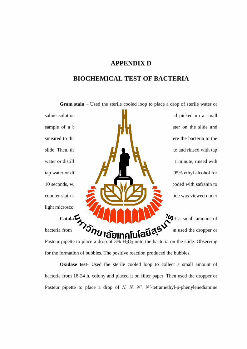
APPENDIX D
BIOCHEMICAL TEST OF BACTERIA
Gram stain – Used the sterile cooled loop to place a drop of sterile water or
saline solution on the slide. The loop was sterilized again and picked up a small
sample of a bacterial colony and stirred into the drop of water on the slide and
smeared to thin layer. The smeared slide was heat fixed to adhere the bacteria to the
slide. Then, the slide was flooded with crystal violet for 1 minute and rinsed with tap
water or distilled water. Followed by Gram's Iodine solution for 1 minute, rinsed with
tap water or distilled water again. The slide was decolorized by 95% ethyl alcohol for
10 seconds, washed off with tap water. Finally, the slide was flooded with safranin to
counter-stain for 1 minute and rinsed with tap water, the dried slide was viewed under
light microscope with oil-immersion.
Catalase test – Used the sterile cooled loop to collect a small amount of
bacteria from 18- 24 h. colony and placed it onto the slide. Then used the dropper or
Pasteur pipette to place a drop of 3% H2O2 onto the bacteria on the slide. Observing
for the formation of bubbles. The positive reaction produced the bubbles.
Oxidase test- Used the sterile cooled loop to collect a small amount of
bacteria from 18-24 h. colony and placed it on filter paper. Then used the dropper or
Pasteur pipette to place a drop of N, N, N’, N’-tetramethyl-p-phenylenediamine

88
dihydrochloride onto the bacteria on filter paper. Observing color changed within 10
seconds. The positive reaction was purple color.
Motility indole lysine – Used the sterile needle to inoculate the
suspected bacteria once by stabbed in the semi-agar media through the bottom of the
tube. After 18-24 h. of incubation at 35 °C, the motility and lysine decarboxylase and
deaminase activity were read before testing for indole test. The positive result of
motility was observed by the radiated movement from central of inoculation. If non-
motile, the bacteria grew only along the line of inoculation. The presence of lysine
decarboxylase caused the entire tube to revert to purple and if caused the yellow
bottom with purple at the top of the tube indicated a negative test for lysine
decarboxylase. The presence of lysine deaminase caused the top of the tube to turn
deep red, the top remained purple in a negative test. The indole test was done by
added 3-4 drops of Kovac’s reagent to the medium. The positive reaction was red to
pink and the negative reaction was yellow layer.
OF-glucose test – Used the sterile cooled to needle inoculate the
suspected bacteria into the OF-glucose test medium tube by stabbed half way to the
bottom of the tube. The medium tube was incubated at 35 °C for 48 h. The positive
result for fermentation of glucose was turned to yellow color.
Simmons citrate – Used the sterile cooled loop to inoculate the
suspected bacteria on Simmons citrate by streaked on the surface of the agar slant.
The agar was incubated at 35 °C for 24 h. and observed the changed color of the agar.
The positive result was deep blue color.

89
Triple sugar iron agar (TSI) – Used the sterile cooled needle to
inoculate the suspected bacteria in the TSI agar by stabbed through the bottom center
of the medium tube and then streaked on the surface of the agar slant. The agar was
incubated at 35 °C for 24 h. and observed the changed color of the agar. If the
organism fermented glucose but did not fermented lactose and/or sucrose, the slant
became red and butt indicated the yellow color (K/A). If the organism fermented
glucose, lactose and/or sucrose, the organism turned the phenol red indicator to
yellow both in butt and in slant (A/A). Some organisms generated gases, which
produced bubble in the medium. If the organism was non-fermenter, the slant
indicated the red color while there was no change in the color of the butt. (K/NC).

CURRICULUM VITAE
Name Miss TANYAPORN KULLAKET
Date of birth 10 September 1989
Place of birth Nakhon Ratchasima, Thailand.
Education and work experience
2008-2011 Bachelor of science: Veterinary Technology (GPA: 3.25)
Second-class honor, Kasetsart University.
2012-2013 Veterinary Technician at Thai Vet Laboratory Limited
Company Bangkok.
2013-2016 Teacher Assistant (TA) of Principle Biology Laboratory and
Microbiology Laboratory courses.