Detection of aneuploidy in a single cell using the Ion ......Detection of aneuploidy in a single...
6
APPLICATION NOTE Ion PGM System Detection of aneuploidy in a single cell using the Ion ReproSeq PGS View Kit • Ion Reporter Software facilitates genome-wide aneuploidy detection in multiple samples in parallel, with three sensitivity options, so that copy number analysis workflows can be tuned to achieve desired levels of sensitivity and specificity. Aneuploidy detection in preimplantation embryos Copy number variations (CNVs) represent a class of variation in which segments of the genome have been duplicated (gains) or deleted (losses). Large, genomic copy number imbalances can range from subchromosomal regions to entire chromosomes. Inherited and de novo CNVs (up to 10 Mb) have been associated with many disease Key findings • The Ion PGM ™ System, in concert with the Ion ReproSeq ™ PGS View Kit and Ion Reporter ™ Software, enables the fastest next-generation sequencing (NGS) workflow (as little as 8 hours) for detection of aneuploidies in preimplantation embryos, with single-cell input DNA amounts in a simple and cost-effective workflow. • The Ion PGM System detected 100% of aneuploidies previously detected by array comparative genomic hybridization (aCGH) in a control set of 134 samples. The system sensitivity allows not only for detection of whole-arm translocations or chromosome gains and losses but also for subchromosomal alterations tens of megabases (Mb) in size. 19 20 21 22 X Y 16 17 18 7 8 9 10 11 12 6 1 2 3 4 5 13 14 15 13 14 15 Figure 1. Aneuploidy in genomes. Most somatic cells contain an exact diploid set of chromosomes; genomes that lack or contain additional chromosomes are termed aneuploid. Chromosomal gains (trisomies of chromosomes 14 and 15) are highlighted in blue, while a loss of chromosome 13 is indicated in red. conditions, and they may impact human embryo viability as well. This application note will discuss the use of Ion Torrent ™ semiconductor sequencing technology and Ion Reporter Software for the detection of aneuploidies. Most normal human somatic cells contain a diploid [2N] set of autosomes (non-sex chromosomes) and a pair of sex chromosomes. Cells that do not contain an exact diploid set are termed aneuploid (Figure 1). Common types of aneuploidy that survive to term are monosomy (the loss of one chromosome) of the X chromosome in females, known as Turner syndrome (45, X), and some trisomies, i.e., three copies of a given chromosome in a diploid cell. Live births are possible with trisomies of autosomes 13 (Patau syndrome), 18 (Edwards syndrome), and 21 (Down syndrome), as with the presence of extra sex chromosomes such as in Klinefelter syndrome (47, XXY) and Triple X syndrome (47, XXX).
Transcript of Detection of aneuploidy in a single cell using the Ion ......Detection of aneuploidy in a single...
APPLICATION NOTE Ion PGM System
Detection of aneuploidy in a single cell using the Ion ReproSeq PGS View Kit
• Ion Reporter Software facilitates genome-wide aneuploidy detection in multiple samples in parallel, with three sensitivity options, so that copy number analysis workflows can be tuned to achieve desired levels of sensitivity and specificity.
Aneuploidy detection in preimplantation embryosCopy number variations (CNVs) represent a class of variation in which segments of the genome have been duplicated (gains) or deleted (losses). Large, genomic copy number imbalances can range from subchromosomal regions to entire chromosomes. Inherited and de novo CNVs (up to 10 Mb) have been associated with many disease
Key findings• The Ion PGM™ System, in concert
with the Ion ReproSeq™ PGS View Kit and Ion Reporter™ Software, enables the fastest next-generation sequencing (NGS) workflow (as little as 8 hours) for detection of aneuploidies in preimplantation embryos, with single-cell input DNA amounts in a simple and cost-effective workflow.
• The Ion PGM System detected 100% of aneuploidies previously detected by array comparative genomic hybridization (aCGH) in a control set of 134 samples. The system sensitivity allows not only for detection of whole-arm translocations or chromosome gains and losses but also for subchromosomal alterations tens of megabases (Mb) in size.
19 20 21 22 X Y
16 17 18
7 8 9 10 11 126
1 2 3 4 5
13 14 15
13 14 15
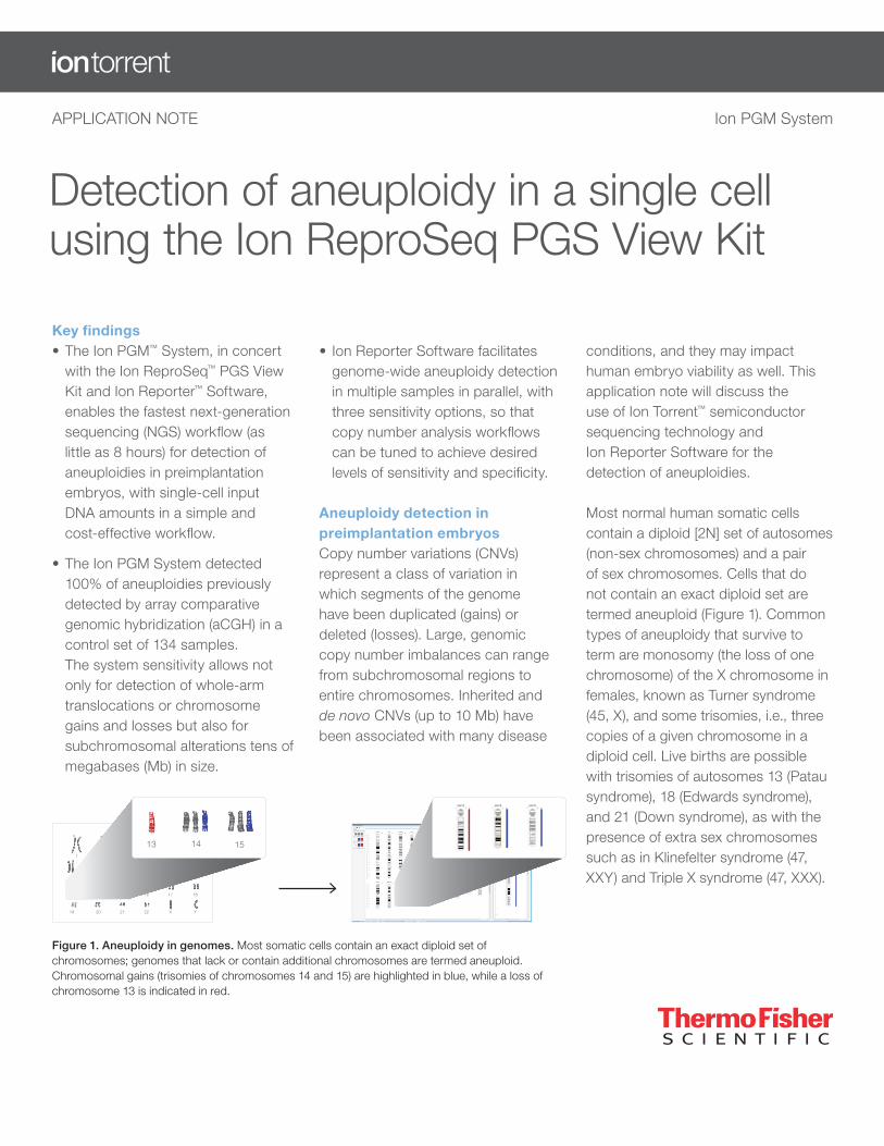
Figure 1. Aneuploidy in genomes. Most somatic cells contain an exact diploid set of chromosomes; genomes that lack or contain additional chromosomes are termed aneuploid. Chromosomal gains (trisomies of chromosomes 14 and 15) are highlighted in blue, while a loss of chromosome 13 is indicated in red.
conditions, and they may impact human embryo viability as well. This application note will discuss the use of Ion Torrent™ semiconductor sequencing technology and Ion Reporter Software for the detection of aneuploidies.
Most normal human somatic cells contain a diploid [2N] set of autosomes (non-sex chromosomes) and a pair of sex chromosomes. Cells that do not contain an exact diploid set are termed aneuploid (Figure 1). Common types of aneuploidy that survive to term are monosomy (the loss of one chromosome) of the X chromosome in females, known as Turner syndrome (45, X), and some trisomies, i.e., three copies of a given chromosome in a diploid cell. Live births are possible with trisomies of autosomes 13 (Patau syndrome), 18 (Edwards syndrome), and 21 (Down syndrome), as with the presence of extra sex chromosomes such as in Klinefelter syndrome (47, XXY) and Triple X syndrome (47, XXX).
Research samples can be analyzed for aneuploidy using molecular techniques such as quantitative fluorescence PCR (QF-PCR) and aCGH. These analyses are complementary to cytogenetics-based tests such as full karyotype analysis of metaphase chromosomes and fluorescence in situ hybridization (FISH) karyotyping.
Interest in applying NGS technologies to aneuploidy detection has grown considerably, primarily due to the technical ability to simultaneously analyze multiple samples rapidly and more affordably. Additionally, the massively parallel DNA sequencing allows for concurrent mitochondrial DNA (mtDNA) analysis, which is of interest as recent reports suggest an association between higher mtDNA levels and aneuploidy in preimplantation embryos [1].
Cost-effective and rapid detection of aneuploidies in multiple embryos, with low amounts of input DNA Aneuploidy analysis in a developing preimplantation embryo requires assessing a single cell or multiple cells. Time-to-results is essential when determining which preimplantation embryo has a complete chromosomal complement, as the window for implantation is relatively small, and freezing might not be an option. The requirements for aneuploidy analysis of preimplantation embryos include a rapid, comprehensive examination of all 23 pairs of chromosomes, and a cost-effective, accurate method enabled through sample multiplexing.
The Ion ReproSeq PGS View Kit, combined with the Ion PGM System and Ion Reporter Software, delivers
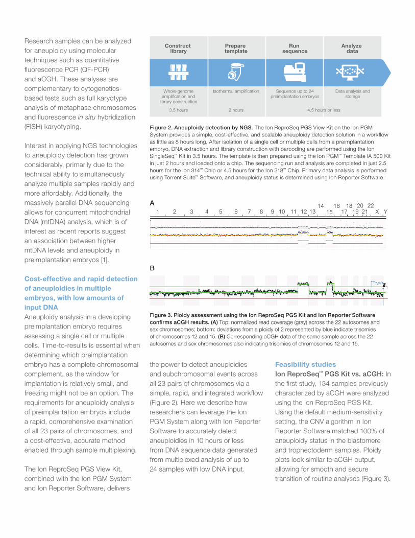
Figure 2. Aneuploidy detection by NGS. The Ion ReproSeq PGS View Kit on the Ion PGM System provides a simple, cost-effective, and scalable aneuploidy detection solution in a workflow as little as 8 hours long. After isolation of a single cell or multiple cells from a preimplantation embryo, DNA extraction and library construction with barcoding are performed using the Ion SingleSeq™ Kit in 3.5 hours. The template is then prepared using the Ion PGM™ Template IA 500 Kit in just 2 hours and loaded onto a chip. The sequencing run and analysis are completed in just 2.5 hours for the Ion 314™ Chip or 4.5 hours for the Ion 318™ Chip. Primary data analysis is performed using Torrent Suite™ Software, and aneuploidy status is determined using Ion Reporter Software.
the power to detect aneuploidies and subchromosomal events across all 23 pairs of chromosomes via a simple, rapid, and integrated workflow (Figure 2). Here we describe how researchers can leverage the Ion PGM System along with Ion Reporter Software to accurately detect aneuploidies in 10 hours or less from DNA sequence data generated from multiplexed analysis of up to 24 samples with low DNA input.
Construct library
Prepare template
Run sequence
Analyze data
Whole-genome amplification and
library construction
3.5 hours
Isothermal amplification
2 hours
Sequence up to 24 preimplantation embryos
Data analysis and storage
4.5 hours or less
Feasibility studiesIon ReproSeq™ PGS Kit vs. aCGH: In the first study, 134 samples previously characterized by aCGH were analyzed using the Ion ReproSeq PGS Kit. Using the default medium-sensitivity setting, the CNV algorithm in Ion Reporter Software matched 100% of aneuploidy status in the blastomere and trophectoderm samples. Ploidy plots look similar to aCGH output, allowing for smooth and secure transition of routine analyses (Figure 3).
A
B
Figure 3. Ploidy assessment using the Ion ReproSeq PGS Kit and Ion Reporter Software confirms aCGH results. (A) Top: normalized read coverage (gray) across the 22 autosomes and sex chromosomes; bottom: deviations from a ploidy of 2 represented by blue indicate trisomies of chromosomes 12 and 15. (B) Corresponding aCGH data of the same sample across the 22 autosomes and sex chromosomes also indicating trisomies of chromosomes 12 and 15.
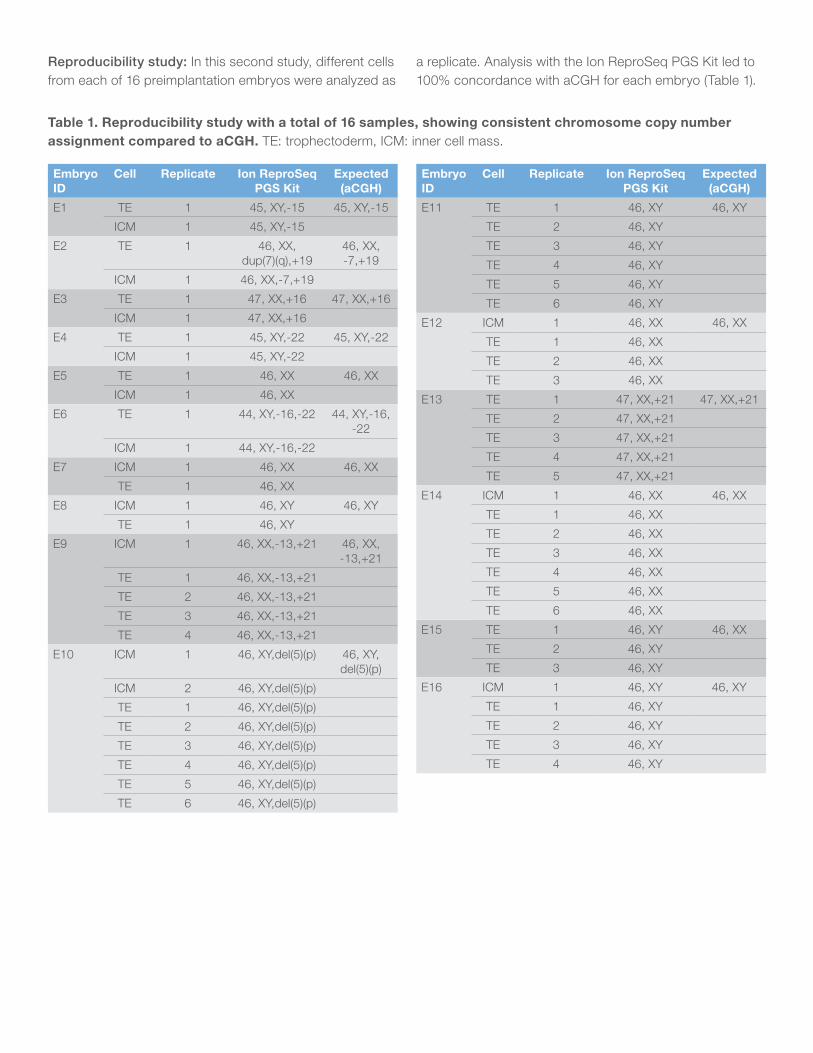
Reproducibility study: In this second study, different cells from each of 16 preimplantation embryos were analyzed as
Table 1. Reproducibility study with a total of 16 samples, showing consistent chromosome copy number assignment compared to aCGH. TE: trophectoderm, ICM: inner cell mass.
Embryo ID
Cell Replicate Ion ReproSeq PGS Kit
Expected (aCGH)
E1 TE 1 45, XY,-15 45, XY,-15
ICM 1 45, XY,-15
E2 TE 1 46, XX, dup(7)(q),+19
46, XX, -7,+19
ICM 1 46, XX,-7,+19
E3 TE 1 47, XX,+16 47, XX,+16
ICM 1 47, XX,+16
E4 TE 1 45, XY,-22 45, XY,-22
ICM 1 45, XY,-22
E5 TE 1 46, XX 46, XX
ICM 1 46, XX
E6 TE 1 44, XY,-16,-22 44, XY,-16, -22
ICM 1 44, XY,-16,-22
E7 ICM 1 46, XX 46, XX
TE 1 46, XX
E8 ICM 1 46, XY 46, XY
TE 1 46, XY
E9 ICM 1 46, XX,-13,+21 46, XX, -13,+21
TE 1 46, XX,-13,+21
TE 2 46, XX,-13,+21
TE 3 46, XX,-13,+21
TE 4 46, XX,-13,+21
E10 ICM 1 46, XY,del(5)(p) 46, XY, del(5)(p)
ICM 2 46, XY,del(5)(p)
TE 1 46, XY,del(5)(p)
TE 2 46, XY,del(5)(p)
TE 3 46, XY,del(5)(p)
TE 4 46, XY,del(5)(p)
TE 5 46, XY,del(5)(p)
TE 6 46, XY,del(5)(p)
Embryo ID
Cell Replicate Ion ReproSeq PGS Kit
Expected (aCGH)
E11 TE 1 46, XY 46, XY
TE 2 46, XY
TE 3 46, XY
TE 4 46, XY
TE 5 46, XY
TE 6 46, XY
E12 ICM 1 46, XX 46, XX
TE 1 46, XX
TE 2 46, XX
TE 3 46, XX
E13 TE 1 47, XX,+21 47, XX,+21
TE 2 47, XX,+21
TE 3 47, XX,+21
TE 4 47, XX,+21
TE 5 47, XX,+21
E14 ICM 1 46, XX 46, XX
TE 1 46, XX
TE 2 46, XX
TE 3 46, XX
TE 4 46, XX
TE 5 46, XX
TE 6 46, XX
E15 TE 1 46, XY 46, XX
TE 2 46, XY
TE 3 46, XY
E16 ICM 1 46, XY 46, XY
TE 1 46, XY
TE 2 46, XY
TE 3 46, XY
TE 4 46, XY
a replicate. Analysis with the Ion ReproSeq PGS Kit led to 100% concordance with aCGH for each embryo (Table 1).
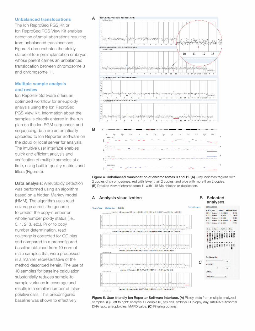
Unbalanced translocationsThe Ion ReproSeq PGS Kit or Ion ReproSeq PGS View Kit enables detection of small aberrations resulting from unbalanced translocations. Figure 4 demonstrates the ploidy status of four preimplantation embryos whose parent carries an unbalanced translocation between chromosome 3 and chromosome 11.
Multiple sample analysis and reviewIon Reporter Software offers an optimized workflow for aneuploidy analysis using the Ion ReproSeq PGS View Kit. Information about the samples is directly entered in the run plan on the Ion PGM sequencer, and sequencing data are automatically uploaded to Ion Reporter Software on the cloud or local server for analysis. The intuitive user interface enables quick and efficient analysis and verification of multiple samples at a time, using built-in quality metrics and filters (Figure 5).
Data analysis: Aneuploidy detection was performed using an algorithm based on a hidden Markov model (HMM). The algorithm uses read coverage across the genome to predict the copy-number or whole-number ploidy status (i.e., 0, 1, 2, 3, etc.). Prior to copy number determination, read coverage is corrected for GC bias and compared to a preconfigured baseline obtained from 10 normal male samples that were processed in a manner representative of the method described herein. The use of 10 samples for baseline calculation substantially reduces sample-to-sample variance in coverage and results in a smaller number of false-positive calls. This preconfigured baseline was shown to effectively
Figure 4. Unbalanced translocation of chromosomes 3 and 11. (A) Gray indicates regions with 2 copies of chromosomes, red with fewer than 2 copies, and blue with more than 2 copies. (B) Detailed view of chromosome 11 with ~18 Mb deletion or duplication.
Figure 5. User-friendly Ion Reporter Software interface. (A) Ploidy plots from multiple analyzed samples. (B) Left to right: analysis ID, couple ID, sex call, embryo ID, biopsy day, mtDNA:autosomal DNA ratio, aneuploidies, MAPD value. (C) Filtering options.
A B
C
Analysis visualization Selected analyses
A
B
determine ploidy state regions even for samples that were prepared across different labs using the low-coverage, whole-genome aneuploidy workflow. Using the HMM framework, the algorithm utilizes coverage information for a sample in a statistical model that determines the likelihood for a particular genomic region belonging to an alternate ploidy status.
Tunable aneuploidy detection sensitivity: The analysis methods provide tunable scaling factors. Algorithm parameters have been tuned for low-pass, whole-genome sequencing data using technical sample replicate data as well as samples with known aneuploidies. Users may select from three levels of sensitivity: low, medium, and high. High sensitivity enables more permissive aneuploidy assessment for smaller chromosomal segments and aneuploidies in noisy regions, but with a higher probability of false positives. The medium setting is the default and is the recommended initial starting point for aneuploidy detection. Low sensitivity is the most stringent and will only detect regions with strong evidence of aneuploidy.
Confidence and precision quality metrics for aneuploidy prediction: In addition to predicting copy number across chromosomal regions and the associated ploidy, the algorithm calculates two quality metrics for each region: confidence and
precision. Confidence is defined as a log ratio between the called ploidy state likelihood of the region and the expected ploidy state likelihood. Large confidence values indicate the algorithm is very certain that the ploidy state differs from the expected. Precision is defined as a log ratio between the likelihood of the assigned ploidy state and a next-closest state. Low precision (<10) denotes uncertainty in the absolute ploidy value assignment. It is possible that high ploidy states (e.g., copy number state >5) will have low precision and high confidence, indicating that the algorithm is uncertain about the absolute ploidy value but quite certain about the existence of a ploidy state that is different from the expected (for example, high confidence that a copy number increase is present, but uncertainty in whether the exact copy number is 5 or 6).
Overall sample quality metric: The median of the absolute values of all pairwise differences (MAPD) score is reported on aneuploidy analyses. MAPD is used to assess sample variability and define whether the data are useful for copy number analysis. Samples with an MAPD score above 0.3 should be carefully reviewed before validating a ploidy call.
Aneuploidy sample sex calling: Samples from preimplantation embryos are of unknown sex. However, identification of copy
number aberrations in sex chromosomes requires measuring the expected ploidy for the X and Y chromosomes in a sample. The presence or absence of the Y chromosome in a sample is used to make the sex call.
ConclusionThe Ion ReproSeq PGS View Kit, in combination with the Ion PGM System and Ion Reporter Software, provides a robust method for the detection of aneuploidies in as little material as a single cell, using low amounts of input DNA in a simple, rapid workflow. Independent testing by customers detected 100% of the known copy number changes present in 134 samples. The results were 100% concordant with data obtained from aCGH analysis. However, in contrast to aCGH, multiplexing with barcoding allows for simultaneous analysis of up to 24 samples in a single, low-pass, whole-genome sequencing run. Each Ion PGM System supports three different chip types that scale to different throughput needs. This throughput flexibility reduces the need for batching to achieve cost efficiency, and enables aneuploidy analysis as soon as samples are available.
AcknowledgmentsData used in this application note were kindly provided by Dr. Luis Alcaraz, Bioarray, Spain.
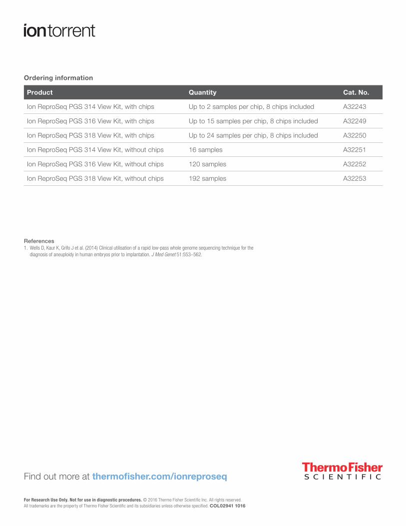
Ordering information
Product Quantity Cat. No.
Ion ReproSeq PGS 314 View Kit, with chips Up to 2 samples per chip, 8 chips included A32243
Ion ReproSeq PGS 316 View Kit, with chips Up to 15 samples per chip, 8 chips included A32249
Ion ReproSeq PGS 318 View Kit, with chips Up to 24 samples per chip, 8 chips included A32250
Ion ReproSeq PGS 314 View Kit, without chips 16 samples A32251
Ion ReproSeq PGS 316 View Kit, without chips 120 samples A32252
Ion ReproSeq PGS 318 View Kit, without chips 192 samples A32253
References1. Wells D, Kaur K, Grifo J et al. (2014) Clinical utilisation of a rapid low-pass whole genome sequencing technique for the
diagnosis of aneuploidy in human embryos prior to implantation. J Med Genet 51:553–562.
Find out more at thermofisher.com/ionreproseq
For Research Use Only. Not for use in diagnostic procedures. © 2016 Thermo Fisher Scientific Inc. All rights reserved. All trademarks are the property of Thermo Fisher Scientific and its subsidiaries unless otherwise specified. COL02941 1016