Desulfovibrio desulfuricans LSt
6
APPLIED AND ENVIRONMENTAL MICROBIOLOGY, Jan. 1993, p. 290-295 0099-2240/93/010290-06$02.00/0 Copyright ©) 1993, American Society for Microbiology Cobalamin-Mediated Mercury Methylation by Desulfovibrio desulfuricans LSt SUNG-CHAN CHOI AND RICHARD BARTHA* Department of Biochemistry and Microbiology, Cook College, Rutgers University, New Brunswick, New Jersey 08903-0231 Received 14 September 1992/Accepted 9 November 1992 The prominence of sulfate reducers in mercury biomethylation prompted the examination of the methyl carrier and mercury methylation activity of Desulfovibrio desulfuricans LS. There was a low degree of mercury tolerance and a high degree of methylation during fermentative growth; the opposite was true during sulfate reduction. During 2 days of fermentative growth, up to 37% of HgCl2 was methylated at 0.1 ,ug/ml, but obly 1.5% was methylated at 10.0 ,ug/ml. Less than 1% of the added HgCl2 was methylated under sulfate-reducing conditions. D. desulfuricans LS radioimmunoassay results were positive for cobalamin. The addition of CoCI2 and benzimidazole to fermentative cultures increased methylation activity. From D. desulfuricans LS grown in the presence of 57CoC12, a corrinoid was extracted and purified. High-performance liquid chromatography analysis of the purified extract yielded a single peak with the retention time of cobalamin, and 97% of the 57Co radioactivity was associated with this peak. Fast atom bombardment and UV and visible spectra of the isolated corrinoid matched those of cobalamin. When methylated with 14CH31, the isolated corrinoid methylated Hg2' with a 93.9%o preservation of '4C specific activity. We conclude that D. desu(furicans LS methylates mercury via cobalamin (vitamin B12). Under physiological conditions, the enzymatic catalysis of this reaction is likely. Monomethylmercury is a highly potent and biomagnifica- tion-prone neurotoxin. In the Minamata incident, which involved a local discharge of methylmercury, tainted sea- food caused 2,200 poisonings with 750 fatalities (20). Subse- quently, methylmercury was recognized as a global health hazard due to the environmental conversion of less hazard- ous mercury pollutants to this very toxic form (12, 19). Even in pristine freshwater lakes of Wisconsin that receive atmo- spheric mercury inputs only, large fish accumulate methylm- ercury in excess of the 0.5-ppm Food and Drug Administra- tion limit (10). Recent work identified sulfate reducers of anoxic aquatic sediments as the principal environmental methylators of mercury (7, 8). Mercury-methylating strains of Desulfovibrio desulfuncans were isolated and described (7, 8), but much remains to be learned about the biochemistry of the mercury methylation process by these microorganisms. Methylcobal- amin, which under appropriate conditions spontaneously methylates mercuric ions (6, 25), has long been suspected but never proven to mediate environmental mercury meth- ylation. Paradoxically, methylcobalamin was identified in some acetogens and methanogens (22) which were ruled out as environmental methylators of mercury (7). Cobalt por- phyrins were identified in Desulfovibrio gigas and D. desul- furicans Norway (11, 16), but these porphyrins do not appear to be methyl carriers. 5-Methylbenzimidazolyl coba- mide, in its structure and function closely related to cobal- amin, was identified in some sulfate-reducing bacteria (13). It is not known at this time whether this carrier can methylate mercuric ions. Radioimmunoassays of a mercury-methylating strain of D. desulfuricans, strain LS, indicated the presence of tetrahy- drofolate and cobalamin (3). The carbon flow in mercury * Corresponding author. t New Jersey Agricultural Experiment Station publication D-01408-01-92. biomethylation suggested that the methyl group donated to mercury originates as C-3 of pyruvate and is donated as C-3 of serine by serine hydroxymethyl transferase to tetrahydro- folate. From here, cobalamin or a closely related methyl carrier was postulated to transfer the methyl group to mercuric ions (3). The aim of the study presented here was to positively identify the methyl carrier of D. desulfuricans LS and to document its involvement in the mercury methylation pro- cess. MATERIALS AND METHODS Cell growth and methylmercury analysis. The D. desulfu- ncans strain used in this study was previously isolated from an anoxic, low-salinity (0.4%) salt marsh sediment in Cheesequake State Park, N.J. (7). The strain designation of LS (for low salinity) was added to identify this isolate. When grown in lactate-sulfate medium, cells were motile and vibrio-shaped with the dimensions 0.5 by 1.0 ,Lm. Media utilized in this study for supporting sulfate-reducing and fermentative growth were Postgate's lactate-sulfate medium C and pyruvate medium D, respectively (17). The purity of the strain was routinely monitored with ferrous sulfate- containing diagnostic medium, by tests for the presence of desulfoviridin, growth inhibition by molybdate, and methy- lation of mercury, and by microscopic observation (8). The maintenance of culture purity was a high priority since contaminants can easily take over under fermentative growth conditions. Cell growth was routinely determined by protein measurement with the dye-binding method of Brad- ford (4). For the determination of monomethylmercury synthesis, either medium C or D received an appropriate concentration of HgCl2 at the time of inoculation. After incubation at 27°C for 1 to 2 days, the cell suspensions were extracted by the method of Longbottom et al. (15), and monomethylmercury levels were measured with a Hewlett-Packard 5890 gas 290 Vol. 59, No. 1
Transcript of Desulfovibrio desulfuricans LSt

APPLIED AND ENVIRONMENTAL MICROBIOLOGY, Jan. 1993, p. 290-2950099-2240/93/010290-06$02.00/0Copyright ©) 1993, American Society for Microbiology
Cobalamin-Mediated Mercury Methylation byDesulfovibrio desulfuricans LSt
SUNG-CHAN CHOI AND RICHARD BARTHA*
Department ofBiochemistry and Microbiology, Cook College,Rutgers University, New Brunswick, New Jersey 08903-0231
Received 14 September 1992/Accepted 9 November 1992
The prominence of sulfate reducers in mercury biomethylation prompted the examination of the methylcarrier and mercury methylation activity ofDesulfovibrio desulfuricans LS. There was a low degree of mercurytolerance and a high degree of methylation during fermentative growth; the opposite was true during sulfatereduction. During 2 days of fermentative growth, up to 37% of HgCl2 was methylated at 0.1 ,ug/ml, but obly1.5% was methylated at 10.0 ,ug/ml. Less than 1% of the added HgCl2 was methylated under sulfate-reducingconditions. D. desulfuricans LS radioimmunoassay results were positive for cobalamin. The addition of CoCI2and benzimidazole to fermentative cultures increased methylation activity. From D. desulfuricans LS grown inthe presence of 57CoC12, a corrinoid was extracted and purified. High-performance liquid chromatographyanalysis of the purified extract yielded a single peak with the retention time of cobalamin, and 97% of the 57Coradioactivity was associated with this peak. Fast atom bombardment and UV and visible spectra of the isolatedcorrinoid matched those of cobalamin. When methylated with 14CH31, the isolated corrinoid methylated Hg2'with a 93.9%o preservation of '4C specific activity. We conclude that D. desu(furicans LS methylates mercuryvia cobalamin (vitamin B12). Under physiological conditions, the enzymatic catalysis of this reaction is likely.
Monomethylmercury is a highly potent and biomagnifica-tion-prone neurotoxin. In the Minamata incident, whichinvolved a local discharge of methylmercury, tainted sea-
food caused 2,200 poisonings with 750 fatalities (20). Subse-quently, methylmercury was recognized as a global healthhazard due to the environmental conversion of less hazard-ous mercury pollutants to this very toxic form (12, 19). Evenin pristine freshwater lakes of Wisconsin that receive atmo-spheric mercury inputs only, large fish accumulate methylm-ercury in excess of the 0.5-ppm Food and Drug Administra-tion limit (10).
Recent work identified sulfate reducers of anoxic aquaticsediments as the principal environmental methylators ofmercury (7, 8). Mercury-methylating strains of Desulfovibriodesulfuncans were isolated and described (7, 8), but muchremains to be learned about the biochemistry of the mercurymethylation process by these microorganisms. Methylcobal-amin, which under appropriate conditions spontaneouslymethylates mercuric ions (6, 25), has long been suspectedbut never proven to mediate environmental mercury meth-ylation. Paradoxically, methylcobalamin was identified insome acetogens and methanogens (22) which were ruled outas environmental methylators of mercury (7). Cobalt por-phyrins were identified in Desulfovibrio gigas and D. desul-furicans Norway (11, 16), but these porphyrins do notappear to be methyl carriers. 5-Methylbenzimidazolyl coba-mide, in its structure and function closely related to cobal-amin, was identified in some sulfate-reducing bacteria (13). Itis not known at this time whether this carrier can methylatemercuric ions.Radioimmunoassays of a mercury-methylating strain ofD.
desulfuricans, strain LS, indicated the presence of tetrahy-drofolate and cobalamin (3). The carbon flow in mercury
* Corresponding author.t New Jersey Agricultural Experiment Station publication
D-01408-01-92.
biomethylation suggested that the methyl group donated tomercury originates as C-3 of pyruvate and is donated as C-3of serine by serine hydroxymethyl transferase to tetrahydro-folate. From here, cobalamin or a closely related methylcarrier was postulated to transfer the methyl group tomercuric ions (3).The aim of the study presented here was to positively
identify the methyl carrier of D. desulfuricans LS and todocument its involvement in the mercury methylation pro-cess.
MATERIALS AND METHODS
Cell growth and methylmercury analysis. The D. desulfu-ncans strain used in this study was previously isolated froman anoxic, low-salinity (0.4%) salt marsh sediment inCheesequake State Park, N.J. (7). The strain designation ofLS (for low salinity) was added to identify this isolate. Whengrown in lactate-sulfate medium, cells were motile andvibrio-shaped with the dimensions 0.5 by 1.0 ,Lm. Mediautilized in this study for supporting sulfate-reducing andfermentative growth were Postgate's lactate-sulfate mediumC and pyruvate medium D, respectively (17). The purity ofthe strain was routinely monitored with ferrous sulfate-containing diagnostic medium, by tests for the presence ofdesulfoviridin, growth inhibition by molybdate, and methy-lation of mercury, and by microscopic observation (8). Themaintenance of culture purity was a high priority sincecontaminants can easily take over under fermentativegrowth conditions. Cell growth was routinely determined byprotein measurement with the dye-binding method of Brad-ford (4).For the determination of monomethylmercury synthesis,
either medium C or D received an appropriate concentrationof HgCl2 at the time of inoculation. After incubation at 27°Cfor 1 to 2 days, the cell suspensions were extracted by themethod of Longbottom et al. (15), and monomethylmercurylevels were measured with a Hewlett-Packard 5890 gas
290
Vol. 59, No. 1
COBALAMIN-MEDIATED MERCURY METHYLATION 291
chromatograph equipped with a capillary column (0.53-mminside diameter by 10 m long; Alltech AT-35, Deerfield, Ill.).Operating conditions were as follows: 95:5 Ar-CH4 (vol/vol)carrier gas (Matheson Gas Products, East Rutherford, N.J.)at 40 ml/min, injector at 210°C, oven at 100°C, and electroncapture detector at 250°C. Monomethylmercury peak (reten-tion time of 1.25 min) areas were recorded by a Hewlett-Packard 3392A integrator.Comnoid extraction and purification. For determination of
cobalamin in whole cells, a vitamin B12-folate dual radioim-munoassay kit was used according to the instructions of themanufacturer (Amersham Inc., Arlington Heights, Ill.). Tofacilitate monitoring of the corrinoid during the purification,57Co label (57CoC12, specific activity of 5.21 mCi/,ug of Co;Amersham Inc.) was incorporated into the cells. Understrictly anaerobic conditions, 19 liters of lactate-sulfatemedium in a 20-liter carboy received 0.2 mCi of 57CoC12 andwas incubated at 27°C for 4 days. About 50 g (wet weight) ofcells, including some inorganic sulfide precipitate, was har-vested by centrifugation. Extraction with boiling KCN at afinal concentration of 10 mM converted all of the corrinoidinto the more stable cyano form. After removal of cell debrisby centrifugation at 15,000 x g for 30 min, further purifica-tion was performed through a precleaned nonionic AmberliteXAD-4 (Supelco Inc., Bellefonte, Pa.) column (1.5 by 30 cm)and the same-sized aluminum oxide column on the basis ofthe procedure described by Stupperich et al. (24). A Rotava-por apparatus (RE 121; Buchi, Flawil, Switzerland) was usedfor the evaporation of methanol at a temperature not exceed-ing 40°C. The residue was dissolved in water. At each step,57Co radioactivity was measured with a gamma counter(model 1191; T. M. Analytic, Elk Grove Village, Ill.) withover 90% counting efficiency. Purification was carried out inthe dark or under a dim red light to minimize any photodeg-radation of the corrinoid.HPLC analysis. High-performance liquid chromatography
(HPLC) was performed with a Partisphere 5-,um C18 column(0.46-cm inside diameter by 25 cm long; Whatman Inc.,Clifton, N.J.), and a solvent gradient was generated with anIntelligent pump (model L-600; Hitachi). A UV-visible spec-trophotometer (model L-4200; Hitachi) operating at 546 nmconnected with a Chromato-integrator (model D-2000; Hita-chi) was used for the detection of the corrinoid. The mobilephases were A, 0.1% acetic acid, and B, 100% methanol.The column was eluted at a flow rate of 1 ml/min with aninitial 0- to 5-min isocratic elution (75% A-25% B) followedby a linear gradient to 35% A-65% B within 25 min.Cyanocobalamin standard was purchased from Sigma Chem-ical Co., St. Louis, Mo., and was, at times, injected togetherwith the corrinoid extract. Eluted solvent was collected in1.0-ml fractions, and the corresponding 57Co radioactivitywas measured with a gamma counter.FAB-MS and UV-visible spectral analysis. Fast atom bom-
bardment-mass spectrometry (FAB-MS) analysis was per-formed with a ZAB-T high-resolution mass spectrometer(model 8058; Vacuum Generators, Manchester, England)with a technique known as liquid secondary ionization massspectrometry. The sample was dissolved in "magnetic bul-let" matrix, which is a 3:1 mixture of dithiothreitol anddithioerythritol dissolved in methanol. Experimental condi-tions were 10 kV of accelerating potential and 35 kV ofcesium ion gun. Data were acquired in the continuum mode,and the signal was averaged over the entire analysis (100scans).UV and visible absorption spectra were obtained in water
with a Shimadzu spectrophotometer UV-265.
Transmethylation. By using the previously described spec-trophotometer, spontaneous transmethylation by methylco-balamin was recorded as a time lapse (cycle time of 10 min,15 cycles) spectral change to hydroxycobalamin in a wave-length range of 280 to 400 nm. The reaction mixture con-tained 30 F.M methylcobalamin and 60 ,LM HgCl2 in 0.1 MNa-acetate buffer, pH 4.5. The reaction was initiated by adirect addition of concentrated HgCl2 solution into a quartzcuvette.For the demonstration of mercury-methylating activity
with the purified corrinoid, 50 ,uCi of 1 CH3I (specificactivity of 8 mCi/mmol; New England Nuclear Corp., Bos-ton, Mass.) diluted in 100 mg of unlabeled CH3I was addedto 20 ml of corrinoid extract, obtained from a cell massequivalent to 40 mg of protein. The reaction was allowed toproceed at room temperature under reducing conditionsgenerated by a gentle stream of N2 gas (Matheson GasProducts) and the addition of 20 mg of sodium borohydride.The cleanup included the extraction of the methylcobalamininto the phenol phase and back-extraction from the phenol.Details for the exchange of the -CN to the -CH3 groupwere the same as described for the chemical synthesis ofmethylcobalamin from cyanocobalamin (9). The methylatedcorrinoid extract in 0.1 M Na-acetate buffer (pH 4.5) re-ceived 1 mg of HgCl2 and was allowed to stand at roomtemperature and in the dark for 6 h. After extraction ofmonomethylmercury, the final benzene extract was analyzedby gas chromatography and the transfer of the 14C label wasmeasured by a liquid-scintillation counter (BetaTrac 6895; T.M. Analytic). Counting efficiency was determined by theexternal standard ratio method. A control experiment wasperformed by using the same procedure, except the purifiedcorrinoid was omitted.
RESULTS AND DISCUSSION
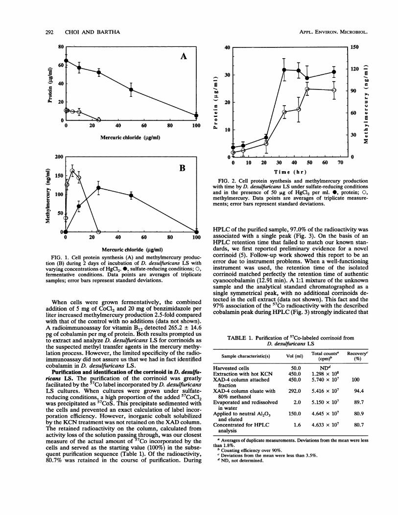
Culture conditions and methylmercury synthesis. The ab-solute amounts of methylmercury produced and the percent-age of the mercuric ion methylated by D. desulfuncans LSstrongly depended on the culture conditions and on thelevels of the mercury added. It was reported earlier thatmore methylmercury was produced under fermentative thanunder sulfate-reducing conditions (7). When in the presentstudy three serial transfers on pyruvate medium excludedsulfide carryover, this effect was much more pronounced(Fig. 1). From the data presented, we calculated that on thebasis of micrograms of protein, up to 17.1 and 2.9 ng ofmethylmercury were produced during 2 days of incubationunder fermentative and under sulfate-reducing conditions,respectively. Under fermentative conditions, 37.0, 12.6, and1.5% of 0.1, 1.0, and 10.0 p.g of HgCl2 per ml, respectively,were transformed to methylmercury. Under sulfate-reducingconditions, 0.7, 0.5, and 0.2% of 10, 25, and 50 ,ug of HgCl2per ml were methylated, respectively. In the fermentativeculture, HgCl2 levels above 10 p.g/ml strongly inhibited bothcell growth and methylation activity. In the sulfate-reducingculture, however, the absolute amounts of methylmercuryproduced increased up to 25 ,ug of HgCl2 added per ml anddeclined above this HgCl2 level only gradually. Cell yieldswere still substantial at 50 p.g of HgCl2 per ml. Of course,under sulfate-reducing conditions, the numbers reflect onlythe added rather than the available HgCl2 concentrationsbecause most of the mercuric ions precipitated as insolubleHgS. When added within the tolerance range, methylmer-cury synthesis generally paralleled the increase in cell pro-tein, as illustrated in Fig. 2.
VOL. 59, 1993
292 CHOI AND BARTHA
I--I
.0
go
A60'
40-1
20
0.-0 20 40 60 80 10
-
9
OD
3-
*
ro
'o
Mercuric chloride (pg/ml)
100
Mercuric chloride (.ug/mi)FIG. 1. Cell protein synthesis (A) and methylmercury produc-
tion (B) during 2 days of incubation of D. desulfuricans LS withvarying concentrations of HgCl2. 0, sulfate-reducing conditions; 0,
fermentative conditions. Data points are averages of triplicatesamples; error bars represent standard deviations.
When cells were grown fermentatively, the combinedaddition of 5 mg of CoCl2 and 20 mg of benzimidazole perliter increased methylmercury production 2.5-fold comparedwith that of the control with no additions (data not shown).A radioimmunoassay for vitamin B12 detected 265.2 + 14.6pg of cobalamin per mg of protein. Both results prompted usto extract and analyze D. desulfuricans LS for corrinoids asthe suspected methyl transfer agents in the mercury methy-lation process. However, the limited specificity of the radio-immunoassay did not assure us that we had in fact identifiedcobalamin in D. desulfuricans LS.
Purification and identification of the corrinoid in D. desujfu-ricans LS. The purification of the corrinoid was greatlyfacilitated by the 7Co label incorporated byD. desulfuicansLS cultures. When cultures were grown under sulfate-reducing conditions, a high proportion of the added 5'CoCl2was precipitated as 57CoS. This precipitate sedimented withthe cells and prevented an exact calculation of label incor-poration efficiency. However, inorganic cobalt solubilizedby the KCN treatment was not retained on the XAD column.The retained radioactivity on the column, calculated fromactivity loss of the solution passing through, was our closestmeasure of the actual amount of 7Co incorporated by thecells and served as the starting value (100%) in the subse-quent purification sequence (Table 1). Of the radioactivity,80.7% was retained in the course of purification. During
150
120 -
9Ogo5"
90U
60 *Is0
30 Io:9
01: ' I
0 10 20 30 40 50 60 70
Time (hr)FIG. 2. Cell protein synthesis and methylmercury production
with time by D. desulfuricans LS under sulfate-reducing conditionsand in the presence of 50 ,ug of HgCl2 per ml. *, protein; 0,methylmercury. Data points are averages of triplicate measure-ments; error bars represent standard deviations.
HPLC of the purified sample, 97.0% of the radioactivity wasassociated with a single peak (Fig. 3). On the basis of anHPLC retention time that failed to match our known stan-dards, we first reported preliminary evidence for a novelcorrinoid (5). Follow-up work showed this report to be anerror due to instrument problems. When a well-functioninginstrument was used, the retention time of the isolatedcorrinoid matched perfectly the retention time of authenticcyanocobalamin (12.91 min). A 1:1 mixture of the unknownsample and the analytical standard chromatographed as asingle symmetrical peak, with no additional corrinoids de-tected in the cell extract (data not shown). This fact and the97% association of the 57Co radioactivity with the describedcobalamin peak during HPLC (Fig. 3) strongly indicated that
TABLE 1. Purification of "Co-labeled corrinoid fromD. desulfuricans LS
Sample characteristic(s) Vol (ml) Total countse Recovery'(cpm)' (%
Harvested cells 50.0 NDdExtraction with hot KCN 450.0 1.298 x 10⁸XAD-4 column attached 450.0 5.740 x 10⁷ 100
fractionXAD-4 column eluate with 292.0 5.416 x 10⁷ 94.480% methanol
Evaporated and redissolved 2.0 5.150 x i07 89.7in water
Applied to neutral A1203 150.0 4.645 x 10⁷ 80.9and eluted
Concentrated for HPLC 1.6 4.633 x 10⁷ 80.7analysisI Averages of duplicate measurements. Deviations from the mean were less
than 1.8%.b Counting efficiency over 90%.cDeviations from the mean were less than 3.5%.d ND, not determined.
200
IIIV3-
APPL. ENVIRON. MICROBIOL.

COBALAMIN-MEDIATED MERCURY METHYLATION 293
0.15 300I
0.1 200C 0~~~~~~~~~~~~~~~~~~~P
0.05 100
v i . 00 5 10 15 20 25 30
Retention time (min) I FractionsFIG. 3. HPLC separation of the D. desulfuncans LS corrinoid
and the association of the "Co radioactivity (bars) with the collectedfractions. The slight offset is due to volume holdup between detectorand collection tubes.
cobalamin was the only corrinoid present in D. desulfuricansLS.The identity of the isolated corrinoid was confirmed by
FAB-MS. The positive ion spectrum of the unknown corri-noid indicated a molecular weight of 1,354, identical to thatof cyanocobalamin (Fig. 4). Loss of the a axial ligand (cyanogroup) resulted in an intense peak at mlz 1329. An interpre-tation of the fragmentation is as follows: m/z 1286 [1329 -CONH]+, m/z 1270 [1329 - CH3CONH2]+ or [1329 - Co]',mlz 1257 [corrin-dimethylbenzimidazole-sugar-OPO2OCH(CH3)CH2]+, m/Z 1199 [corrin-dimethylbenzimidazole-sug-ar-OP02], m/Z 1183 [1329 - dimethylbenzimidazole]+, mlz1069 [corrin-CH2CH2CONHCH2CH(CH3)OPO20H + H]+,mlz 989 [corrin-CH2CH2CONHCH2CH(CH3)OH]J, and m/z971 (989 - H2O)+. These data are in good agreement withthe ones published by Barber et al. (1).The UV and visible absorption spectra showed a series of
spectral bands at wavelengths of 275, 360, 480, and 540 nm,with a relative peak height ratio of 1:0.46:0.06:0.09. Again,
100
80
;60._
40 . >
20 _ o
0950 1050 11
400 TABLE 2. Conversion of D. desulfuricans LS corrinoid to its4C-methyl form and spontaneous methylation of Hg2+
by this methylcorrinoid
Radioactivity (dpm)Exptl step
Control' Corrinoid extract
Corrinoid methylation andremoval of 14CH3I
Addition of 14CH3I 1.11 x 10⁸ 1.11 x 10⁸Water extract of organic layer 2.03 x 10⁵ 1.20 x 10⁶
Mercury methylation andmethylmercury extraction
Starting sample for Hg2+ 3.00 x 10⁴ 9.31 x 10⁵methylation
Toluene extract 3.40 x 10² 6.18 x 10⁵Sodium thiosulfate extract 0 5.04 x 10⁴Benzene extract 0 4.25 x 10⁴
a Identical treatment, except that the D. desulfuricans LS corrinoid wasomitted.
this spectrum is in good agreement with published data forcyanocobalamin (21).The described characteristics confirm that the corrinoid
isolated from D. desulfuricans LS is identical to cobalamin.Role of cobalamin in the methylation of mercury by D.
desulfuricans LS. Cyanocobalamin isolated from D. desulfu-ricans LS was methylated by '4CH31, and any residual14CH3I was carefully removed. When the prepared 14CH3-cobalamin was allowed to react with mercuric ions in a pH4.5 acetate buffer, 0.038% of the original "4CH3I labelpartitioned into benzene and was determined by gas chro-matography as 99 Ftg of methylmercury. No radioactivityremained in the negative control (Table 2). The correspond-ing specific activity ratio (specific activity of the methylmer-cury produced/specific activity of the added 14CH31) was93.9%, indicating that the methylated D. desulfuricans LScorrinoid spontaneously transferred its methyl group tomercuric ion. Thus, in addition to spectral evidence, thefunctional identity of the D. desulfuricans LS corrinoid tomethylcobalamin was also established.Under the appropriate conditions, such as low-pH acetate
buffer, the transfer of the methyl group from methylcobal-amin to mercuric ions occurred spontaneously and quite
I.
150 1250 1350m/z
FIG. 4. The positive ion FAB mass spectrum of the purified corrinoid extract from D. desulfuricans LS. Numbers represent the positionsof peaks of interest (see text).
VOL. 59, 1993
294 CHOI AND BARTHA
0.5
.0
.0
0
280 300 350 400
Wavelength (nm)
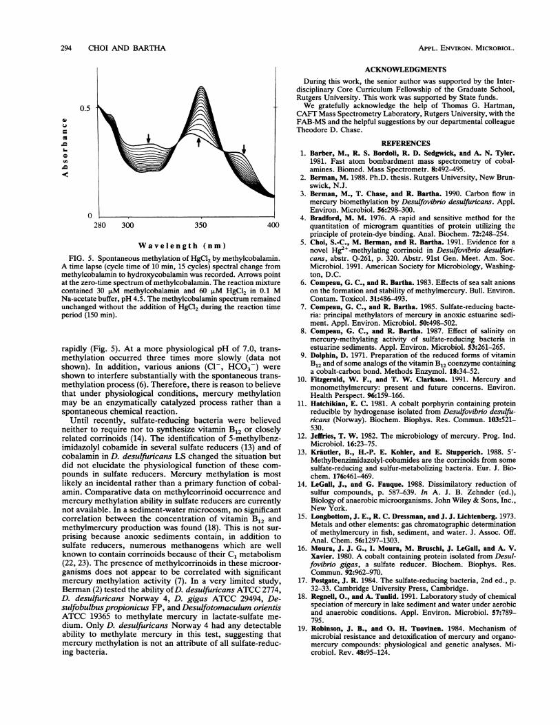
FIG. 5. Spontaneous methylation of HgCl2 by methylcobalamin.A time lapse (cycle time of 10 min, 15 cycles) spectral change frommethylcobalamin to hydroxycobalamin was recorded. Arrows pointat the zero-time spectrum of methylcobalamin. The reaction mixturecontained 30 ,uM methylcobalamin and 60 ,M HgCl2 in 0.1 MNa-acetate buffer, pH 4.5. The methylcobalamin spectrum remainedunchanged without the addition of HgCl2 during the reaction timeperiod (150 min).
rapidly (Fig. 5). At a more physiological pH of 7.0, trans-methylation occurred three times more slowly (data notshown). In addition, various anions (Cl-, HCO3-) wereshown to interfere substantially with the spontaneous trans-methylation process (6). Therefore, there is reason to believethat under physiological conditions, mercury methylationmay be an enzymatically catalyzed process rather than a
spontaneous chemical reaction.Until recently, sulfate-reducing bacteria were believed
neither to require nor to synthesize vitamin B12 or closelyrelated corrinoids (14). The identification of 5-methylbenz-imidazolyl cobamide in several sulfate reducers (13) and ofcobalamin in D. desulfuricans LS changed the situation butdid not elucidate the physiological function of these com-pounds in sulfate reducers. Mercury methylation is mostlikely an incidental rather than a primary function of cobal-amin. Comparative data on methylcorrinoid occurrence andmercury methylation ability in sulfate reducers are currentlynot available. In a sediment-water microcosm, no significantcorrelation between the concentration of vitamin B12 andmethylmercury production was found (18). This is not sur-prising because anoxic sediments contain, in addition tosulfate reducers, numerous methanogens which are wellknown to contain corrinoids because of their C1 metabolism(22, 23). The presence of methylcorrinoids in these microor-ganisms does not appear to be correlated with significantmercury methylation activity (7). In a very limited study,Berman (2) tested the ability ofD. desulfuricans ATCC 2774,D. desulfuricans Norway 4, D. gigas ATCC 29494, De-sulfobulbus propionicus FP, and Desulfotomaculum orientisATCC 19365 to methylate mercury in lactate-sulfate me-dium. Only D. desulfuricans Norway 4 had any detectableability to methylate mercury in this test, suggesting thatmercury methylation is not an attribute of all sulfate-reduc-ing bacteria.
ACKNOWLEDGMENTS
During this work, the senior author was supported by the Inter-disciplinary Core Curriculum Fellowship of the Graduate School,Rutgers University. This work was supported by State funds.We gratefully acknowledge the help of Thomas G. Hartman,
CAFT Mass Spectrometry Laboratory, Rutgers University, with theFAB-MS and the helpful suggestions by our departmental colleagueTheodore D. Chase.
REFERENCES1. Barber, M., R. S. Bordoli, R. D. Sedgwick, and A. N. Tyler.
1981. Fast atom bombardment mass spectrometry of cobal-amines. Biomed. Mass Spectrometr. 8:492-495.
2. Berman, M. 1988. Ph.D. thesis. Rutgers University, New Brun-swick, N.J.
3. Berman, M., T. Chase, and R. Bartha. 1990. Carbon flow inmercury biomethylation by Desulfovibrio desulfuricans. Appl.Environ. Microbiol. 56:298-300.
4. Bradford, M. M. 1976. A rapid and sensitive method for thequantitation of microgram quantities of protein utilizing theprinciple of protein-dye binding. Anal. Biochem. 72:248-254.
5. Choi, S.-C., M. Berman, and R. Bartha. 1991. Evidence for anovel Hg2+-methylating corrinoid in Desulfovibrio desulfuri-cans, abstr. Q-261, p. 320. Abstr. 91st Gen. Meet. Am. Soc.Microbiol. 1991. American Society for Microbiology, Washing-ton, D.C.
6. Compeau, G. C., and R. Bartha. 1983. Effects of sea salt anionson the formation and stability of methylmercury. Bull. Environ.Contam. Toxicol. 31:486-493.
7. Compeau, G. C., and R. Bartha. 1985. Sulfate-reducing bacte-ria: principal methylators of mercury in anoxic estuarine sedi-ment. Appl. Environ. Microbiol. 50:498-502.
8. Compeau, G. C., and R. Bartha. 1987. Effect of salinity onmercury-methylating activity of sulfate-reducing bacteria inestuarine sediments. Appl. Environ. Microbiol. 53:261-265.
9. Dolphin, D. 1971. Preparation of the reduced forms of vitaminB12 and of some analogs of the vitamin B12 coenzyme containinga cobalt-carbon bond. Methods Enzymol. 18:34-52.
10. Fitzgerald, W. F., and T. W. Clarkson. 1991. Mercury andmonomethylmercury: present and future concerns. Environ.Health Perspect. 96:159-166.
11. Hatchikian, E. C. 1981. A cobalt porphyrin containing proteinreducible by hydrogenase isolated from Desulfovibno desulfu-ricans (Norway). Biochem. Biophys. Res. Commun. 103:521-530.
12. Jeifries, T. W. 1982. The microbiology of mercury. Prog. Ind.Microbiol. 16:23-75.
13. Krautler, B., H.-P. E. Kohler, and E. Stupperich. 1988. 5'-Methylbenzimidazolyl-cobamides are the corrinoids from somesulfate-reducing and sulfur-metabolizing bacteria. Eur. J. Bio-chem. 176:461-469.
14. LeGall, J., and G. Fauque. 1988. Dissimilatory reduction ofsulfur compounds, p. 587-639. In A. J. B. Zehnder (ed.),Biology of anaerobic microorganisms. John Wiley & Sons, Inc.,New York.
15. Longbottom, J. E., R. C. Dressman, and J. J. Lichtenberg. 1973.Metals and other elements: gas chromatographic determinationof methylmercury in fish, sediment, and water. J. Assoc. Off.Anal. Chem. 56:1297-1303.
16. Moura, J. J. G., I. Moura, M. Bruschi, J. LeGall, and A. V.Xavier. 1980. A cobalt containing protein isolated from Desul-fovibrio gigas, a sulfate reducer. Biochem. Biophys. Res.Commun. 92:962-970.
17. Postgate, J. R. 1984. The sulfate-reducing bacteria, 2nd ed., p.32-33. Cambridge University Press, Cambridge.
18. Regnell, O., and A. Tunlid. 1991. Laboratory study of chemicalspeciation of mercury in lake sediment and water under aerobicand anaerobic conditions. Appl. Environ. Microbiol. 57:789-795.
19. Robinson, J. B., and 0. H. Tuovinen. 1984. Mechanism ofmicrobial resistance and detoxification of mercury and organo-mercury compounds: physiological and genetic analyses. Mi-crobiol. Rev. 48:95-124.
APPL. ENvIRON. MICROBIOL.
COBALAMIN-MEDIATED MERCURY METHYLATION 295
20. Sakamoto, M., A. Nakano, Y. Kinjo, H. Higashi, and M.Futatsuka. 1991. Present mercury levels in red blood cells ofnearby inhabitants about 30 years after the outbreak of Mina-mata disease. Ecotoxicol. Environ. Saf. 22:58-66.
21. Schneider, Z., and A. Stroiniski. 1987. Spectroscopy of corni-noids, p. 44-55. In Comprehensive B12. Walter de Gruyter, Inc.,New York.
22. Stupperich, E., H.-J. Eisinger, and S. Schurr. 1990. Corrinoids
in anaerobic bacteria. FEMS Microbiol. Rev. 87:355-360.
23. Stupperich, E., and B. Krautler. 1988. Pseudo vitamin B12 or5-hydroxybenzimidazolyl cobamide are the corrinoids found inmethanogenic bacteria. Arch. Microbiol. 149:268-271.
24. Stupperich, E., I. Steiner, and M. Ruhlemann. 1986. Isolationand analysis of bacterial cobamides by high-performance liquidchromatography. Anal. Biochem. 155:365-370.
25. Thayer, J. S. 1981. Effect of halide ions on the rates of reactionof methylcobalamin with heavy-metal species. Inorg. Chem.20:3573-3576.
VOL. 59, 1993


![Diverse hydrogen production and consumption pathways … · ing dissimilatory sulfate reduction (e.g., Desulfovibrio desulfuricans)[50, 51], fumarate and nitrate reduction (e.g.,](https://static.fdocuments.net/doc/165x107/5f478d2813f91a6e9825a474/diverse-hydrogen-production-and-consumption-pathways-ing-dissimilatory-sulfate-reduction.jpg)