Desmoplastic Ameloblastoma: A Diagnostic Dilemma · Desmoplastic ameloblastoma shows marked...
8
Oral and Maxillofacial Pathology Journal, January-June 2018;9(1):23-30 23 Desmoplastic Ameloblastoma: A Diagnostic Dilemma 1 Narendra T Chaudhari, 2 Jagdish V Tupkari, 3 Manisha S Ahire, 4 Tabita Joy ABSTRACT Introduction: Desmoplastic ameloblastoma (DA) is a rare variant of ameloblastoma that shows unique features, behavior, and presentation. Earlier it was believed to be less aggressive but this tumor has gained importance, as recent reports suggest increased incidence of destruction and recurrence. It is most commonly confused with fibro-osseous lesions and only histo- pathological examination helps to establish the final diagnosis. Due to varied clinical and radiographic appearance this lesion can be misdiagnosed, hence its adequate knowledge is imperative for dental health professionals to ensure appropriate management. Case report: Herewith, we present a case of DA affecting the maxilla. Based on clinical, radiographic, computed tomography (CT) scan, magnetic resonance imaging (MRI), fine needle aspiration biopsy (FNAB) findings along with histotopathologi- cal evaluation using special and immunohistochemistry (IHC) stains, the present case was diagnosed as DA. Management: The tumor was treated by partial maxillectomy along with removal of the involved teeth and two bone plates were used to reconstruct the left maxillary anterior region and these plates were attached to the bone with monocortical screws. The postoperative healing phase was without any complications. Conclusion: Some researchers suggest that DA might be less biologically aggressive than conventional ameloblastoma as the desmoplasia in DA might act as a limiting barrier for local spread of the DA tumor cells. But recently, few cases showed that DA tends to exhibit rapid growth and progressive behavior, with incidence of recurrence the same as conventional ameloblas- tomas. The radiological and histological findings of ill-defined borders and poor encapsulation require a long-term follow-up. Keywords: Ameloblastoma, Desmoplastic, Dilemma, Maxilla, Recurrence. How to cite this article: Chaudhari NT, Tupkari JV, Ahire MS, Joy T. Desmoplastic Ameloblastoma: A Diagnostic Dilemma. Oral Maxillofac Pathol J 2018;9(1):23-30. Source of support: Nil Conflict of interest: None OMPJ CASE REPORT 10.5005/jp-journals-10037-1124 1 Dental Surgeon and Consulting Oral Pathologist, 2 Professor and Head, 3 Assistant Professor, 4 Associate Professor 1 Dental Surgeon and Consulting Oral Pathologist at Shri Sai Dental Multispeciality Clinic, Mumbai, Maharashtra, India 2-4 Department of Oral Pathology and Microbiology, Government Dental College & Hospital, Mumbai, Maharashtra, India Corresponding Author: Narendra T Chaudhari, Nirman Park Phase 3, A-6, Room no 4, Santoshimata road, Kalyan West 421301, Mumbai, Maharashtra, India, Phone: +919969529473 e-mail: [email protected] INTRODUCTION Ameloblastoma is the second most common benign odontogenic tumor 1 that locally manifests as an invasive, destructive, 2 and aggressive lesion. 3 Apart from malig- nant ameloblastoma and ameloblastic carcinoma, many researchers believe the conventional ameloblastoma to be a low-grade malignant tumor. 4 Solid or multicystic, unicystic, and extraosseous are the clinical types of ameloblastoma, while the radio- graphic presentation is usually in the form of a unilocular or multilocular radiolucency. 1 The term “ameloblastoma” encompasses several histological patterns, 5 of which follicular and plexiform are the most common variet- ies, followed by the acanthomatous and granular cell types. Uncommon variants include desmoplastic, basal cell, clear cell, keratoameloblastoma, and papilliferous keratoameloblastoma. 2 Desmoplastic ameloblastoma shows marked varia- tions in its anatomical location, morphology and radio- graphic appearance as compared with the other forms of ameloblastoma, hence there was a need to classify it as a separate clinicopathologic type. 1 Subsequently, DA was included as a distinct variant of ameloblastoma in the World Health Organization classification of odontogenic tumors in 2005. 4,6 The worldwide incidence of DA is about 0.9 to 12.1% of total ameloblastoma. In general, there are two histo- logic variants of DA, simple DA (88.0%) and DA with osteoplasia (12.0%). 7 The radiographic picture of DA is not specific 1 and can be confused with fibro-osseous lesions or malignant tumors. 4 The DA can resemble odontogenic fibroma, squamous odontogenic tumor, ameloblastic fibroma, and squamous cell carcinoma in its histopatho- logical presentation. 1 Desmoplastic ameloblastoma shows a more aggres- sive behavior than other types of ameloblastoma, which may be due to its potential to grow to a large size, peculiar anatomic location in anterior maxilla leading to an early invasion of adjoining structures and bone. 8 We report here a case of DA affecting the anterior maxilla, in a bid to assist the dental health professionals in better understanding of this unusual odontogenic tumor. CASE REPORT A 16-year-old male patient visited our outpatient depart- ment with a chief complaint of asymptomatic, slowly
Transcript of Desmoplastic Ameloblastoma: A Diagnostic Dilemma · Desmoplastic ameloblastoma shows marked...

Desmoplastic Ameloblastoma: A Diagnostic Dilemma
Oral and Maxillofacial Pathology Journal, January-June 2018;9(1):23-30 23
OMPJ
Desmoplastic Ameloblastoma: A Diagnostic Dilemma1Narendra T Chaudhari, 2Jagdish V Tupkari, 3Manisha S Ahire, 4Tabita Joy
ABSTRACT
Introduction: Desmoplastic ameloblastoma (DA) is a rare variant of ameloblastoma that shows unique features, behavior, and presentation. Earlier it was believed to be less aggressive but this tumor has gained importance, as recent reports suggest increased incidence of destruction and recurrence. It is most commonly confused with fibro-osseous lesions and only histo-pathological examination helps to establish the final diagnosis. Due to varied clinical and radiographic appearance this lesion can be misdiagnosed, hence its adequate knowledge is imperative for dental health professionals to ensure appropriate management.
Case report: Herewith, we present a case of DA affecting the maxilla. Based on clinical, radiographic, computed tomography (CT) scan, magnetic resonance imaging (MRI), fine needle aspiration biopsy (FNAB) findings along with histotopathologi-cal evaluation using special and immunohistochemistry (IHC) stains, the present case was diagnosed as DA.
Management: The tumor was treated by partial maxillectomy along with removal of the involved teeth and two bone plates were used to reconstruct the left maxillary anterior region and these plates were attached to the bone with monocortical screws. The postoperative healing phase was without any complications.
Conclusion: Some researchers suggest that DA might be less biologically aggressive than conventional ameloblastoma as the desmoplasia in DA might act as a limiting barrier for local spread of the DA tumor cells. But recently, few cases showed that DA tends to exhibit rapid growth and progressive behavior, with incidence of recurrence the same as conventional ameloblas-tomas. The radiological and histological findings of ill-defined borders and poor encapsulation require a long-term follow-up.
Keywords: Ameloblastoma, Desmoplastic, Dilemma, Maxilla, Recurrence.
How to cite this article: Chaudhari NT, Tupkari JV, Ahire MS, Joy T. Desmoplastic Ameloblastoma: A Diagnostic Dilemma. Oral Maxillofac Pathol J 2018;9(1):23-30.
Source of support: Nil
Conflict of interest: None
OMPJ
Case RePORt10.5005/jp-journals-10037-1124
1Dental Surgeon and Consulting Oral Pathologist, 2Professor and Head, 3Assistant Professor, 4Associate Professor1Dental Surgeon and Consulting Oral Pathologist at Shri Sai Dental Multispeciality Clinic, Mumbai, Maharashtra, India2-4Department of Oral Pathology and Microbiology, Government Dental College & Hospital, Mumbai, Maharashtra, India
Corresponding Author: Narendra T Chaudhari, Nirman Park Phase 3, A-6, Room no 4, Santoshimata road, Kalyan West 421301, Mumbai, Maharashtra, India, Phone: +919969529473 e-mail: [email protected]
INTRODUCTION
Ameloblastoma is the second most common benign odontogenic tumor1 that locally manifests as an invasive, destructive,2 and aggressive lesion.3 Apart from malig-nant ameloblastoma and ameloblastic carcinoma, many researchers believe the conventional ameloblastoma to be a low-grade malignant tumor.4
Solid or multicystic, unicystic, and extraosseous are the clinical types of ameloblastoma, while the radio-graphic presentation is usually in the form of a unilocular or multilocular radiolucency.1 The term “ameloblastoma” encompasses several histological patterns,5 of which follicular and plexiform are the most common variet-ies, followed by the acanthomatous and granular cell types. Uncommon variants include desmoplastic, basal cell, clear cell, keratoameloblastoma, and papilliferous keratoameloblastoma.2
Desmoplastic ameloblastoma shows marked varia-tions in its anatomical location, morphology and radio-graphic appearance as compared with the other forms of ameloblastoma, hence there was a need to classify it as a separate clinicopathologic type.1 Subsequently, DA was included as a distinct variant of ameloblastoma in the World Health Organization classification of odontogenic tumors in 2005.4,6
The worldwide incidence of DA is about 0.9 to 12.1% of total ameloblastoma. In general, there are two histo-logic variants of DA, simple DA (88.0%) and DA with osteoplasia (12.0%).7 The radiographic picture of DA is not specific1 and can be confused with fibro-osseous lesions or malignant tumors.4 The DA can resemble odontogenic fibroma, squamous odontogenic tumor, ameloblastic fibroma, and squamous cell carcinoma in its histopatho-logical presentation.1
Desmoplastic ameloblastoma shows a more aggres-sive behavior than other types of ameloblastoma, which may be due to its potential to grow to a large size, peculiar anatomic location in anterior maxilla leading to an early invasion of adjoining structures and bone.8 We report here a case of DA affecting the anterior maxilla, in a bid to assist the dental health professionals in better understanding of this unusual odontogenic tumor.
CASE REPORT
A 16-year-old male patient visited our outpatient depart-ment with a chief complaint of asymptomatic, slowly

Narendra T Chaudhari et al
24
enlarging swelling in his left maxilla since 1 year. There was no history of trauma and his past medical history was unremarkable.
Extraoral examination showed a diffuse swelling in the left maxillary region extending from the ala of nose till zygomatic region anteroposteriorly and superoinferiorly from ala–tragus line to the occlusal plane (Fig. 1). The ala and floor of the nose appeared to be raised on the affected side. The swelling was oval and had a smooth surface. The skin over the swelling appeared to be normal. There was no tenderness on palpation. Intraorally, a firm to hard swelling was seen in the left maxillary region extending from 21 to 25 buccally and superoinferiorly from the ves-tibular mucosa to the marginal gingiva of the teeth on the buccal side. Expansion of only the buccal cortex was noted in the affected area. The swelling was approximately 4 × 2 × 2 cm in size, diffuse, oval in shape, and had a smooth surface (Fig. 2). The mucosa over the swelling appeared normal. On palpation, the swelling was nontender, and the inspection findings were confirmed. The swelling was
bony hard in consistency and was neither fluctuant nor compressible. It was nonpulsatile and was not fixed to the overlying mucosa and appeared to arise from within the bone.
The teeth in the affected area were not sensitive to percussion and no mobility could be demonstrated except slight displacement was noticed in 21, 22, and 23. Electric pulp vitality testing revealed that all the teeth in the vicinity were vital.
Orthopantomograph (OPG) revealed an ill-defined radiolucent lesion with wispy striae-like septae in the left maxillary region. The lesion was approximately 2.5 × 2 cm in size. It caused displacement of 21, 22, 23, and 24. There was no displacement of floor of the maxillary sinus (Fig. 3). The MRI of maxilla showed a well-defined expans-ile mass with abnormal marrow intensity in left maxillary alveolar process and adjacent anterior wall of maxillary sinus. The lesion was hypointense on T1-weighted (T1W) and T2-weighted (T2W) images with thin internal septae (Fig. 4). A mild heterogeneous postcontrast enhancement
Fig. 1: Extraoral view showing diffuse swelling in left anterior maxilla
Fig. 2: Intraoral view showing expansion of buccal cortex with slight teeth displacement
Fig. 3: Orthopantomograph view: an ill-defined radiolucent lesion with wispy striae-like septae in the left maxillary anterior region
Fig. 4: The MRI showing a well-defined expansile mass which was hypointense on T1W and T2W images with thin internal septae

Desmoplastic Ameloblastoma: A Diagnostic Dilemma
Oral and Maxillofacial Pathology Journal, January-June 2018;9(1):23-30 25
OMPJ
measuring 3 × 2.3 × 2.7 cm extended along the roots of adja-cent teeth and caused splaying of the lateral incisor, canine, and 1st premolar. Three-dimensional (3D) CT scan view showed a moderately expansile lesion causing destruc-tion of bone in left anterior aspect of maxillary alveolus. Thinning of overlying cortical bone with few, irregular internal trabculae and no obvious extension into the sinus was also noted (Fig. 5). Based on clinical and radiological findings, a diagnosis of fibro-osseous lesion was made.
Fine needle aspiration biopsy displayed spindle- and oval-shaped odontogenic epithelial cells (Fig. 6) sugges-tive of “benign epithelial odontogenic tumor.” The com-plete hemogram showed values within the normal range. Incisional biopsy was performed to establish a definitive diagnosis which showed histopathological features like thin, long, and compressed strands of odontogenic cells surrounded by dense collagen fiber bundles and areas of homogenization with presence of peripheral bony trabeculae consistent with those of DA (Fig. 7).
The tumor was treated by partial maxillectomy along with removal of the involved teeth and two bone plates were used to reconstruct the left maxillary anterior region and these plates were attached to the bone with mono-cortical screws (Fig. 8). The postoperative healing phase was without any complications.
The surgical specimen consisted of a portion of the maxilla along with multiple teeth, extending from left central incisor to left maxillary second premolar. The specimen was subjected to histopathological examination. Microscopic examination revealed numerous irregular islands and cords of odontogenic epithelium with periph-eral tall columnar ameloblast-like cells showing reverse polarity. These islands enclosed stellate reticulum-like cells consisting of few areas of squamous metaplasia, which were present in a dense collagenous stroma com-prised of compactly arranged numerous collagen fiber bundles and plump to spindle-shaped fibroblasts. A few compressed strands of odontogenic cells were also noted.
Fig. 5: Three-dimensional CT scan showing a moderately expansile lesion causing destruction of bone with few, irregular internal trabculae
Fig. 6: Fine needle aspiration biopsy showing spindle- and oval-shaped odontogenic epithelial cells (10×)
Fig. 7: Incisional biopsy showing thin, long, and compressed strands of odontogenic cells surrounded by dense collagen fiber bundles (hematoxylin and eosin, 10×)
Fig. 8: Postoperative 3D CT showing maxillary reconstruction using two bone plates and monocortical screws
Narendra T Chaudhari et al
26
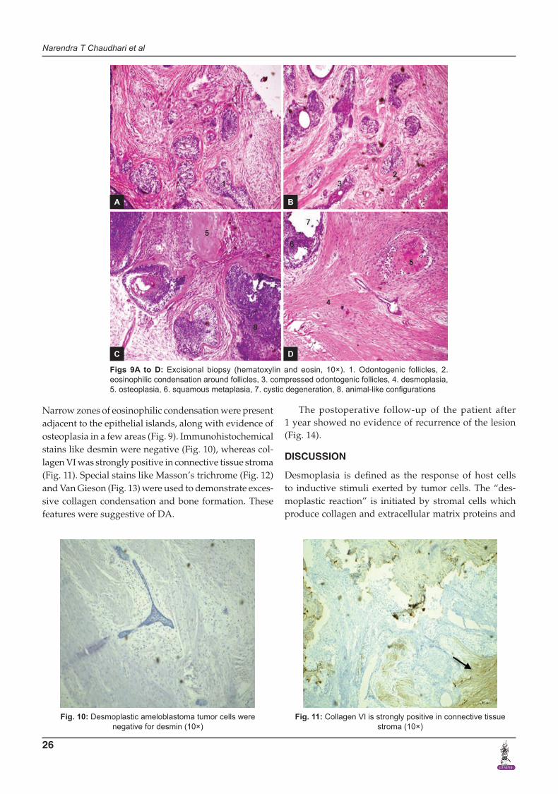
Narrow zones of eosinophilic condensation were present adjacent to the epithelial islands, along with evidence of osteoplasia in a few areas (Fig. 9). Immunohistochemical stains like desmin were negative (Fig. 10), whereas col-lagen VI was strongly positive in connective tissue stroma (Fig. 11). Special stains like Masson’s trichrome (Fig. 12) and Van Gieson (Fig. 13) were used to demonstrate exces-sive collagen condensation and bone formation. These features were suggestive of DA.
The postoperative follow-up of the patient after 1 year showed no evidence of recurrence of the lesion (Fig. 14).
DISCUSSION
Desmoplasia is defined as the response of host cells to inductive stimuli exerted by tumor cells. The “des-moplastic reaction” is initiated by stromal cells which produce collagen and extracellular matrix proteins and
Figs 9A to D: Excisional biopsy (hematoxylin and eosin, 10×). 1. Odontogenic follicles, 2. eosinophilic condensation around follicles, 3. compressed odontogenic follicles, 4. desmoplasia, 5. osteoplasia, 6. squamous metaplasia, 7. cystic degeneration, 8. animal-like configurations
Fig. 10: Desmoplastic ameloblastoma tumor cells were negative for desmin (10×)
Fig. 11: Collagen VI is strongly positive in connective tissue stroma (10×)
A
C
B
D

Desmoplastic Ameloblastoma: A Diagnostic Dilemma
Oral and Maxillofacial Pathology Journal, January-June 2018;9(1):23-30 27
OMPJ
may facilitate the invasion process in cancer.9 The DA is a rare unusual variant of ameloblastoma, which has a low incidence rate3 and shows significant stromal desmopla-sia histologically.2 It was first described by Eversole et al, who labeled it as an “ameloblastoma with pronounced desmoplasia or DA.”8,10 Till date, about 150 cases are reported in the literature.8 Desmoplastic ameloblastoma was initially defined as a central lesion but recently, two cases of peripheral DA (PDA) were reported.11 The PDA is believed to be arising as hamartomatous or neo-plastic proliferations of the dental lamina, odontogenic remnants, or pluripotent cells in the basal cell layer of the mucosa. The peripheral variant exhibits the same histopathologic features as intraosseous DA. However, the peripheral variant being less aggressive, requires a conservative approach for treatment as compared with the intraosseous variant.12
“Hybrid” lesion is an extremely unusual variant and was first described in detail by Waldron and El-Mofty.13
It is a tumor in which histologically, areas of follicular or plexiform ameloblastoma coexist with characteristic areas of DA.14
Clinical Features
The DA is predominantly seen in the 2nd to 5th decade of life and most commonly occurs in Japanese and Chinese populations. Usually there is equal gender distribution but some reports suggest male preponderance.3,8 The DA tends to affect the anterior premolar region of the jaws,6 with similar frequency in maxilla and mandible.15 This is in contrast to the location of the classic types of ameloblastoma, which usually are found in the posterior area of the mandible.4 In our case, the patient was male, belonging to second decade, and the lesion involved the left anterior region of maxilla.
In DA, the patient’s first complaint is usually an asymptomatic swelling with cortical plate expansion16 mainly in the alveolar region and usually occupying the tooth-bearing area.17 Teeth displacement (92% of the cases) and root resorption (33% of cases) are routinely noted in DA.15 In case of maxillary sinus involvement, nasal and pharyngeal obstruction does occur.5 Maxillary lesions are more dangerous, due to the vicinity of vital structures and the maxillary sinus. They are associated with rapid spread due to very thin cortical bone.1 The growth of DA is very fast when it involves the maxillary sinus18 and also the treatment is complicated due to penetrating nature of DA in the surrounding bone.8 Slowly enlarging, painless bony swelling with buccal cortex expansion and teeth displace-ment were the clinical findings in our case.
Radiological Features
The most common radiographic presentation of DA is a mixed radiolucent/radiopaque lesion due to osseous
Fig. 12: Masson’s trichrome stain (10×). 1. Collagen fibers (mature and immature), 2. trabecular bone, 3. fibrous bone
Fig. 13: Van Gieson’s stain (10×). 1. Odontogenic follicle, 2. collagen fibers
Fig. 14: Postoperative follow-up after 1 year

Narendra T Chaudhari et al
28
metaplasia within the dense fibrous septa resembling a benign fibro-osseous lesion. Also the borders of the lesion are poorly demarcated without capsule, suggestive of an infiltrative process1 affecting the prognosis.3 These features are not specific and can resemble other mixed radiolucent lesions.1 The DA has a tendency for de novo synthesis of extracellular fibrous protein which could serve as nidus for calcification seen in the DA with osteo-plasia.7 The disappearance of the lamina dura and the periodontal ligament space can be seen in the early stages of tumor development.19 Our case showed tooth displace-ment and ill-defined radiolucency on OPG, whereas CT and MRI scan revealed the extent and pattern of bone destruction along with internal architecture of the lesion.
This radiographic appearance may indicate that DA is more aggressive than other variants of ameloblastoma and warrants a radical approach to treatment.5 The CT and MRI can be used to distinguish DA from other fibro-osseous lesions by the detection of thick, bony trabeculae situated peripherally between the tumor elements.15 The differential diagnosis of a mixed radiodense-radiolucent lesion with diffuse borders includes fibro-osseous lesions (cemento-ossifying fibroma, cementoblastoma, and fibrous dysplasia), calcifying odontogenic cyst, and chronic sclerosing osteomyelitis. A definitive diagnosis prior to surgery requires histopathology to aid proper management.1,2
Histological Features
The classical histopathological features of DA include extreme stromal desmoplasia in the form of moderately cellular, fibrous connective tissue with abundant col-lagen,1 which squeezes or compresses the odontogenic islands and cords resulting in pointed, stellate, or “kite-like” appearance. The epithelial cells at the periphery of the islands are cuboidal, although columnar cells showing reversed nuclear polarity with hyperchromatic nuclei are seldom seen. The center of the islands may contain spindle-shaped or squamoid epithelial cells.16
Prominent osteoplasia in the form of metaplastic woven or mature bony trabeculae containing osteocytes and lined by plump active osteoblasts is also described, which can be attributed to stimulation of osteoblasts by tumor cell for formation of new bone. Granular cell transformation; follicular, plexiform, acanthomatous, and basaloid changes; and cystic degeneration within the tumor have also been reported.16 Microcysts that contain eosinophilic amorphous deposits or appear empty may occur centrally.17 A rare phenomenon of mucous cell differentiation is also reported.13 In our case, some areas showed odontogenic follicles containing peripheral ameloblast-like cells, central area resembling stellate
reticulum-like cells, squamous metaplasia, and cystic degeneration, whereas other areas consisted of com-pressed odontogenic islands with peripheral cuboidal cells which contained indistinct cells in the central region. Irregular odontogenic islands which were pointed, stel-late, bizzare animal-like configuration were also seen in many places.10 Marked stromal desmoplasia along with eosinophilic condensation around follicles and osteopla-sia in the form of woven as well as mature trabeculae was suggestive of DA.
Histopathology of DA is characterized by its infiltra-tion into bone marrow spaces without a fibrous con-nective tissue capsule.19 The precise diagnosis of DA depends on the identification of the typical ameloblastic areas, which may require examination of more tissue or a repeated biopsy.1
Immunohistochemistry/Special Stains
In the past few IHC stains have been used to study tumor microenvironment. The stroma in DA shows positive stain for laminin V and type IV collagen, suggesting an inductive effect of the epithelium over the fibrous stroma, resulting in a duplicated basal lamina. This is manifested in the form of deposits of an acellular eosinophilic matrix associated with epithelial islands.11 Expression of type III collagen indicates an ectomesenchymal origin for the stromal cells of the DA. Also strong immunolocalization of bone morphogenetic proteins 2, 3, 4, and 7 in the mes-enchymal tissues explains the formation of hard tissues in this neoplasm.20
Type VI collagen staining adjacent to tumor islands in DA indicates an active synthesis of extracellular matrix proteins leading to newly produced connective tissue and not a scar tissue. Transforming growth factor (TGF)-β, which is a potent local factor for modulating extracellular matrix formation and its marked immunoexpression, suggests that TGF-β produced by DA tumor cells plays a part in desmoplastic matrix formation.13
The immunoexpression of S-100 and desmin is vari-able, while keratin immunoreactivity is irregularly seen in tumor cells showing squamous differentiation. Vimentin is not expressed by either squamatoid or spindle-shaped cells. This may be due to different factors, such as dedif-ferentiation or the rate of proliferation of the neoplastic cells, inherent cellular potentials, or extracellular media-tors.21 In our case, the tumor stroma showed strong positivity for collagen VI, but desmin was negative. We preferred collagen VI over TGF-β as TGF-β regulates only extracellular matrix formation, whereas collagen VI demonstrates recently formed connective tissue, indicat-ing active matrix synthesis leading to excessive stromal desmoplasia.

Desmoplastic Ameloblastoma: A Diagnostic Dilemma
Oral and Maxillofacial Pathology Journal, January-June 2018;9(1):23-30 29
OMPJ
The studies related to special stains are scarce in DA. Special stains like Van Gieson to study desmoplasia4 and Masson’s trichrome to differentiate between fibrous/mature bone have only been used in the past.16 We used Van Gieson and Masson’s trichrome stains in the present case. Staining with Van Gieson showed abundant, dense red-colored collagen fiber bundles throughout the stroma and around the odontogenic follicles. The cells in the fol-licle showed black nucleus and the cytoplasm was yellow. Bone-stained red and numerous fine yellow-colored fibers were seen interspersed between the collagen fibers, sug-gestive of reticular or elastic fibers. Reticular fibers are fine, delicate fibers that provide support to coarse colla-gen network. Elastic fibers have microfibrillar structure and consist of three types—oxytalan, elaunin, and elastic fibers.22 The presence of oxytalan fibers in the stromal tissue points the origin of DA from the epithelial rests of Malassez in the periodontal membrane.7
Masson’s trichrome stains are used for selective demonstration of muscle, collagen fibers, fibrin, and erythrocytes. It is based on the principle that smaller dye molecule will penetrate and stain a tissue element, but whenever a larger dye molecule penetrates the same element it will then replace the smaller molecule.22 In the present case, this stain demonstrated numerous bluish-green-colored collagen fiber bundles intermixed with reddish-pink-colored fibers. This differential staining could be attributed to difference in pore size between mature and immature fibers as diameter of fibril in col-lagen is up to 0.4 µm, whereas microfibril has diameter about 40 nm.23 Collagen VI is a type of collagenous protein referred to as “high molecular weight aggregate” and “short chain collagen”24 having less pore volume. This indicates excessive new collagen formation leading to desmoplasia. Mature bone appeared reddish-pink in color, whereas fibrous bone was bluish-green in color, indicating active osteoplasia within the stroma.
Management and Recurrence
The interface between the lesion and normal bone is usually difficult to locate, also DA tends to infiltrate between bony trabeculae; hence, curettage/enucleation often results in recurrence. Therefore, block excision is the most widely accepted form of treatment.2,5 Ill-defined borders suggest an infiltration process and an aggressive biological behavior, so it requires an extensive treatment.6
Some researchers suggest that DA might be less bio-logically aggressive than conventional ameloblastoma as the desmoplasia in DA might act as a limiting barrier for local spread of the DA tumor cells.7 But recently, few cases showed that DA tends to exhibit rapid growth and
progressive behavior, with incidence of recurrence same as conventional ameloblastomas.19 The radiological and histological findings of ill-defined borders and poor encapsulation require a long-term follow-up.
The panoramic radiography cannot help to distin-guish DA from fibro-osseous lesions due to superimpo-sition; hence, cone beam CT scans are recommended as they provide more accurate visualization of the internal structure and expansion of maxilla–mandible tumors.25
CONCLUSION
We report a case of DA in a male patient affecting the anterior maxilla. A brief review of DA is discussed and findings of various investigations like clinical, radio-graphic (MRI and CT scan), and histological examination (IHC and special stains) were helpful for diagnosis and confirmation of DA.
REFERENCES 1. Sheikh S, Pallagatti S, Singla I, Kalucha A. Desmoplastic
ameloblastoma: a case report. J Dent Res Dent Clin Dent Prospects 2011 Winter;5(1):27-32.
2. Rastogi R, Jain H. Case report: desmoplastic ameloblastoma. Indian J Radiol Imaging 2008 Feb;18(1):53-55.
3. Desai H, Sood R, Shah R, Cawda J, Pandya H. Desmoplastic ameloblastoma: report of a unique case and review of litera-ture. Indian J Dent Res 2006 Jan-Mar;17(1):45-49.
4. Yazdi I, Seyedmajidi M, Foroughi R. Desmoplastic amelo-blastoma (a hybrid variant): report of a case and review of the literature. Arch Iranian Med 2009 May;12(3):304-308.
5. Sun ZJ, Wub YR, Cheng N, Zwahlen RA, Zhao YF. Desmoplastic ameloblastoma—a review. Oral Oncol 2009 Sep;45(9):752-759.
6. Amaral MB, Freire-Maia B, Serpa MR, Mesquita RA. A case report of desmoplastic ameloblastoma. J Clin Exp Dent 2010 Jun;2(3):e149-e152.
7. Effiom OA, Odukoya O. Desmoplastic ameloblastoma: analy-sis of 17 Nigerian cases. Oral Surg Oral Med Oral Pathol Oral Radiol Endod 2011 Jan;111(1):e27-e31.
8. Meena V, Sachin B, Chaitanya H, Hemant B. Desmoplastic ameloblastoma of mandible—a rare variant. UJMDS 2014 Jan-Mar;2(1):71-75.
9. Kawashiri S, Tanaka A, Noguchi N, Hase T, Nakaya H, Ohara T, Kato K, Yamamoto E. Significance of stromal desmopla-sia and myofibroblast appearance at the invasive front in squamous cell carcinoma of the oral cavity. Head Neck 2009 Oct;31(10):1346-1353.
10. Reichart PA, Philipsen HP. Odontogenic tumors and allied lesions. Hanover Park (IL): Quintessence Publishing; 2004. pp. 117-118.
11. Bologna-Molina R, Mosqueda-Taylor A, de Almeida-Oslei P, Toral-Rizo V, Martínez-Mata G. Peripheral desmoplastic ameloblastoma: histopathological and immunohistochemi-cal profile of a case. Med Oral Patol Oral Cir Bucal 2010 Nov;15(6):e846-e849.
12. Oteri G, Lentini M, Pisano M, Cicciù M. Peripheral desmo-plastic ameloblastoma in adolescent age: clinico-pathological and immunohistochemical analysis of a case. Open Dent J 2014 Sep;8:159-163.

Narendra T Chaudhari et al
30
13. Angadi PV, Kale A, Hallikerimath S, Kotrashetti V, Mane D, Bhatt P, Shukla D. “Hybrid” desmoplastic ameloblastoma: an unusual case report with immunohistochemical investigation for TGF-β and review of literature. East J Med 2011;16(1):9-17.
14. dos Santos JN, De Souza VF, Azevêdo RA, Sarmento VA, Souza LB. “Hybrid” lesion of desmoplastic and conventional ameloblastoma: immunohistochemical aspects. Rev Bras Otorrinolaringol 2006 Sep-Oct;72(5):709-713.
15. Shashikanth MC, Neetha MC, Ali IM, Shambulingappa P. Desmoplastic ameloblastoma in the maxilla: a case report and review of literature. Indian J Dent Res 2007 Oct-Dec;18(4): 214-217.
16. Savithri V, Janardhanan M, Suresh R, Vinod Kumar RB. Desmoplastic ameloblastoma with osteoplasia: review of literature with a case report. J Oral Maxillofac Pathol 2013 Oct;17(2):298-301.
17. Majumdar S, Uppala D, Kotina S, Veera SK, Boddepalli R. Desmoplastic ameloblastoma. Int J Appl Basic Med Res 2014 Sep;4(Suppl 1):S53-S55.
18. Kato H, Nomura J, Matsumura Y, Tagawa T. A case of desmo-plastic ameloblastoma occupying maxillary sinus. Contemp Clin Dent 2011 Jul-Sep;2(3):234-236.
19. Katsura K, Maruyama S, Suzuki M, Saku T, Takagi R, Hayashi T. Case of desmoplastic ameloblastoma arising in the maxillary
alveolus: the origin and time-course changes in the early stage of tumor development observed on dental radiographs. Dentomaxillofac Radiol 2011 Feb;40(2):126-129.
20. Inoue M, Nagatsuka H, Tamamura R, Chong HS, Tsujigiwa H, Borkosky S, Fujii M, Nagai N, Setsu K. Localization of Oxytalan fiber, Type III Collagen and BMP family in conven-tional and desmoplastic ameloblastoma. J Hard Tissue Biol 2008 Apr;17(1):23-30.
21. Siar CH, Ng KH. Patterns of expression of intermediate filaments and S-100 protein in desmoplastic ameloblastoma. J Nihon Univ Sch Dent 1993 Jun;35(2):104-108.
22. Suvarna SK, Layton C, Bancroft JD. Theory and practice of histological techniques. 7th ed. London: Churchill Livingstone; 2012. pp. 190-200.
23. Culling CFA, Allison RT, Barr WT. Cellular pathology tech-nique. 4th ed. St. Louis (MO): Butterworth-Heinemann; 1985; p. 165.
24. Hessle H, Engvall E. Type VI collagen. Studies on its local-ization, structure, and biosynthetic form with monoclonal antibodies. J Biol Chem 1984 Mar;259(6):3955-3961.
25. Luo J, You M, Zheng G, Xu L. Cone beam computed tomog-raphy signs of desmoplastic ameloblastoma: review of 7 cases. Oral Surg Oral Med Oral Pathol Oral Radiol 2014 Oct;118(4):e126-e133.