Design of Peptides That Bind in the Minor Groove of DNA at S-(A,T ...
12
J. Am. Chem. SOC. 1992, 114, 8783-8794 8783 CI,H19N30,S: C, 52.50; H, 6.44; N, 14.13; S, 10.78. Found: C, 52.29; H, 6.55; N, 13.98; S, 10.56. NMR Studies. Nuclear Overhauser effects (NOE’s) were detected using the Varian 300-MHz instrument. Difference NOE measurements were measured as negative enhancements upon irradiation of the proton of interest. The experiment was repeated in triplicate for each compound at 21 OC with varying d2 delay times. The decoupler power was set to 18 dB and the 90-deg pulse width was measured for each compound and set appropriately. The FIDs were multiplied by a heavy line broadening (Ib = 5) apodization function before subtraction. Temperature studies were performed on the GE 3WMHz instrument at a sample concentration of 25 mM. The temperature was set to 295 K, and a spectrum was taken at this temperature and at 5-deg intervals up to 325 K. The chemical shifts of the amide hydrogen versus tem- perature were plotted, and the temperature coefficients were measured as ppb/K from the slope of the line. TFE titration studies were performed on the same instrument at 25 “C. A spectrum was first taken in DMSO; then TFE was added in portions so as to increase the amount of TFE by 10% with each addition. After each addition, another spectrum was recorded. This was repeated to a final concentration of 70% TFE in DMSO. The change in chemical shifts (A6) of the amide hydrogen was then measured. Molecular Modeling Studies. Energy minimization studies were per- formed as described by Ferguson and RaberI6J7 using the Random In- cremental Pulse Search (RIPS) method. Eight energy-minimized structures were generated, and the figures were displayed using a Ma- cintosh Quadra 700 computer with Alchemy 111 software, TRIPOS Associates, Inc. 1992. Hz, 1 H, lactam P-CH,), 2.35-2.46 (m, 1 H, pro @-CH,), 2.69 (dd, J = 14.1 and 7.8 Hz, 1 H, lactam &CH2), 2.77 (d, J = 4.8 Hz, 3 H, NCH3), 3.46 (dd, J = 8.0 and 5.6 Hz, 2 H, pro 6-CH2), 3.56-3.58 (m, 2 H, SCH2), 4.80-4.85 (m, 1 H, thiazolidine a-CH), 5.18 (dd, J = 7.4 and 5.0 Hz, 1 H, SCHN), 7.49 (br s, 1 H, CONH); NMR (75.5 MHz, CDCl,, pro = pyrrolidine) 6 23.61 (pro y-C), 26.23 (pro P-C), 28.36 (Boc CH,), 36.52 (lactam 8-C), 39.38 and 39.54 (SCH2 and NCH,), 47.79 (pro 6-C), 57.42 (thiazolidine a-C), 62.69 (SCHN), 69.52 (pro a-C), 80.64 (Boc C-0), 153.72 (Boc C=O), 169.47 (CONH), 172.54 (lactam C 4); FAB MS m/z 356 [MH]+. Anal. Calcd for C16H25N304S: C, 54.06; H, 7.09; N, 11.82; S, 9.02. Found: C, 53.84; H, 7.05; N, 11.64; S, 8.88. [3’- (S )-[ 3’a,6’a(R *),7’aa]]- 1 -Acetyltetrahydro-5’-oxospiro[pyrrol- idine-~6’-(5’H)-pyrrolo(2,l-~~~azoli~~3’-~-me~yl~r~~~de (6). N-Methylamide 13 (40 mg, 0.1 1 mmol) was deprotected in 4 N HC1/ dioxane (5 mL) at room temperature for 1 h. The solvent was removed in vacuo; and the residue was taken up in CH2C12, whereupon the solvent was again removed in vacuo. The residue was dried in vacuo overnight. It was then dissolved in Ac20 (10 mL) and cooled in an ice bath, whereupon NEt, (1 equiv) and DMAP (cat.) were added. The solution was stirred at room temperature overnight. The solvent was evaporated in vacuo, and the residue was chromatographed on a 1.5 X 45 cm silica gel flash column using CH2C12/MeOH as the eluting solvent. The product (6) was isolated as an oil: [ala +139.2O (c 0.90, MeOH); ’H NMR (300 MHz, CDCI,, pro = pyrrolidine) 6 1.90-2.07 (m, 2 H, pro yCH2), 2.09 (s, 3 H, COCH,), 2.16-2.22 (m, 2 H, @-CH2), 2.31-2.45 (m, 1 H, lactam 8-CH2), 2.72 (dd, J = 14.1 and 8.1 Hz, 1 H, lactam 8-CH2),2.82 (d, J = 5.1 Hz, 3 H, NCH,), 3.56-3.65 (m, 4 H, pro 6-CH, and SCH2),4.85 (dd, J = 8.5 and 5.0 Hz, 1 H, thiazolidine a-CH), 5.15 (dd, J = 8.0 and 4.4 Hz, 1 H, SCHN), 7.24 (br s, 1 H, CONH); I3C NMR (75.5 MHz, CDCI,, pro = pyrrolidine) 6 23.60 (pro y-C), 24.13 (COCH,), 26.50 (pro FC), 36.31 (lactam @-C), 38.34 (SCH2), 39.1 1 (NCH,), 48.87 (pro 6-C), 57.70 (thiazolidine a-C), 62.67 (SCHN), 69.50 (pro a-C), 169.57 and 169.72 (CONH and COCH,), 173.04 (lactam C 4 ) ; FAB MS m/z 298 [MH]+. Anal. Calcd for Acknowledgment. This research was supported in part by an NIH grant (NS20036) to RLJ and an NIH predoctoral train- eeship (GM07994) to MJG. The authors gratefully acknowledge the assistance of Dr. David M. Ferguson in the molecular modeling studies. Design of Peptides That Bind in the Minor Groove of DNA at S-(A,T)G(A,T)C(A,T)-3’ Sequences by a Dimeric Side-by-Side Motif Warren S. Wade, Milan Mrksich, and Peter B. Dervan* Contribution from the Arnold and Mabel Beckman Laboratories of Chemical Synthesis, California Institute of Technology, Pasadena, California 91125. Received May 11, 1992 Abstract: The designed peptides pyridine-2-carboxamide-netropsin (2-PyN) and 1 -methylimidazole-2-carboxamide-netropsin (2-1”) are crescent-shaped synthetic analogs of the natural products netropsin (N) and distamycin A (D). Footprinting experiments indicate that the peptides 2-PyN and 2-1” bind specifically the 5 base pair sequence 5’-TGTCA-3’. Affinity cleaving data suggest that the complexes, 2-ImN.5’-TGTCA-3’ and 2-PyN.5’-TGTCA-3’, are composed of two equivalent orientations which disfavor a 1:l model. The footprinting and affinity cleaving data are in accord with a 2:l complex where a novel side-by-side antiparallel dimer binds in the minor groove of double-helical DNA. Netropsin and Mstamycin A. Netropsin (N) and distamycin A (D) are natural products that bind in the minor groove of double-helical DNA at sites of 4 or 5 successive A,T base pairs (Figure 1).l4 X-rays and NMR6 studies of netropsin-DNA and (1) For reviews, see: (a) Dervan, P. B. Science 1986, 232, 464-471. (b) Zimmer, C.; Wahnert, U. Prog. Biophys. Molec. Biol. 1986, 47, 31-112. (2) (a) Krylov, A. S.; Grokhovsky, S. L.; Zasedatelev, A. S.; Zhuze, A. L.; Gursky, G. V.; Gottikh, B. P. Nucleic Acids Res. 1979, 6, 289-304. (b) Zasedatelev, A. S.; Gursky, G. V.; Zimmer, Ch.; Thrum, H. Mol. Biol. Rep. 1974, I, 337-342. (c) Zasedatelev, A. S.; Zhuze, A. L.; Zimmer, Ch.; Grokhovsky, S. L.; Tumanyan, V. G.; Gursky, G. V.; Gottikh, B. P. Dokl. Akad. Nauk SSSR 1976, 231, 1006-1009. (3) (a) Van Dyke, M. W.; Hertzberg, R. P.; Dervan, P. B. Proc. Natl. Acad. Sei. U.S.A. 1982, 79, 5470-5474. (b) Van Dyke, M. W.; Dervan, P. B. Cold Spring Harbor Symposium on Quantitative Biology 1982, 47, 347-353. (c) Van Dyke, M. W.; Dervan, P. B. Biochemistry 1983, 22, 2373-2377. (d) Harshman, K. D.; Dervan, P. B. Nucleic Acids Res. 1985, 13, 4825-4835. (e) Fox, K. R.; Waring, M. J. Nucleic Acids Res. 1984, 12, 9271-9285. (f) Lane, M. J.; Dobrowiak, J. C.; Vournakis, J. Proc. Narl. Acad. Sci. U.S.A. 1983, 80, 3260-3264. distamycin-DNA complexes reveal how sequence specificity is accomplished. The crescent-shaped di- and tripeptides are bound in the middle of the minor groove of an A,T-rich sequence. The amide hydrogens of the N-methylpyrrolecarboxamides form bi- furcated hydrogen bonds with the N 3 of adenine and the 02 of (4) (a) Schultz, P. G.; Taylor, J. S.; Dervan, P. B. J. Am. Chem. Soc. 1982, 104, 6861-6863. (b) Taylor, J. S.; Schultz, P. G.; Dervan, P. B. Tetrahedron 1984,40,457-465. (c) Schultz, P. G.; Dervan, P. B. J. Biomol. Srruct. Dyn. (5) (a) Kopka, M. L.; Yoon, C.; Goodsell, D.; Pjura, P.; Dickerson, R. E. Proc. Narl. Acad. Sci. U.S.A. 1985,82, 1376-1380. (b) Kopka, M. L.; Yoon, C.; Goodsell, D.; Pjura, P.; Dickerson, R. E. J. Mol. Biol. 1985, 183, 553-563. (c) Coll, M.; Frederick, C. A.; Wang, A. H.4.; Rich, A. Proc. Natl. Acad. Sci. U.S.A. 1987, 84, 8385-8389. (6) (a) Patel, D. J.; Shapiro, L. J. Biol. Chem. 1986, 261, 1230-1240. (b) Klevitt, R. E.; Wemmer, D. E.; Reid, B. R. Biochemistry 1986, 25, 3296-3303. (c) Pelton, J. G.; Wemmer, D. E. Biochemistry 1988, 27, 1984, I, 1133-1147. 8088-8096. 0002-7863/92/1514-8783$03.00/0 0 1992 American Chemical Society
Transcript of Design of Peptides That Bind in the Minor Groove of DNA at S-(A,T ...

J. Am. Chem. SOC. 1992, 114, 8783-8794 8783
CI,H19N30,S: C, 52.50; H, 6.44; N, 14.13; S, 10.78. Found: C, 52.29; H, 6.55; N, 13.98; S, 10.56.
NMR Studies. Nuclear Overhauser effects (NOE’s) were detected using the Varian 300-MHz instrument. Difference NOE measurements were measured as negative enhancements upon irradiation of the proton of interest. The experiment was repeated in triplicate for each compound at 21 OC with varying d2 delay times. The decoupler power was set to 18 dB and the 90-deg pulse width was measured for each compound and set appropriately. The FIDs were multiplied by a heavy line broadening (Ib = 5) apodization function before subtraction.
Temperature studies were performed on the GE 3WMHz instrument at a sample concentration of 25 mM. The temperature was set to 295 K, and a spectrum was taken at this temperature and at 5-deg intervals up to 325 K. The chemical shifts of the amide hydrogen versus tem- perature were plotted, and the temperature coefficients were measured as ppb/K from the slope of the line.
TFE titration studies were performed on the same instrument at 25 “C. A spectrum was first taken in DMSO; then TFE was added in portions so as to increase the amount of TFE by 10% with each addition. After each addition, another spectrum was recorded. This was repeated to a final concentration of 70% TFE in DMSO. The change in chemical shifts (A6) of the amide hydrogen was then measured.
Molecular Modeling Studies. Energy minimization studies were per- formed as described by Ferguson and RaberI6J7 using the Random In- cremental Pulse Search (RIPS) method. Eight energy-minimized structures were generated, and the figures were displayed using a Ma- cintosh Quadra 700 computer with Alchemy 111 software, TRIPOS Associates, Inc. 1992.
Hz, 1 H, lactam P-CH,), 2.35-2.46 (m, 1 H, pro @-CH,), 2.69 (dd, J = 14.1 and 7.8 Hz, 1 H, lactam &CH2), 2.77 (d, J = 4.8 Hz, 3 H, NCH3), 3.46 (dd, J = 8.0 and 5.6 Hz, 2 H, pro 6-CH2), 3.56-3.58 (m, 2 H, SCH2), 4.80-4.85 (m, 1 H, thiazolidine a-CH), 5.18 (dd, J = 7.4 and 5.0 Hz, 1 H, SCHN), 7.49 (br s, 1 H, CONH); NMR (75.5 MHz, CDCl,, pro = pyrrolidine) 6 23.61 (pro y-C), 26.23 (pro P-C), 28.36 (Boc CH,), 36.52 (lactam 8-C), 39.38 and 39.54 (SCH2 and NCH,), 47.79 (pro 6-C), 57.42 (thiazolidine a-C), 62.69 (SCHN), 69.52 (pro a-C), 80.64 (Boc C-0), 153.72 (Boc C=O), 169.47 (CONH), 172.54 (lactam C 4 ) ; FAB MS m / z 356 [MH]+. Anal. Calcd for C16H25N304S: C, 54.06; H, 7.09; N, 11.82; S, 9.02. Found: C, 53.84; H, 7.05; N, 11.64; S, 8.88.
[3’- (S )-[ 3’a,6’a(R *),7’aa]]- 1 -Acetyltetrahydro-5’-oxospiro[pyrrol- idine-~6’-(5’H)-pyrrolo(2,l-~~~azoli~~3’-~-me~yl~r~~~de (6). N-Methylamide 13 (40 mg, 0.1 1 mmol) was deprotected in 4 N HC1/ dioxane (5 mL) at room temperature for 1 h. The solvent was removed in vacuo; and the residue was taken up in CH2C12, whereupon the solvent was again removed in vacuo. The residue was dried in vacuo overnight. It was then dissolved in Ac20 (10 mL) and cooled in an ice bath, whereupon NEt, (1 equiv) and DMAP (cat.) were added. The solution was stirred at room temperature overnight. The solvent was evaporated in vacuo, and the residue was chromatographed on a 1.5 X 45 cm silica gel flash column using CH2C12/MeOH as the eluting solvent. The product (6) was isolated as an oil: [ala +139.2O ( c 0.90, MeOH); ’H NMR (300 MHz, CDCI,, pro = pyrrolidine) 6 1.90-2.07 (m, 2 H, pro yCH2) , 2.09 (s, 3 H, COCH,), 2.16-2.22 (m, 2 H, @-CH2), 2.31-2.45 (m, 1 H, lactam 8-CH2), 2.72 (dd, J = 14.1 and 8.1 Hz, 1 H, lactam 8-CH2), 2.82 (d, J = 5.1 Hz, 3 H, NCH,), 3.56-3.65 (m, 4 H, pro 6-CH, and SCH2), 4.85 (dd, J = 8.5 and 5.0 Hz, 1 H, thiazolidine a-CH), 5.15 (dd, J = 8.0 and 4.4 Hz, 1 H, SCHN), 7.24 (br s, 1 H, CONH); I3C NMR (75.5 MHz, CDCI,, pro = pyrrolidine) 6 23.60 (pro y-C), 24.13 (COCH,), 26.50 (pro F C ) , 36.31 (lactam @-C), 38.34 (SCH2), 39.1 1 (NCH,), 48.87 (pro 6-C), 57.70 (thiazolidine a-C), 62.67 (SCHN), 69.50 (pro a-C), 169.57 and 169.72 (CONH and COCH,), 173.04 (lactam C 4 ) ; FAB MS m / z 298 [MH]+. Anal. Calcd for
Acknowledgment. This research was supported in part by an NIH grant (NS20036) to RLJ and an NIH predoctoral train- eeship (GM07994) to MJG. The authors gratefully acknowledge the assistance of Dr. David M. Ferguson in the molecular modeling studies.
Design of Peptides That Bind in the Minor Groove of DNA at S-(A,T)G(A,T)C(A,T)-3’ Sequences by a Dimeric Side-by-Side Motif Warren S. Wade, Milan Mrksich, and Peter B. Dervan* Contribution from the Arnold and Mabel Beckman Laboratories of Chemical Synthesis, California Institute of Technology, Pasadena, California 91125. Received May 1 1 , 1992
Abstract: The designed peptides pyridine-2-carboxamide-netropsin (2-PyN) and 1 -methylimidazole-2-carboxamide-netropsin (2-1”) are crescent-shaped synthetic analogs of the natural products netropsin (N) and distamycin A (D). Footprinting experiments indicate that the peptides 2-PyN and 2-1” bind specifically the 5 base pair sequence 5’-TGTCA-3’. Affinity cleaving data suggest that the complexes, 2-ImN.5’-TGTCA-3’ and 2-PyN.5’-TGTCA-3’, are composed of two equivalent orientations which disfavor a 1:l model. The footprinting and affinity cleaving data are in accord with a 2:l complex where a novel side-by-side antiparallel dimer binds in the minor groove of double-helical DNA.
Netropsin and Mstamycin A. Netropsin ( N ) and distamycin A (D) are natural products that bind in the minor groove of double-helical DNA a t sites of 4 or 5 successive A,T base pairs (Figure 1) . l4 X-rays and NMR6 studies of netropsin-DNA and
(1) For reviews, see: (a) Dervan, P. B. Science 1986, 232, 464-471. (b) Zimmer, C.; Wahnert, U. Prog. Biophys. Molec. Biol. 1986, 47, 31-112.
(2) (a) Krylov, A. S.; Grokhovsky, S. L.; Zasedatelev, A. S.; Zhuze, A. L.; Gursky, G. V.; Gottikh, B. P. Nucleic Acids Res. 1979, 6 , 289-304. (b) Zasedatelev, A. S.; Gursky, G. V.; Zimmer, Ch.; Thrum, H. Mol. Biol. Rep. 1974, I, 337-342. (c) Zasedatelev, A. S.; Zhuze, A. L.; Zimmer, Ch.; Grokhovsky, S. L.; Tumanyan, V. G.; Gursky, G. V.; Gottikh, B. P. Dokl. Akad. Nauk SSSR 1976, 231, 1006-1009.
(3) (a) Van Dyke, M. W.; Hertzberg, R. P.; Dervan, P. B. Proc. Natl. Acad. Sei. U.S.A. 1982, 79, 5470-5474. (b) Van Dyke, M. W.; Dervan, P. B. Cold Spring Harbor Symposium on Quantitative Biology 1982, 47, 347-353. (c) Van Dyke, M. W.; Dervan, P. B. Biochemistry 1983, 22, 2373-2377. (d) Harshman, K. D.; Dervan, P. B. Nucleic Acids Res. 1985, 13, 4825-4835. (e) Fox, K. R.; Waring, M. J . Nucleic Acids Res. 1984, 12, 9271-9285. ( f ) Lane, M. J.; Dobrowiak, J. C.; Vournakis, J. Proc. Narl. Acad. Sci. U.S.A. 1983, 80, 3260-3264.
distamycin-DNA complexes reveal how sequence specificity is accomplished. The crescent-shaped di- and tripeptides are bound in the middle of the minor groove of an A,T-rich sequence. The amide hydrogens of the N-methylpyrrolecarboxamides form bi- furcated hydrogen bonds with the N 3 of adenine and the 0 2 of
(4) (a) Schultz, P. G.; Taylor, J. S.; Dervan, P. B. J . Am. Chem. Soc. 1982, 104, 6861-6863. (b) Taylor, J. S.; Schultz, P. G.; Dervan, P. B. Tetrahedron 1984,40,457-465. (c) Schultz, P. G.; Dervan, P. B. J. Biomol. Srruct. Dyn.
( 5 ) (a) Kopka, M. L.; Yoon, C.; Goodsell, D.; Pjura, P.; Dickerson, R. E. Proc. Narl. Acad. Sci. U.S.A. 1985,82, 1376-1380. (b) Kopka, M. L.; Yoon, C.; Goodsell, D.; Pjura, P.; Dickerson, R. E. J . Mol. Biol. 1985, 183, 553-563. (c) Coll, M.; Frederick, C. A.; Wang, A. H . 4 . ; Rich, A. Proc. Natl. Acad. Sci. U.S.A. 1987, 84, 8385-8389.
(6) (a) Patel, D. J.; Shapiro, L. J . Biol. Chem. 1986, 261, 1230-1240. (b) Klevitt, R. E.; Wemmer, D. E.; Reid, B. R. Biochemistry 1986, 25, 3296-3303. (c) Pelton, J. G.; Wemmer, D. E. Biochemistry 1988, 27,
1984, I , 1133-1147.
8088-8096.
0002-7863/92/1514-8783$03.00/0 0 1992 American Chemical Society
8784 J . Am. Chem. SOC., Vol. 114, No. 23, 1992 Wade et ai.
I ,N<HCI ,,,T2
t HN
HN
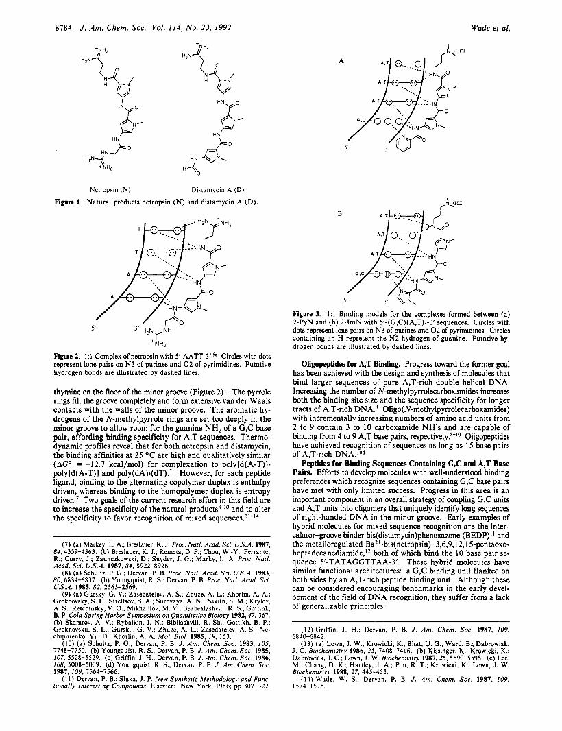
Nctropsin (N) Dislamycin A (D) Figure 1. Natural products netropsin (N) and distamycin A (D).
B
1 - 5 ' 3 ' H , N ~ N H
+NH,
Figure 2. 1:l Complex of netropsin with 5'-AATT-3'.5a Circles with dots represent lone pairs on N 3 of purines and 0 2 of pyrimidines. Putative hydrogen bonds are illustrated by dashed lines.
thymine on the floor of the minor groove (Figure 2). The pyrrole rings fill the groove completely and form extensive van der Waals contacts with the walls of the minor groove. The aromatic hy- drogens of the N-methylpyrrole rings are set too deeply in the minor groove to allow room for the guanine NH2 of a G,C base pair, affording binding specificity for A,T sequences. Thermo- dynamic profiles reveal that for both netropsin and distamycin, the binding affinities at 25 OC are high and qualitatively similar (AGO = -12.7 kcal/mol) for complexation to poly[d(A-T)]. poly[d(A-T)] and ~oly(dA).(dT).~ However, for each peptide ligand, binding to the alternating copolymer duplex is enthalpy driven, whereas binding to the homopolymer duplex is entropy drivene7 Two goals of the current research effort in this field are to increase the specificity of the natural productss-IO and to alter the specificity to favor recognition of mixed
(7) (a) Markey, L. A.; Breslauer, K. J. Proc. Natl. Acad. Sci. U.S.A. 1987, 84,4359-4363. (b) Breslauer, K. J.; Remeta, D. P.; Chou, W.-Y.; Ferrante, R.; Curry, J.; Zaunczkowski, D.; Snyder, J. G.; Marky, L. A. Proc. Narl. Acad. Sci. U.S.A. 1987, 84, 8922-8926.
(8) (a) Schultz, P. G.; Dervan, P. B. Proc. Natl. Acad. Sci. U.S.A. 1983, 80,6834-6837. (b) Youngquist, R. S.; Dervan, P. B. Proc. Natl. Acad. Sci. U.S.A. 1985, 82. 2565-2569.
(9) (a) Gursky, G. V.; Zasedatelev, A. S.; Zhuze, A. L.; Khorlin, A. A,; Grokhovsky, S. L.; Streltsov, S. A,; Surovaya, A. N.; Nikitn, S. M.; Krylov, A. S.; Retchinsky, V. 0.; Mikhaillov, M. V.; Beabealashvili, R. S.; Gottihk, B. P. Cold Spring Harbor Symposium on Quantitative Biology 1982,47, 367. (b) Skamrov, A. V.; Rybalkin, I. N.; Bibilashvili, R. Sh.; Gottikh, B. P.; Grokhovskii, S. L.; Gurskii, G. V.; Zhuze, A. L.; Zasedatelev, A. S.; Ne- chipurenko, Yu. D.; Khorlin, A. A. Mol. Biol. 1985, 19, 153.
(IO) (a) Schultz, P. G.; Dervan, P. B. J . Am. Chem. SOC. 1983, 105, 7748-7750. (b) Youngquist, R. S.; Dervan, P. B. J . Am. Chem. SOC. 1985, 107, 5528-5529. (c) Griffin, J. H.; Dervan, P. B. J . Am. Chem. SOC. 1986, 108, 5008-5009. (d) Youngquist, R. S.; Dervan, P. B. J. Am. Chem. SOC.
( I 1) Dervan, P. B.; Sluka, J. P. New Synthetic Methodology and Func- lionally Interesting Compounds; Elsevier: New York. 1986; pp 307-322.
1981, 109,1564-7566.
I ,N;HCI
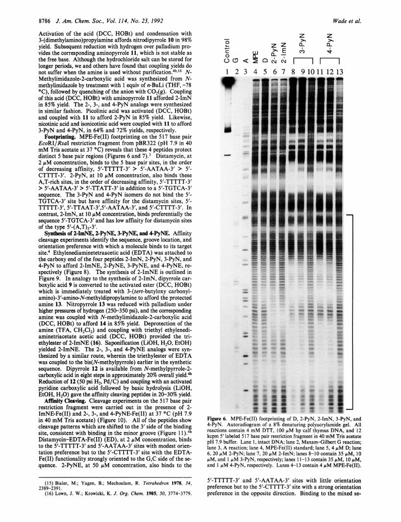
Figure 3. 1:l Binding models for the complexes formed between (a) 2-PyN and (b) 2-1" with 5'-(G,C)(A,T),-3' sequences. Circles with dots represent lone pairs on N 3 of purines and 0 2 of pyrimidines. Circles containing an H represent the N 2 hydrogen of guanine. Putative hy- drogen bonds are illustrated by dashed lines.
Ongopeptide for A,T Binding. Progress toward the former goal has been achieved with the design and synthesis of molecules that bind larger sequences of pure A,T-rich double helical DNA. Increasing the number of N-methylpyrrolecarboxamides increases both the binding site size and the sequence specificity for longer tracts of A,T-rich DNA.s Oligo(N-methylpyrrolecarboxamides) with incrementally increasing numbers of amino acid units from 2 to 9 contain 3 to 10 carboxamide NH's and are capable of binding from 4 to 9 A,T base pairs, respectively.8-10 Oligopeptides have achieved recognition of sequences as long as 15 base pairs of A,T-rich DNA.lOd
Peptides for Binding Sequences Containing G,C and A,T Base Pairs. Efforts to develop molecules with well-understood binding preferences which recognize sequences containing G,C base pairs have met with only limited success. Progress in this area is an important component in an overall strategy of coupling G,C units and A,T units into oligomers that uniquely identify long sequences of right-handed DNA in the minor groove. Early examples of hybrid molecules for mixed sequence recognition are the inter- calator-groove binder bis(distamycin)phenoxazone (BEDP)" and the metalloregulated Ba2+.bis(netropsin)-3,6,9,1 2,l S-pentaoxo- heptadecanediamide,'* both of which bind the 10 base pair se- quence 5'-TATAGGTTAA-3'. These hybrid molecules have similar functional architectures: a G,C binding unit flanked on both sides by an A,T-rich peptide binding unit. Although these can be considered encouraging benchmarks in the early devel- opment of the field of DNA recognition, they suffer from a lack of generalizable principles.
(12) Griffin, J . H.; Dervan, P. B. J . Am. Chem. SOC. 1987, 109, 6840-6842.
(13) (a) Lown, J. W.; Krowicki, K.; Bhat, U. G.; Ward, B.; Dabrowiak, J. C. Biochemistry 1986, 25, 7408-7416. (b) Kissinger, K.; Krowicki, K.; Dabrowiak, J . C.; Lown, J. W. Biochemistry 1987, 26, 5590-5595. (c) Lee, M.; Chang, D. K.; Hartley, J. A.; Pon, R. T.; Krowicki, K.; Lown, J. W. Biochemistry 1988, 27. 445-455.
(14) Wade, W. S.; Dervan, P. B. J. Am. Chem. SOC. 1987, 109, 1574-1575.

Peptides That Bind in the Minor Groove of DNA J . Am. Chem. SOC., Vol. 114 , No. 23, 1992 8785
2 - P y N 2-1”
HN
3-PyN 4 - P y N
Figure 4. Synthetic analogs pyridine-2-carboxamide-netropsin (2-PyN), 1-methylimidazole-2-carboxamide-netropsin (2-ImN), pyridine-3- carboxamide-netropsin (3-PyN), and pyridine-4-carboxamide-netropsin (4-PyN).
An alternative approach is the systematic substitution of the tris (N-methylpyrrolecarboxamide) framework of distamycin to search for altered base pair ~ p e c i f i c i t y . ~ ~ . ~ ~ In the minor groove
of DNA, the major difference between edges of A,T and G,C base pairs is the presence of the guanine 2-amino group protruding from the floor. By replacing the pyrrole CH with a heteroatom capable of forming a hydrogen bond to the guanine 2-amino group, for example, a steric clash might be turned into a stabilizing inter- action. Molecules containing such a substitution display an in- creased tolerance for G,C base pairs in their binding sites, with an overall loss in specificity.I3
Design Rationale: The 1:l Complex. Our approach is illustrated in Figure 3A, wherein we expected that replacement of the ter- minal N-methylpyrrolecarboxamide of D with pyridine-2- carboxamide would afford an analog, pyridine-2-carboxamide- netropsin (2-PyN) (Figure 4), that would specifically bind to 1 G,C base pair followed by 3 A,T base pairs. In analogy with distamycin binding A,T-rich sites, it was anticipated that the amide hydrogens would form bifurcated hydrogen bonds with adenine N3 and thymine 0 2 atoms, while the pyridine nitrogen would participate in a key hydrogen bond with the NH2 group of the G,C base pair (Figure 3). The 3-carboxamide and Ccarboxamide isomers (3-PyN and 4-PyN) serve as controls for the placement of the ring nitrogen (Figure 4). We found that 2-PyN bound 3 A,T-rich sites 5 base pairs in size, 5’-TTTTT-3’, 5’-AATAA-3’, 5’-CTTTT-3’, and an unanticipated mixed sequence 5’-TGTCA- 3’.14 Furthermore, affinity cleaving experiments revealed no orientation preference for binding to the 5’-TGTCA-3’ sequence. In contrast, pyridine-3-carboxamide-netropsin (3-PyN) and pyridine-4-carboxamide-netropsin (4-PyN) preferred A,T-rich sequences, suggesting that the placement of the ring nitrogen is important for the recognition of the sequence 5’-TGTCA-3’.I4 In an effort to explore the structural limitations of binding to the sequence 5’-TGTCA-3’, the pyridine ring was replaced with N-methylimidazole to afford 1-methylimidazole-2-carboxamide netropsin (2-1”) (Figures 3b and 4). Footprinting and affinity cleaving experiments indicate that this synthetic peptide binds the 5‘-TGTCA-3‘ site with higher sequence specificity than 2-PyN.
Results Synthesis of > I d , 2-PyN, 3-PyN, and 4“. The syntheses
of 2-1” and the 2-, 3-, and 4PyN analogs are outlined in Figure 5 . N-Methyl-4-(N-methyl-4-nitropyrrole-2-carboxamide)- pyrrole-2-carboxylic acid (9) is available in five steps starting from N-methylpyrrole-2-carboxylic acid in 20% overall yield.’s*’6
10: R1 =NO2 R o = H b L 1 l : R,=NH2 R 2 = H
12: R1 L NO2 R2 = CH2CH2NHCO- CH,N(CH2C02Et)CHr CH2N(CH2C02Et)2
11
H \
I O \
4 3 Figure 5. Synthetic scheme for 2-, 3-, and 4-PyN and 2-1”. (a) (i) DCC, HOBt; (ii) 3-(dimethy!amino)propylamine; (b) 100 psi H,, 10% Pd/C; (c) N-methylimidazo!e-2-carboxylic acid, DCC, HOBt; (d) picolinic acid, DCC, HOBt; (e) nicotinic acid, DCC, HOBt; (f) isonicotinic acid, DCC, HOBt.
8786 J. Am. Chem. SOC., Vol. 114, No. 23, 1992 Wade et al.
Activation of the acid (DCC, HOBt) and condensation with 3-(dimethy1amino)propylamine affords nitrodipyrrole 10 in 98% yield. Subsequent reduction with hydrogen over palladium pro- vides the corresponding aminopyrrole 11, which is not stable as the free base. Although the hydrochloride salt can be stored for longer periods, we and others have found that coupling yields do not suffer when the amine is used without p~r i f ica t ion .~~. '~ N- Methylimidazole-2-carboxylic acid was synthesized from N- methylimidazole by treatment with 1 equiv of n-BuLi (THF, -78 "C), followed by quenching of the anion with C02(g). Coupling of this acid (DCC, HOBt) with aminopyrrole 11 afforded 2-1" in 85% yield, The 2-, 3-, and 4-PyN analogs were synthesized in similar fashion. Picolinic acid was activated (DCC, HOBt) and coupled with 11 to afford 2-PyN in 85% yield. Likewise, nicotinic acid and isonicotinic acid were coupled with 11 to afford 3-PyN and 4-PyN, in 64% and 72% yields, respectively.
Footprinting. MPEeFe(I1) footprinting on the 517 base pair EcoRIIRsaI restriction fragment from pBR322 (pH 7.9 in 40 mM Tris acetate at 37 "C) reveals that these 4 peptides protect distinct 5 base pair regions (Figures 6 and 7).3 Distamycin, at 2 pM concentration, binds to the 5 base pair sites, in the order of decreasing affinity, 5'-TTTTT-3' > 5'-AATAA-3' > 5'- CTTTT-3'. 2-PyN, at 10 pM concentration, also binds these A,T-rich sites, in the order of decreasing affinity, 5'-TTTTT-3' > 5'-AATAA-3' > 5'-TTATT-3' in addition to a 5'-TGTCA-3' sequence. The 3-PyN and 4-PyN isomers do not bind the 5'- TGTCA-3' site but have affinity for the distamycin sites, 5'- " T - 3 ' , 5'-TTAAT-3',5'-AATAA-3', and 5'-CTTTT-3'. In contrast, 2-ImN, at 10 p M concentration, binds preferentially the sequence 5'-TGTCA-3' and has low affinity for distamycin sites of the type 5'-(A,T)5-3'.
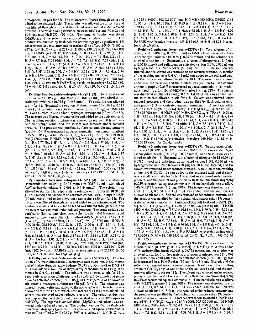
Syntaesi of 2-ImNE, 2PyNE, SPyNE, and 4PyNE. Affinity cleavage experiments identify the sequence, groove location, and orientation preference with which a molecule binds to its target site.4 Ethylenediaminetetraacetic acid (EDTA) was attached to the carboxy end of the four peptides 2-ImN, 2-PyN, 3-PyN, and 4-PyN to afford 2-ImNE, 2-PyNE, 3-PyNE, and 4-PyNE, re- spectively (Figure 8). The synthesis of 2-ImNE is outlined in Figure 9. In analogy to the synthesis of 2-ImN, dipyrrole car- boxylic acid 9 is converted to the activated ester (DCC, HOBt) which is immediately treated with 3-(tert-butyloxy carbonyl- amino)-3'-amino-N-methyldipropylamine to afford the protected amine 13. Nitropyrrole 13 was reduced with palladium under higher pressures of hydrogen (250-350 psi), and the corresponding amine was coupled with N-methylimidazole-2-carboxylic acid (DCC, HOBt) to afford 14 in 85% yield. Deprotection of the amine (TFA, CH2C12) and coupling with triethyl ethylenedi- aminetriacetate acetic acid (DCC, HOBt) provided the tri- ethylester of 2-ImNE (16). Saponification (LiOH, H20, EtOH) yielded 2-ImNE. The 2-, 3-, and 4-PyNE analogs were syn- thesized by a similar route, wherein the triethylester of EDTA was coupled to the bis(N-methylpyrrole) earlier in the synthetic sequence. Dipyrrole 12 is available from N-methylpyrrole-2- carboxylic acid in eight steps in approximately 2W0 overall Reduction of 12 (50 psi H2, Pd/C) and coupling with an activated pyridine carboxylic acid followed by basic hydrolysis (LiOH, EtOH, H20) gave the affinity cleaving peptides in 20-30% yield. Affinity Cleaving. Cleavage experiments on the 5 17 base pair
restriction fragment were carried out in the presence of 2- ImNEeFe(I1) and 2-, 3-, and 4-PyNE-Fe(II) at 37 OC (pH 7.9 in 40 mM Tris acetate) (Figure 10). All of the peptides show cleavage patterns which are shifted to the 3' side of the binding site, consistent with binding in the minor groove (Figure 1 l).4b Distamycin-EDTAeFe(I1) (ED), at 2 pM concentration, binds to the 5'-TTTTT-3' and 5'-AATAA-3' sites with modest orien- tation preference but to the S-CTTTT-3'site with the EDTA. Fe(I1) functionality strongly oriented to the G,C side of the se- quence. 2-PyNE, at 50 p M concentration, also binds to the
(15) Bialer, M.; Yagen, B.; Mechoulam, R. Tetrahedron 1978, 34,
(16) Lown, J. W.; Krowicki, K. J . Org. Chem. 1985, 50, 3774-3779. 2389-239 1.
1 2 3 4 5 6 7
Z >. h c9
I
n
Z )r
9 P
n 8 910111213
Figure 6. MPE-Fe(I1) footprinting of D, 2-PyN, 2-ImN, 3-PyN, and 4-PyN. Autoradiogram of a 8% denaturing polyacrylamide gel. All reactions contain 4 mM DTT, 100 pM bp calf thymus DNA, and 12 kcpm 5' labeled 5 17 base pair restriction fragment in 40 mM Tris acetate pH 7.9 buffer. Lane 1 , intact DNA; lane 2, Maxam-Gilbert G reaction; lane 3, A reaction; lane 4, MPEeFe(I1) standard; lane 5, 4 p M D; lane 6 ,20 pM 2-PyN; lane 7 ,20 pM 2-1"; lanes 8-10 contain 35 pM, 10 pM, and 1 pM 3-PyN, respectively; lanes 11-13 contain 35 pM, 10 pM, and 1 p M 4-PyN, respectively. Lanes 4-13 contain 4 p M MPEeFe(I1).
5'-TTm-3' and 5'-AATAA-3' sites with little orientation preference but to the 5'-CTTTT-3'site with a strong orientation preference in the opposite direction. Binding to the mixed se-

Peptides That Bind in the Minor Groove of DNA J . Am. Chem. SOC., Vol. 114, No. 23, 1992 8181
D a t 2 p M
& a 3 2 p 5 ' - C C C C T A T T T T T A C C T T A A T C T C A T C A ATAA C C T T T C T T A C A C C T C A C C T C C C T T T CCCC-3'
3'- C C C C A a A T C C A AT T A C A C T A C T g y g C C A A A C A A T C T C C A C T C C A CC C m C C C C-5' - 2 - P y N at 10 pM
I 32p 5'- C C C C T A$I";rl̂ T A C C T TA f i $ C AT A AT A AT C C T T T C T T A G A C C T C A C C T C C C A C T T T T C C C C C-3'
3 ' -CCCCA A A T A T C C A A T A A C C T A T T A T T A C C A A A C A A T C T C C A C T C C A C C C T C A A A A C C C C C - 5 ' w - 2 - J m N at 10 LIM
3 2 p 5 ' - C C C C T A T T T T T A T A C G T T A T C T C A CATAATAATCCTTTCTTACACCTCACCTCCCACTTTTCCCCC-3' 3'- C C C C A T A A A A AT A T C C A AT $=$ C T A T T A T T A C C A A A C A A T C T C C ACT C C A C C C T C A A A A C C C C C-5'
3 - P y N at 10 pM
- A -. 3 2 p 5 ' - C C C C T A T T T T T A C C TAA T C A T C A ATAA C C T T T C T T A C A C C T C A C C T C C C A T T T CCCC-3'
3'- C C C C A &=%A T C C F g A C T A C T g $ f i C C A A A C A A T C T C C A C T C C A C C C a C C C C-5' - - w
4 - P y N at 10 pM
&& a
Figure 7. MPE protection patterns for D, 2-PyN, 2-ImN, 3-PyN, and 4-PyN on bp 4268-4335 of ~ B R 3 2 2 . ~ ' Data from 5' end labeled fragment shown in Figure 5 . Bar heights are proportional to the protection from cleavage at each band. Boxes represent equilibrium binding sites determined by the published model.'
quence, 5'-TGTCA-3', results in equal cleavage intensities on each be a combination of the ED and 2-ImNE specificities (Figure 12). side of the binding site, suggesting two equivalent orientations. To determine the specificity of 2-ImNE, the size in base pairs of 2-ImNE, at IO pM concentration, shows this same behavior at each band is calculated by linear interpolation from the sizes of the 5'-TGTCA-3' site, while binding to the 5'-CTTTT-3' site the standards (Figure 13). Sequence analysis of the plasmid occurs with the same orientation preference exhibited by 2-PyNE. reveals 27 resolvable 5'-WGWCW-3' sites (W = A or T),'* and In common with distamycin, 3-PyNE and 4-PyNE bind to the remarkably, all are within 35 base pairs of observed 2-ImNE 5'-TTTTT-3' and 5'-AATAA-3' sites with similar orientation cleavage sites (Table I).I9 High-resolution analysis of eight of distributions but bind the 5'-CTTTT-3'site with less orientation these sites confirms binding to a 5'-WGWCW-3' sequence with preference. two equivalent orientations.20 With regard to binding to the
Sequence Specificity. In the resolvable region of the 517 base 5'-WGWCW-3' sites, A,T base pairs on either side of the sequence pair restriction fragment, only a small fraction of the 512 possible generally result in higher binding affinities.*O 5 base pair sites are present. To be confident that the observed Discussion
1:l Model. Binding of 2-PyN and 2-1" to the 5'-CTTTT-3' strong binding sites are representative of all binding sites, the double-strand cleavage reactions were performed on DNA 4363 base pairs in length. Cleavage of st,,I linearized pBR32217 by site is consistent with the design rationale model (Figure 3). Both ED, 2-PyNE, and 2-ImNE reveals markedly different cleavage
(18) Cornish-Bowden, A. Nucleic Acids Res. 1985, 13, 3021-3030. (19) There are three other 5'-WGWCW-3' sites, but these are within 700 patterns for ED and 2-ImNE, while the 2-PyNE lane appears to
base pairs of the labeled end and are not resolved on the gel. Calculation of (17) By actual count, pBR322 contains 99% of the 512 unique 5 bp se- the sizes of the bands has an error of f40 base pairs.
(20) Wade, W. S. Ph.D. Thesis, California Institute of Technology, 1989. quences.
8188 J . Am. Chem. Soc., Vol. 114, No. 23, 1992
0 9 Table I. Probable 2-ImNE Binding Sites on pBR322”
Wade et al.
2-ImNE 2-PyNE Figure 8. Affinity cleaving analogs 1 -methylimidazole-2-carboxamide- netropsin-EDTAaFe(I1) (2-ImNE) and pyridine-2-carboxamide-net- ropsin-EDTAeFe(I1) (2-PyNE).
compounds cleave this site with a strong preference for the ori- entation which places the heterocyclic nitrogen near the G,C base pair. The observations that the distamycin analog ED binds in the opposite orientation and that 3-PyNE and 4-PyNE do not have a strong orientation preference at this site support the 1:l model. It is clear, however, that 5’-CTTTT-3’ is not the highest affinity binding site for 2-PyN and 2-1”.
Both 3-PyN and 4-PyN bind strongly to A,T-rich sites with orientation preferences and relative site preferences similar to those of distamycin. This is presumably a consequence of the ring nitrogens being in the wrong positions to favorably hydrogen bond with guanine 2-amino groups. Binding of 2-PyN to these same sites likely arises from inadequate positioning of the pyridine nitrogen, with the pyridine ring rotated relative to the carboxamide. This conformation of 2-PyN, with the ring nitrogen facing away from the minor groove, more closely resembles the 3-PyN and 4-PyN analogs and can display analogous binding properties. Presumably the other conformer, with the pyridine nitrogen positioned along the concave edge of the molecule, is the con- formation which recognizes the mixed sequence 5’-TGTCA-3’. The observation that 2-1” does not recognize A,T-rich sites supports this model since positioning the imidazole nitrogen away from the groove places the bulky methyl group toward the floor of the minor groove, which is expected to disrupt binding.
cleavage site relative positionb position( sequence strength
29 1 I d
86 71d 196 212 666 682d 730 711d
732d 736*
1935 1969 1993 202 1 2137 2106
2139 2163
2215 2224 2359 2374 2573 2585 2844 2861 3269 3289 3326 3335
3362 3513 3534 3601 3624 3749 3742 3823 3808
3837 3858
4215 4 1 99d 4332 431 I d
S
m W
W m
m m m
S
W
m m
m
m
m m
S
W
m m
ATGTTTGACAGCTTA AACGCAGTCAGGCAC TCGCCAGTCACTATG AACCCAGTCAGCTCC GGGCATGACTATCGT ACTTATGACTGTCTT ATGACTGTCTTCTTT AAGCC AGACATTAAC CAGGCAGACATCTGT ACCTCTGACACATGC CAGCTTGTCTGTAAG GGAGCAGACAAGCCC GACCCAGTCACGTAG CTCACTGACTCGCTG GCTCAAGTCAGAGGT CGGTAAGACACGACT TGGTCTGACAGTTAC CG ATCTGTCTATTTC TTGCCTGACTCCCCG CATCCAGTCTATTAA CGTGGTGTCACGCTC ATCGTTGTC AGAAGT CTTACTGTCATGCCA TTCTGTGACTGGTGA A ACC AAGTCATTCTG GTTATTGTCTCATGA T ATCATGAC ATTAAC
(I Underlined sequences represent putative or actual binding sites. *Calculated cleavage band positions averaged between labels, pBR322 n~mbering.~’ (Lowest numbered base pair of the putative binding site.
Cleavage site observed on a high-resolution sequencing gel.
21 Model. Binding of 2-PyN and 2-1” to the 5’-TGTCA-3’ site was unanticipated. In early 1:l models, the amide bonds were rotated, presenting carbonyl lone pairs which could participate in hydrogen bonds with guanine 2-amino groups to explain the presence of G,C base pairs in both the second and fourth positions of the binding site.14 Another unusual feature of this complex was revealed in affinity cleaving experiments. The cleavage in- tensities on either side of the binding site were nearly equal, in contrast to distamycin analogs binding to A,T-rich sites, where it has been found that the orientation preferences are dependent on the flanking sequence^.^ Recognition by 2-1” or 2-PyN of a DNA sequence containing 2 G,C base pairs separated by 1 A,T base pair with no orientation preference was difficult to rationalize with a 1:l model.
NMR studies have revealed that distamycin, at millimolar concentrations, is capable of binding to the minor groove of an 5‘-AAATT-3’ sequence as a side-by-side dimer.*’ Consideration
13 I
14: R = COp-r-Bu C C 15: R = H
16: R = Et eC 6: R P H Figure 9. Synthetic scheme for 2-ImNE. (a) (i) DCC, HOBt; (ii) H2N(CH2)3NCH3(CH2)3NHBoc; (b) (i) 300 psi H2, 10% Pd/C; (ii) N-methyl- imidazole-1-carboxylic acid, DCC, HOBt; (c) 20% TFA/CH2C12; (d) (Et02CCH2)2N(CH2)2N(CH2C02Et)(CH2C02H), DCC, HOBt; (e) LiOH, EtOH, H20.

Peptides That Bind in the Minor Groove of DNA
w w z z w w z z % %
m w P P
1 2 3 4 5 6 7 8
-
Figure 10. Specific cleavage by the ED, 2-PyNE, 2-ImNE, 3-PyNE, and 4-@NE analogs. Autoradiogram of a 8% denaturing polyacrylamide gel. All reactions contain 4 mM DTT, 100 pM bp calf thymus DNA, and 12 kcpm 5’ labeled 5 17 base pair restriction fragment in 40 mM Tris acetate pH 7.9 buffer. Lane 1, intact DNA; lane 2, Maxam-Gilbert G reaction; lane 3, A reaction; lane 4, 2 pM ED; lane 5, 50 pM 2-PyNE; lane 6,70 pM 2-ImNE; lane 7, 10 pM 3-PyNE; lane 8, 7 pM 4-PyNE.
of such a 2:l binding model could explain the recognition of 2 G,C base pairs in the 5’-TGTCA-3’ site by 2-1” or 2-PyN, with each guanine amino group hydrogen bonding to imidazole or
(21) (a) Pelton, J. G.; Wemmer, D. E. Proc. Natf. Acad. Sci. U S A . 1989, 86,5723-5727. (b) Pelton, J. G.; Wemmer, D. E. J. Am. Chem. SOC. 1990, 11 2, 1393-1 399.
J. Am. Chem. SOC., Vol. 114, No. 23, 1992 8789
pyridine ring nitrogens on different ligands (Figure 14). Fur- thermore, the (2-ImNE),.5’-TGTCA-3’ and (2-PyNE)#- TGTCA-3’ complexes position a cleaving function on each side of the binding site, consistent with the affinity cleaving results. In collaboration with the Wemmer group, we have recently characterized the complex between 2-1” with d(GCAT- GACTCGG)d(CCGAGTCATGC) by two-dimensional NMR spectro~copy.~~ 2-1” binds as an antiparallel, sideby-side dimer with high cooperativity in the minor groove of DNA. Energy minimization of the complex with constraints from NOESY ex- periments affords a model wherein hydrogen bonds are likely formed between the imidazole nitrogens and the guanine 2-amino groups (Figure 14).22
Sequence Specificity for 5’-WGWCW3’ Sites. 2-1” and 2-PyN bind to 5’-WGWCW-3’ sites but not to the related se- quences 5’-WGWGW-3‘ or 5‘-WCWGW-3‘. This observation can be rationalized by analysis of the disposition of hydrogen binding sites in the minor groove of these sequences. The hy- drogen-bonding character of an A-T base pair resembles that of a T-A base pair due to the approximate minor groove symmetry of the adenine N3 and thymine 0 2 atoms. In a G,C base pair, the guanine 2-amino group lies closer to the guanine-containing strand and as a result, the relative positions of the two guanine amino groups in the three sequences 5’-GTC-3’, 5’-GTG-3’, and 5’-CTG-3’ are very different. As a consequence of the helicity of B-DNA, the amino groups are separated by a larger distance in the 5’-GTC-3’ sequence relative to the 5’-CTG-3’ sequence. The 5’-GTG-3’ sequence is clearly different from the other two, since both amino groups derive from guanine residues on the same strand. This distinction, in combination with restrictions on lig- and-ligand stacking interactions and other ligand-DNA contacts, favors binding to the 5’-WGTCW-3’ sequence over the similar 5’-WGTGW-3‘ and 5’-WCTGW-3’ sequences.
Biological Implications of the 5’-WGWCW-3’ Sequence. A review of DNA-binding proteins revealed that a 5’-WGWCW-3‘ sequence is present in the concensus recognition sites of the transcriptional activator GCN423 and the oncogenic proteins jun and Current models suggest that these leucine zipper proteins bind as Y-shaped dimers in the major groove of DNA, with no minor groove contacts by the protein^.^^.^^ There exists the possibility that 2-1” can bind as a dimer in the minor groove simultaneous with binding in the major groove by the dimeric proteins. Related work from this laboratory has shown that 2-1“ and GCN4(226-281)26 can bind a 5’-CTGACTAAT-3’ sequence ~imultaneously.~~
Implications for the Design of Minor Groove Binding Molecules. The most significant finding of this study is that 2-1” shows an apparent preference for cooperative 2:l binding to the 5‘- TGTCA-3’ sequence over 1:l binding. The width of the minor roove for B-DNA is sequence dependent and can vary from 3-4 1 for A,T-rich DNA to 5-6 A for G,C-containing regiom2*
X-ray structures of 1 : 1 complexes formed between netropsin and distamycin with short oligonucleotides reveal that the natural products sit deeply in the minor groove and make extensive van der Waals contacts to both walls of the binding site, contributing favorably to the total free energy of binding.5 The minor groove width for 1:l complexes of A,T-rich DNA with distamycin and netropsin is approximately 3.4 A5 compared to 6.8 A from models of 2: 1 complexes.21.22 Certain sequences with inherently wider
(22) Mrksich, M.; Wade, W. S.; Dwyer, T. J.; Geierstanger, B. H.; Wemmer, D. E.; Dervan, P. B. Proc. Natl. Acad. Sci. U.S.A. 1992, 89,
(23) (a) Hill, D. E.; Hope, I. A.; Macke, J. P.; Struhl, K. Science 1986, 224,451-457. (b) Hope, I. A.; Struhl, K. Cell 1985,43, 177-188.
(24) (a) Bohmann, D. T.; Bos, T. J.; Admon, A.; Nishimura, T.; Vogt, P. K.; Tjian, R. Science 1987,238, 1386-1392. (b) Rauscher, F. J.; Sambucetti, L. C.; Curran, T.; Distel, R. J.; Spiegelman, B. M. Cell 1988,52,471-480.
(25) Vinson, C. R.; Sigler, P. B.; McKnight, S . L. Science 1989, 246, 91 1.-916.
(26) Oakley, M. G.; Dervan, P. B. Science 1990, 248, 847-850. (27) Oakley, M. G.; Mrksich, M.; Dervan, P. B. Biochemistry, in press. (28) Yoon, C.; PrivE, G. G.; Goodsell, D. S.; Dickerson, R. E. Proc. Natl.
7586-7590.
Acad. Sci. U.S.A. 1988, 85, 6332-6336.

8790 J. Am. Chem. SOC., Vol. 114, No. 23, 1992
ED at 2 pM
Wade et aL
s a p S ' - C C C C T T T T T A C C T T A A T C T C A T C A A T A C C T T T C T T A C A C C T C A C C T C G C T T T GGGG-3' A T C C A AT T A C AC T A C T $ T $ C C A A A C A A T C T GC AC 1 C C AC C C&A$CCCC-S' 3'- C C C C A t t t t t t t 4tt+ t t t t t
I I I t 2 - P y N E at 50 pM
$4 C C C C T A T T T TACCTTA C T C A C A A T A C C T T T C T T A C A C C T C A C C T C G C T T T CCCC-3'
A T C C A AT $y& C T $ F $ C C A A A C A A T C T C C A C T C C A C C C C C C C-5' 32p $1 C C C C A t t t t ++
+++ ++t&t t+ + + + 32p C C C C T A T T T T T A C C T A A T C A T C A A T A A C C T T T C T T A C A C C T C A C C T C C C TTT CCGC-3'
A T C C F g A C T A C T $y& C C A A A C A A T C T C C A C T C C A C C C a C C C C-5' C C C C A t t t t t f t t t t t t t
4 - P y N E at 10 FM
trttttr4rt+ 3 a p S ' - C C C C T A T T T T A C C TAA T C A T C A A T A C C T T T C T T A C A C C T C A C C T C G C T T T CCCC-3'
3'- C C C C A a A T C C L g A C T AC T $ F $ C C A A A C A A T C T C C A C T C C A C C G & cc c c-Y t t t t t t t t t
Figure 11. Cleavage of the 517 base pair fragment of pBR322, base pairs 4268-4335,)' by the ED, 2-PyNE, 2-ImNE, 3-PyNE, and 4-PyNE analogs. Data from 5' end labeled fragment shown in Figure 10. Arrows are proportional to the maximum densities of the cleavage bands. Boxes represent binding sites determined by the published method.4
minor groove widths may favor dimeric binding. We cannot distinguish at this time the importance of sequence dependent flexibility of the minor groove and sequence dependent groove width. We would anticipate that if the guanine NH, protruding from the floor of the minor groove is participating in a specific hydrogen bond with the imidazole N3 of 2-ImN, the peptide ligand is not set as deeply in the minor groove as distamycin. A shallow penetration into the minor groove for 2-1" and 2-PyN in complex with 5'-TGTCA-3' sequences would suggest that these 2:l complexes would have overall lower free energy (and likely lower enthalpy) than that observed for distamycin in complex with
Finally, dimeric motifs seem unlikely to be limited to N - methylpyrrolecarboxamide analogs. For example, chromomycin, an oligosaccharide-chromophore, binds in the minor groove as a 2:l complex.29 Interestingly, the sugar of one ligand of
Poly [d(A-T)I*polY [d(A-T)I .'
chromomycin stacks on the chromophore unit of the other ligand. The increased width of the chromomycin dimer requires the DNA to undergo a major structural transition, resulting in a wider and more shallow minor groove.29 It is reasonable to expect a larger class of structure types to form dimeric structures which can recognize double-helical DNA with high sequence specificity. Experimental Section 'H NMR spectra were recorded either at 400 MHz on a JEOL-GX
400 or at 300 MHz on a General Electric-QE 300 NMR spectrometer in DMSO-& Chemical shifts are reported in parts per million relative to residual DMSO. IR spectra were recorded on a Perkin-Elmer FTIR spectrometer. High-resolution mass spectra (HRMS) were recorded using electron ionization (El) or fast atom bombardment (FAB) tech-
(29) (a) Gao, X.; Patel, D. J. Biochemisrry 1989, 28, 751-762. (b) Gao. X.; Mirau, P.; Patel, D. J . J. Mol. Biol. 1992, 223, 259-279.

Peptides That Bind in the Minor Groove of DNA J. Am. Chem. SOC., Vol. 114, No. 23, 1992 8791
1 2
W L U z z
n 6 7 8
A \ 0 N *HCl
I
B
Figure 12. Sitespecific double-stranded cleavage of pBR322 (4363 base pairs) by ED, 2-PyNE, and 2-ImNE. Autoradiogram of a 1% agarose gel. All reactions contain 1 mM sodium ascorbate, 100 pM bp calf thymus DNA, and 24 kcpm 3' labeled pBR322 linearized with Sty1 and labeled only at one terminus with Klenow fragment. Lanes 1-5 are labeled on the clockwise strand with [ c Y - ~ ~ P I ~ A T P . Lanes 6-8 are labeled on the counterclockwise strand with [cY-~~PITTP. Lane 1, intact DNA; lane 2, molecular weight standards; lanes 3 and 6 ,2 pM E D lanes 4 and 7, 50 pM 2-PyNE; lanes 5 and 8, 70 pM 2-ImNE.
1 i
0 1000 2000 3000 4000 4363 EDat2p.M
I I *t + t + dl 5'
It t 4 + 2-PyNE at 50 p.M
2-ImNE at 70 p.M
Figure 13. Cleavage sites on pBR322 by ED, 2-PyNE, and 2-ImNE. Arrows represent the extent of cleavage in lanes 3-8 of Figure 12. Band densities are averaged between labels and then normalized to the most intense band. Arrow positions represent approximate binding site loca- tions accurate to within 40 base pairs. Cleavage bands within 700 base pairs of the labeled ends are not resolved on the gel.
niques at the Mass Spectrometry Laboratory at the University of Cali- fornia, Riverside. Reagent grade chemicals were used as received unless otherwise noted. Tetrahydrofuran (THF) was distilled under nitrogen from sodium/benzophenone ketyl. Dichloromethane and triethylamine were distilled under nitrogen from powdered calcium hydride. Di- methylformamide (DMF) was purchased as an anhydrous solvent from Aldrich. NJV'Xarbonyldiimidazole was sublimed at 100 "C and 1 Torr. Flash chromatography was carried out using EM Science Kieselgel 60 (230-400) mesh.30 Thin-layer chromatography was performed on EM Reagents silica gel plates (0.2-mm thickness). All the pyrrole derivatives were visualized with short-wave ultraviolet light.
N-Methylimidazole-2-carboxylic Acid. To a solution of N-methyl- imidazole (1.00 g, 12.2 mmol) in THF (20 mL), cooled to -70 "C, was added n-BuLi (5.2 mL, 13.0 mmol in hexanes), and the solution was allowed to stir for 90 min. Dry carbon dioxide was bubbled through the solution for 30 min, and the solution was allowed to stir while warming
Figure 14. 2:l Binding models for the complexes formed between (a) 2-PyN and (b) 2-1" with the 5'-TGTCA-3' sequence. Circles with dots represent lone pairs on N3 of purines and 0 2 of pyrimidines. Circles containing an H represent the N2 hydrogen of guanine. Putative hy- drogen bonds are illustrated by dashed lines.
to room temperature. The reaction was quenched with HCl (lN, 3 mL), at which time a yellow oil separated. Lyophilization of this oil afforded the imidazolecarboxylic acid (1.3 g, 96%) as a white solid: 'H NMR
(KBr) 3450 (w), 2600 (m), 1980 (w), 1652 (s), 1514 (m), 1462 (m), 1402 (s), 1354 (s), 1312 (s), 1276 (m), 1172 (w), 1014 (w), 906 (w), 800 (s), 778 (m), 632 (w) cm-'; EIMS m/e (relative intensity) 126.0428 (17, M+, 126.0430 calcd. for C&N202), 109 (6, M - OH), 108 (7, M -
3-[4- (N-Methyl-4-nitropy~ole-2-carboxamido)-N-metbylpy~ol~2- carboxamidoldimethylpropylamine (10). To a solution of nitrodipyrrole 9 (2.5 g, 8.56 mmol), N-hydroxybenzotriazole hydrate (1.35 g, 10.0 mmol), and 3-(dimethylamino)propylamine (1.3 mL, 10.2 mmol) in DMF (70 mL) at 0 "C was added dicyclohexylcarbodiimide (1.90 g, 9.09 mmol) in CH2C12 (5 mL). The reaction mixture was allowed to warm to room temperature and was stirred for 20 h. The mixture was filtered through celite, and the solvent was removed under reduced pressure. The product was purified by flash column chromatography (1 % concentrated aqueous ammonia in methanol) to give 10 (2.85 g, 88%): 'H NMR (400
1 H, J = 6.7 Hz), 7.53 (d, 1 H, J = 1.8 Hz), 7.17 (d, 1 H, J = 1.8 Hz), 6.77 (d, 1 H, J = 1.8 Hz), 3.90 (s, 3 H), 3.77 (s, 3 H), 3.2 (m, 2 H), 2.21 (t, 2 H, J = 7.4 Hz), 2.1 1 (s, 6 H), 1.59 (m, 2 H); FABMS m/e (relative intensity) 377.1925 (20, M + H, 377.1937 calcd. for C17H2SN604).
Pyridine-2-carboxamide-netropsin (2-4.N) (1). To a solution of pi- colinic acid (0.075 g, 0.610 mmol) and N-hydroxybenzotriazole hydrate (0.090 g, 0.666 mmol) in DMF (2 mL) was added a solution of di- cyclohexylcarbodiimide (0.130 g, 0.630 mmol) in CH2C12 (2 mL). The solution was allowed to stir for 40 min. Separately, a solution of nitro-
(400 MHz, DMSO-d6) 6 3.99 (s, 3 H), 7.27 (s, 1 H), 7.50 (s, 1 H); IR
H2O), 82 (100, M - C02).
MHz, DMSO-d,) 6 10.23 (s, 1 H), 8.13 (d, 1 H, J = 1.8 Hz), 8.07 (t,
dipyrrole 10 (0.10 g, 0.266 mmol) and palladium-on activated carbon (lo%, 20 mg) in DMF (10 mL) was allowed to stir under a hydrogen (30) Still, W. C.; Kahn, M.; Mitra, A. J. Org. Chem. 1978,43,2923-2925.
8192 J . Am. Chem. SOC., Vol. 114, No. 23, 1992
atmosphere (50 psi) for 5 h. The mixture was filtered through celite and added to the activated acid. The solution was allowed to stir for 4 h and was filtered through celite, and the solvent was removed under reduced pressure. The residue was partitioned between ethyl acetate (30 mL) and 10% aqueous NaHC03 (20 mL). The organic fraction was dryed (MgSOJ, and the solvent was removed under reduced pressure. The product was purified by flash column chromatography (gradient 0-1% concentrated aqueous ammonia in methanol) to afford 2-PyN (0.102 g,
nm; ' H NMR (400 MHz, DMSO-d,) 8 10.73 (s, 1 H), 9.95 (s, 1 H), 8.71 (dd, 1 H, J = 6.5, 1.8 Hz), 8.17 (t, 1 H, J = 7.6 Hz), 8.11 (d, 1 H, J = 7.7 Hz), 8.05 (ddd, 1 H, J = 7.7, 7.8, 1.8 Hz), 7.64 (ddd, 1 H, J = 7.6, 4.8, 1.8 Hz), 7.37 (d, 1 H, J = 1.8 Hz), 7.24 (d, 1 H, J = 1.8 Hz),7.18(d,lH,J=1.8Hz),6.93(d,lH,J=1.8Hz),3.85(s,3H), 3.81 (s, 3 H), 3.23 (q, 2 H, J = 7.4 Hz), 3.02 (t, 2 H, J = 7.6 Hz), 2.74 (s, 6 H), 1.84 (quint, 2 H, J = 7.6 Hz); IR (KBr) 2910 (w), 2700 (w), 1640 (s), 1580 (s), 1530 (s), 1460 (m), 1432 (s), 1400 (m), 1260 (m), 1200 (w), 1120 (w) cm-I; FABMS m/e (relative intensity) 452.2427 (60, M + H, 452.2410 calcd. for C23H30N703), 350 (28, M - CSHI4N2), 228 (46).
Pyridine-3-carboxamide-netropsin (3-PyN) (3). To a solution of nicotinic acid (0.057 g, 0.464 mmol) in DMF (3 mL) was added N,N'- carbonyldiimidazole (0.075 g, 0.463 mmol). The solution was allowed to stir for 1 h. Separately, a solution of nitrodipyrrole 10 (0.081 g, 0.215 mmol) and palladium on activated carbon (lo%, 15 mg) in DMF (10 mL) was allowed to stir under a hydrogen atmosphere (50 psi) for 5 h. The mixture was filtered through celite and added to the activated acid. The resulting reaction mixture was allowed to stir for 18 h and was filtered through celite, and the solvent was removed under reduced pressure. The product was purified by flash column chromatography (gradient 0-1% concentrated aqueous ammonia in methanol) to afford
300 (23 500) nm; ' H NMR (400 MHz, DMSO-d,) 6 10.64 (s, 1 H), 9.98 (s, 1 H), 9.77 (bs, 1 H), 9.14 (d, 1 H, J = 2.5 Hz), 8.77 (dd, 1 H, J = 6.2, 5.5 Hz), 8.38 (d, 1 H, J = 8.8 Hz), 8.17 (t, 1 H, J = 5.5 Hz), 7.64 (dd, 1 H, J = 8.8, 5.5 Hz), 7.35 (d, 1 H, J = 1.8 Hz), 7.19 (d, 1 H, J = 1.8 Hz), 7.10 (d, 1 H, J = 1.8 Hz), 6.93 (d, 1 H, J = 1.8 Hz), 3.87 (s, 3 H), 3.81 (s, 3 H), 3.24 (q, 2 H, J = 5.2 Hz), 3.05 (dt, 2 H, J = 8.3, 6.7 Hz), 2.76 (d, 6 H, J = 5.5 Hz), 1.84 (quint, 2 H, J = 7.8 Hz); IR (KBr) 3100 (w), 2940 (w), 2700 (w), 1670 (m), 1632 (s), 1575 (s), 1550 (s), 1530 (s), 1464 (m), 1434 (s), 1422 (s), 1260 (m), 1200 (w), 1120 (w) an- ' ; FABMS m / e (relative intensity) 452.2399 (2, M + H, 452.2410 calcd. for CZ3H3,N7O3).
Pyridine-4-carboxamide-netropsin (4-PyN) (4). To a solution of isonicotinic acid (0.052 g, 0.423 mmol) in DMF (3 mL) was added N,N'-carbonyldiimidazole (0.068 g, 0.419 mmol). The solution was allowed to stir for 1 h. Separately, a solution of nitropyrrole 10 (0.080 g, 0.212 mmol) and palladium on activated carbon (lo%, 15 mg) in DMF (10 mL) was stirred under a hydrogen atmosphere (50 psi) for 5 h. The mixture was filtered through celite and added to the activated acid. The solution was allowed to stir for 18 h and was filtered through celite, and the solvent was removed under reduced pressure. The crude residue was purified by flash column chromatography (gradient e l % concentrated aqueous ammonia in methanol) to afford 4-PyN (0.069 g, 72%): UV (H20) A,,, (e ) 209 (22 SOO), 241 (17 500), 293 (22 500) nm; 'H NMR
J = 6.2 Hz), 8.18 (t, 1 H, J = 7.6 Hz), 8.01 (d, 2 H, J = 5.5 Hz), 7.38 (d, 1 H, J = 1.8 Hz), 7.20 (d, 1 H, J = 1.8 Hz), 7.13 (d, 1 H, J = 1.8 Hz), 6.93 (d, 1 H, J = 1.8 Hz), 3.87 (s, 3 H), 3.81 (s, 3 H), 3.23 (q, 2 H, J = 7.6 Hz), 3.02 (t, 2 H, J = 7.4 Hz), 2.74 (s, 6 H), 1.84 (quint, 2 H, J = 7.5 Hz); IR (KBr) 3100 (w), 2950 (w), 2700 (w), 1660 (m), 1640 (s), 1573 (s), 1542 (s), 1462 (m), 1434 (s), 1402 (s), 1288 (w), 1260 (m), 1202 (w) cm-I; FABMS m / e (relative intensity) 452.2431 (6, M + H, 452.2410 calcd. for C23H30N703).
1-Methylimidazole-2-carboxamide-netropsin (2-1") (2). To a so- lution of N-methylimidazole-2-carboxylic acid (0.06 mg, 0.532 mmol) and N-hydroxybenzotriazole hydrate (0.075 g 0.55 mmol) in DMF (3 mL) was added a solution of dicyclohexylcarbcdiimide (0.1 15 g, 0.55 mmol) in CH2CI2 (2 mL). The mixture was allowed to stir for 12 h. Separately, a solution of nitropyrrole 10 (0.40 g, 1.06 mmol) and palla- dium on activated carbon (lo%, 0.06 g) in DMF (5 mL) was allowed to stir under a hydrogen atmosphere (50 psi) for 6 h. The mixture was filtered through celite and added to the activated acid. The solution was allowed to stir for 12 h at which time methanol (4 mL) was added and solvent was removed under reduced pressure. The crude residue was taken up in ethyl acetate (25 mL) and washed once with 10% aqueous NaHCO,. The organic layer was dried (MgS04), and solvent was re- moved under reduced pressure. The product was purified by flash col- umn chromatography (gradient 0-1% concentrated aqueous ammonia in methanol) to afford 2-1" (0.19 g, 79%) as a yellow oil: UV (H20) A,,
85%): UV (H2O) A,,, (c) 223 (Sh, 21 000), 238 (22000), 296 (26000)
3-PyN (0.062 g, 64%): UV (H2O) Amax (c) 213 (19500), 246 (19 500),
(400 MHz, DMSO-$) 8 10.82 (s, 1 H), 10.00 (s, 1 H), 8.84 (d, 2 H,
Wade et al.
(c) 255 (19000), 302 (26000) nm; 'H NMR (400 MHz, DMSO-d,) 6 10.92 (bs, 1 H), 10.05 (bs, 1 H), 9.89 (s, 1 H), 8.18 (t, 1 H, J = 6.6 Hz), 7.58 (s, 1 H), 7.35 (s, 1 H), 7.32 (d, 1 H, J = 1.8 Hz), 7.18 (d, 1 H, J = 1.8 Hz), 7.14 (d, 1 H, J = 1.8 Hz), 6.92 (d, 1 H, J = 1.8 Hz), 4.02 (s, 3 H), 3.85 (s, 3 H), 3.80 (s, 3 H), 3.24 (q, 2 H, J = 6.4 Hz), 3.04 (m, 2 H), 2.74 (d, 6 H, J = 5.8 Hz), 1.85 (quint, 2 H, J = 7.4 Hz); FABMS m/e (relative intensity) 455.2519 (0.8, M + H, 455.2519 calcd.
Pyridine-2-carboxamid-netropin-EDTA (5). To a solution of pi- colinic acid (0.0095 g, 0.0772 mmol) in DMF (1 mL) was added N,- N'-carbonyldiimidazole (0.0125 g, 0.0772 mmol), and the solution was allowed to stir for 1 h. Separately, a solution of nitropyrrole 12 (0.061 g, 0.0785 mmol) and palladium on activated carbon (IO%, 0.030 g) was hydrogenated in a Parr Rocker (50 psi) for 18 h. The solution was filtered, and the solvent was removed under reduced pressure. A solution of the resulting amine in CH2CI2 (2 mL) was added to the activated acid, and the mixture was allowed to stir for 18 h. The solvent was removed under reduced pressure, and the product was purified by flash column chromatography (0.25% concentrated aqueous ammonia in 1:l metha- no1:ethanol) to afford 2-1"-EDTA triester (14 mg, 62%). The triester was dissolved in ethanol (1 mL), 0.5 M LiOH (1 mL) was added, and the solution was allowed to stir for 1 h. Solvent was removed under reduced pressure, and the product was purified by flash column chro- matography (1% concentrated aqueous ammonia in 1:l methanoketha- nol) to afford 2-PyNE (3.9 mg, 62%): UV (H20) A,,, (e) 245 (21 500), 305 (26000) nm; 'H NMR (400 MHz, DMSO-d, + TFA) 8 10.78 (s, 1 H), 9.95 (s, 1 H), 9.33 (bs, 1 H), 8.70 (dd, 1 H, J = 4.2, 1.7 Hz), 8.48 ( t , l H , J = 6 . 8 H z ) , 8 . 1 8 ( m , l H),8.15(d,l H,J=7.8Hz),8.06(ddd, 1 H, J = 7.9, 7.7, 1.8 Hz), 7.64 (ddd, 1 H, J = 7.8, 4.9, 1.7 Hz), 7.37 (d, 1 H, J = 1.8 Hz), 7.24 (d, 1 H, J = 1.8 Hz), 7.17 (d, 1 H, J = 1.8 Hz), 6.96 (d, 1 H, J = 1.8 Hz), 4.01 (s, 2 H), 3.88 (s, 2 H), 3.85 (s, 3 H), 3.80 (s, 7 H), 3.30-3.00 (m, 12 H), 2.75 (d, 3 H, J = 5.4 Hz), 1.82 (m, 4 H); FABMS m/e (relative intensity) 769.3634 (2, M + H, 769.3633 calcd. for C35H49NloOlo).
Pyridine-3-carboxamide-netropsin-EDTA (7). To a solution of nic- otinic acid (0.0095 g, 0.0772 mmol) in DMF (1 mL) was added N,N'- carbonyldiimidazole (0.0125 g, 0.0772 mmol), and the solution was al- lowed to stir for l h. Separately, a solution of nitropyrrole 12 (0.061 g, 0.0785 mmol) and palladium on activated carbon (lo%, 0.030 g) was hydrogenated in a Parr Rocker (50 psi) for 18 h, and filtered, and the solvent was removed under reduced pressure. A solution of the resulting amine in CH2CI2 (2 mL) was added to the activated acid, and the mix- ture was allowed to stir for 18 h. The solvent was removed under reduced pressure, and the product was purified by flash column chromatography (0.25% concentrated aqueous ammonia in 1:l methano1:ethanol) to afford 3-PyN-EDTA triester (13 mg, 59%). The triester was dissolved in eth- anol (1 mL), 0.5 M LiOH (1 mL) was added, and the solution was allowed to stir for 1 h. Solvent was removed under reduced pressure, and the product was purified by flash column chromatography (1% concen- trated aqueous ammonia in 1:l methano1:ethanol) to afford 3-PyNE (3.8 mg, 66%): UV (HzO) A,, (e) 238 (20000), 296 (24000) nm; 'H NMR
1 H), 9.32 (s, 1 H), 9.01 (d, 1 H, J = 5.7 Hz), 8.88 (dd, 1 H, J = 7.7, 1.7 Hz), 8.47 (t, 1 H, J = 7.4 Hz), 8.18 (t, 1 H, J = 7.5 Hz), 8.08 (dd, 1 H, J = 7.7, 5.8 Hz), 7.36 (d, 1 H, J = 1.8 Hz), 7.17 (d, 1 H, J = 1.8 Hz),7.12(d,lH,J=1.8Hz),6.96(d,lH,J=1.8Hz),4.01(s,3H), 3.88 (s, 5 H), 3.81 (s, 4 H), 3.80 (s, 3 H), 3.30-3.00 (m, 12 H), 2.76 (d, 3 H, J = 5.2 Hz), 1.83 (m, 4 H); FABMS m/e (relative intensity) 769.3606 (10, M + H, 769.3633 calcd. for C35H49N10010), 743 (20, M
Pyridine-4-carboxamidenetropin-EDTA (8). To a solution of iso- nicotinic acid (0.0095 g, 0.0772 mmol) in DMF (1 mL) was added N,N'-carbonyldiimidazole (0.0125 g, 0.0772 mmol), and the solution was allowed to stir for 1 h. Separately, a solution of nitropyrrole 12 (0.061 g, 0.0785 mmol) and palladium on activated carbon (10%. 0.030 g) was hydrogenated in a Parr Rocker (50 psi) for 18 h and filtered, and the solvent was removed under reduced pressure. A solution of the resulting amine in CHZCl2 (2 mL) was added to the activated acid, and the mix- ture was allowed to stir for 18 h. The solvent was removed under reduced pressure, and the product was purified by flash column chromatography (0.25% concentrated aqueous ammonia in 1:l methanokethanol) to afford 4-PyN-EDTA triester (1 1 mg, 50%). The triester was dissolved in eth- anol (1 mL), 0.5 M LiOH (1 mL) was added, and the solution was allowed to stir for 1 h. Solvent was removed under reduced pressure, and the product was purified by flash column chromatography (1% concen- trated aqueous ammonia in 1:l methanokethanol) to afford 4-PyNE (3.1 mg, 63%): UV (H20) A,, (c) 245 (18000), 305 (22500) nm; 'H NMR
1 H), 9.08 (d, 1 H, J = 7.5 Hz), 8.49 (t, 1 H, J = 7.3 Hz), 8.46 (d, 1 H, J = 7.3 Hz), 8.18 (bs, 1 H), 7.38 (d, 1 H, J = 1.8 Hz), 7.15 (d, 1
for C22H31N803).
(400 MHz, DMSO-d, + TFA) 8 10.88 (s, 1 H), 9.99 (s, 1 H), 9.35 (bs,
+ H - CN).
(400 MHz, DMSO& + TFA) 8 11.09 (s, 1 H), 9.98 (s, 1 H), 9.33 (bs,

Peptides That Bind in the Minor Groove of DNA
H, J = 1.8 Hz), 6.97 (d, 1 H, J = 1.8 Hz), 4.04 (s, 2 H), 3.91 (s, 2 H), 3.88 (s, 3 H), 3.86 (s, 4 H), 3.79 (s, 3 H), 3.40-3.00 (m, 12 H), 2.75 (d, 3 H, J = 5.2 Hz), 1.82 (m, 4 H); FABMS m / e (relative intensity) 769.3638 (0.4, M + H, 769.3633 calcd. for C35H49N10010).
3'-[4-( N-Methyl-4-nitropyrrole-2-carboxamido)-N-methylpyrrole-2- carboxamido)-3-[ tert-butyloxycarbonylamino]methyldipropylamine (13). To a solution of nitrodipyrrole 9 (0.850 g, 2.71 mmol), N-hydroxy- benzotriazole hydrate (0.37 1 g, 2.75 mmol), and 3-(tert-butyloxy- carbonylamino)-3'-amino-N-methyldipropylamine (1.50 g, 6.1 2 mmol) in DMF (5 mL) at 0 OC was added dicyclohexylcarbodiimide (0.575 g, 2.75 mmol) in CH2C12 (5 mL). The reaction mixture was allowed to warm to room temperature and was stirred for 15 h. The mixture was filtered, and the solvent was removed under reduced pressure. The product was purified by flash column chromatography (25% methylene chloride in methanol) to give 10 (1.10 g, 78%): ' H NMR (300 MHz,
8.11 (d, 1 H, J = 1.8 Hz), 7.58 (d, 1 H , J = 1.8 Hz), 7.19 (d, 1 H, J = 1.8 Hz), 6.90 (d, 1 H, J = 1.8 Hz), 3.94 (s, 3 H), 3.81 (s, 3 H), 3.38 (m, 2 H), 3.03-3.15 (b, 6 H), 2.70 (d, 3 H , J = 7.3 Hz), 1.89 (m, 2 H), 1.78 (m, 2 H), 1.40 (s, 9 H); IR 3317 (m), 2966 (m), 2358 (w), 1694 (s), 1652 (s), 1574 (m), 1538 (s), 1520 (s), 1310 (s), 1215 (m), 1167 (m), 11 16 (w) cm-l; FABMS m / e (relative intensity) 520.2862 (27, M + H, 520.2884 calcd. for C17H25N604), 420 (8, M + H - C,H,O,).
l -Methy l imidazo le -2 -carboxamid~e~o~ i~ ia~oa~heptane- tBoc (14). To a solution of N-methylimidazole-2-carboxylic acid (0.465 g, 4.19 mmol) and N-hydroxybenzotriazole hydrate (0.930 g, 6.88 mmol) in DMF (10 mL), at 0 OC, was added a solution of 1,3-dicyclohexyl- carbodiimide (1.00 g, 4.85 mmol) in CH,CI, (10 mL). The solution was allowed to warm to room temperature and was stirred an additional 10 h. Separately, a solution of nitropyrrole 13 (0.50 g, 0.962 mmol) and palladium on activated carbon (lo%, 0.080 g) was allowed to stir under a hydrogen atmosphere (300 psi) in a Parr bomb apparatus for 5 h. The mixture was filtered through celite and added to the activated acid, and the solution was allowed to stir for 2 h. Methanol (3 mL) was added, and the solvent was removed under reduced pressure. The residue was slurried in 10% aqueous NaHCO, (50 mL) and washed with CH,CI, (2 X 50 mL). The combined organic layers were dried (NaSO,), concen- trated, and purified by flash column chromatography (methanol) to afford 14 (0.490 g, 85%) as a yellow solid: ' H NMR (400 MHz,
7.38 (s, 1 H), 7.27 (d, 1 H , J = 1.8 Hz), 7.17 (d, 1 H , J = 1.8 Hz), 7.14 (d, 1 H, J = 1.8 Hz), 6.82 (d, 1 H , J = 1.8 Hz), 6.77 (t, 1 H, J = 5.8 Hz), 3.98 (s, 3 H), 3.83 (s, 3 H), 3.78 (s, 3 H), 3.17 (q, 2 H , J = 6.8 Hz), 2.93 (q, 2 H, J = 6.9 Hz), 2.28 (t, 2 H , J = 7.2 Hz), 2.27 (t, 2 H, J = 7.3 Hz), 2.10 (s, 3 H), 1.59 (quint, 2 H, J = 7.2 Hz), 1.50 (quint, 2 H, J = 7.3 Hz), 1.35 (s, 9 H); IR 3322 (m), 2948 (m), 1643 (s), 1582 (m), 1538 (s), 1470 (s), 1428 (s), 1365 (m), 1254 (mi, 1168 (m), 1122 (m) cm-I; FABMS m/e (relative intensity) 598.3447 (41, M + H, 598.3464 calcd. for C29H44N905), 498 (19, M + H - C,H,O,).
l - M e t b y l i m i d a z o l e 2 - c a r b o x a m i d e - n e t r o p s i n n e ( 15). To a solution of 14 (0.230 g, 0.385 mmol) in CH,CI, (8 mL) was added trifluoroacetic acid (2 mL), and the resulting mixture was allowed to stir 25 min. The reaction mixture was triturated with diethyl ether (100 mL), and the residue was dissolved in 1% concentrated aqueous ammo- nium hydroxide in methanol (30 mL), and the solvent was removed under reduced pressure. The product was purified by flash column chroma- tography (6% concentrated aqueous ammonium hydroxide in methanol) to afford the primary amine (185 mg, 97%): IH NMR (400 MHz,
7.37 (d, 1 H, J = 1.8 Hz), 7.27 (d, 1 H, J = 1.8 Hz), 7.16 (d, 2 H, J = 1.8 Hz), 7.13 (s, 1 H), 7.02 (d, 1 H, J = 1.8 Hz), 3.97 (s, 3 H), 3.82 (s, 3 H), 3.77 (s, 3 H), 3.30 (q, 2 H, J = 7.2 Hz), 3.17 (q, 2 H, J = 6.8 Hz), 2.50 (m, 2 H), 2.28 (t, 2 H, J = 7.0 Hz), 2.10 (s, 3 H), 1.59 (quint, 2 H, J = 7.2 Hz), 1.47 (quint, 2 H, J = 7.1 Hz); IR 3315 (m), 2880 (w), 1642 (s), 1603 (m), 1462 (m), 1433 (m), 1259 (m) cm-l; FABMS m / e (relative intensity) 498.2947 (22, M + H, 498.2941 calcd. for C24H36N903).
l-Methylimidazole-2-carboxamidenetropsin-EDTA, Triethyl Ester (16). A solution of EDTA triethyl ester (0.128 g, 0.34 mmol) and N,N'-carbonyldiimidazole (55 mg, 0.34 mmol) in CH2C12 (4 mL) was allowed to stir for 1 h. Primary amine 15 (0.066 g, 0.133 mmol) was added, and the resulting solution was allowed to stir for 12 h. Solvent was removed under reduced pressure, and the product was purified by flash column chromatography (0.25% concentrated aqueous ammonia in 1:l methano1:ethanol) to afford the triester (0.017 g, 15%): ' H NMR
= 6.8 Hz), 7.95 (t, 1 H, J 7.1 Hz), 7.39 (s, 1 H), 7.28 (d, 1 H, J = 1 . 8 H z ) , 7 . 1 7 ( d , l H , J = l . 8 H z ) , 7 . 1 3 ( d , 1 H , J = 7 . 3 H z ) , 7 . 0 3 ( d , 1 H, J = 1.8 Hz), 6.83 (d, 1 H, J = 1.8 Hz), 4.05 (q, 6 H, J = 6.9 Hz), 3.98 (s, 3 H), 3.88 (s, 3 H), 3.74 (s, 3 H), 3.49 (s, 4 H), 3.43 (s, 2 H),
DMSO-d,) 6 10.25 (s, 1 H), 10.12 (bs, 1 H), 8.25 (t, 1 H, J = 6.5 Hz),
DMSO-d,) 6 10.44 (S , 1 H), 9.89 (s, 1 H), 8.01 (t, 1 H, J = 7.3 Hz),
DMSO-d6) 6 10.44 (s, 1 H), 9.92 (s, 1 H), 8.03 (t, 1 H, J = 7.2 Hz),
(400 MHz, DMSO-d,) 6 10.43 ( S , 1 H), 9.90 (s, 1 H), 8.01 (t, 1 H, J
J . Am. Chem. SOC., Vol. 114, No. 23, 1992 8193
3.18 (s, 2 H), 3.15 (m, 2 H), 3.11 (q, 2 H, J = 7.2 Hz), 2.69 (m, 4 H), 2.31 (t, 2 H, J = 7.1 Hz), 2.29 (t, 2 H, J = 7.1 Hz), 2.13 (s, 3 H), 1.6 (m, 4 H), 1.16 (t. 9 H, J = 7.3 Hz).
1-Methylimidazole-2-carboxamidenetropirEDTA (6). A solution of triethyl ester 16 (14 mg, 0.016 mmol) in ethanol (2 mL) and 0.5 M LiOH ( 1 mL) was allowed to stir for 16 h. The solvent was removed under reduced pressure, and the product was purified by flash chroma- tography (1% concentrated aqueous ammonia in 1: 1 methano1:ethanol) to afford 2-ImNE (0.0034 g, 27%): UV ( H 2 0 ) A,,, ( 6 ) 252 (19000),
1 H), 9.98 (s, 1 H), 9.36 (bs, 1 H), 8.47 (t, 1 H, J = 6.8 Hz), 8.19 (t, 1 H, J = 7.2 Hz), 7.70 (s, 1 H), 7.54 (s, 1 H), 7.32 (d, 1 H, J = 1.8 Hz), 7.16 (d, 1 H, J = 1.8 Hz), 7.12 (d, 1 H, J = 1.8 Hz), 6.95 (d, 1 H, J = 1.8 Hz), 4.02 (s, 5 H), 3.87 (s, 5 H), 3.81 (s, 4 H), 3.79 (s, 2 H), 3.78 (s, 3 H), 3.30-3.00 (m, 12 H), 2.75 (s, 3 H), 1.84 (m, 4 H); FABMS
304 (26500) nm; ' H NMR (400 MHz, DMSO-d, + TFA) 6 10.96 ( s ,
m/e (relative intensity) 772.3746 (14, M + H, 772.3742 calcd. for CuHwN,iOin). 549 (39). 507 (17).
-DNA Reagents and Materials. All water was distilled, filtered through an organic removal cartidge (Corning), and redistilled. For DNA ma- nipulations, the water and all buffers used were autoclaved for 20 min at 160 OC. Acrylamide was purchased as a 30% solution from National Diagnostics. To each liter was added 15 g (1.5%) N,N'-methylenebis- acrylamide. Calf thymus DNA was sonicated for 1 min and phenol extracted. The solution was exhaustively dialyzed against water and diluted to 1 mM bp. Sigma type XX tRNA was deproteinized by phenol extraction and diluted to 1 mg/mL. Plasmid pBR322 was grown in HBlOl cells and purified by CsCl gradient.3'.32 Chemical sequencing reactions were performed according to published method^.'^^'^ All other reagents and materials were used as received.
Sample Preparation. Milligram quantities of the compounds were weighed on a Sartorius microbalance and diluted to 100 mL with water. The average molar extinction coefficient from three measurements was used to determine the concentration of a stock solution, which was lyo- philized in 600 pL double-lock eppendorf tubes and stored dry at -20 OC.
MPE.Fe(II) Footprinting.' A 20 pM MPEsFe(I1) solution was pre- pared by mixing 10 pL of a 1 mM MPE solution with 10 pM of a freshly prepared 1 mM ferrous ammonium sulfate solution, then diluting to 500 pL. Solutions were prepared containing 1 pL/tube 20X TA buffer (800 mM Tris, 100 mM sodium acetate, pH 7.9), 2 pL/tube I mM bp calf thymus DNA, labeled restriction fragment, and water to make 8 pL/tube total solution. 4 pL of a 5X solution of the compound was added, and the tubes were incubated in the dark for 30 min at 37 "C. To each tube was added 4 pL of the 20 pM MPEsFe(I1) solution followed by 4 pL of a freshly prepared 20 mM DTT solution. Final concentrations were 40 mM Tris acetate (pH 7.9), 5 mM sodium acetate, 100 pM bp DNA, 4 pM MPE.Fe(II), and 4 mM DTT, in 20 pL. The reactions were incu- bated at 37 OC for 10 min, lyophilized, and electrophoresed.
Affinity Clea~age.~ Solutions were prepared containing 1 pL/tube 20X TA buffer, 2 pL/tube I mM bp calf thymus DNA, labeled re- striction fragment, and water to make 12 pL/tube. The compounds to be examined were loaded with Fe2+ by adding 10 pL of a freshly prepared 2 mM ferrous ammonium sulfate solution to 10 p L of a 2 mM solution of the compound followed by appropriate dilution with water. 4 p L of the compound was added to each tube, and the solutions were incubated at 37 OC for 30 min in the dark. DTT (4 pL, 20 mM) was added, and the reactions were incubated in the dark at 37 "C for 30 min. Final concentrations were 40 mM pH 7.9 Tris acetate, 100 pM bp DNA, and 4 mM DTT, in a total volume of 20 pL. The reactions were lyophilized and electrophoresed.
Preparation of Linearized pBR322 Labeled at Only One Terminus. pBR322 plasmid was digested with Sty1 and then labeled on the 3' terminus of the clockwise strand with [a-'*P]dATP or on the 3' terminus of the counterclockwise strand with [a-)*P]TTP using the Klenow frag- ment of DNA polymerase I.'* The radiolabeled plasmid was purified on a 0.7% agarose gel containing ethidium bromide. The band containing linearized plasmid was excised, diluted with 500 pL TE buffer (20 mM Tris, 1 mM EDTA, pH 7.9), melted at 70 OC for 10 min, phenol ex- tracted, and ethanol precipitated.
Molecular Weight Standards. Equal amounts of the [a-32P]dATP and [cY-~*P]TTP labeled plasmid were combined with 1 pg pBR322. Indi- vidual samples were digested with the restriction endonucleases EcoRI,
(31) (a) Sutcliffe, J. G. Cold Spring Harbor Symposium on Quantitative Biology 1978, 43, 77-90. (b) Peden, K. W. C. Gene 1982, 22, 277-280.
(32) Maniatis, T.; Fritsch, E. F.; Sambrook, J . Molecular Cloning, A Laboratory Manual; Cold Spring Harbor Laboratory: New York, 1982; pp 1-542.
(33) Iverson, B. L.; Dervan, P. B. Nucleic Acids Res. 1987, 15, 7823-7830. (34) Maxam, A. M.; Gilbert, W. S . Methods in Enzymol. 1980, 65,
499-560.
8194 J . Am. Chem. SOC. 1992, 114, 8794-8799
XmnI, BamHI, and PsrI or PuuII. The reactions were ethanol precipi- tated, counted, and diluted to the same concentration (cpm/pL), and equal volumes were combined with the same cpm of intact pBR322.
Cleavage of Linear pBR322. Solutions were prepared containing 0.75 pL/tube 20X TA buffer, 1.5 pL 1 mM bp calf thymus DNA, radio- labeled pBR322, and water to make 9 pL. 3 pL of a 5X solution of compoundeFe(I1) was added, and the solutions were incubated at 37 OC for 30 min in the dark. 3 pL of a 5 mM sodium ascorbate solution was added, and the reactions were incubated at 37 O C for 2 h. Final con- centrations were 40 mM pH 7.9 Tris acetate, 5 mM sodium acetate, 100 pM bp DNA, and 1 mM sodium ascorbate, in a total volume of 15 pL. The reactions were diluted with Ficoll loading buffer and electrophoresed on a 1% vertical agarose gel.
Densitometry. Footprinting and affinity cleaving autoradiograms were scanned on a LKB XL laser densitometer. The scans were output to an IBM printer, and footprints were determined by comparison to the MPEaFe(I1) standard lane. A horizontal line was drawn from the top of the unprotected band nearest the footprint, and the distance in ab-
sorbance units from this line to the maximum peak height was deter- mined for each band and plotted as a histogram. Affinity cleavage patterns were measured in similar fashion, using peak heights of sites without specific cleavage as a baseline. To determine the sites of cleavage in the double strand cleavage assay, the cleavage lanes were scanned as above and output to the Hoeffer program GS370 through an analog to digital converter. Cleavage band molecular weights were determined by comparison to the molecular weight standard lane, and positions on pBR322 were calculated. The positions were averaged between labels and used, along with the average relative area of the peaks, to generate histograms.
Acknowledgment. W e are grateful to the National Institutes of Health (GM-27681) and to Burroughs Wellcome for research support, to the Nat ional Science Foundation for a predoctoral fellowship to W.S.W., and to the Nat ional Institutes of Heal th for a Research Service Award to M.M.
What Causes Aqueous Acceleration of the Claisen Rearrangement?
Christopher J. Cramer*,+ and Donald G. Truhlar*q* Contribution from the US. Army Chemical Research Development and Engineering Center, Aberdeen Proving Ground, Maryland 21010-5423, and Department of Chemistry and Supercomputer Institute, University of Minnesota, Minneapolis, Minnesota 55455-0431. Received May 26, I992
Abstract: We report the results of applying a new self-consistent-field solvation model to the Claisen rearrangement of allyl vinyl ether, all possible methoxy-substituted derivatives, two alkylated derivatives, and one carboxymethylated derivative in order to understand the effects of aqueous solvation on the reaction rates. We have employed the AM1-SM2 version of the model to calculate the changes in free energies of solvation in passing from the lowest-energy conformations of the starting materials to both chair and boat transition states. The hydrophobic effect is always accelerative but always small and not very structure sensitive. Other first-hydration-shell effects attributable to hydrophilic parts of the reagents are more sensitive to the substitution pattern. The polarization contributions to the activation energies are usually larger. A favorable polarization contribution is found to be associated with efficient sequestration of charges of opposite sign into separated regions of space. We conclude that aqueous acceleration of the Claisen rearrangement is caused by electric polarization and first-hydration-shell hydrophilic effects, with the relative magnitudes and even the signs of these effects being quite sensitive to substitution pattern.
Introduction Pericyclic reactions in aqueous solution can exhibit both rate
acceleration and improved diastereoselection.' Increased rates have been in part attributed to hydrophobic interactions of non- polar solutes with each other and with water. However, Blake and Jorgensen have suggested that improved solvation of a more polar transition state (TS) relative to starting material may also play a significant role in one particular class of pericyclic processes, Diels-Alder reactions.*
T h e Claisen rearrangement (Chart I) is a different example of a pericyclic reaction, a [3,3] sigmatropic shift, which also shows considerable acceleration on going from nonpolar to aqueous solvent^.^" By its very nature as a n oxa-Cope rearrangement, even for the rearrangement of the prototype allyl vinyl ether, it is clear that both polar and hydrophobic (or hydrophilic) effects may readily contribute to the overall observed acceleration. Experimentally, it is difficult to discern the relative contributions to transition-state lowering from these effects.
W e have recently described's8 a semiempirical, effective-Ham- iltonian SCF approach9 for modeling aqueous solvation. T h e
'Correspondence may be addressed to either author at University of
'US. Army Chemical Research Development and Engineering Center and
?Department of Chemistry and Supercomputer Institute, University of
Minnesota.
De artment of Chemistry, University of Minnesota.
Minnesota.
Chart I
[3'31 O 3 7. R b . R
0-
method incorporates solvent-induced polarizationt0 and structural relaxation" of the solute (whether equilibrium structure or
(1) (a) Breslow, R. Acc. Chem. Res. 1991, 24, 159. (b) Grieco, P. A. Aldrichim. Acta 1991, 24, 59. (c) Blokzijl, W.; Blandamer, M. J.; Engberts, J . B. F. N. J. Am. Chem. SOC. 1991, 113, 4241. (d) Cativiela, C.; Garcia, J. 1.; Mayoral, J. A.; Avenoza, A.; Peregrina, J. M.; Roy, M. A. J . Phys. Org. Chem. 1991, 4, 48.
(2) Blake, J. F.; Jorgensen, W. L. J . Am. Chem. SOC. 1991, 113, 7430. (3) White, W. N.; Wolfarth, E. F. J. Org. Chem. 1970, 35, 2196, 3585. (4) Coates, R. M.; Rogers, B. D.; Hobbs, S. J.; Peck, D. R.; Curran, D.
( 5 ) Gajewski, J. J.; Jurayj, J.; Kimbrough, D. R.; Gande, M. E.; Ganem,
(6) Brandes, E.; Grieco, P. A.; Gajewski, J. J. J . Org. Chem. 1989,54, 515. (7) Cramer, C. J.; Truhlar, D. G. J . Am. Chem. SOC. 1991, 113, 8305,
9901 (E). (8) Cramer, C. J.; Truhlar, D. G. Science 1992, 256, 213. (9) Tapia, 0. In Quantum Theory of Chemical Reactions; Daudel, R.,
Pullman, A., Salem, L., Veillard. A., Eds.; Reidel: Dordrecht, 1980; Vol. 2, p 25.
(IO) Aleman, C.; Maseras, F.; Lledb, A.; Duran, M.; Bertrin, J. J . Phys. Chem. 1989, 2, 611. Alagona, G.; Ghio, C.; Igual, J.; Tomasi, J. J. Mol. Slrucf. (Theochem) 1990, 204, 253.
P. J. Am. Chem. SOC. 1987, 109, 1160.
B.; Carpenter, B. K. J . Am. Chem. SOC. 1987, 109, 1170.
0002-7863192115 14-8794%03.00/0 0 1992 American Chemical Society

![Phenylalanine-Rich Peptides Potently Bind ESAT6, a ...repository.ias.ac.in/74232/1/1-pub.pdfesp. BCG [19]. Numerous structural and biochemical studies on this Esat6:Cfp10 complex bear](https://static.fdocuments.net/doc/165x107/6086f2f1c1159576226f2ea4/phenylalanine-rich-peptides-potently-bind-esat6-a-esp-bcg-19-numerous-structural.jpg)


![Generation of a … · 2020-03-21 · small molecules and biomolecules (e.g., proteins, peptides and nucleic acids) bind to their targets in specifi c signaling cas-cades. [2 ] While](https://static.fdocuments.net/doc/165x107/5f0841357e708231d42119eb/generation-of-a-2020-03-21-small-molecules-and-biomolecules-eg-proteins-peptides.jpg)