Department of Radiation Oncology and BIO-X, School of Medicine Stanford University Molecular Imaging...
13
Department of Radiation Oncology and BIO-X, School of Medicine Stanford University Molecular Imaging Program at Stanford MIPS Monte Carlo treatment planning for small animal microCT-based radiotherapy M. Bazalova, H. Zhou, P. Keall, E. Graves Stanford University, CA, USA
-
Upload
francis-burke -
Category
Documents
-
view
216 -
download
0
Transcript of Department of Radiation Oncology and BIO-X, School of Medicine Stanford University Molecular Imaging...

Department of Radiation Oncology and BIO-X, School of Medicine
Stanford UniversityMolecular Imaging Program at Stanford
MIPS
Monte Carlo treatment planning for small animal
microCT-based radiotherapy
M. Bazalova, H. Zhou, P. Keall, E. GravesStanford University, CA, USA
Department of Radiation Oncology and BIO-X, School of Medicine
Stanford UniversityMolecular Imaging Program at Stanford
MIPS
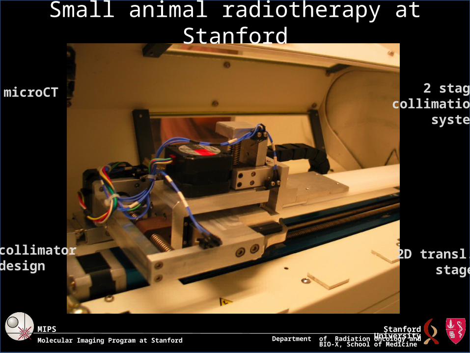
Small animal radiotherapy at Stanford
microCT
2D transl.stage
2 stagecollimation
system
collimatordesign
Department of Radiation Oncology and BIO-X, School of Medicine
Stanford UniversityMolecular Imaging Program at Stanford
MIPS
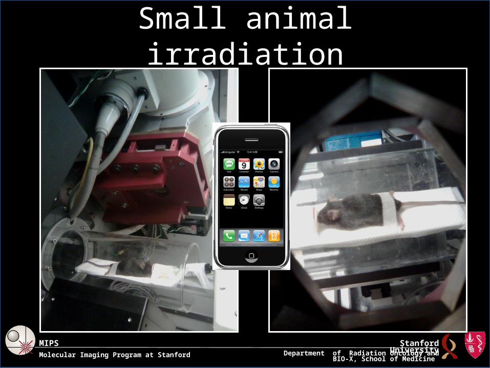
Small animal irradiation
Department of Radiation Oncology and BIO-X, School of Medicine
Stanford UniversityMolecular Imaging Program at Stanford
MIPS
Motivation
• For kilovoltage photon beams, the dose has to be calculated by the Monte Carlo (MC) method.
• The aim of this work is to investigate – the efficiency of MC dose calculation for kV photon beams
in submillimeter voxels – the importance of tissue segmentation for kV photon beam
• BEAMnrc and DOSXYZnrc EGSnrc codes are used.• Two methods for dose calculations are studied and
optimized.• Dual-energy microCT imaging is studied.
Department of Radiation Oncology and BIO-X, School of Medicine
Stanford UniversityMolecular Imaging Program at Stanford
MIPS
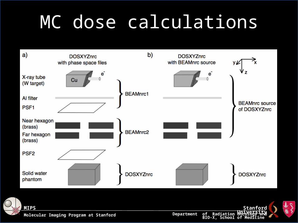
MC dose calculations

Department of Radiation Oncology and BIO-X, School of Medicine
Stanford UniversityMolecular Imaging Program at Stanford
MIPS
Two calculation approaches: comparison
For (0.2×0.2×0.2) mm3, 120 kVp beam
• beam < 30 mm phase-space files
• beam > 30 mmBEAMnrc source
3.0 GHz machine
Department of Radiation Oncology and BIO-X, School of Medicine
Stanford UniversityMolecular Imaging Program at Stanford
MIPS
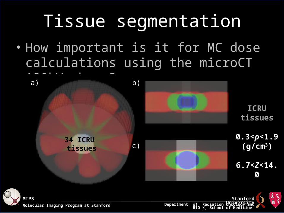
Tissue segmentation• How important is it for MC dose calculations
using the microCT 120kVp beam?a) b)
c)34 ICRU tissues
ICRU tissues
0.3<ρ<1.9(g/cm3)
6.7<Z<14.0

Department of Radiation Oncology and BIO-X, School of Medicine
Stanford UniversityMolecular Imaging Program at Stanford
MIPS
Tissue segmentation - Results
Need to know Z.

Department of Radiation Oncology and BIO-X, School of Medicine
Stanford UniversityMolecular Imaging Program at Stanford
MIPS
Dual-energy microCT (DEmCT) imaging
• DEmCT is based on a parameterization of the linear attenuation coefficient.
• Results in ρ and Z of each voxel.
• Tested on a mouse phantom and a calibration phantom with 70 and 120 kVp beams.

Department of Radiation Oncology and BIO-X, School of Medicine
Stanford UniversityMolecular Imaging Program at Stanford
MIPS
DEmCT phantom: Results
• ρ and Z extracted with a reasonable accuracy
• beam hardening does seem to play a role (will be investigated)
• noise is an important issue, image quality should be improved
Department of Radiation Oncology and BIO-X, School of Medicine
Stanford UniversityMolecular Imaging Program at Stanford
MIPS
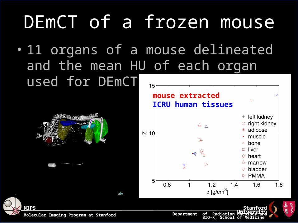
DEmCT of a frozen mouse• 11 organs of a mouse delineated and the
mean HU of each organ used for DEmCT
mouse extractedICRU human tissues
Department of Radiation Oncology and BIO-X, School of Medicine
Stanford UniversityMolecular Imaging Program at Stanford
MIPS
Conclusions
• Monte Carlo dose calculations using the EGSnrc codes for microCT-based small animal radiotherapy have been investigated.
• For beams with diameters larger than 30 mm, BEAMnrc sources should be used as opposed to phase-space files.
• Tissue segmentation is an important factor. the absorbed dose in a tissue correlates with the atomic number Z of the tissue.
• Dual energy microCT is a promising technique for tissue segmentation for small animal radiotherapy dose calculations.

Department of Radiation Oncology and BIO-X, School of Medicine
Stanford UniversityMolecular Imaging Program at Stanford
MIPS
Acknowledgements• Geoff Nelson• Manuel Rodriguez• Rahil Jogani• Fred van den Haak• The work is supported by NIH grant R01
CA131199.