Department of Orthopaedic Surgery, Duke …€ Department of Orthopaedic Surgery, Duke University...
14
AOFAS Public Education Forum (FootCareMD): Ankle Arthroscopy Authors: William R. Mook, MD†, Selene G. Parekh, MD, MBA†‡ †Department of Orthopaedic Surgery, Duke University Medical Center, Durham, NC ‡The Fuqua School of Business, Duke University, Durham, NC
Transcript of Department of Orthopaedic Surgery, Duke …€ Department of Orthopaedic Surgery, Duke University...

AOFAS Public Education Forum (FootCareMD): Ankle Arthroscopy
Authors: William R. Mook, MD†, Selene G. Parekh, MD, MBA†‡
†Department of Orthopaedic Surgery, Duke University Medical Center, Durham, NC
‡The Fuqua School of Business, Duke University, Durham, NC
What is ankle arthroscopy?
Ankle arthroscopy is a minimally invasive surgical technique that utilizes the technology
of fiberoptics, magnifying lenses, and digital video monitors to allow the surgeon to directly
visualize the inside of an ankle through small incisions. Several incisions, approximately half a
centimeter in length, are fashioned about the ankle to allow for the insertion of an arthroscope, or
small fiberoptic video camera, and/or special arthroscopic instruments. Sterile fluid is also
circulated through the ankle to distend the joint, creating more space for the arthroscope and
instruments. This also allows for better visibility within the ankle, space to maneuver
instruments, and clearance of debris.
Figure 1. Anterior ankle arthroscopy set-‐up
How is ankle arthroscopy performed?
Ankle arthroscopy is generally performed as an outpatient surgery under general
anesthesia with or without a regional pain block or epidural anesthetic with sedation. After

adequate anesthesia is established, a tourniquet is applied to the leg and the leg is prepped and
draped in a sterile fashion. Mechanical distraction devices are sometimes used to help surgeons
temporarily enlarge the potential space of the ankle. After the foot and ankle are appropriately
positioned, at least two, approximately 0.5mm incisions are made in the ankle. These incisions
become the entry sites into the ankle, or portals, for the arthroscopic camera and instruments.
These portals are placed strategically in an effort to avoid vessels and nerves. The incisions are
made in the front or back of the ankle, or a combination of these. Sterile fluid is then allowed to
flow through the ankle to further open the joint. The camera and instruments can then be
exchanged between portals to perform the surgery. At the conclusion of the procedure, small
sutures are placed in the skin to close the portals. A sterile compressive dressing, and sometimes
a splint or boot (based on the nature of the procured), are then applied. The patient is brought to
the recovery area and is usually discharged home the same day with specific weight bearing and
dressing care instructions.
What conditions is ankle arthroscopy used to treat?
Ankle arthroscopy can sometimes be used as an alternative to open ankle surgery, which
is a surgical approach utilizing larger incisions to access the inside of the ankle. It can be used to
diagnose and treat different disorders of the ankle joint. The list of problems that this technology
can be used for is constantly evolving, but includes:
1. Osteochondral defect of the talus (also referred to as osteochondritis dessicans,
OCDs, osteochondral fractures)- These are lesions of the articular cartilage lining the joint that
can be caused by both acute and/or chronic trauma. This includes acute ankle sprains and
repetitive ankle injuries caused by chronic instability. Atraumatic causes of OCDs include
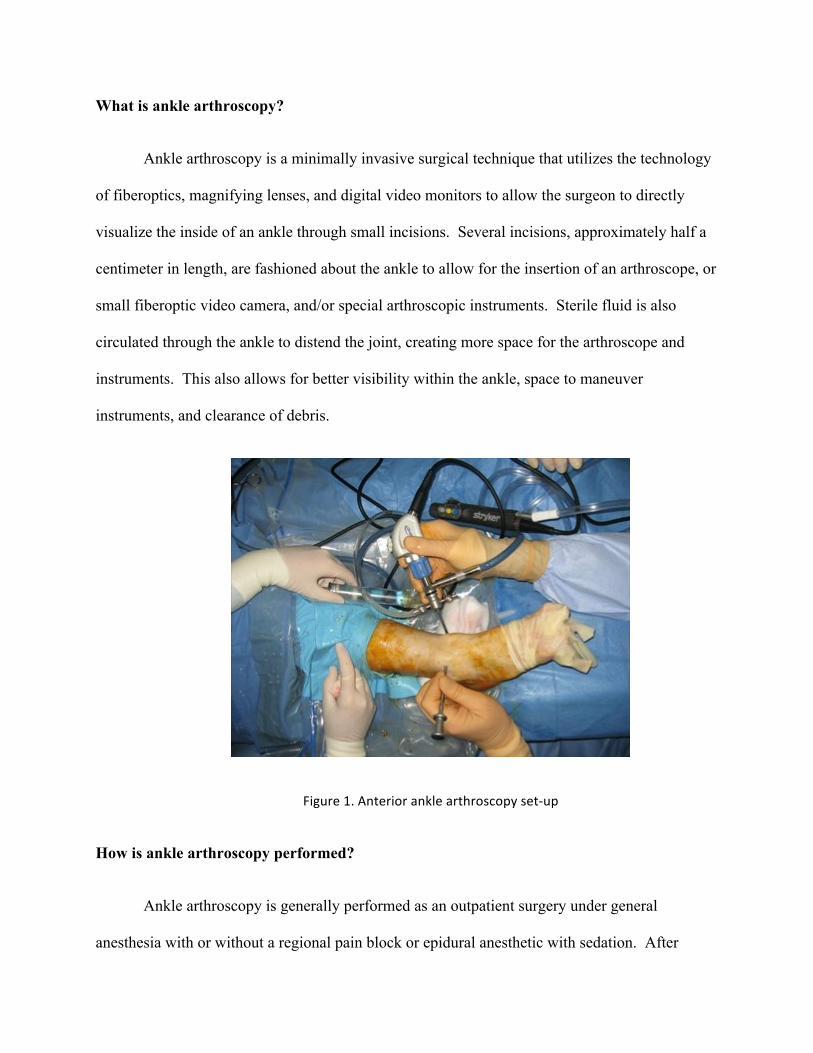
vascular insults, genetic predisposition, degeneration, and metabolic abnormalities.12 Patients
will often present with complaints of persistent and progressive ankle pain and swelling. This
can be associated with mechanical symptoms of catching, clicking, or popping, and decreased
range of motion. The diagnosis is made with the combination of physical exam and diagnostic
imaging including x-rays, MRI, and/or CT scan. The treatment will be based on the size and
location of the OCD, associated symptoms, patient demographics, and activity demands of the
patient. After the diagnosis is made arthroscopically, treatment options include microfracture,
subchondral drilling, abrasion arthroplasty, fragment fixation, and bone grafting procedures.
Thorough discussion with your surgeon is necessary to determine which option is most
appropriate for you.
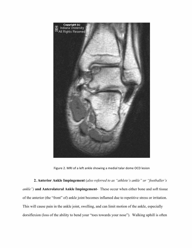
Figure 2. MRI of a left ankle showing a medial talar dome OCD lesion
2. Anterior Ankle Impingement (also referred to as “athlete’s ankle” or “footballer’s
ankle”) and Anterolateral Ankle Impingement- These occur when either bone and soft tissue
of the anterior (the “front” of) ankle joint becomes inflamed due to repetitive stress or irritation.
This will cause pain in the ankle joint, swelling, and can limit motion of the ankle, especially
dorsiflexion (loss of the ability to bend your “toes towards your nose”). Walking uphill is often

painful. This is common in soccer players and any athlete with recurrent ankle sprains. The
diagnosis of anterior ankle impingement can be made by identifying osteophytes, or “bone
spurs,” on standard x-rays of the ankle. Sometimes a MRI is necessary if bone spurs are not
present. MRI can identify redundant and inflamed soft tissue in the anterolateral gutter of the
ankle not seen with standard x-rays. This is considered anterolateral ankle impingement. If
nonoperative measures fail to relieve symptoms of either of these conditions, ankle arthroscopy
can be used to shave away redundant soft tissues and/or bone spurs.
Figure 3. Arthroscopic view of the ankle showing anterolateral impingement
3. Posterior Ankle Impingement- This occurs when the bone and soft tissue of the
hindfoot (the “back” of the ankle) becomes inflamed due to repetitive stress. This will cause pain
in the ankle joint, swelling, and often times limited motion of the ankle, especially plantarflexion
(loss of the ability to “press on the gas”). This overuse syndrome occurs most commonly in
Anterolateral Impingement

ballet dancers, but can also be seen in other athletes. Like anterior ankle impingement, it is
usually associated with bone issues, in the posterior part of the ankle (the “back” of the ankle). It
can also be associated with an accessory bone, which is not found in all patients that is referred
to as an os trigonum. Surgical treatment involves placing arthroscopic incisions in the back of
the ankle to access the painful area. Bone spurs, inflamed soft tissue, and if present, the os
trigonum, can then be removed arthroscopically.
Figure 4. Lateral radiograph showing an os-‐trigonum in the posterior ankle
3. Synovitis- Synovitis is inflammation of the soft tissue lining of the ankle joint
(synovium) that will often manifest as pain, swelling, and loss of motion. This can occur due to
an acute trauma, inflammatory arthritis (i.e. rheumatoid arthritis), overuse, and degenerative joint

disease (osteoarthritis).9 If nonsurgical treatment options fail to provide relief, ankle arthroscopy
can be used to surgically remove inflamed synovium.
4. Loose Bodies- Articular cartilage and/or scar tissue following trauma to the ankle can
become free floating in the joint and form what is referred to as a “loose body”. These can also
occur with in the setting of a condition called synovial chondromatosis, where the lining of the
joint becomes redundant for unexplained reasons. These loose bodies can cause problems such
as clicking, catching, and frank locking that often lead to pain, swelling, and loss of motion.
Occasionally loose bodies can be identified with standard x-rays or a CT scan, but frequently
require an MRI is needed to visualize the culprit. Ankle arthroscopy can be used to find and
remove the loose body.
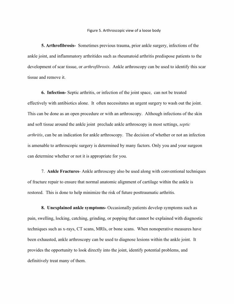
Figure 5. Arthroscopic view of a loose body
5. Arthrofibrosis- Sometimes previous trauma, prior ankle surgery, infections of the
ankle joint, and inflammatory arthritides such as rheumatoid arthritis predispose patients to the
development of scar tissue, or arthrofibrosis. Ankle arthroscopy can be used to identify this scar
tissue and remove it.
6. Infection- Septic arthritis, or infection of the joint space, can not be treated
effectively with antibiotics alone. It often necessitates an urgent surgery to wash out the joint.
This can be done as an open procedure or with an arthroscopy. Although infections of the skin
and soft tissue around the ankle joint preclude ankle arthroscopy in most settings, septic
arthritis, can be an indication for ankle arthroscopy. The decision of whether or not an infection
is amenable to arthroscopic surgery is determined by many factors. Only you and your surgeon
can determine whether or not it is appropriate for you.
7. Ankle Fractures- Ankle arthroscopy also be used along with conventional techniques
of fracture repair to ensure that normal anatomic alignment of cartilage within the ankle is
restored. This is done to help minimize the risk of future posttraumatic arthritis.
8. Unexplained ankle symptoms- Occasionally patients develop symptoms such as
pain, swelling, locking, catching, grinding, or popping that cannot be explained with diagnostic
techniques such as x-rays, CT scans, MRIs, or bone scans. When nonoperative measures have
been exhausted, ankle arthroscopy can be used to diagnose lesions within the ankle joint. It
provides the opportunity to look directly into the joint, identify potential problems, and
definitively treat many of them.
9. Tibiotalar arthritis- Ankle fractures, infection, osteonecrosis, and arthritis may
eventually lead to chronic pain and stiffness that can not be controlled with non-operative
measures. Ankle fusion is a treatment option appropriate for many patients in this situation.
When performed by an experienced surgeon, ankle arthroscopy offers a minimally invasive way
to perform ankle fusion that may yield results that are equal to or better than conventional open
techniques.4,6 This procedure has its limitations. Your surgeon can determine if this procedure
is appropriate option for you.
Recovery: How do I care for my ankle after surgery?
This will ultimately depend on the type of problem and nature of the arthroscopic
procedure used to treat the problem. Patients can expect pain and swelling following surgery
that necessitates elevation of the leg and oral pain medication for at least several days. The type
of procedure performed will determine whether or not your ability to bear weight on the affected
leg will be restricted after surgery. This can range from progressive immediate weight bearing
with crutches, to a period of strict non-weight bearing for one to two months. If ankle
arthroscopy is used as an adjunct to conventional fracture fixation, this period of non-weight
bearing may be longer depending on your body’s ability to heal the fracture. Your dressing will
be left in place until follow-up with your surgeon, and sutures will be removed one to two weeks
after surgery. Active range of motion is generally allowed immediately. After the swelling and
soft tissue reaction subsides, a progressive strengthening routine may be implemented. It will be
up to your surgeon when each of these activities is allowed and whether or not a formal physical
therapy referral is necessary.
Outcomes
Many factors will contribute to the outcome of your ankle arthroscopy procedure. These
include, but are not limited to: your expectations, the severity of your condition, complexity of
the procedure performed, as well as postoperative compliance, rehabilitation and motivation. The
literature shows that an average of greater than 70-90% of patients undergoing ankle arthroscopy
for the most common indications achieve good or excellent results.1,2,10,11,13,14
What are the advantages of ankle arthroscopy?
Ankle arthroscopy makes possible direct visualization of the inside of the ankle without
large cosmetically unsightly scars. It minimizes other problems encountered with large incisions
around the ankle such as pain, bleeding, wound breakdown, and infection. The procedure can be
performed as an outpatient because of its minimally invasive nature. Patients may be able to
begin rehabilitation sooner, rehabilitate more functionally, and return to high level activities such
as sports.3-5,7
Who is not eligible for ankle arthroscopy?
Elective arthroscopy is contraindicated in patients with soft tissue infections of the ankle
such as cellulitis, acute and chronic open wounds, and dermatitis overlying the ankle. Patients
with severe arthritic changes with loss of the joint space are not good candidates for arthroscopic
debridement procedures. Patients with severe peripheral vascular disease, peripheral neuropathy,
reflex sympathetic dystrophy/complex regional pain syndrome, and edema may not be eligible
for ankle arthroscopy. It is important to thoroughly discuss your individual risks, potential
benefits, and the alternatives to ankle arthroscopy with your surgeon.
Complications

Potential complications of ankle arthroscopy include, but are not limited to, injury to
nerves, vessels, tendons, ligaments or cartilage about the ankle, deep and superficial infections,
scarring, reflex sympathetic dystrophy/complex regional pain syndrome, missed diagnoses,
broken instruments, and anesthetic complications.8 It is important to attend follow-up
appointments with your surgeon following surgery as recommended.
The following symptoms should be urgently reported to your surgeon, as they may be an
indication of a complication:
-pain not controlled by pain medication
-constitutional symptoms including nausea, vomiting, fevers, or chills
- wound redness, swelling, warmth or drainage
- new numbness, weakness, or tingling.
FAQ:
When can I safely return to driving?
This will be determined by the type of procedure you undergo and your surgeon’s
evaluation of your progress. When you are able to bear weight without limitation and are no
longer taking narcotic pain medication you will likely be cleared to return to driving. This can
be as soon as several days after surgery or may take one to two months.
When can I expect to return to work/sports?

This will be determined by the type of procedure you undergo and your surgeon’s
evaluation of your progress. If your mobility allows you to safely complete your job duties,
there is the possibility of returning to work several days after surgery. Most patients can expect
to be out of work for at least one to two weeks while they recover. It is possible to return to high
level sports following ankle arthroscopy.3,5 This will depend on your ability to protect yourself
effectively and perform during your particular sporting activity. Athletes could be cleared to
return to play at as early as one to two weeks, but in all likelihood can expect an excess of four to
six weeks.
Additional Resources
1. Amendola, A.; Petrik, J.; and Webster-‐Bogaert, S.: Ankle arthroscopy: outcome in 79 consecutive patients. Arthroscopy, 12(5): 565-‐73, 1996.
2. Baums, M. H.; Kahl, E.; Schultz, W.; and Klinger, H. M.: Clinical outcome of the arthroscopic management of sports-‐related "anterior ankle pain": a prospective study. Knee Surg Sports Traumatol Arthrosc, 14(5): 482-‐6, 2006.
3. Calder, J. D.; Sexton, S. A.; and Pearce, C. J.: Return to training and playing after posterior ankle arthroscopy for posterior impingement in elite professional soccer. Am J Sports Med, 38(1): 120-‐4.
4. Cutsuries, A. M.; Saltrick, K. R.; Wagner, J.; and Catanzariti, A. R.: Arthroscopic arthroplasty of the ankle joint. Clin Podiatr Med Surg, 11(3): 449-‐67, 1994.
5. DeBerardino, T. M.; Arciero, R. A.; and Taylor, D. C.: Arthroscopic treatment of soft-‐tissue impingement of the ankle in athletes. Arthroscopy, 13(4): 492-‐8, 1997.
6. Myerson, M. S., and Quill, G.: Ankle arthrodesis. A comparison of an arthroscopic and an open method of treatment. Clin Orthop Relat Res, (268): 84-‐95, 1991.
7. Scranton, P. E., Jr., and McDermott, J. E.: Anterior tibiotalar spurs: a comparison of open versus arthroscopic debridement. Foot Ankle, 13(3): 125-‐9, 1992.
8. Stetson, W. B., and Ferkel, R. D.: Ankle Arthroscopy: I. Technique and Complications. J Am Acad Orthop Surg, 4(1): 17-‐23, 1996.
9. Stetson, W. B., and Ferkel, R. D.: Ankle Arthroscopy: II. Indications and Results. J Am Acad Orthop Surg, 4(1): 24-‐34, 1996.
10. van Dijk, C. N.: Hindfoot endoscopy. Foot Ankle Clin, 11(2): 391-‐414, vii, 2006. 11. van Dijk, C. N.; Tol, J. L.; and Verheyen, C. C.: A prospective study of prognostic factors
concerning the outcome of arthroscopic surgery for anterior ankle impingement. Am J Sports Med, 25(6): 737-‐45, 1997.
12. van Dijk, C. N., and van Bergen, C. J.: Advancements in ankle arthroscopy. J Am Acad Orthop Surg, 16(11): 635-‐46, 2008.
13. Verhagen, R. A.; Struijs, P. A.; Bossuyt, P. M.; and van Dijk, C. N.: Systematic review of treatment strategies for osteochondral defects of the talar dome. Foot Ankle Clin, 8(2): 233-‐42, viii-‐ix, 2003.
14. Willits, K.; Sonneveld, H.; Amendola, A.; Giffin, J. R.; Griffin, S.; and Fowler, P. J.: Outcome of posterior ankle arthroscopy for hindfoot impingement. Arthroscopy, 24(2): 196-‐202, 2008.