DEPARTMENT OF BIOTECHNOLOGY FACULTY OFENGINEERING …
20
DEPARTMENT OF BIOTECHNOLOGY FACULTY OFENGINEERING & TECHNOLOGY
Transcript of DEPARTMENT OF BIOTECHNOLOGY FACULTY OFENGINEERING …

DEPARTMENT OF BIOTECHNOLOGYFACULTY OFENGINEERING & TECHNOLOGY

Tumor Suppressor Gene
Content Outline1. What is tumor suppressor gene?
2. Tumor Suppressor gene from humans
3. Retinoblastoma: Function and Mechanism
4. P53 gene
5. Cell cycle regulation
6. Test your understanding
7. References & Further reading
Content Outline1. What is tumor suppressor gene?
2. Tumor Suppressor gene from humans
3. Retinoblastoma: Function and Mechanism
4. P53 gene
5. Cell cycle regulation
6. Test your understanding
7. References & Further reading

•A gene that codes for regulator proteins that prevent the cell from undergoing uncontrolled
division
•Oncogenes drive abnormal cell proliferation as a consequence of genetic alterations that either
increase gene expression or lead to uncontrolled activity of the oncogene-encoded proteins.
•Tumor suppressor genes represent the opposite side of cell growth control, normally acting to
inhibit cell proliferation and tumor development.
•Tumor-suppressor genes normally restrain growth, so mutations that inactivate them allow
inappropriate cell division. This gene encodes cellular proteins that normally function to prevent
tumor development, and retinoblastoma typifies the behavior of a classical tumor suppressor
gene.
•Normally, Fusion of tumor cells with normal cells yields hybrids that contain chromosomes from
both parents. Such hybrids are usually non-tumorigenic. The first tumor suppressor gene was
identified by studies of retinoblastoma, a rare childhood eye tumor.
What is tumor suppressor gene?
•A gene that codes for regulator proteins that prevent the cell from undergoing uncontrolled
division
•Oncogenes drive abnormal cell proliferation as a consequence of genetic alterations that either
increase gene expression or lead to uncontrolled activity of the oncogene-encoded proteins.
•Tumor suppressor genes represent the opposite side of cell growth control, normally acting to
inhibit cell proliferation and tumor development.
•Tumor-suppressor genes normally restrain growth, so mutations that inactivate them allow
inappropriate cell division. This gene encodes cellular proteins that normally function to prevent
tumor development, and retinoblastoma typifies the behavior of a classical tumor suppressor
gene.
•Normally, Fusion of tumor cells with normal cells yields hybrids that contain chromosomes from
both parents. Such hybrids are usually non-tumorigenic. The first tumor suppressor gene was
identified by studies of retinoblastoma, a rare childhood eye tumor.
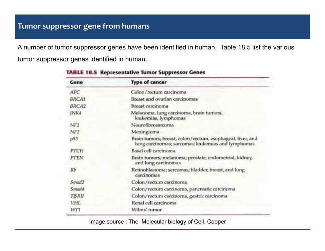
Tumor suppressor gene from humans
A number of tumor suppressor genes have been identified in human. Table 18.5 list the various
tumor suppressor genes identified in human.
Image source : The Molecular biology of Cell, Cooper
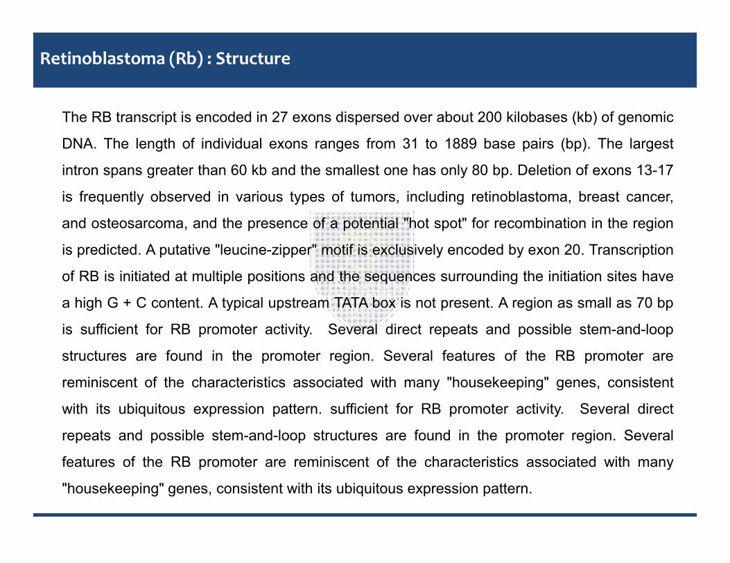
Retinoblastoma (Rb) : Structure
The RB transcript is encoded in 27 exons dispersed over about 200 kilobases (kb) of genomic
DNA. The length of individual exons ranges from 31 to 1889 base pairs (bp). The largest
intron spans greater than 60 kb and the smallest one has only 80 bp. Deletion of exons 13-17
is frequently observed in various types of tumors, including retinoblastoma, breast cancer,
and osteosarcoma, and the presence of a potential "hot spot" for recombination in the region
is predicted. A putative "leucine-zipper" motif is exclusively encoded by exon 20. Transcription
of RB is initiated at multiple positions and the sequences surrounding the initiation sites have
a high G + C content. A typical upstream TATA box is not present. A region as small as 70 bp
is sufficient for RB promoter activity. Several direct repeats and possible stem-and-loop
structures are found in the promoter region. Several features of the RB promoter are
reminiscent of the characteristics associated with many "housekeeping" genes, consistent
with its ubiquitous expression pattern. sufficient for RB promoter activity. Several direct
repeats and possible stem-and-loop structures are found in the promoter region. Several
features of the RB promoter are reminiscent of the characteristics associated with many
"housekeeping" genes, consistent with its ubiquitous expression pattern.

Size and structure of Rb genes

•The Rb protein is a tumor suppressor, which plays a pivotal role in the negative control of the cell cycle and in
tumor progression. It has been shown that Rb protein (pRb) is responsible for a major G1 checkpoint, blocking
S-phase entry and cell growth.
•The retinoblastoma family includes three members, Rb/p105, p107 and Rb2/p130, collectively referred to as
'pocket proteins'. The pRb protein represses gene transcription, required for transition from G1 to S phase, by
directly binding to the transactivation domain of E2F and by binding to the promoter of these genes as a
complex with E2F.
•pRb represses transcription also by remodeling chromatin structure through interaction with proteins such as
hBRM, BRG1, HDAC1 and SUV39H1, which are involved in nucleosome remodeling, histone
acetylation/deacetylation and methylation, respectively. Loss of pRb functions may induce cell cycle
deregulation and so lead to a malignant phenotype.
•Visible deletions of chromosome 13q14 were found in some retinoblastomas, suggesting that loss (rather than
activation) of the Rb gene led to tumor development.
Function and mechanism of Rb gene
•The Rb protein is a tumor suppressor, which plays a pivotal role in the negative control of the cell cycle and in
tumor progression. It has been shown that Rb protein (pRb) is responsible for a major G1 checkpoint, blocking
S-phase entry and cell growth.
•The retinoblastoma family includes three members, Rb/p105, p107 and Rb2/p130, collectively referred to as
'pocket proteins'. The pRb protein represses gene transcription, required for transition from G1 to S phase, by
directly binding to the transactivation domain of E2F and by binding to the promoter of these genes as a
complex with E2F.
•pRb represses transcription also by remodeling chromatin structure through interaction with proteins such as
hBRM, BRG1, HDAC1 and SUV39H1, which are involved in nucleosome remodeling, histone
acetylation/deacetylation and methylation, respectively. Loss of pRb functions may induce cell cycle
deregulation and so lead to a malignant phenotype.
•Visible deletions of chromosome 13q14 were found in some retinoblastomas, suggesting that loss (rather than
activation) of the Rb gene led to tumor development.

Figure: Rb deletions in retinoblastoma: Many retinoblastomas have deletions of thechromosomal locus (13q14) that contains the Rb gene.
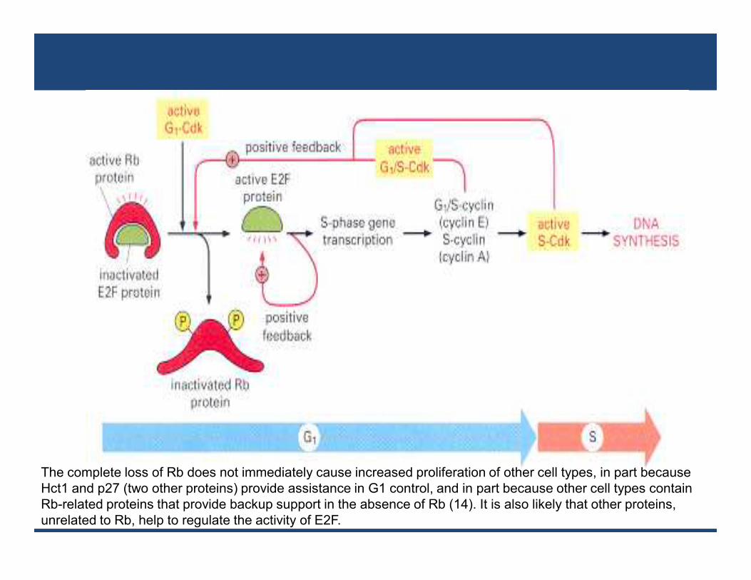
The complete loss of Rb does not immediately cause increased proliferation of other cell types, in part becauseHct1 and p27 (two other proteins) provide assistance in G1 control, and in part because other cell types containRb-related proteins that provide backup support in the absence of Rb (14). It is also likely that other proteins,unrelated to Rb, help to regulate the activity of E2F.
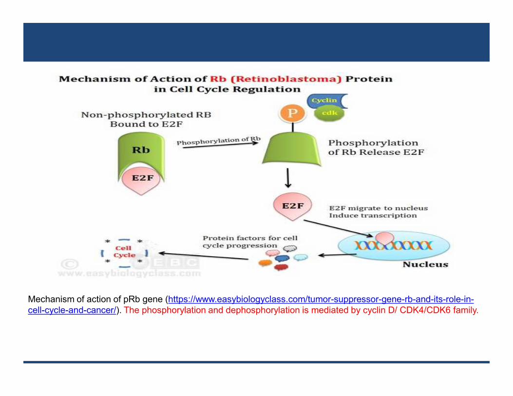
Mechanism of action of pRb gene (https://www.easybiologyclass.com/tumor-suppressor-gene-rb-and-its-role-in-cell-cycle-and-cancer/). The phosphorylation and dephosphorylation is mediated by cyclin D/ CDK4/CDK6 family.

P53 Tumor suppressor gene
• p53, also known as TP53 or tumor protein (EC :2.7.1.37) is a gene that codes for a protein
that regulates the cell cycle and hence functions as a tumor suppression. It is very important for
cells in multicellular organisms to suppress cancer. P53 has been described as "the guardian of
the genome", referring to its role in conserving stability by preventing genome mutation. The
name is due to its molecular mass: it is in the 53 kilodalton fraction of cell proteins. The human
p53 gene is located on the seventeenth chromosome (17p13.1).
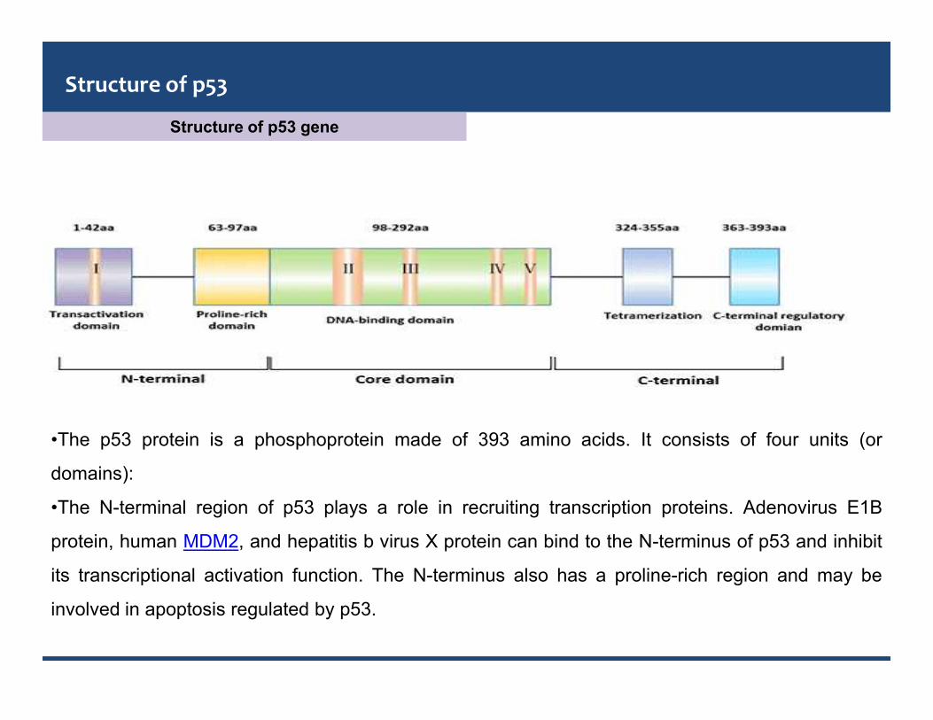
Structure of p53 gene
Structure of p53
•The p53 protein is a phosphoprotein made of 393 amino acids. It consists of four units (or
domains):
•The N-terminal region of p53 plays a role in recruiting transcription proteins. Adenovirus E1B
protein, human MDM2, and hepatitis b virus X protein can bind to the N-terminus of p53 and inhibit
its transcriptional activation function. The N-terminus also has a proline-rich region and may be
involved in apoptosis regulated by p53.
•The intermediate region of p53 protein (102 ~ 292aa) contains four conserved regions, where
80% ~ 90% of the mutations caused by tumor cells occur.
•The C-terminus of p53 (300-393aa) contains a variable junction region that connects the
central core region to the C-terminal region; there is also a tetramerization region. There are
also three nuclear localization signals (NLS) at the C-terminus of p53: NLS1 (316-325aa),
NLS2 (369-375aa), and NLS3 (379-384aa). The C-terminus is a flexible unfolded region that
binds to DNA non-specifically and acts as a repressor in the regulation of other genes.
•The spatial structure of p53 protein determines its biological function
•Wild-type p53 is a labile protein, comprising folded and unstructured regions which function in
a synergistic manner.p53 protein has been voted molecule of the year
Function and mechanism of p53
It plays an important role in cell cycle control and apoptosis. Defective p53 could allow
abnormal cells to proliferate, resulting in cancer. As many as 50% of all human tumors contain
p53 mutants. In normal cells, the p53 protein level is low. DNA damage and other stress
signals may trigger the increase of p53 proteins, which have three major functions: growth
arrest, DNA repair and apoptosis (cell death). The growth arrest stops the progression of cell
cycle, preventing replication of damaged DNA. During the growth arrest, p53 may activate the
transcription of proteins involved in DNA repair. Apoptosis is the "last resort" to avoid
proliferation of cells containing abnormal DNA.
Role of p53
p53 and Cell Cycle Regulation
•In addition to being in the form of p53 protein, p53 is present in cells as a potential untranscribed
form. The untranscribed form of p53 is only activated to produce biological functions. p53 protein
can be activated by DNA damage. The upstream factor of p53 recognizes DNA-damaged proteins.
•In addition, p53 can also bind directly to the ends and damage sites of DNA, thereby being
phosphorylated or receiving other signals to generate activity. In addition to DNA damage, tissue
hypoxia also increases and activates p53 concentration.
•p53-mediated downstream processes mainly include:
•Make the cells stop at the cell cycle test point
•Make the cells go to apoptosis
•p53 gene has the function of blocking cell cycle. The expression or ectopic expression of p53 after
DNA damage keeps the cell cycle at the G1/S test point.

https://opentextbc.ca/biology/chapter/6-3-cancer-and-the-cell-cycle/
(a) The role of p53 is to monitor DNA. If damage is detected, p53 triggers repair mechanisms. If repairs areunsuccessful, p53 signals apoptosis. (b) A cell with an abnormal p53 protein cannot repair damaged DNA andcannot signal apoptosis. Cells with abnormal p53 can become cancerous.

p53 and DNA Damage Repair
The p53-regulated DNA damage repair process mainly includes Nucleotide Excision repair (NER),
Base excision repair (BER), Nonhomologous end-joining (NHEJ) and transcriptional activation
pathways and Homologous recombination by both transactivation-dependent and-independent
pathways
p53 and Apoptosis•p53 gene has the effect of promoting cell apoptosis. p53 protein can induce the apoptosis of
malignant tumor cells, thus damaging the function of tumor cells and eventually causing tumor
atrophy or disappearance. It can regulate apoptosis through proteins such as Bax/Bcl-2, Fas/Apo1,
and induce apoptosis through death signal receptor proteins such as TNF receptor and Fas protein.
p53 can also directly stimulate mitochondria to release highly toxic oxygen free radicals to induce
apoptosis .
•The role of p53 in promoting apoptosis is closely related to its tumor suppressor function. Apoptotic
mechanisms regulated by p53 include transcriptional activation and non-transcriptional activation
pathways.

Test your understanding
________ are changes to the nucleotides in a segment of DNA that codes for a protein.a. Proto-oncogenesb. Tumor suppressor genesc. Gene mutationsd. Negative regulatorsA gene that codes for a positive cell cycle regulator is called a(n) ________.a. kinase inhibitorb. tumor suppressor genec. proto-oncogened. OncogeneWhich of the following could be coded by a tumor-supressor gene?
a) A protein that helps prevent progression through cell cycleb) A protein that helps prevent apoptosisc) A protein that codes for a DNA repair enzymed) A protein that forms part of a growth factor signaling pathway
Which property of p53 enables it to prevent the development of cancer?a) It is a transcription factor that causes protein production which stimulates the cell cycleb) It prevents replication of cells with damaged DNAc) It prevents cells from triggering apoptosisd) It stimulates synthesis of DNA repair enzymes that replace telomere sequence lost during cell division
Which of the following about Rb tumor suppressor protein is correct?a) It binds E2F transcription factor and prevents cell from entering S phase until a mitogenic signal is receivedb) It is activated when phosphorylated by Cdkc) It is a transcription factord) When a mitogenic signal is received, it binds the transcription factor E2F and thus stimulates the cell toenter S phase
References1. http://dpuadweb.depauw.edu/cfornari_web/DISGEN/retinoblastoma_website/public_html/protein.htm
2. https://www.ncbi.nlm.nih.gov/books/NBK9894/
3. https://www.slideshare.net/gangadharchatterjee/molecular-basis-of-cancer-part-1
4. The molecular biology of cell, 2nd edition Geoffrey cooper
Further reading1. King R.J.B., Cancer Biology, Addision Wesley Longmann Ltd, U.K., 1996.
2. Ruddon.R.W., Cancer Biology, Oxford University Press, Oxford, 1995.
3. Maly B.W.J., Virology a practical approach, IRL press, Oxford, 1987.
4. Dunmock. N.J and Primrose S.B., Introduction to modern Virology, Blackwell Scientific Publications, Oxford,
1988.
References & Further reading
References1. http://dpuadweb.depauw.edu/cfornari_web/DISGEN/retinoblastoma_website/public_html/protein.htm
2. https://www.ncbi.nlm.nih.gov/books/NBK9894/
3. https://www.slideshare.net/gangadharchatterjee/molecular-basis-of-cancer-part-1
4. The molecular biology of cell, 2nd edition Geoffrey cooper
Further reading1. King R.J.B., Cancer Biology, Addision Wesley Longmann Ltd, U.K., 1996.
2. Ruddon.R.W., Cancer Biology, Oxford University Press, Oxford, 1995.
3. Maly B.W.J., Virology a practical approach, IRL press, Oxford, 1987.
4. Dunmock. N.J and Primrose S.B., Introduction to modern Virology, Blackwell Scientific Publications, Oxford,
1988.