Dental Pulp - Seminar2 / orthodontic courses by Indian dental academy
98
DENTAL PULP Seminar by Dr. SATHYA KUMAR.S Postgraduate Student
-
Upload
indian-dental-academy -
Category
Documents
-
view
231 -
download
3
Transcript of Dental Pulp - Seminar2 / orthodontic courses by Indian dental academy
DENTAL PULP
Seminar by
Dr. SATHYA KUMAR.S
Postgraduate Student
DEPARTMENT OF CONSERVATIVE DENTISTRY & ENDODONTICS
SRI RAMACHANDRA DENTAL COLLEGE AND HOSPITALS
CHENNAI
CONTENTS
IntroductionThe Pulp as a Connective TissueThe Cells of the PulpFibersGround SubstanceSystemic Factors that affect the PulpThe Circulation of the PulpClinical CorrelationsAgingPeriodontal DiseaseChanges due to InflammationDental Caries Defense against CariesThe dynamics of the Development of Pulpitis from Dental CariesIndirect Pulp CappingRelationship of Depth of Preparation to Reparative Dentin FormationOther Physical TraumaPolishing of RestorationsDentin Sterilizing AgentsPermanent Restorative Materials Pulp Capping and PulpotomyDrugs in Pulp Capping and PulpotomyLateral & Accessory Canal Anatomic ConsiderationsEffects of Periodontal Treatment & Local MedicationCorrelation of Periodontal Involvement with PainRetrogressive and Age Changes of the Dental PulpDecrease in Cellular Components Local Factors INTRODECTION
The living pulp creates and shapes its own local in the center of the
tooth. The pulp under normal conditions tends to form dentin eventually,
faciolingullay and mesiodistally. The pulp therefore tends to lie in the center
of the tooth and shapes itself to a miniaturization. The pulp is bunch of
tissues made of nerves, blood vessels, connective tissues
THE PULP AS A CONNECTIVE TISSUE
The dental pulp is composed of cells, ground substance and fibers.
The cells manufacture a fundamental matrix, which then acts as site and
precursor for the fiber complex. The fiber complex is composed of collagen
and reticulum.
THE CELLS OF THE PULP
Fibroblasts
The basic cells of the pulp are fibroblasts, which are similar to
connective tissue fibroblasts found elsewhere in the body. In the young pulp
there is a great preponderance of fibroblasts as compared with collagen
fibers.
Fibroblasts are active in collagen synthesis. The fibroblasts reveal
well-developed organelles with extensive, dense, rough-surfaced
endoplasmic reticulum in the form of dilated cisternae.
Rows and clusters of ribosomes are present. The Golgi apparatus has
an extensively developed stack and a large number of vesicies and vacuoles.
The mitochondria are large with straight cristae running transversely across
the matrix. A cilium is frequently found near the nucleus and an additional
centriole may be located perpendicular to the long axis of the cilium.
Collagen fibers are present on the cell body and its processes.
Dental pulp synthesize at least six glycoproteins, the major one being
fibronectin.
Pulp fibroblasts also synthesize and secrete chondroitin sulfate a
major sulfated In addition, they manufacture heparin and dermatan sulfates.
The fibroblasts exhibit faint metachromasia, and phosphatase and
adenosine triphosphate activity. (Lipid) particles are present in their
cytoplasm. As the individual gets older, glycogen and other periodic acid-
Schiff Positive materials increase, In older tissues, the cellular elements
begin to decrease. This fiber increase and cell reduction has clinical
implications, in that a more fibrous pulp is less able to defend itself against
irritants than is a young, highly cellular pulp.
Both fibroblasts and odontoblasts are derived from the mesenchyme,
but odontoblasts are more highly differentiated cells than are fibro-blasts.
ODONTOBLASTS
The odontoblast is a highly differentiated cell in the pulp. The main
function of the odontoblasts is the production of dentin. Morphologic
variations occur in the odontoblasts, ranging from the tall columnar cells in
the crown of the tooth to a low columnar type in the middle of the root. In
the root portion of the tooth, the odontoblasts are shorter and are more or
less cuboidal. Toward the apex, they are flattened and look more like
fibroblasts.
In the coronal portion of the pulp, odontoblasts are more columnar,
they elaborate regular dentin with regular dentinal tubules.
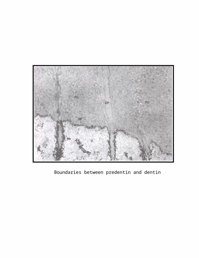
Boundaries between predentin and dentin
ELECTRON MICROSCOPE FINDINGS
In Scanning electron micrographs, odontoblasts appear as large,
closely aligned, multilayered, sweet potato-shaped cells. Nucleus. The
nucleus of a typical odontoblast is ellipsoidal and contains chromatin and
nucleoli. Two-thin membranes surround the nucleus, the inner membrane
appears to be continuous, but the outer one is interrupted at points by
openings.
NUCLEOLUS
In electron microscope examinations of human teeth, differentiated
odontoblasts contained one to four nucleoli. The nucleoli from fully
developed teeth were ring-shaped. In less developed teeth, compact
nucleoli, representative of types active in RNA production,
CYTOPLASM
Most of the cytoplasm is occupied by an extensive rough-surfaced
endoplasmic reticulum and by numerous transitional vesicles of the rER.
Fine fibrillar material is present within the cisternae of the rER. The
central portion of the odontoblast is occupied by a large Golgi apparatus.
Vesicles are concentrated near each immature face of the Golgi apparatus.
Membrane-bound granules of varying sizes and shapes, possibly lysosomes
containing a highly dense material are found in the cytoplasm. Secretory
granules containing mineral deposits are present in the area of the Golgi
complex. Mitochondria are evenly distributed throughout most of the cell
body, Centrioles are present throughout the region of the Golgi complex.
Large numbers of microtubules are also present.
The odontoblasts are lined up in palisade formation along the
predentin border. Generally, the odontoblastic layer is about six to eight
cells deep. The organelles, which are situated in the cell body, extend as far
as the level of the modified terminal bar apparatus,
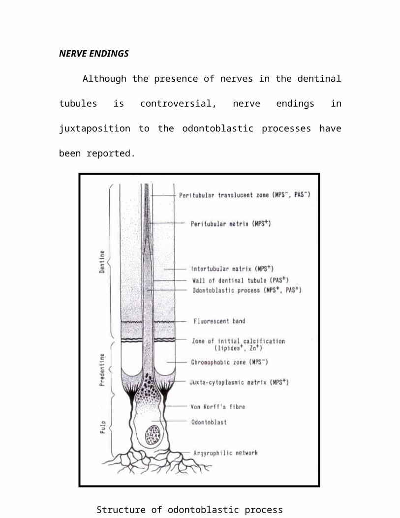
THE ODONTOBLASTIC PROCESS
Each odontoblastic process traverses the predentin, and then occupies
a canaliculus in the dentin. The odontoblastic processes (called dentinal
fibers, or Tomes fibers: are cytoplasmic tubular projections.
The odontoblastic processes are usually devoid of major cytoplasmic
organelles. Dense (secretory) granules and lysosome like bodies are also
present. Numerous filaments are oriented parallel to the cell membrane.
These fine filaments are the most characteristic feature of the odontoblastic
process and its branches. Groups of microvesicles similar to those seen in
the Golgi apparatus, are situated near the plasma membrane.
INTERELLULAR JUNCTIONS.
Small regions of the plasma membranes between cells are visible only
by electron microscopy. There are three types of intercellular junctions: (1)
impermeable, (2) adhering, and (3) communicating.
IMPERMEABLE JUNCTIONS also known as tight junctions, help
the cell maintain a distinct internal environment.
ADHERING JUNCTIONS are maintained by desmosomes, which
are the intercellular bridges seen in light microscopy. There are three types
of desmosomes; belt, spot, and hemidesmosomes.
COMMUNICATING JUNCTIONS, also known as gap junctions, are
structures that mediate direct transfer of chemical messages between cells.
They enable cells to exchange nutrients and signal molecules for
coordination of function.
ODONTOBLASTIC JUNCTIONAL COMPLEXES.
Surface epithelial cells possess terminal bars at their apical
extremities. The terminal bars are seen to consist of several components,
designated as junctional complexes.
In the border region between odontoblastic processes and cell bodies,
neighboring odontoblasts are in close contact with each other and with other
pulp cells. Such junctions permit the passage of small substances between
cells. Thus, the junctional complex present at the apical end of the
odontoblast may not be entirely similar to that seen at the apical ends of
epithelial cells.
Tight adhesion occurs between odontoblasts, and they are not easily
separated.
NERVE ENDINGS
Although the presence of nerves in the dentinal tubules is
controversial, nerve endings in juxtaposition to the odontoblastic processes
have been reported.
Structure of odontoblastic process
ODONTOBLASTIC COMMUNICATIONS
The odontoblastic nuclei always remain at the inner border of the
dentin, and unlike the osteoblasts they do not become buried unless they are
pathologically involved. The odontoblastic processes are in contact with
adjacent processes through an extensive lateral branch. The odontoblasts
may be regarded as part of a mesenchymal syncytium.
If an odontoblast is injured, other odontoblasts are affected. The
cytoplasm of the odontoblasts contains a basophilic stippling attributable to
the presence of RNA. Minute sudanophilic (probably lipid; granules and
vacuoles are scattered throughout the cytoplasm and the dentinal fibers.
These findings these cells are capable of glycolysis and fatty acid
metabolism plus a functional citric acid cycle and pentose shunt. Esterases
of various types have been detected in the odontoblasts and subodontoblastic
layers of the pulp and in pulp homogenates, indicating intracellular anabolic
or digestive functions.
The function of odontoblasts is the secretion of ground substance and
collagen. Under the layer of odontoblasts in the coronal portion of the tooth,
there is a cell-free zone that contains nerve elements.
DEFENSE AND OTHER CELLS
Some of the cells in the pulp are defense cells. Histiocytes, or resting
wandering cells, are usually found near the blood vessels.
Undifferentiated mesenchymal cells are capable of becoming
macrophages during injury. They also may become fibroblasts,
odontoblasts, or osteoclasts. Before injury, they appear elongated; following
injury, they differentiate into macrophages and, as such can ingest foreign
material. Fat cells are not ordinarily found in the pulp.
Lymphocytes are not found usually in the un-inflamed pulp, Plasma
cells and eosinophils are found there following injury.
Pericytes are found in the walls of the precapillaries and metarterioles
the function of pericytes is to manufacture the connective tissue of the
precapillary region.
FIBERS
The fibers of the pulp are the same as those of other connective
tissues. Reticular fibers are found around blood vessels and around the
odontoblasts in the pulp.
Collagen fibers are by the pulpal fibroblasts. There are two prominent
patterns of collagen deposition in dental pulp: diffuse, in which the
collagenous fibers lack of definite orientation, and bundle type; in which
large, coarse bundles run parallel to nerves or independently. In young
pulps, few collagen fibers are found. As the pulp gets older more and more
collagen is elaborated.
Pulp collagen fiber turnover is fairly high compared with that of other
dental tissues.Regardless of age, the apical portion of the pulp is usually
more fibrous than the coronal portion.
GROUND SUBSTANCE
The ground substance of the pulp is part of the system of ground
substances in the body. It influences the spread of infection, metabolic
changes in cells, stability of crystalloids, and the effects of hormones,
vitamins, and other metabolic substances.
The ground substance of the pulp is similar to that of connective tissue
elsewhere in the body; it is composed of protein associated with
glycoproteins and acid mucopolysaccharides 20% of the pulpal carbohydrate
is in glycosaminoglycans.
The following properties have been attributed to glycosaminoglycans
water retention, ion binding and electrolyte distribution during
mineralization (Bowness, 1968), and influence on collagen fibrillogenesis
The pulps of growing teeth stain metachromatically, The metabolism
of the cells and the fibers of the pulp is mediated through the ground
substance. The ground substance, a viscid fluid, through which metabolites
pass from the circulation to the cells and through which breakdown products
from the cells come back to the venous circulation. There is no way for
nutrients to get fro the blood supply to the cells other than through the
ground substance.
In a similar manner, substances excreted by the cell must go through
the ground substance to get into the efferent circulation.
Depolymerization by enzymes elaborated by microorganisms found in
pulp inflammation may change the ground substance of the pulp.
Mucopolysaccharidase activity has been detected , by histochemical
techniques, within the pulps of resorbing deciduous teeth, (1980) these
enzymes are capable of degrading the ground substance of the pulp and
dentin by disrupting the glycosaminoglycan-collagen linkage. Thus, the
ground substance plays a significant role in health and disease of the pulp
and dentin.
SYSTEMIC FACTORS THAT AFFECT THE PULP
Certain systemic conditions affect the cells, fibers, and ground
substance of the connective tissue of the pulp.
VITAMIN DEFICIENCY
Deficiency of certain vitamins, notably vitamin C affects fibroblasts
generally and specifically affects the fibroblasts in the dental pulp.
HORMONES AND HORMONAL IMBALANCE
STEROIDS. Systemic administration of high doses of corticosteroids
to rabbit’s collagen synthesis in the dental pulps.
DIABETES. Diabetics tend to age more quickly because of
obliterative endarteritis. There is impairment of nutrition and metabolic
processes. Tissue repair is interfered with in diabetics.
Thyroid Deficiency.
There was rapid deposition of dentin, which narrowed the lumen of
the pulp, and all tissues showed a decreased amount of cellular elements.
PROTEIN DEFICIENCY
Dietary proteins are essential for the formation and maintenance of
tissue structure; they constitute the main sources of amino acids and
nitrogen, Dietary proteins may also serve as energy sources.
HEREDITARY DISEASES
A number of diseases have been reported to affect the dental pulp.
These include diseases of the blood.
THE CIRCULATION OF THE PULP
The circulation of the blood is the transportation system by means of
which the various cells of the body are supplied with nutrients and the waste
products from the cells are removed for elimination from the body.
The development of the vascular system structurally and functionally
is related directly to the needs of the tissues.
MICROCIRCULATION
The primary function of microcirculation is to transport nutrients to,
and to remove metabolic waste products from, the tissues.. The major
microcirculatory vessels are the arterioles. the capillaries, and the venules.
The arterioles are resistance vessels measuring approximately 50um in
diameter and have several layers of smooth muscle, which provide for
control of vascular geometry.
The arterioles, the capillaries, and the venules are, to some extent,
able to respond to the variations in the requirements of the functioning
tissues.
CAPILLARIES
Capillaries have an average diameter of 8 um to 10um. They are the
exchange vessels responsible for the transport of materials between blood
and tissues.
Capillaries are surrounded by a loose group of reticular and collagenous
fibers.
The main morphologic characteristic of the capillaries is the general
absence of smooth muscle cells.
The luminal surface of the endothelial cell lined by a thin layer of
glycoprotein, and the tissue side is covered by the basement membrane.
TYPES OF CAPILLARIES
Depending on its morphologic features, the capillary endothelium can
be classified into three basic types: continuous, fenestrated, and
discontinuous.
Continuous endothelium is devoid of fenestrations. Fenestrated
endothelium has regional thinning (fenestrations) of the capillary walls.
Fenestrated capillaries exist in the renal glomerulus, in the intestinal mucosa,
in the sulcular gingiva, and in the pulp among odontoblasts near the
predentin. The basement membrane is discontinuous. Discontinuous
endothelium is found in the spleen, liver, and bone marrow. In the venous
system, capillaries coalesce into postcapillary venules.
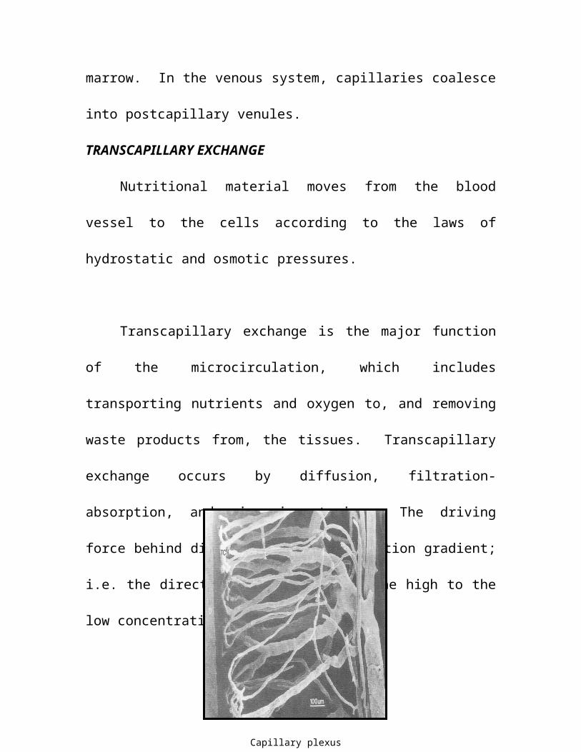
TRANSCAPILLARY EXCHANGE
Nutritional material moves from the blood vessel to the cells
according to the laws of hydrostatic and osmotic pressures.
Transcapillary exchange is the major function of the microcirculation,
which includes transporting nutrients and oxygen to, and removing waste
products from, the tissues. Transcapillary exchange occurs by diffusion,
filtration-absorption, and micropinocytosis. The driving force behind
diffusion is a concentration gradient; i.e. the direction of flux is from the
high to the low concentration.
Capillary plexus
Water-soluble substances do not diffuse readily through the
membrane; Normally, the higher pressure in the arteriolar capillaries favors
filtration, whereas the lower pressure in the venular capillaries causes fluid
absorption. The number of capillaries in a given area depends on the number
of cells in that area.
CONTROL OF BLOOD FLOW
The blood supply to any given area is controlled by nerve impulses
and humoral agents. A hormonal mechanism also is involved in the control
of blood flow. Epinephrine, which is liberated from the adrenal medulla,
causes vasoconstriction. thereby limiting the flow of blood.
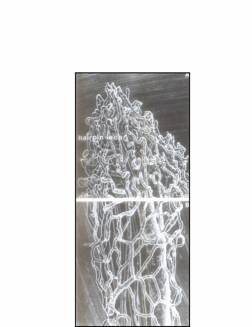
STUDIES OF PULPAL VESSELS
Pulpal and periodontal blood vessels have been studied by a variety of
techniques including cinematography and optical, electron, and x-ray
microscopy. The casts showed that the main feeding arterioles entered the
root canal through the apical foramina and traversed the central portions to
reach the coronal portion of the pulp Some arterioles looped in a U-turn
configuration.
Pulp chamber arterioles could be separated into two groups; one
advanced coronally toward the pulp horn, branching and forming a dense
terminal capillary network toward the dentin. The other ran between the
floor and the roof of the pulp chamber, also branching into a dense terminal
capillary network. The terminal capillaries drained into the venules beneath
the dentin, and merged to form the primary venules. Arteriovenous and
veno-venous anastomoses and U-turn loops appeared to be unique features
of the pulpal vessels.
ULTRASTRUCTURE OF THE PULP CAPILLARY
They found that the cytoplasm of the endothelial cells contained rough
endoplasmic reticulum (rER), a small Golgi complex, occasional
mitochondria, and filaments.
REGULATION OF PULPAL HEMODYNAMICS
Chemical Regulation
Nerves help to regulate the blood supply to the dental pulp.
Sympathetic nerve fibers liberate norepinephrine, which constricts the
vessels.
The catecholamines, called adrenoreceptors. There are two types of
adrenoreceptors, alpha (a) and beta (B), The blood vessels of the pulp
contain both A and B adrenoreceptors. The A receptors are responsible for
contraction of the vascular musculature and produce vasoconstriction.
Stimulation of B receptors causes a relaxation of the vascular musculature.
Circulating catecholamines such as adrenaline or noradrenaline exert less of
a vasoconstrictor effect than local sympathetic nerve activation.
VASOCONSTRICTION
Intra-arterial administration of norepinephrine decreases pulpal blood
flow. The flow reduction was blocked by the A-adrenergic antagonist
phenoxybenzamine (PBZ), indicating the presence of an A-adrenergic
system that was responsible for the decrease in pulpal blood flow.
Sympathetic adrenergic vasoconstrictor system causes variations in systemic
hemodynamics, which, in turn, influence pulpal hemdynamics. In addition
to the sympathetic adrenergic system, other chemical mediators cause
vasoconstriction.
VASODILATION
Pulpal vessels are apparently equipped with B adrenergic receptors,
Activation of B receptors by intra-arterial injection of isoproterenol caused
a paradoxical reduction of pulpal blood flow the flow response to ISO was
blocked by the B-antagonist propranolol; propranolol alone caused no flow
changes in the pulp. These microcirculatory responses to ISO were blocked
by propranolol.
Effects of various humoral substances and biogenic amines on pulp
blood flow have been studied, with conflicting results.
RATES OF PULPAL BLOOD FLOW
Pulp blood flow has been measured in the teeth of experimental
animals. It can be seen that blood flow in the pulp is relatively high,
compared to that of other oral tissues and skeletal muscle.
STRUCTURAL AND FUNCTIONAL HETEROGENEITY IN PULPAL
CIRCULATION
The anatomic heterogeneity of the vascular network within the pulp is
closely related to the heterogeneous regional flow distribution. (Meyer and
Path 1979, Kim 1981). The highest capillary density occurs in the
peripheral layer of the coronal region. The core of the apical region has the
lowest density.
INTRAVITAL MOICROSCOPIC STUDY
Many investigators have observed the living pulp circulation directly.
Kim et al (1983) studied microcirculatory dynamics of the pulp in Kim et al
found that the fastest mean intravascular flow velocity in a 42 Um arteriole
was 2.1 mm/sec; the slowest mean intravascular flow velocity (0.11 mm/sec)
was measured in an 11 Um postcapillary venule.
LYMPHATICS
The lymphatic system is a second circulatory system whose primary
function is to recirculate the interstitial fluid to the bloodstream.. The
lymphatic system also serves as a transport system for the products of cells
into the blood circulation.
Lymph and fluid from the teeth and subcutaneous tissues drain into
the submaxillary and submental glands and eventually to superficial and
deep cervical glands that are distributed along the external and internal
jugular veins. From these ducts the fluid is returned to the bloodstream at
the junctions of the left and the right internal jugular and subclavian veins.
LYMPHATICS IN THE DENTAL PULP
The presence of lymphatic vessels in the dental pulp has been the
subject of controversy, because of the close morphologic resemblance of
lymphatic vessels and veins or capillaries.
The main structural differences between the lymphatic vessels and
capillaries are the lack of a basement membrane and the absence of
fenestration in the endothelial cells. Brown et al (1969) have claimed that a
recording of osmotic pressure in the pulp is indirect evidence that pulpal
lymphatics do exist. Bernick (1977) showed that lymph capillaries
originated as blind openings near the Zone of Weil and the odontoblastic
layer. The collecting vessels then passed apically in the pulp, accompanying
blood vessels and nerves.
INTRAPULPAL PRESSURE
Leakage of blood proteins and other substances through the walls of
the capillaries into the tissue spaces produces interstitial fluid. The results
indicated that the tooth pulp pressure was pulsatile, with the number of
pulses perminute corresponding to the dog”s heart rate. However, the
pressure was less than that of the systemic arterial pulse. The pulpal blood
vessel response is due to activation, of adrenoreceptors.
Increased tissue pressure apparently occurs e to pulp inflammation.
pulp pressure increases up to 50mm Hg when pulps are exposed. The
pressure drops dramatically to between 5 and 15 mm Hg when the pulp
tissue at the exposure site becomes necrotic.
CLINICAL CORRELATIONS
Local Anesthetics
Vasoconstrictors are added to a local anesthetic agent for the purpose
of prolonging the anesthetic state and for obtaining a deeper anesthesia by
confining the anesthetic to the injection site.
LIGAMENTAL INJECTION
Recently, the ligamental injection technique has become popular.
Neither carbocaine nor saline solutions anesthetize the pulp when injected
ligamentally. The anesthetic must contain epinephrine if the ligamental
injection is to be effective. Cardiovascular changes may follow the injection
of an epinephrine-containing anesthetic agent. Thus, the wisdom of using
the ligamental injection technique is open to question.
GENERAL ANESTHETICS
General anesthetics apparently do have an effect on the velocity of
blood flow in the pulp. whether or not such blood flow interferences would
adversely affect the pulp remain conjectural.
TEMPERATURE CHANGES AND DRUGS
Temperature changes and drugs applied to the dentin affect the
microcirculation of the dental pulp.
TEMPERATURE ELEVATION
A 10cc to 15oc increase in pulp temperature induced by a heating wire
wrapped around the tooth, caused arteriolar dilation and a linear increase in
intrapulpal pressure of 2.5 mm hg per degree centigrade.
Heat generated by tooth preparation may cause pulpal inflammatioon
and thereby affect pulpal blood flow. tooth preparation with copious water
spray to the dentin from all angles resulted in insignificant changes in pulpal
blood flow. However, tooth preparation without water spray caused
considrable reduction in pulpal blood flow.
TEMPERATURE REDUCTION
At temperatures lower than –2c, the pulp tissues exhibit immediate
pulpal pathology, such as vascular engorgement and necrosis. Substances
such as hydrogen peroxide and carbon dioxides produce gas emboli in the
capillaries of the odontoblastic layer and reduce the blood flow.
ENDODONTIC THERAPY
During endodontic therapy, if only part of the pulp is extirpated,
profuse hemorrhage occurs, because of the increased diameters of the
vessels in the central part of the pulp From a clinical standpoint, there
would be less hemorrhage if the pulp were extirpated closer to the apex of
the tooth. Excessive bleeding during instrumentation of the canal may
indicate that some pulp tissue has remained in the apical third of the root
canal.
AGING
In older pulps, circulation is decreased. Atherosclerotic changes take
place in the blood vessels, which narrow and become increasingly calcified.
PERIODONTAL DISEASE
Periodontal disease also causes reduction of the circulation in the
dental pulp. As a consequence, degenerative pulp changes may take place
(Fig.5-26). Reparative processes in older fibrotic pulps are diminished as
result of a reduction in the blood supply. Excessive irradiation also produces
a marked degree of arteriosclerosis and arteriolosclerosis, resulting in pulp
necrosis.
ANTERIOR OSTEOTOMY
Occlusal abnormalities may be corrected by maxillary or mandibular
segmental osteotomies. The results indicate that, of all the tissues, pulpal
blood flow was most severely decreased immediately after surgery
CHANGES DUE TO INFLAMMATION
In acute inflammation, chemical mediators released from injured cells
excite sensory nerve fibers, which then act on the muscular elements of the
blood vessels and cause dilation of the vessels. During chronic inflammation
pulp, tissue pressure is elevated, although reduced from the high pressures
resulting from acute inflammation. During inflammation, the effects of
infiltration anesthesia are diluted, which results in a diminution of
anesthesia.
PULP IRITANTS
MICROBIAL
Dental caries
Dental caries is a localized, progressive decay of the teeth. The
disease is initiated by demineralization of the surface of tooth by organic
acids such as lactic acid produced by microorganisms. The acid is produced
by several different microorganisms, most notably streptococcus mutans.
Thus caries of the enamel results from contact of acids and enzymes
that accumulate in plaques of microorganisms. The disintegrating substances
remain in contact with the tooth surface for an extended period of time.
Carious dentin
Carious dentin consists of two layers :
1. The outer layer, in which there is irreversible denaturation and
infection.
2. The inner layer, in which the denaturation is reversible and there is no
infection. This layer can be remineralised physiologically.
In the early stages, carious dentin has the morphologic patter typical
of sound dentin. As decalcification progresses, the fibrils of the intertubular
matrix are destroyed and the dentinal tubules are distended.
MICROORGANISMS IN CARIOUS DENTIN
Microorganisms or their products most commonly impinge on the
dental pulp during the process of dental caries formation. They are always
found in caries of the enamel and the dentin and are involved in both
demineralization’s.
Bacterial invasion of the dentin occurs in two waves. In the primary
invasion, the dentin structure is altered by predominant lactobacilli. A mixed
bacterial invasion in the secondary wave is associated with gross dentinal
destruction.
EFFECTS OF DRUGS
The use of “cavity –sterilizing agents” to kill microorganisms in the
dentinal tubules is to be discouraged. It is difficult, if not impossible, to
sterilize the base of a cavity with medicaments. Since the medicaments are
often more damaging to the dental pulp than are the microorganisms, the use
of cavity –sterilizing agents should be avoided. Far greater harm to the pulp
results from the use of irritating drugs than from leaving the few
microorganisms that may be present within the dentinal tubules.
DEFENSE AGAINST CARIES
The pulp defends itself if against dental caries by producing changes
in the primary dentin, by elaborating new dentin, and by inflammatory and
immunologic reactions.
The extension of Tomes fibers, form peritubular dentin. The matrix
immediately surrounding the odontoblastic process is highly mineralized,
sclerosis of the dentin –an increase in peritubular dentin constitutes the
initial defense of the pulp against dental caries, tending to slow down the
decay.
The pulp underlying reparative dentin remains relatively normal until
the carious process comes close to it. Just prior to actual exposure by caries,
inflammatory changes become manifest. Thus before exposure the pulp
demonstrates adequate defense capacity against caries. The pulp does not
become inflamed until reparative dentin is invaded and wide areas of
dentinal tubules are dematerialized.
IMMUNOLOGIC REACTIONS TO CARIES
In addition to the dentinal changes, the pulp apparently manufactures
antibodies against the antigenic components of dental caries. These
immunoglobulins are capable of migrating into the dentin. Immunoglobulins
IgG, IgM, IgA, complement components C3, and C4 and secretory
component.
THE DYNAMICS OF THE DEVELOPMENT OF PULPITIS FROM
DENTAL CARIES
The persistence of dental caries for weeks, months, or years provides
a continuous stimulus for an inflammatory response within the dental pulp.
The pulp protects itself adequately in several ways, depending on the type of
caries and its penetration, the structure of the tooth, the reaction of the
underlying dentinal tubules and the age of the patient. The pulp reacts to the
process of dental caries by forming sclerotic dentin in the primary dentinal
tubules and also by the elaboration of reparative dentin under the region of
the involved tubules.
INDIRECT PULP CAPPING
What procedure should be employed in deep seated caries where there
is danger of exposing the pulp if all of the decay is excavated? Should all of
the decay be excavated, resulting in exposure, or should some decay be left,
in order to avoid exposure of the pulp? The latter procedure is referred to as
indirect pulp capping. Pulps that are uninflammated, in a transitional stage,
or in a stage of chronic partial pulpitis without liquefaction necrosis. Are
amenable to indirect pulp capping procedures.
The number of microorganisms remaining in the dentin is reduced by
the use of indirect pulp capping agents, such as zinc oxide eugenol or
calcium hydroxide. In deeply carious teeth, it may be advisable to use hand
instruments to remove undermined enamel walls. The decay then may be
excavated and zinc oxide –eugenol placed into the cavity, provided the
operator is sure that a pulp exposure is not present.
Severe pulp damage may develop originally normal pulp after full
crown preparation.
ODENTOGENIC PULPITIS
Some commonly used operative procedures endanger the health of
tooth more than do the disease processes they are intended to correct. Pulp
inflammation for which the dentists own procedures are responsible may
well be designated “dentistogenic pulpitis” (i.e dentist-induced” pulpitis).
REASONS FOR PREVENTING PULPITIS
The effect of postoperative inflammation may be to shorten the period
during which the pulp remains vital and to impair resistance of the pulp to
subsequent irritation. If the dentist by modifying his operative techniques,
can reduce damage to the pulp.
Cutting of the dentinal tubules or odontoblastic processes causes
changes in the injured protoplasm that result in leakage of fluid. This
outward flow of fluids causes displacement of the odontoblastic nuclei into
the dentinal tubules through capillary action. When the pulp of a tooth is
examined histological after an operative procedure such as cutting of the
dentin, the odontoblastic layer subjacent to the cavity preparation typically
exhibits changes attributable to the fluid exudates; these include
displacement of odontoblastic nuclei, disturbance of the pulpodentinal
membrane and various degrees of inflammation of the pulp.
DEPTH OF CAVITY PREPARATIONS
Cavity preparation causes an increased rate of dentin collagen
turnover and some odontoblastic cell damage protein synthesis by the
odontoblasts directly under, and adjacent to the cavity preparation is
curtailed the deeper a cavity is cut and therefore the closer the odontoblastic
nucleus is approached the more severe is the injury to the odontoblasts. A
superficial cavity preparation that cuts the odontoblastic processes close to
the dentinoenamel junction usually produces only mild irritation.
RELATIONSHIP OF DEPTH OF PREPARATION TO REPARATIVE
DENTIN FORMATION
The relationship between increased rate of reparative dentin formation
and increased depth of cavity preparations holds only if the dentin remaining
between the pulp and the dentinal enamel junction is at least half the original
thickness. At that level, the maximal threshold of stimulation is reached.
Further cutting of the dentin causes greater injury to the odontoblasts.
SPEED OF ROTATION
When dentin is cut by instruments at the injury varies in degree only.
The greatest amount of odontoblastic damage occurs are speeds up to
50,000 rpm, provided that a coolant is used properly. High speed cutting is
disadvantageous when burs are countersunk into the dentin, since water is
excluded in a confined region.
Usually, heat is generated in the tooth in operative procedures by
cutting instruments and impression materials. Heat may also be delivered to
the tooth and by electrosurgical procedures for gingivoplasty. Factors in the
production of heat within the dental pulp as a result of cavity preparation are
the depth of the preparation the speed of rotation.
Grinding and drilling of tooth structure without a coolant produces
both reversible and irreversible pulp changes. The circulation of the dental
pulp is affected by elevation of temperature. The intrapulpal pressure is also
affected by excessive heat generation. That the thermal damage was greater
with steel burs than with carbide burs. With proper cooling, carbide burs
produce negligible pulp damage. However uncooled carbide burs and
diamond instruments produce severe damage to the dental pulp which is
uncompensated for by intermittent grinding or variations in preparation time.
Even with the use of a coolant, diamond instruments are capable of
producing damage to the pulp.
EFFECTS OF HEAT FROM TOOTH BLEACHING
A rise in pulp temperature that occurs in some tooth bleaching
procedures did not produce deleterious pulp changes.
EFFECTS OF HEAT FROM ELECTROSURGERY
Heat may be delivered to the pulp by electro surgical gingivoplasty.
Even the placement of a calcium hydroxide base, covered by copal varnish,
under the metallic restorations did not prevent pulpal damage.
SIZE OF WHEELS AND BURS
The larger sizes produce greater pulp damage. When a large
instrument is used, a greater area at a time is cut. The coolant cannot get to
the tooth as readily, resulting in more severe reactions.
SMEAR LAYER
When cavities are prepared an amorphous layer of dentinal debris
remains attached to the dentin. The nature of cutting instrument is unrelated
to the development of this layer.
COOLANTS
In order to reduce or eliminate the heat generated by the cutting
procedures, coolants must be employed.
AIR SPRAY
The average of observed temperature rise on all cuts was without
coolant. When an air coolant was employed, the average rise in temperature
was reduced to 170C. Through were no significant differences in the cooling
effectiveness between air-water spray and air alone.
DRYING OF DENTIN
Air blast are damaging to the pulp. It has been a blast of air on the
dentin, with either an ordinary chip syringe or compressed air, for 10
seconds is enough to produce displacement of odontoblastic nuclei. Thus the
use of air spray or air coolant, especially during deep cavity preparation,
presents a potential hazard to the pulp, and therefore cavity cutting should
not be performed with air-cooling alone. During cavity toilet, the cavity
should not be dried with air blasts cotton pellets should be used instead.
WATER SPRAY
Immediate damage to the dental pulp was greater in air-cooled than in
water-cooled teeth up to 15 days postoperatively. Full crown preparations
both with and without water spray. The pulpal blood flow was reduced by
12% in the tooth prepared with water spray. After 1 hour, the blood flow
returned to within 7% of the control. Without water spray, there was a 44%
reduction of blood flow, after 1 hour, pulpal blood flow was even further
decreased.
To be effective, the water should be delivered directly at the point of
contact between the bur and the tooth. High speed cutting should be done
with a brush stroke similar to that employed by a painter using water colors.
TEMPERATURE OF COOLANTS
The temperature of the water-cooling used during cavity preparation
apparently has little significant effect on the pulp. Thus it appears that the
temperature of coolants used clinically would have to significant effects on
the dental pulp.
OTHER PHYSICAL TRAUMA
Blows
Physical trauma such as a blow, with or without fracture, may cause
hemorrhage in the pulp, resulting in nutritional disturbances to the cells,
hyalination of the pulp tissue, excessive mineralization and tooth
discoloration. The pulp may recover completely or it may become necrotic,
depending on the severity of the hemorrhage, on whether or not the apex is
completed and on the establishment of infection.
Fracture
The changes for pulp survival increase when the crown is fractured,
compared to traumatized teeth without crown fractures. Should the crown
fracture exposure the pulp, pulp capping or pulpotomy is usually highly
successful.
Healing with calcification of the pulp; healing with interposition of
the pulp
Healing with interposition of connective tissue between the fragments,
Healing with interposition of bone and connective tissue and
Persistence of granulation tissue between the fragments.
Higher recovery or survival rates of pulps occur in permanent teeth
with transverse IARFs than in traumatized teeth with no root fractures.
Traumatic occlusion
Light occlusal forces produced by the placement of high amalgam
restorations in rats’ teeth has not caused significant pulpal changes over
short periods.
POLISHING OF RESTORATIONS
Polishing of restorations without taking precautions for dissipation of
heat is dangerous to the pulp. A significant elevation in temperature occurs
as a result of friction. Sandpaper discs or rubber cups, rub dry at high speeds,
can generate sufficient heat to damage the pulp.
DENTIN STERILIZING AGENTS
Many compounds (each with its advocates) have been used for
sterilization of dentin phenols and phenolic derivatives such as thymol,
eugenol and beech word creosotes silver nitrate and combination of drugs,
such as parachlorophenol and penicillin.
Phenol
It has been stated that phenol combines with organic matter in the
dentinal tubules and forms a coagulam that blocks the tubules and limits the
action of the phenol. Demonstrated that the phenol actually increased rather
than decreased, the permeability of the dentinal tubules.
Silver Nitrate
Silver salts diffuse rapidly through the dentinal tubules and regardless
of the depth of the cavity, eventually reach the pulp tissue.
Eugenol
Eugenol, mixed with zinc oxide in a paste form is often applied to
deep cavities to allay pain associated with inflammation of the pulp.
Varnishes
Cavity liners of the varnish type have limited value for protection of
pulp against silicates or cements. There is evidence that some liners reduce
the degree of dentin dehydration by various filling materials and aid in the
prevention of recurrent caries around amalgam restorations observed that
teeth with cavities that were lined only with varnish and filled with silicates
remained sensitive for varying periods of time. In many the pulps became
necrotic, and periapical areas of rarefaction developed.
Calcium hydroxide
The presence of the Ca2+ ion may activate adenosine triphosphatase,
which may then enhance dentin mineralization. When applied to dentin may
also stimulate mitosis of pulp fibroblasts. When applied to neutralizer for the
acidity of silicate and zinc phosphate cements and prevents penetration of
the acid into the pulp.
Calcium hydroxide is an insoluble base that dissociates, to a limited
degree, into Ca+ and OH ions. The hydroxyl ions are available for
neutralizing the H+ ions from the acids of the cements. There is a significant
elevation of pH in the dentin subjacent to calcium hydroxide application
after 1 to 3 days. Larger silicate restorations the amount of hydroxyl ions
liberated by the calcium hydroxide may not be sufficient to neutralize the
acidity of silicates. Some free acid may remain unneutralized.
Zinc oxide eugenol is a temporary filling material that is also used
frequently as liner under other restorative materials. Zinc oxide eugenol base
under amalgam restorations is a better thermal insulator than calcium
hydroxide or zinc phosphate cements. Zinc oxide eugenol toxic to
pulp cells and some reports have suggested that zinc oxide eugenol may be
more irritating to the pulp than zinc phosphate cement.
The greater the amount of free eugenol in the mixture, the greater the
chance of in the mixture, the greater the chance of the pulpal irritation. There
is litter likelihood that a thick mixture of zinc oxide eugenol will irritate the
pulp.
GUTTA-PERCHA
This sensitivity is attributable to marginal leakage that per must oral
fluids to penetrate the freshly cut dentin. Gutta percha does not seal the
dentinal tubules fluids and bacteria from the mouth are pumped into the
dentin and the odontoblasts are injured.
The chances for marginal leakage were reduced by covering the
applied gutta percha with zinc oxide eugenol pulp inflammation,
characterized by displaced odontoblastic nuclei and the presence of
polymorph nuclear leukocytes, lymphocytes, plasma cells, macrophages and
circulatory disturbances was induced.
PERMANENT RESTORATIVE MATERIALS
Silicates
Silicate cements were the most popular anterior esthetic restorative
material. Its popularly has declined because of its high solubility, color
instability and tendency to produce severe damage to the pulp, especially
when used without a liner.
The penetrations was associated with the liberation of dioxide that
entered the pulp and resulted in thrombosis of the entire vascular system of
the pulp. Silicates also cause a centrifugal flow of fluid in the dentinal
tubules, which may result in displacement of the odontoblasts.
Bacteria have also been demonstrated to persist in the dentinal tubules
under silicate restorations. Thus, it appears as if pulpal damage under
silicates is not due to chemical is not due solely to chemical irritation of the
filling but results from irritation caused by bacterial walls in the gap between
the filling and cavity walls.
Tests of tooth-pulp reactions have not been made for all composite
resins. It may generally be concluded that all composite resins, with or
without resin liners, irritate the dental pulp.
Adhesive restorative resins
In addition to conventional restorative resins, several new resins are
reported to be adhesive to both enamel and dentin. Etching of the tooth
structure with phosphoric or citric acid is supposed to enhance adhesion.
Several reports indicate that with the use of these newer adhesive
resins, pulpal damage is slight to absent. Furthermore, etched dentin with a
liner such as calcium hydroxide is both undesirable and contraindicates.
Copper amalgam
High copper-content amalgam alloys have been increasingly utilized
in dentist because of their higher compressive strengths and resistance to
creep and corrosion. However, copper has been found to be toxic in various
cell culture systems.
The pulps under high-copper amalgam alloys exhibited slight
inflammation and extensive irregular reparative dentin formation. The
disparity in the results of the two studies may be due to the fact that in the
latter study the cavities were lined.
PULP CAPPING AND PULPOTOMY
Pulp capping is the covering of an exposed pulp with a medicated
dressing in an attempt to preserve vitality. Pulpotomy is the removal of the
coronal portion of the pulp and the covering of the remaining pulp stump
with a medicated dressing in order to maintain the vitality of the radicular
pulp tissue.
Pulp capping has been employed after carious pulp exposures,
mechanical exposure of the pulp during operative procedures, and traumatic
exposures resulting from tooth fractures.
Physical phenomena associated with mechanical pulp exposures
When a dental pulp is exposed mechanically, a number of physical
phenomena occur in the coronal pulp tissue that influence the subsequent
reactions and prognosis. The phenomena involved are :
Heat
The closer a cavity preparation is to the pulp, the greater is the
likelihood of heat injury. Dentin is an effective insulator; as more and more
dentin is removed heat damage to the underlying pulp tissue becomes more
likely unless adequate cooling has been employed.
Pressure
When the pulp is exposed pressure is transmitted directly to the pulp
by the bur or hand instrument. Pressure is damaging the greater the pressure,
the less favorable the prognosis. Pulp tissue pressure is also increased
intrinsically by pulp exposure.
Crushing of pulp tissue
Pulp tissue is inevitably contused by the exposure or surgical removal
of a portion of the pulp.
Hemorrhage
Exposure of the pulp invariably results in some hemorrhage from the
capillaries in the odontoblastic layer and sometimes the underlying pulp
tissue.
Intrusion of dentin chips
As a result of exposure or pulpotomy, dentin debris (chips) from the
grinding of the dentin is pushed into the remaining pulp tissue.
Exposure
To saliva influences the end result. Short periods of exposure are not
as harmful as long periods.
Marginal leakage
Is the another important factor to be considered in pulp repair
following pulp capping and pulpotomy. If the restoration leaks,
inflammation persists and repair cannot occur. The end result is likely to be
pulp necrosis.
Drugs in pulp capping and pulpotomy
There has been too much emphasis on drugs employed in pulp
capping and pulpotomy rather than on diagnosis.
Calcium hydroxide
Calcium hydroxide has been the drug of choice for use in pulp
capping and pulpotomy. It has antibacterial activity. Their experiments
suggested that the calcium in the granulations observed in the calcium
hydroxide group came not from the bloodstream but from the capping
material.
Tricalcium Phosphate
Another calcium-containing compound, tricalcium phosphate has
been reported to be nonirritating to connective tissue. When implanted into
bone, it stimulated new bone formation.
Collagen preparations
Enriched collagen gels have been reported to enhance wound healing
by promotin fibroblastic proliferation.
Formaldehyde preparations
Formocresol (Buckley’s formula) consists of formaldehyde, 19% and
cresol, 35% in a vehicle of 15% glycerin in water.* It and other
formaldehyde-containing pastes have been advocated for pulpotomy
procedures in both primary and permanent teeth. However the reports on
efficacy are contra indicatory.
LATERAL & ACCESSORY CANAL ANATOMIC CONSIDERATIONS
Lateral and accessory canals are ubiquitous and their distribution is
readily observed in histologic specimens of anterior and posterior human
teeth.
In molars, a great many accessory canals were seen these canals were
present in both the apical third and the coronal portions of the teeth. The
tissue in the lateral canals was composed of capillaries, pulp cells, ground
substance, and fibers and was confluent with the pulp tissue proper.
Blood supply
The main blood supply of the pulp enters through the apical foramen
or foramina. In addition to the vessels entering the apical foramina, other
vessels occasionally come into the pulp in the furcation areas.
The effect of periodontal disease on the dental pulp
A definite relationship appears to exist between the presence of
periodontal lesions and the status of the pulp tissue first by Turner and Drew
bacteria were present in periodontally involved teeth but absent in normal
teeth inflammatory pulp changes to ingress of toxins through these lateral
canals. The incidence of pulp inflammation was slightly higher, and the
incidence of pulp degeneration was distinctly greater in periodontally
involved teeth.
Atrophic changes (Pulposis)
Atrophic pulps are present in many periodontally involved teeth, the
largest number in any specific diagnostic category. Atrophic pulpal changes
twice as often in periodontally involved teeth with caries.
The atropic pulps invariably had fewer than the number of cells in
both the coronal and the radicular portions of the pulp. Collagen deposition
was increased. Abundant dystrophic pulp tissue.
Mechanisms
The mechanisms for the production of atrophy within the pulps appear
to be interference with the blood supply through the lateral canals, both
within the furcation regions and along the sides of the roots. Loss of the
blood supply to a small region of the pulp tissue leads to the death of the
pulp tissue leads to the death of the pulp cells supplied by the affected
capillaries.
EFFECTS OF PERIODONTAL TREATMENT & LOCAL
MEDICATION
Deep scaling
Deep scaling and curettage in periodontal treatment may possibly be
instrumental in causing pulp damage. Deep scaling may sever blood vessels,
especially in the furcation regions of molars. The consequent loss of blood
supply to a small region of the pulp can produce a pain spasm and ultimately
death of the pulp cells supplied by the affected capillaries – a phenomenon
comparable to a cardiac anginal attack.
The effect of pulp lesions on periodontal lesions
Granulomas develop in the apical periodontal ligament following
inflammation of the pulp from caries, trauma, or restorative procedures.
Interradicular periodontal lesions can be initiated and perpetuated by
inflamed or necrotic pulps. Extensive pulp lesions cause periodontal changes
through the lateral and the accessory foramina and also, through the crestal
extension of the periapical granulomatous lesions.
CORRELATION OF PERIODONTAL INVOLVEMENT WITH PAIN
Incidence
Pain is felt in periodontally involved teeth that have no caries or
restorations but less frequently than in periodontally involved teeth with
caries or restorations. Atrophy or inflammation of the pulp is responsible for
the highest incidence of pain in periodontally involved teeth. The
superimposition of periodontal lesions appeared to increase the incidence of
pain to 55%. In the teeth with periodontal disease but no caries or
restorations, the incidence of pain was 40%.
THE PULPAL-PERIODONTAL SYNDROME
The etiology of periodontal lesions can be either pulpal or periodontal.
Furthermore, a particular aggregation of signs and symptoms, radiographic
findings, and test results, has led to the identification of a clinical entity, the
pulpal periodontal syndrome. This syndrome may be defined as one
involving inflammation or degeneration of the pulp. With a clinical pocket
of the same tooth, and it can be initiated by either pulpal or periodontal
disease.
RETROGRESSIVE AND AGE CHANGES OF THE DENTAL PULP
The science of gerontology is receiving more attention as the life span
of the individual increases. Aging of human tissues is genetically controlled.
It has often been said in jest that living to a ripe old age involves selection of
the proper parents. Many theories about the causes of aging have been
advanced.
The wear and tear theory simply postulates that the organism wears
out with use. Each cell is endowed with specific amounts of vital substances,
such as enzymes. When these substances are used up, they are not replaced.
Eventually, death of the cells and the organism ensues. Mathematical
theories have been postulated in which an empirical mortality curve fits into
a formulated equation. The cellular interaction theory is based on every
other part.
The collagen theory postulates that collagen fibers form continuously
at a slow rate and the collagen is eliminated slowly or not at all. In the waste
product theory, metabolic waste products are not readily excreted from the
cells or intercellular fluids. In the endocrine theory, endocrine functions
slowly decrease and cell metabolism is gradually affected adversely. The
calcium theory suggest that aging is caused by a defect in calcium
metabolism.
Many of these theories can be applied to concepts concerning
retrogressive and age changes in the dental pulp. Among the age changes
that have been reported to occur in the pulp and dentin are
Decrease in cellular components
Dentinal sclerosis
Decrease in number and quality of blood vessels and nerves
Reduction in size and volume of the pulp, owing to continued
(secondary) dentin deposition and to reparative dentin formation
Increase in number and thickness of collagen fibers.
Increase of pulp stones and dystrophic mineralization
DECREASE IN CELLULAR COMPONENTS
Fibroblasts
Aging effects a reduction in the number of cells of the pulp possibly
as a consequence of reduced circulation. There is also significant decrease
in the number of regenrable cells with increasing maturity the pulpal
fibroblasts exhibit a decreased oxygen uptake.
Odontoblasts
The odontoblasts appear to undergo degenerative changes with
advancing age. Gradually, the odontoblasts atrophy and disappear over some
or all areas of the dental pulp.
Dentinal sclerosis
The primary dentinal tubules are also affected by aging. Increases in
peritubular dentin. The dentinal tubules are ultimately occluded, a condition
called sclerosis of dentin. Sclerosis of the dentin is also initiated by dental
caries. Dental caries elicits reactions within the primary dentinal tubules
which tend to slow down the progress of the disease.
DECREASE IN NUMBER AND QUALITY OF BLOOD VESSELS AND
NERVES
Blood vessels
Aging has an adverse effect on the number and quality of blood vessels
supplying the dental pulp. Blood vessels of aged pulps undergo
arteriosclerotic changes, resulting a diminished blood supply to the cells of
the coronal portions of the pulps. The pulpal arterioles from young teeth
typically consisted of endothelial layers abutting directly on a thin internal
elastic membrane. In contrast, the arterioles of the older pulps exhibited
hyperplasia of the intima, resulting in a narrowing of the vessel lumen.
Formation of secondary dentin
Continuous deposition of the dentin, which tends to reduce the volume of
the dental pulp, takes place throughout life. As an individual ages, the
tubules become less regular and more wavy and they change direction. This
indicates that changes are occurring in the odontoblasts, possibly as a result
of continuous ionic exchange from the saliva.
Reparative dentin in Root Canals
Reparative dentin is present in significant amounts in the root canals
of all teeth that are chronically inflamed and especially in those that are
periodontally involved. There are always some viable tissue elements
remaining within the root canal.
Formation of dentin matri
The pulp cells secrete collagen precursors. The collagen precursors found
within the cells are no sulfated amino-polysaccharides associated with
protein. Pulp cells other than odontoblasts are capable of elaborating dentin
matrix.
Interference with matrix formation and mineralization
Interference with the formation of reparative dentin may occur under
certain local and systemic conditions
LOCAL FACTORS
Severe pulp inflammation
When pulp inflammation is severe, there may be interference with both
the elaboration and the mineralization of the predentin. The quantity of
dentin formed depends on the severity of the injury. The rate of production
of dentin alters the morphology of the odontoblasts, which, inturn, affects
the structuring of the dentin.
Operative manipulations
Operative procedures involving the dentin cause degeneration of the
odontoblasts. After the initial injury to the odontoblasts caused by cutting of
a cavity or other operative procedures, certain subtle changes occur. The
injured odontoblasts may recover, but in all probability most undergo
necrosis.
Operations on the dentin, with resultant damage to the involved
odontoblasts, cause temporary derangement in mineralization shown by the
formation of a basophilic line. This has been defined as a calciotraumatic
reaction.
The calciotraumatic response is a record within the dentin, showing
that there was an interference with the mineralization of the dentinal matrix
at the time of the cutting procedure.