DENT5102, Spring, 2007 Unit2. Restorative Materials Unit3. Dental Caries Unit5. Periodontal and...
144
DENT5102, Spring, 2007 Unit2. Restorative Materials Unit3. Dental Caries Unit5. Periodontal and Periapical Unit6. General Principles of Interpretation in Osseous Structures
-
date post
19-Dec-2015 -
Category
Documents
-
view
221 -
download
2
Transcript of DENT5102, Spring, 2007 Unit2. Restorative Materials Unit3. Dental Caries Unit5. Periodontal and...

DENT5102, Spring, 2007
Unit2. Restorative Materials Unit3. Dental Caries Unit5. Periodontal and Periapical Unit6. General Principles of Interpretation
in Osseous Structures

DENT5102 quiz #1 is posted at the following web address:
http://www1.umn.edu/dental/courses/dent_5102/Quiz1/quiz07.html.
Restorative Materials
According to radiographic density beginning with most radiopaque
Group I. Gold alloys, amalgam,silver Gr.II. Gutta percha, zinc oxyphosphate or
other base materials, composite with opacifier, rubber base impression material, calcium hydroxide with opacifier
Gr.III. Porcelain

Restorative Materials (Cont.)
Gr. IV. Radiolucent. Calcium hydroxide, composite, resin


Dental Caries
Severity 1st degree (early, incipient, enamel only) 2nd degree (moderate, to DEJ) 3rd degree (advanced, into dentin) 4th degree (extensive, extending to pulp)

Caries Progression

Dental Caries (Cont.)
Location Occlusal, incisal Lingual, palatal Buccal, facial Proximal (mesial, distal) Cemental (root) Recurrent

Dental Caries
Most common location for proximal caries: just apical to the contact area.Enamel caries is usually triangular in shape, occasionally rounded.
Radiographically, occlusal caries can be seen only when it is in dentin (3rd degree).


Incipient Caries


Dental Caries (Cont.)
Cemental (root)


Dental Caries (Cont.)
Recurrent


Recurrent Caries
Caries immediately next to a restoration Inadequate margins or excavation Metallic restorations often hide Clinical examination


Dental Caries (Cont.)
Adumbrasion (cervical radiolucency, cervical burnout).


Adumbration
Between CEJ and alveolar crest Diffuse radiolucency Ill-defined borders Presence of the edge of root Clinical evaluation

Caries: Xerostomia
Therapeutic radiation Sjogren’s syndrome Caries begins at cervical region Extensive decay


Periodontal And Periapical Diseases

Periodontal Disease
Usefulness of Radiographs
Amount of bone present Condition of alveolar crest Bone loss in furcation areas Width of periodontal ligament Local factors: calculus, overhanging
restorations Crown/root ratio
Limitations of Radiographs
No indication of morphology of bony defects
No indication of successful management No indication of hard/soft tissue
relationship, I.e., depth of pockets

Normal Alveolar Crest
1.0-1.5 mm apical to cemento-enamel junction
Parallel to line joining the CEJ of adjoining teeth
Smooth Continuation of lamina
dura, has the same radiopacity


Severity or Extent of Bone Loss

Evidence of Early Periodontitis
Localized erosion of crest of bone Blunting of crest- anterior teeth Loss of sharp angle between lamina dura
and crest Widening of pdl near crest


Local Factors
Calculus Overhanging restorations Poor restoration contours

Calculus

Calculus

Overhanging Restoration

Buccal VS Lingual Bone Loss


Direction Of Bone Loss
Horizontal Bone Loss: Crest of bone is parallel to CEJ line between adjoining teeth. The remaining bone is still horizontal but may be positioned apically.


Direction Of Bone Loss
Vertical bone loss
Crest of remaining bone is not parallel to the
CEJ line between adjoining teeth ( displays
an oblique angulation to the CEJ line )


Bone Loss In Bifurcation/trifurcation Areas


Bitewing Radiographs Most Reliable For Crestal Bone Evaluation


Generalized Periodontal Disease
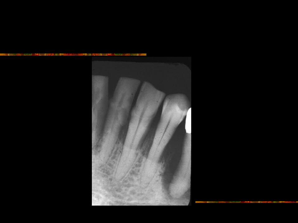
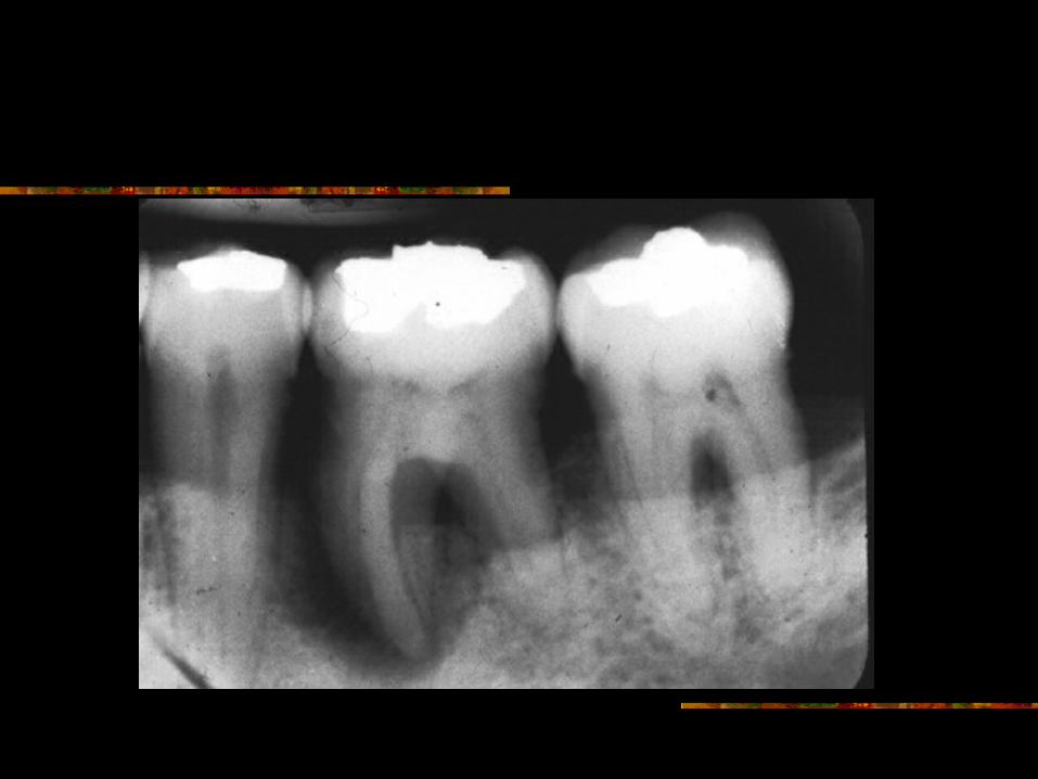
Juvenile Periodontitis(Early-onset Periodontitis, Rapidly Progressing Periodontitis)
Occurs in healthy individuals between puberty and age 25
Amount of bone loss is not consistent with local factors and oral Hygiene habits. Rate of bone loss is 3-4 times faster than in typical periodontitis

Juvenile Periodontitis(cont.)
Typically affects crestal bone of first molars and incisors. Eventually affects greater # of teeth.
Bone loss is progressive and frequently bilaterally symmetrical. Many teeth show vertical bone loss.
Host neutrophil dysfunction has been demonstrated by several investigators.


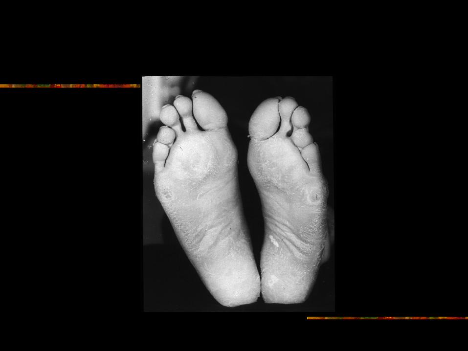
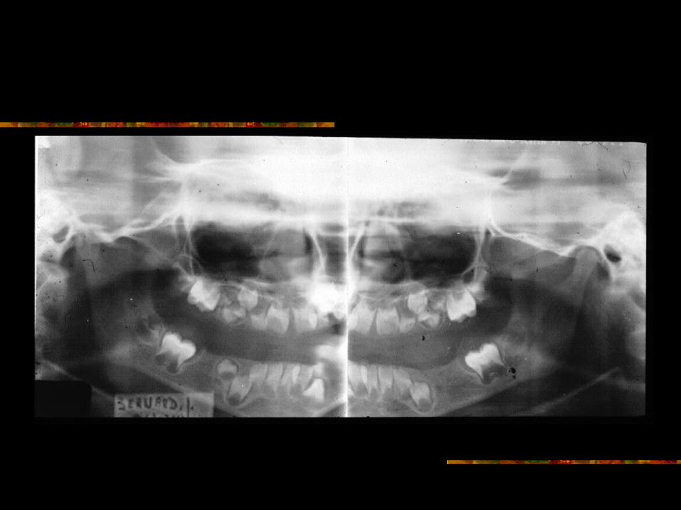
Papillon-Lefevre Syndrome
Autosomal recessive trait Hyperkeratosis of palms and soles Occasional keratosis of other skin
surfaces Calcification in falx cerebri Severe destruction of alveolar bone
involving all deciduous and perm. teeth Exfoliation of teeth


Langerhans’ Cell Histiocytosis (Histiocytosis X)
Complex of three diseases: Eosinophilic granuloma (usually solitary) Hand-Schuller-Christian disease Letterer-Siwe disease Due to abnormal proliferation of
Langerhans’ cells or their precursors

Eosinophilic Granuloma of Bone
Most common in children and young adults
Usually single radiolucency Skull, mandible, vertebra and long bones
commonly involved Painful, mobile teeth and gingival lesions

Hand-Schuller-Christian Disease
Most cases reported in children under 10 years. Has been reported in older individuals
Skeletal and soft tissues may be involved Classic triad of symptoms:
“punched out” destructive bone lesions unilateral or bilateral exophthalmos diabetes insipidus
Complete triad occurs in 25% of patients

Hand-Schuller-Christian (Cont.)
Oral manifestations include: loose teeth exfoliated teeth gingivitis loss of alveolar bone / advanced
periodontitis Sharply outlined multiple radiolucent
lesions in skull, jaws and other bones

Letterer-Siwe Disease
Acute, disseminated form of disease Usually occurs before age 3. Most patients die Involves several bones and organs Skin rash Intermittent fever, enlargement of liver and
spleen, lymphadenopathy common Destructive radiolucencies in jaws Loosening and premature loss of teeth

Hand-Schuller-Christian Disease

Hand-Schuller-Christian Disease

Other Diseases Influencing Course Of Periodontal Disease
Diabetes mellitus Leukemia

Periapical Inflamatory Lesions
Bone destruction around apex of tooth, mostly secondary to pulp exposure due to caries or trauma.
Bacterial invasion of pulp produces toxic metabolites which escape to the periapical bone through apical foramen and cause inflammation. The following may occur:

Periapical Inflamatory Lesions
Periapical granuloma: Localized mass of chronic granulation tissue containing PMN’s, lymphocytes, plasma cells.

Periapical Granuloma
Radiographically, widening of PDL or variable size of periapical radiolucency may be present

Periapical Granuloma

Periapical Granuloma

Periapical Abscess Periapical abscess:
When pus forms in the area. It may develop directly as an acute process or develop in a pre-existing granuloma. Radiographically, appears identical to granuloma.

Periapical Granuloma Or Abscess
Can one differentiate between the two on the basis of radiographs alone?

Periapical Inflamatory Lesions
Radicular cyst (periapical cyst): Cell rests of Mallasez (remnants of epithelial root sheath of Hertwig) proliferate due to inflamatory stimulus of a granuloma or an abscess and provide the epithelial lining. What is the definition of a cyst? “A cyst is an epithelium lined cavity which is filled with fluid or semi-solid material”. Radicular cyst is the ONLY cyst related to non-vital pulp.


Periapical Inflamatory Lesions
Can you definitively differentiate between a periapical granuloma, abscess or radicular cyst on the basis of radiograph alone?


Periapical Inflamatory Lesions(co)
Condensing osteitis ( chronic sclerosing osteomyelitis or osteitis). Occasionally, the reaction to periapical inflammation is predominantly osteoblastic, I.e., more sclerotic bone is formed (radiopaque mass). This usually occurs in children or young adults when the resistance is high. Most common location is mandibular 1st molar.

Condensing Osteitis

(Idiopathic) Osteosclerosis

Osteosclerosis
How do you differentiate between osteosclerosis and condensing osteitis?
In osteosclerosis, the pulp is vital. There are no clinical signs or symptoms. No treatment is necessary.
Condensing osteitis is secondary to pulp exposure. Patient is symptomatic. Endodontic treatment or extraction is indicated.

Calcific Degeneration(Calcific Metamorphosis)
Secondary to Trauma to the Tooth

Calcific Degeneration

Calcific Degeneration

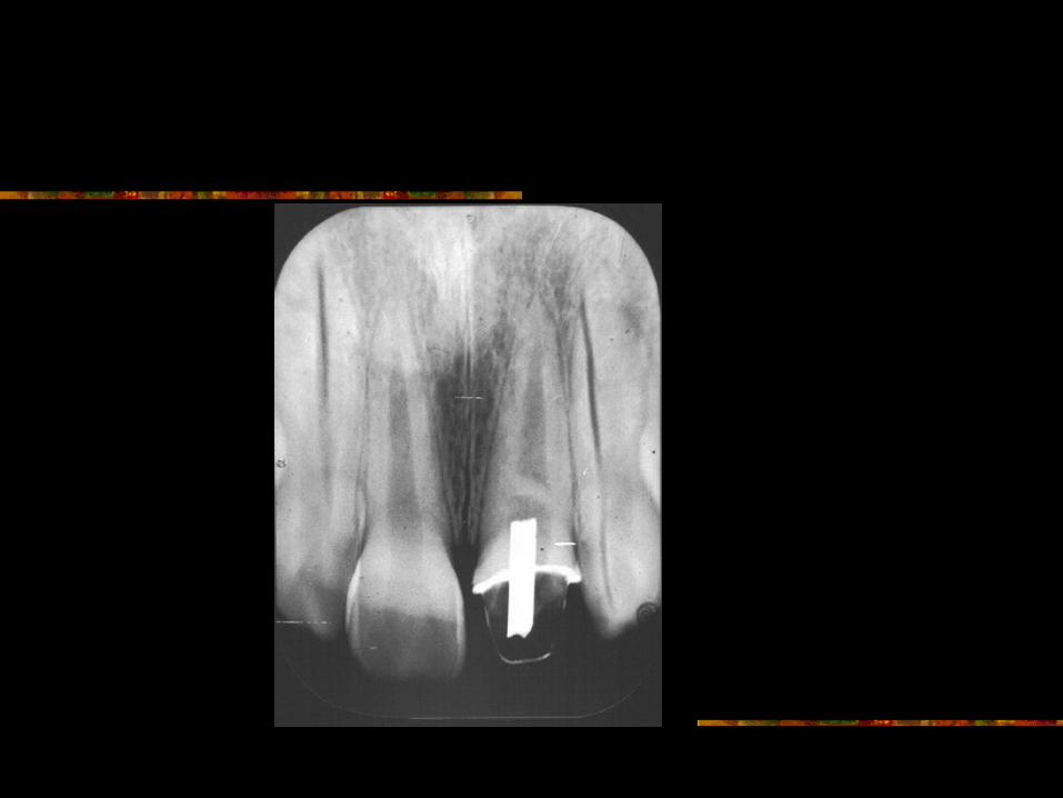
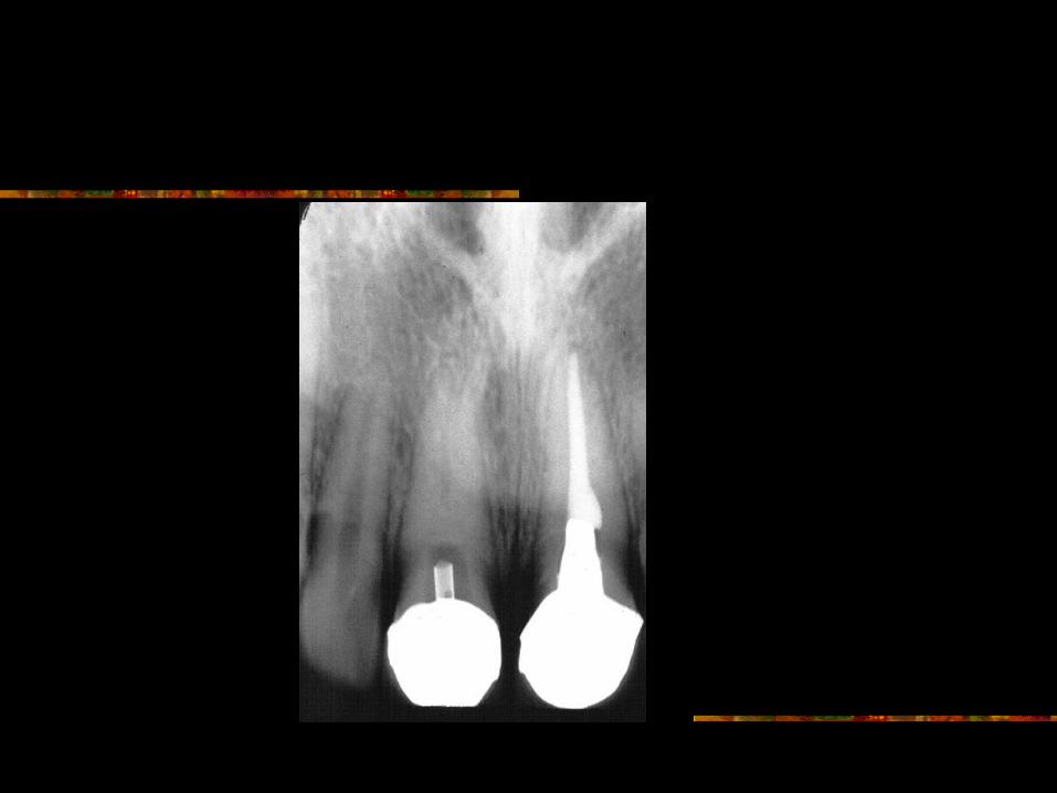
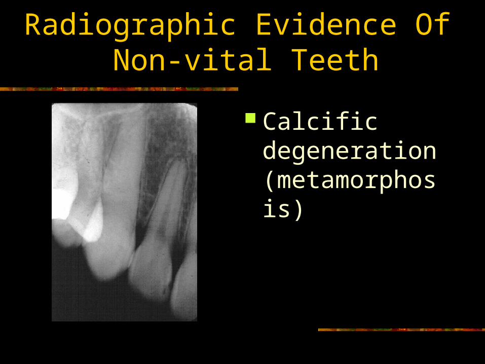
Radiographic Evidence Of Non-vital Teeth
Widening of apical PDL or periapical radiolucency ( associated with indication of pulp exposure)
Discontinuity of lamina dura Displacement of lamina dura Condensing osteitis Calcific degeneration (metamorphosis) Radiographic indication of pulp exposure

Radiographic Evidence Of Non-vital Teeth
Widening of apical PDL or periapical radiolucency ( associated with indication of pulp exposure)

Radiographic Evidence Of Non-vital Teeth
Discontinuity of lamina dura

Radiographic Evidence Of Non-vital Teeth
Displacement of lamina dura

Radiographic Evidence Of Non-vital Teeth
Condensing osteitis
Radiographic Evidence Of Non-vital Teeth
Calcific degeneration (metamorphosis)

Radiographic Evidence Of Non-vital Teeth
Radiographic indication of pulp exposure


Periapical Cemental Dysplasia
Also called Cementoma. Localized alteration in periapical area. Osseous structure is replaced by fibrous tissue, cementum-like material, abnormal bone or combination of these.
Pulp is vital. Patient is asymptomatic. There are no clinical signs.
No treatment is required. Mean age is 39 years.

Periapical Cemental Dysplasia
85% patients are females. 3 times more common in African-americans. Most commonly seen in mandibular anterior
areas. May be multiple. May be bilateral. Well-defined radiolucency, opacity or mixed.


Periapical Cemental Dysplasia
Stage I ( Osteolytic stage ) Stage II ( Osteo or cementoblastic stage) Stage III ( mature stage )

Stage II

Stasge III

Multiple


Apical Scar (Fibrous Scar )
Variation in healing process. Normally surgical site fills with blood clot which organizes and eventually mineralizes and remodels like surrounding bone.
Occasionally, normal mineralization and remodelling fails to occur.
Patient is asymptomatic and no treatment is required.

Apical Scar (Fibrous Scar )

Apical Scar (Fibrous Scar )

Apical Scar (Fibrous Scar )

Periapical Lesions (Bhaskar)
Periapical granuloma 48% Radicular cyst 43% Periapical abscess 1.1% Residual cyst 3.5% Apical scar 3.0% Periapical cemental dysplasia 1.7% Rare lesions 1.0%
Rare Periapical Lesions(Bhaskar)
Central giant cell granuloma Traumatic (simple) bone cyst Hyperparathyroidism

Periapical Lesions(LaLonde and Leubke)
Periapical granuloma 45.2% Radicular cyst 43.8% Periapical abscess 3.0% Other periapical lesions 8.0%

General Principles of Interpretation in Bone

Relative Radiodensity
Radiolucency Radiopacity Mixed (radiolucency and radiopacity)

Peripheral Outline
Borders Well-defined or Ill-defined, Smooth or Ragged?

Expansion of Cortical Plates
Indication of Rate of Growth of Lesion

Effects on Adjacent Structures
Resorption of roots of teeth Mandibular canal ( pain, anesthesia,
paresthesia?)

Location
Mandible Maxilla Anterior Posterior

Multiple-single

Age

Sex