
Dendritic Phosphorescent Probes for Oxygen Imaging in Biological Systems
13
Dendritic Phosphorescent Probes for Oxygen Imaging in Biological Systems Artem Y. Lebedev, † Andrei V. Cheprakov, ‡ Sava Sakadz ˇic ´, § David A. Boas, § David F. Wilson, † and Sergei A. Vinogradov* ,† Department of Biochemistry and Biophysics, University of Pennsylvania, Philadelphia, Pennsylvania 19104, Department of Chemistry, Moscow State University, Moscow, Russia, and Martinos Center for Biomedical Imaging, Massachusetts General Hospital/Harvard Medical School, Charlestown, Massachusetts 02129 ABSTRACT Oxygen levels in biological systems can be measured by the phosphorescence quenching method using probes with controllable quenching parameters and defined biodistributions. We describe a general approach to the construction of phosphorescent nanosensors with tunable spectral characteristics, variable degrees of quenching, and a high selectivity for oxygen. The probes are based on bright phosphorescent Pt and Pd complexes of porphyrins and symmetrically π-extended porphyrins (tetrabenzoporphyrins and tetranaphthoporphyrins). π-Extension of the core macrocycle allows tuning of the spectral parameters of the probes in order to meet the requirements of a particular imaging application (e.g., oxygen tomography versus planar microscopic imaging). Metallopor- phyrins are encapsulated into poly(arylglycine) dendrimers, which fold in aqueous environments and create diffusion barriers for oxygen, making it possible to regulate the sensitivity and the dynamic range of the method. The periphery of the dendrimers is modified with poly(ethylene glycol) residues, which enhance the probe’s solubility, diminish toxicity, and help prevent interactions of the probes with the biological environment. The probe’s parameters were measured under physiological conditions and shown to be unaffected by the presence of biomacromolecules. The performance of the probes was demonstrated in applications, including in vivo microscopy of vascular pO 2 in the rat brain. KEYWORDS: phosphorescence • oxygen • porphyrins • dendrimers • quenching • imaging INTRODUCTION O ptical sensors and imaging agents are rapidly de- veloping areas of research in functional macromo- lecular materials (1). Sensors based on dendrimers attract special interest because, among all types of synthetic polymeric carriers, dendrimers offer the unique advantage of molecular monodispersity (2). In imaging, dendritic poly- mers are typically used for signal amplification or for the construction of controlled microenvironments by way of dendritic encapsulation (3, 4). If an optically active motif is placed inside a dendrimer, the latter forms a protective cage, preventing physical contacts of the core with macromolecu- lar objects and shielding it from small-molecule quenchers in the environment. Optical probes for biological imaging of oxygen present an important example of the encapsulat- ing capability of dendrimers, which play a key role in the construction of these practically useful materials. Oxygen-dependent quenching of phosphorescence (5) is an optical method for oxygen sensing in biological systems (6, 7). Applications of the technique range from local “point” measurements to planar 2D imaging (8) to high-resolution microscopy (9) and near-infrared 3D tomography (10). Phosphorescence quenching offers excellent specificity, sub- millisecond temporal response, high sensitivity, and relative simplicity of implementation, complementing and in many ways surpassing other oximetry methods (11-15). In phosphorescence quenching, probes are the only invasive component of the measurement scheme, and their design is the key to applicability of the method. Early oxygen probes were based on simple water-soluble derivatives of palladium porphyrins (5). These chromophores had to be prebound to bovine serum albumin in order to enhance their solubility and to prevent interactions of the porphyrins with biological molecules. Prebinding was also important because water-soluble porphyrins, modified with either ionic groups (SO 3 - , CO 2 - , NH 3 + ) or neutral residues, e.g., oligo(ethylene glycols) (16), are prone to aggregation. Moreover, when injected systemically, they bind to biological targets and become heterogeneously distributed throughout the tissue. Local environments of probe molecules in different tissue microcompartments are highly heterogeneous, and as a result, the response of phosphorescence to oxygen cannot be interpreted quantitatively. Binding to albumin makes it possible to partially overcome these limitation, but a number of problems arise as a result of the use of an exogenous protein, which constitutes a part of the injected material. Introduction of polyglutamic phosphorescent probes (17) (in the literature known as Oxyphors R2 and G2) (18) eliminated the necessity of prebinding porphyrins to foreign albumin because polyglutamic branches rendered highly water-soluble materials. Still, polyglutamic probes required the endogenous albumin because only albumin complexes were suitable for measurements in the physiological oxygen * E-mail: [email protected]. Received for review March 14, 2009 and accepted May 13, 2009 † University of Pennsylvania. ‡ Moscow State University. § Massachusetts General Hospital/Harvard Medical School. DOI: 10.1021/am9001698 © 2009 American Chemical Society ARTICLE 1292 VOL. 1 • NO. 6 • 1292–1304 • 2009 www.acsami.org Published on Web 06/01/2009
Transcript of Dendritic Phosphorescent Probes for Oxygen Imaging in Biological Systems
Dendritic Phosphorescent Probes for OxygenImaging in Biological SystemsArtem Y. Lebedev,† Andrei V. Cheprakov,‡ Sava Sakadzic,§ David A. Boas,§ David F. Wilson,†and Sergei A. Vinogradov*,†
Department of Biochemistry and Biophysics, University of Pennsylvania, Philadelphia, Pennsylvania 19104,Department of Chemistry, Moscow State University, Moscow, Russia, and Martinos Center for Biomedical Imaging,Massachusetts General Hospital/Harvard Medical School, Charlestown, Massachusetts 02129
ABSTRACT Oxygen levels in biological systems can be measured by the phosphorescence quenching method using probes withcontrollable quenching parameters and defined biodistributions. We describe a general approach to the construction of phosphorescentnanosensors with tunable spectral characteristics, variable degrees of quenching, and a high selectivity for oxygen. The probes arebased on bright phosphorescent Pt and Pd complexes of porphyrins and symmetrically π-extended porphyrins (tetrabenzoporphyrinsand tetranaphthoporphyrins). π-Extension of the core macrocycle allows tuning of the spectral parameters of the probes in order tomeet the requirements of a particular imaging application (e.g., oxygen tomography versus planar microscopic imaging). Metallopor-phyrins are encapsulated into poly(arylglycine) dendrimers, which fold in aqueous environments and create diffusion barriers foroxygen, making it possible to regulate the sensitivity and the dynamic range of the method. The periphery of the dendrimers ismodified with poly(ethylene glycol) residues, which enhance the probe’s solubility, diminish toxicity, and help prevent interactionsof the probes with the biological environment. The probe’s parameters were measured under physiological conditions and shown tobe unaffected by the presence of biomacromolecules. The performance of the probes was demonstrated in applications, including invivo microscopy of vascular pO2 in the rat brain.
KEYWORDS: phosphorescence • oxygen • porphyrins • dendrimers • quenching • imaging
INTRODUCTION
Optical sensors and imaging agents are rapidly de-veloping areas of research in functional macromo-lecular materials (1). Sensors based on dendrimers
attract special interest because, among all types of syntheticpolymeric carriers, dendrimers offer the unique advantageof molecular monodispersity (2). In imaging, dendritic poly-mers are typically used for signal amplification or for theconstruction of controlled microenvironments by way ofdendritic encapsulation (3, 4). If an optically active motif isplaced inside a dendrimer, the latter forms a protective cage,preventing physical contacts of the core with macromolecu-lar objects and shielding it from small-molecule quenchersin the environment. Optical probes for biological imagingof oxygen present an important example of the encapsulat-ing capability of dendrimers, which play a key role in theconstruction of these practically useful materials.
Oxygen-dependent quenching of phosphorescence (5) isan optical method for oxygen sensing in biological systems(6, 7). Applications of the technique range from local “point”measurements to planar 2D imaging (8) to high-resolutionmicroscopy (9) and near-infrared 3D tomography (10).Phosphorescence quenching offers excellent specificity, sub-
millisecond temporal response, high sensitivity, and relativesimplicity of implementation, complementing and in manyways surpassing other oximetry methods (11-15).
In phosphorescence quenching, probes are the onlyinvasive component of the measurement scheme, and theirdesign is the key to applicability of the method. Early oxygenprobes were based on simple water-soluble derivatives ofpalladium porphyrins (5). These chromophores had to beprebound to bovine serum albumin in order to enhance theirsolubility and to prevent interactions of the porphyrins withbiological molecules. Prebinding was also important becausewater-soluble porphyrins, modified with either ionic groups(SO3
-, CO2-, NH3
+) or neutral residues, e.g., oligo(ethyleneglycols) (16), are prone to aggregation. Moreover, wheninjected systemically, they bind to biological targets andbecome heterogeneously distributed throughout the tissue.Local environments of probe molecules in different tissuemicrocompartments are highly heterogeneous, and as aresult, the response of phosphorescence to oxygen cannotbe interpreted quantitatively. Binding to albumin makes itpossible to partially overcome these limitation, but a numberof problems arise as a result of the use of an exogenousprotein, which constitutes a part of the injected material.
Introduction of polyglutamic phosphorescent probes (17)(in the literature known as Oxyphors R2 and G2) (18)eliminated the necessity of prebinding porphyrins to foreignalbumin because polyglutamic branches rendered highlywater-soluble materials. Still, polyglutamic probes requiredthe endogenous albumin because only albumin complexeswere suitable for measurements in the physiological oxygen
* E-mail: [email protected] for review March 14, 2009 and accepted May 13, 2009† University of Pennsylvania.‡ Moscow State University.§ Massachusetts General Hospital/Harvard Medical School.DOI: 10.1021/am9001698
© 2009 American Chemical Society
ARTIC
LE
1292 VOL. 1 • NO. 6 • 1292–1304 • 2009 www.acsami.orgPublished on Web 06/01/2009
range (vide infra). Consequently, oxygen measurementscould be performed only in albumin-rich environments(blood plasma), while incomplete albumin binding, e.g., atthe higher probe concentrations needed to increase thesignal level, could lead to measurement ambiguity (19).
A more comprehensive design of a molecular sensor foroxygen would entail construction of a well-defined microen-vironment around the phosphorescent chromophore withthe purpose of isolating it from interactions with biologicalmolecules. Dendritic encapsulation arguably provides themost straightforward way for the construction of monodis-perse, well-defined molecular jackets around luminescentcenters, although other approaches have been proposed inthe literature (20). In this paper, we present a comprehensivedesign of phosphorescent oxygen nanosensors for biologicalenvironments and describe a family of dendritic oxygenprobes with excitation bands spanning the entire UV-vis-near-infrared (NIR) spectrum.
RESULTS AND DISCUSSIONProbe Parameters. Molecular oxygen, a triplet mol-
ecule in the ground state (O23Σg
-), is able to react withexcited molecules in the environment, quenching theirluminescence (21). Collisional quenching is much less prob-able on the time scale of singlet excited states (nanoseconds)than of triplet states (microseconds to milliseconds), makingphosphorescence much more sensitive to oxygen thanfluorescence. Assuming a large excess of oxygen relative tothe concentration of triplet emitters, a condition typicallymet in biological experiments, the dependence of the phos-phorescence intensity and lifetime on the oxygen concentra-tion follows the Stern-Volmer relationship
where I and τ are the phosphorescence intensity and lifetimeat the oxygen concentration ([O2]) and in the absence ofoxygen (I0 and τ0) and KSV is the Stern-Volmer quenchingconstant. In practice, using lifetime τ as the analytical signalfor [O2] is more convenient because the lifetime is indepen-dent of the probe distribution, provided the chromophoreis in the same molecular environment.
We express the oxygen content in the units of pressure(mmHg) rather than concentration (M) (22) because in themajority of experiments the partial pressure of oxygen (pO2)is the experimental parameter that is actually controlled. Weassume that the Henry’s law holds in the physiological rangeof the oxygen concentrations: [O2] ) RpO2, where R is theoxygen solubility coefficient (M mmHg-1). Considering KSV
) k2τ0, where k2 is the bimolecular rate constant for thequenching reaction, eq 1 can be rewritten as
where kq ) Rk2 and has units of mmHg-1 s-1.Equation 2 contains two probe-specific parameters: con-
stant kq and lifetime τ0. Their interplay defines the sensitivity
and dynamic range of the method. It is desirable that themeasurement parameter, phosphorescence lifetime τ, spansthe largest possible interval of values throughout the rangeof analyte concentrations, assuring the highest possiblemeasurement resolution. To quantify the dynamic range oflifetimes, it is convenient to define parameter R ) (τ0 - τair)/τ0, where τ0 (pO2 ) 0 mmHg) and τair (pO2 ) 159.6 mmHg)are the maximal and minimal values of the phosphorescencelifetimes in physiological experiments. Another importantparameter is the signal-to-noise ratio, which is always higherfor probes with larger emission quantum yields, assumingthe same emission spectra.
Triplet lifetimes of palladium porphyrins in deoxygenatedsolutions at ambient temperatures are in the range ofhundreds of microseconds, and their phosphorescence quan-tum yields are typically 0.05-0.1 (23). Constants kq for“unprotected” metalloporphyrins in aqueous solutions are∼3000 mmHg-1 s-1 (1.9 × 10⁹ M-1 s-1). Given such highquenching constants, the lifetimes and quantum yields ofpalladium porphyrins become extremely low already at lowoxygen concentrations because of excessive quenching (seeFigure S22 in the Supporting Information). Thus, in spite ofthe overall very high signal dynamic range (R ∼ 0.99),palladium porphyrins can be useful only in a very low pO2
range, probably not higher than ∼5 mmHg.
For platinum porphyrins, lifetimes τ0 are typically tensof microseconds and their quantum yields are 0.10-0.25(23, 24). As a result, platinum porphyrins can be used up toabout ∼50 mmHg of pO2, but above that limit, probequantum yields also become low and lifetimes change onlyweakly, causing poor analyte resolution and low sensitivity.Moreover, if quenching constants were to decrease, whichis inevitable upon binding of the probes to biological mac-romolecules, probes with short lifetimes would become verypoorly sensitive to small changes in the analyte concentra-tion [R ∼ 0.5 for platinum porphyrins; compare to R ∼ 0.02for Ru(bpy)3-like complexes (25)].
For higher sensitivity, probes with greater τ0’s are clearlypreferred, but only if their phosphorescence changes gradu-ally instead of being highly quenched already at low oxygenconcentrations. Such an adjustment of the sensitivity can beachieved by tuning constants kq. For example, at kq ∼ 100mmHg-1 s-1, phosphorescence of palladium porphyrinswould change more gradually, permitting measurementseven at air saturation, but the dynamic range would still bequite high (R ∼ 0.9; see Figure S22 in the SupportingInformation). In some cases, when the signal strength isparticularly critical, probes with higher quantum yields (PtP)still would be more useful even at the expense of thesensitivity, especially if only qualitative information aboutpO2 is required.
Overall, it is clear that control over the values of quench-ing constants kq and, most importantly, the ability to keepthem unaltered is the key to accurate oxygen measure-ments. For the majority of phosphorescent chromophores,diffusion of oxygen can be considered the rate-limiting stepfor the quenching reaction (21). Therefore, through alter-
I0/I ) τ0/τ ) 1 + KSV[O2] (1)
1/τ ) 1/τ0 + kqpO2 (2)
ARTIC
LE
www.acsami.org VOL. 1 • NO. 6 • 1292–1304 • 2009 1293

ation of oxygen diffusion in the local environment of phos-phorescent chromophores, constants kq can be regulated.
Considering the above criteria, a molecular design emergesthat comprises a bright phosphorescent chromophore withsufficiently long triplet lifetime τ0 and a protective jacket,whose purpose is to constrain oxygen diffusion in the localenvironment of the chromophore. The periphery of theprobe must be hydrophilic and inert in order to preventinteractions of the probe with components of the biologicalsystem (e.g., biomacromolecules and cellular membranes).
Phosphorescent Chromophores. Relatively fewchromophores exhibit bright (26) phosphorescence at ambi-ent temperatures. Among them, R-diimine complexes of Ru,Ir, and some other transition metals (7a), cyclometalatedcomplexes of Ir and Pt (27), and Pt and Pd complexes ofporphyrins and related tetrapyrroles (5, 23, 28, 29) havebeen used in oxygen sensing, although some other systemshave been proposed (30).
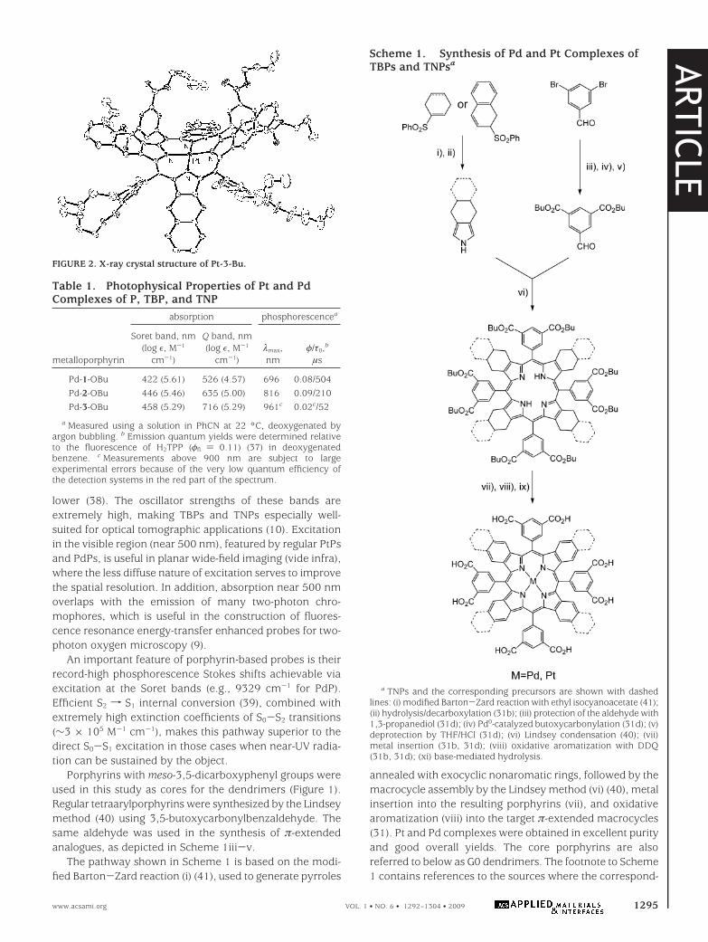
For tissue applications, it is desirable that probes possesabsorption bands in the NIR region. It has been shown thatlateral π-extension of platinum and palladium porphyrins byannealing of their pyrrole residues with external aromaticrings renders chromophores with dramatically red-shiftedabsorption bands and strong room-temperature phospho-rescence (31-33, 28). Structures, absorption and emissionspectra of palladium tetraarylporphyrin (PdP), palladiumtetraaryltetrabenzoporphyrin (PdTBP), and palladium tet-raaryltetranaphthoporphyrin (PdTNP) are shown in Figure1 (34). Spectra of Pt complexes are very similar in shape butare slightly blue-shifted (10-15 nm) compared to those ofthe Pd counterparts (see Table 2). The three basic porphyrin
types, P, TBP, and TNP, are designated in Figure 1 as 1-3,respectively. In this study, we used meso-tetraarylporphy-rins, where aryl (Ar) ) 3,5(RO2C)2C6H3. [To distinguishbetween different groups R and metals (Pt and Pd), prefixesand endings indicating the porphyrin types are added to thenumbers. For example, the Pt complex of TBP with butoxy-carbonyl substituents is abbreviated as Pt-2-OBu.]
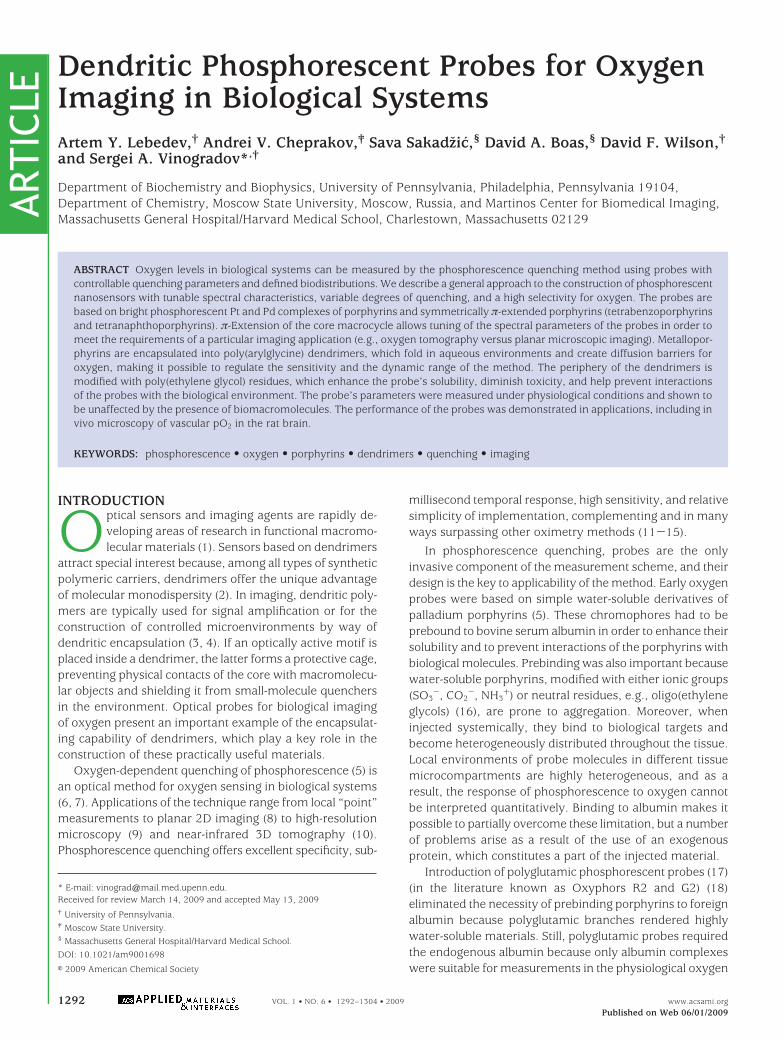
In platinum and palladium porphyrins, S1f T1 intersys-tem crossing is the predominant pathway of deactivation ofthe singlet excited states (S1) (23), and the resulting tripletstates are typically highly emissive (phosphorescent). Inregular (nonextended) porphyrins, macrocycle deviationsfrom planarity dramatically enhance the competing nonra-diative triplet decay and quench phosphorescence (35). Incontrast, platinum and palladium meso-tetraaryltetraben-zoporphyrins (TBPs), although highly nonplanar (31), phos-phoresce with high quantum yields (36). Palladium (31d) andplatinum tetraaryltetranaphthoporphyrins (Figure 2 andSupporting Information) (33b) are also nonplanar and, inaddition, have much narrower T1-S0 gaps (Table 1). Nev-ertheless, they still phosphoresce, although weaker thanTBPs and regular porphyrins. The emission spectra in Figure1b are scaled to reflect the relative phosphorescence quan-tum yields, and the complete photophysical data for thechromophores are summarized in Table 1.
Taken together, the absorption bands of Pt and Pd Ps,TBPs, and TNPs cover practically the entire UV-vis-NIRrange, presenting multiple opportunities for excitation. Theabsorption Q bands of TBPs and TNPs are shifted to the red,i.e., into the region between ∼630 and ∼950 nm, where theabsorption of endogenous chromophores is significantly
FIGURE 1. Three basic structural types of porphyrins that are used as sensing elements of phosphorescent probes. [PEG refers to monomethoxy-oligo(ethylene glycol); av MW 350.] Absorption and emission spectra of complexes Pd-1-OBu, Pd-2-OBu, and Pd-3-OBu in benzonitrile (PhCN)are shown.
ARTIC
LE
1294 VOL. 1 • NO. 6 • 1292–1304 • 2009 Lebedev et al. www.acsami.org
lower (38). The oscillator strengths of these bands areextremely high, making TBPs and TNPs especially well-suited for optical tomographic applications (10). Excitationin the visible region (near 500 nm), featured by regular PtPsand PdPs, is useful in planar wide-field imaging (vide infra),where the less diffuse nature of excitation serves to improvethe spatial resolution. In addition, absorption near 500 nmoverlaps with the emission of many two-photon chro-mophores, which is useful in the construction of fluores-cence resonance energy-transfer enhanced probes for two-photon oxygen microscopy (9).
An important feature of porphyrin-based probes is theirrecord-high phosphorescence Stokes shifts achievable viaexcitation at the Soret bands (e.g., 9329 cm-1 for PdP).Efficient S2 f S1 internal conversion (39), combined withextremely high extinction coefficients of S0-S2 transitions(∼3 × 10⁵ M-1 cm-1), makes this pathway superior to thedirect S0-S1 excitation in those cases when near-UV radia-tion can be sustained by the object.
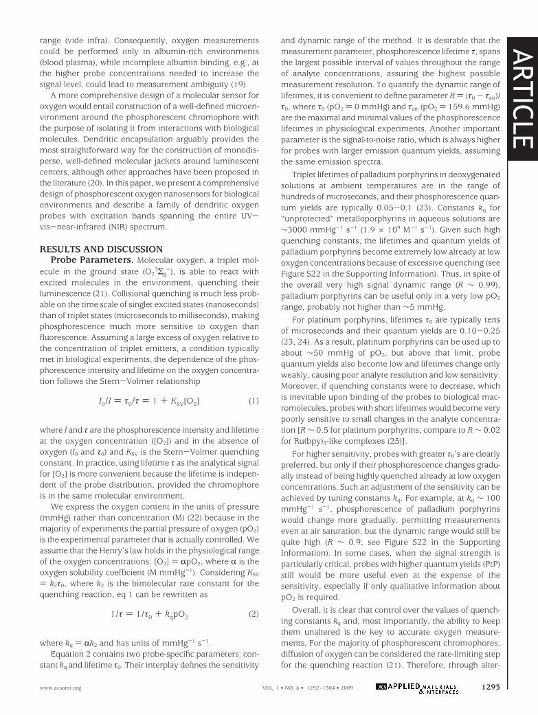
Porphyrins with meso-3,5-dicarboxyphenyl groups wereused in this study as cores for the dendrimers (Figure 1).Regular tetraarylporphyrins were synthesized by the Lindseymethod (40) using 3,5-butoxycarbonylbenzaldehyde. Thesame aldehyde was used in the synthesis of π-extendedanalogues, as depicted in Scheme 1iii-v.
The pathway shown in Scheme 1 is based on the modi-fied Barton-Zard reaction (i) (41), used to generate pyrroles
annealed with exocyclic nonaromatic rings, followed by themacrocycle assembly by the Lindsey method (vi) (40), metalinsertion into the resulting porphyrins (vii), and oxidativearomatization (viii) into the target π-extended macrocycles(31). Pt and Pd complexes were obtained in excellent purityand good overall yields. The core porphyrins are alsoreferred to below as G0 dendrimers. The footnote to Scheme1 contains references to the sources where the correspond-
FIGURE 2. X-ray crystal structure of Pt-3-Bu.
Table 1. Photophysical Properties of Pt and PdComplexes of P, TBP, and TNP
absorption phosphorescencea
metalloporphyrin
Soret band, nm(log ε, M-1
cm-1)
Q band, nm(log ε, M-1
cm-1)λmax,nm
φ/τ0,b
µs
Pd-1-OBu 422 (5.61) 526 (4.57) 696 0.08/504Pd-2-OBu 446 (5.46) 635 (5.00) 816 0.09/210Pd-3-OBu 458 (5.29) 716 (5.29) 961c 0.02c/52
a Measured using a solution in PhCN at 22 °C, deoxygenated byargon bubbling. b Emission quantum yields were determined relativeto the fluorescence of H2TPP (φfl ) 0.11) (37) in deoxygenatedbenzene. c Measurements above 900 nm are subject to largeexperimental errors because of the very low quantum efficiency ofthe detection systems in the red part of the spectrum.
Scheme 1. Synthesis of Pd and Pt Complexes ofTBPs and TNPsa
a TNPs and the corresponding precursors are shown with dashedlines: (i) modified Barton-Zard reaction with ethyl isocyanoacetate (41);(ii) hydrolysis/decarboxylation (31b); (iii) protection of the aldehyde with1,3-propanediol (31d); (iv) Pd0-catalyzed butoxycarbonylation (31d); (v)deprotection by THF/HCl (31d); (vi) Lindsey condensation (40); (vii)metal insertion (31b, 31d); (viii) oxidative aromatization with DDQ(31b, 31d); (xi) base-mediated hydrolysis.
ARTIC
LE
www.acsami.org VOL. 1 • NO. 6 • 1292–1304 • 2009 1295

ing protocols were developed and/or used in similar synthe-ses. Our modifications and improvements of these proce-dures are detailed in the Supporting Information.
Dendritic Cages. Dendritic attenuation of quenchinghas been documented in a number of studies, where quench-ers had different sizes and charges and the quenchingprocesses themselves had different mechanisms (e.g., elec-tron transfer vs energy transfer) (42). If a chromophore isencapsulated inside a dendrimer, the latter forms a protec-tive cage, preventing physical contacts of the core withmacromolecular objects in the environment. However, pro-tecting the chromophore from collisions with small mol-ecules is not as straightforward because the latter caneffectively diffuse through the body of the dendritic matrix.
It is important to realize that quenching constant kq in eq2 is a product of the quencher concentration and thediffusion coefficient, which are both affected by the chro-mophore environment. If the solubility of oxygen in thesolvent (e.g., water) is lower than that in the bulk of thedendrimer, the latter can serve as a “concentrator” foroxygen. Still, a decrease in the rate of oxygen diffusion caneffectively offset an increase in its local concentration, thuslowering the apparent constant kq. Hydrophobic dendriticbranches fold in polar environments (e.g., water), and as aresult, their mobility becomes restricted, preventing oxygenmolecules from freely reaching the phosphorescent core(43). A similar situation occurs in proteins (44). Notably, thedensity of the folded dendritic branches may be lower thanthat of the bulk solvent, and the solubility of oxygen in thefolded dendrimer might be higher than that in the solvent;nevertheless, the constrained dynamics of the branchesaffects the diffusion much more than the density and/or thesolubility.
Dendrimer dynamics is influenced by the interactions ofthe branches with the solvent. In “good” solvents, themobility is higher, and oxygen diffusion to the core isattenuated less than in “bad” solvents. Similarly, for thesame solvent, dendrimers with more solvent-compatiblecomposition limit oxygen access much less than less com-patible dendrimers. In particular, dendrimers composed ofaromatic motifs are most effective in shielding porphyrinsfrom oxygen in aqueous solutions (43).
Among many dendrimers with flexible aromatic skel-etons, dendritic poly(arylglycine) (AG) dendrons (45) (Scheme2) are especially well-suited for the construction of phos-phorescent oxygen probes. AG dendrons offer the advantageof inexpensive starting materials, simplicity of synthesis, andchromatography-free purification. Focal amino groups on AGdendrons complement carboxyls on the core porphyrins,whereas terminal carboxyls on the dendrons provide mul-tipleopportunitiesforfunctionalization.AGdendrons(Scheme2) of three successive generations (G1-G3) were used in thiswork for modification of platinum and palladium porphyrinsand π-extended porphyrins. The resulting dendritic com-pounds are abbreviated as follows:
For dendrons: X-AGnR, where AG denotes the dendriticarylglycine skeleton, n is the dendrimer generation number,
X is the focal functionality, and R is the terminal group. Forexample, a butyl-ester-terminated AG dendron of generation2 with a Boc-protected amino group at the focal point isabbreviated as BocNH-AG2OBu.
For dendrimers: C-(AGnR)m, where C denotes the den-drimer core, AG denotes the dendritic arylglycine skeleton,n is the generation number, R is the terminal group, and mis the number of dendritic wedges attached to the core. Forexample, a generation 2 AG dendrimer consisting of a PdTBPcore and eight AG dendrons terminated by carboxyl groupsis abbreviated as Pt-2-(AG2OH)8.
Scheme 2. Synthesis of AG Dendronsa
a CDMT ) 2-chloro-4,6-dimethoxy-1,3,5-triazine; NMM ) N-meth-ylmorpholine; HBTU ) o-benzotriazole-N,N,N′,N′-tetramethyluroniumhexafluorophosphate; DIPEA ) N,N-diisopropylethylamine.
ARTIC
LE
1296 VOL. 1 • NO. 6 • 1292–1304 • 2009 Lebedev et al. www.acsami.org
The synthesis in Scheme 2 makes use of the Fischerhaloacyl halide method to generate building blocks 4 and 5(45). The following assembly relies on peptide couplingreactions, employing CDMT/NMM and HBTU/DIPEA (seethe Scheme 2 footnote for abbreviations) and permitting thesynthesis of dendrons 6 and 7 in high purity and yield. TheAG dendrons can be produced in multigram quantities andstored for long periods of time without detectable decom-position.
Poly(ethylene glycol) (PEG) Layer. Special require-ments to probes for medical imaging applications includethe lack of toxicity and excretability from the blood uponcompletion of imaging. Globular uncharged molecules withmolecular weights between 1 and 15 kDa are usually ex-cretable by the kidney (46). If a probe satisfies this criterionand remains confined to the intravascular space (does notdiffuse out of the blood vessels), it is likely to be removedfrom the blood by kidney-mediated dialysis, and the pos-sibility of long-term toxicity effects can be avoided.
One way to eliminate interactions of dendritic probeswith biological macromolecules and to avoid toxicity is tomodify their termini with PEG residues. PEGylation of mac-romolecular compounds for drug delivery and related ap-
plications (e.g., artificial blood) is a widely known strategy(47), including PEGylation of dendrimers (48). PeripheralPEG groups on porphyrin dendrimers successfully eliminateinteractions of the probes with proteins, while keeping theprobes highly hydrophilic. Although PEG residues them-selves also contribute to the attenuation of oxygen quench-ing, their effect is small compared to that of the hydrophobicbranches (43). In addition, the effect of PEGs on the acces-sibility of the cores to oxygen saturates with an increase inthe length of linear poly(ethylene oxide) chains (49). As aresult, probe molecules of virtually any size can be generatedwithout significantly changing the quenching properties.
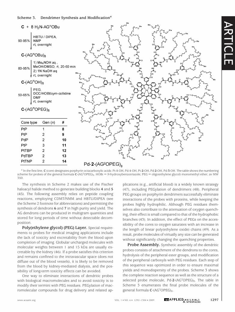
Probe Assembly. Synthetic assembly of the dendriticprobes consists of attachment of the dendrons to the cores,hydrolysis of the peripheral ester groups, and modificationof the peripheral carboxyls with PEG residues. Each step ofthis sequence was optimized in order to ensure maximalyields and monodispersity of the probes. Scheme 3 showsthe complete reaction sequence as well as the structure of aselected probe molecule, Pd-2-(AG2OPEG)8. The table inScheme 3 enumerates the final probe molecules of thegeneral formula C-(AG2OPEG)8.
Scheme 3. Dendrimer Synthesis and Modificationa
a In the first line, C (core) designates porphyrin octacarboxylic acids: Pt-1-OH, Pd-1-OH, Pt-2-OH, Pd-2-OH, Pd-3-OH. The table shows the numberingscheme for probes of the general formula C-(AG2OPEG)8. HOBt ) N-hydroxybenzotriazole; PEG ) oligo(ethylene glycol) monomethyl ether; av MW350.
ARTIC
LE
www.acsami.org VOL. 1 • NO. 6 • 1292–1304 • 2009 1297

Using CDMT as a coupling reagent (50), G1 dendriticbranches (5) were attached to the core porphyrins, yieldingperfect dendrimers C-(AG1OBu)8 in 90-95% yield. Thesedendrimers could be readily purified from an excess of 5 bywashing the crude mixtures with ethanol.
However, CDMT chemistry was not successful in the caseof G2 and G3 dendrimers. Matrix-assisted laser desorptionionization time-of-flight (MALDI-TOF) analysis of the reactionmixtures revealed the presence of dendrimers with four-to-eight AG2OBu branches. Additional experiments confirmedthat lower-molecular-weight peaks in the MALDI spectrawere not caused by fragmentation (see the SupportingInformation).
Among several established peptide-coupling systems(including [(Me2N)2CF]+PF6
-, DCC, CDI, uronium-based re-agents HBTU (O-benzotriazole-N,N,N′,N′-tetramethyluroniumhexafluorophosphate) and HATU (2-(1H-7-azabenzotriazol-1-yl)-1,1,3,3-tetramethyluronium hexafluorophosphate) (51),uronium reagents proved to be superior for the completemodification of porphyrins with AG2 dendrons 6. However,when used under the originally proposed conditions, theystill produced mixtures containing imperfect dendrimers.We have found that the single most important parameterwith respect to complete derivatization of porphyrins withAG dendrons is the choice of solvent. Porphyrin octacar-boxylic acids are soluble only in polar aprotic solvents [e.g.,dimethylformamide (DMF), dimethylacetamide (DMA), N-methylpyrrolidone (NMP), and dimethyl sulfoxide (DMSO)],and NMP was found to be the optimal choice. Possibly,porphyrins are much less aggregated in NMP, and NMPcontains much fewer free-amine impurities than DMF orDMA. Small amine molecules effectively compete with bulkyAG dendrons in the coupling reaction, as evidenced, forexample, by the presence of peaks corresponding toC-(AG2OBu)7NMe2 in the MALDI spectra of reactions carriedout in DMF. Notably, reaction intermediates bearing eightactivated carboxyl groups on the porphyrin core are highlyunstable at room temperature. Reactants (AG dendrons)need to be added to the mixtures immediately following theaddition of DIPEA.
Complete derivatization of porphyrins required a ∼1.5molar excess of dendron 6. The excess reagent was removedusing isothiocyanate-modified resin (Sigma-Aldrich), de-signed specifically to scavenge molecules with free aminogroups. Thus, prepurified dendrimers were subjected to size-exclusion chromatography on SX-1 beads (Biorad) usingtetrahydrofuran (THF) as a mobile phase.
As expected, modification of porphyrins with AG3 den-drons 7 proved to be extremely challenging. So far, the bestresult obtained was a 2:2:1 mixture of dendrimers with six,seven, and eight AG3 branches, respectively, attached to Pt-1-OH. Fortunately, from a practical point of view, modifica-tion of the cores with G2 dendrons was sufficient forattenuation of oxygen sensitivity (vide supra), whereasslightly imperfect G3 dendrimers were adequate for under-standing the trends of the dendrimer behavior at highergenerations.
In spite of the presence of multiple butyl ester groups,solubility of the porphyrin AG dendrimers in commonsolvents, such as CH2Cl2, ether, acetone, and methyl andethyl alcohols, was found to be quite limited; however, AGdendrimers are well-soluble in THF, DMSO, and pyridine.Each of the last three solvents is stable in the presence ofalkali, permitting hydrolytic cleavage of the terminal estersunder basic conditions. However, while attempting to useNaOH in THF/H2O (50:1), we found that MALDI spectra ofthe reaction mixtures showed peaks with lower molecularmasses than was expected for dendrimers polycarboxylicacids, likely because of the partial hydrolysis of anilides inthe body of the dendrimer (52).
In order to remove the peripheral butyl groups withoutaffecting the dendrimer integrity, a two-step scheme wasdevised. At first, poly(butyl ester) dendrimers were treatedwith NMe4OH (∼5 mM) in DMSO/MeOH over a 20-60 minperiod, followed by solvent removal and subsequent hy-drolysis in 0.1 N aqueous NaOH overnight. As a result, puremonodisperse dendrimer carboxylic acids could be isolatedin 80-95% yield. Importantly, when applied to crudemixtures of C-(AG2OBu)8 (Scheme 3), contaminated withunreacted dendrons 6, this two-step sequence yielded com-pletely pure acids C-(AG2OH)8, containing no traces of den-drons H2N-AG2OH. From a practical point of view, this resultis extremely useful because it made it possible to avoidchromatographic purification of the butyl ester dendrimersand thus significantly improve the overall yields. For ex-ample, Pt-1-(AG2OH)8 could be obtained without purificationin 81% yield starting from Pt-1-OH and 6.
At the last stage, the peripheral carboxyl groups on thedendrimers were esterified with monomethoxyoligo(ethyl-ene glycol) residues (av MW 350) in order to obtain water-soluble neutral probes. Esterification was carried out usingthe earlier developed DCC/HOBt chemistry (53). One im-portant practical result of this work is that a convenientwork-up procedure after the esterification reaction wasdeveloped to entirely avoid chromatographic purification. Itwas found that, by simple reprecipitation of PEGylateddendrimers from THF upon the addition of diethyl ether,pure PEGylated dendrimers could be obtained. The yieldsof PEGylation varied in the range of 50-65%. Judging fromthe MALDI spectra, 95-100% of the carboxyl groups wereconverted to the PEG esters.
Photophysical Properties. The photophysical datafor the phosphorescent probes are summarized in Table 2.Spectra of the three selected probes based on platinumporphyrins with changing degrees of π-extension are shownin Figure 3 (54).
Optical properties of the dendrimers in the UV-vis-NIRrange are mostly determined by their porphyrin cores (seeFigure 1 for comparison). The absorption bands of all G2 (9and 12-14) and G3 (11) dendrimers in aqueous solutionsare very close to those of the parent porphyrins (Pt-1-OBu,Pt-2-OBu, and Pt-3-OBu) in PhCN, suggesting that the coresare buried inside the dendritic matrix. In contrast, the Soretbands of the G1 dendrimer (8) and of the unprotected
ARTIC
LE
1298 VOL. 1 • NO. 6 • 1292–1304 • 2009 Lebedev et al. www.acsami.org
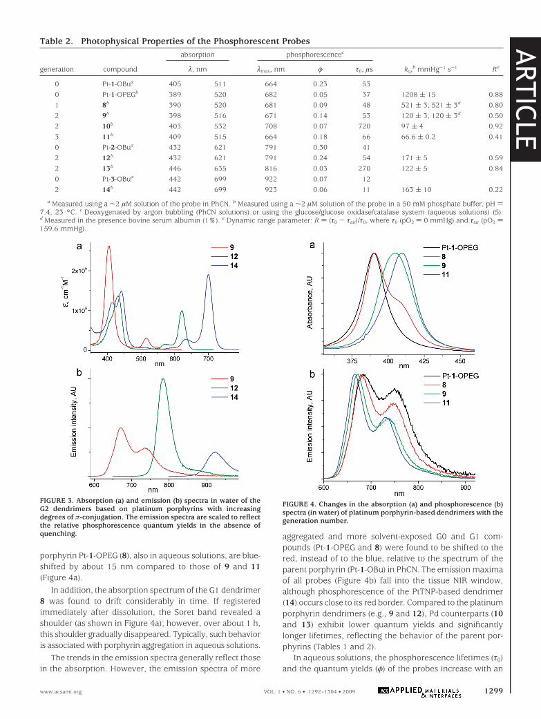
porphyrin Pt-1-OPEG (8), also in aqueous solutions, are blue-shifted by about 15 nm compared to those of 9 and 11(Figure 4a).
In addition, the absorption spectrum of the G1 dendrimer8 was found to drift considerably in time. If registeredimmediately after dissolution, the Soret band revealed ashoulder (as shown in Figure 4a); however, over about 1 h,this shoulder gradually disappeared. Typically, such behavioris associated with porphyrin aggregation in aqueous solutions.
The trends in the emission spectra generally reflect thosein the absorption. However, the emission spectra of more
aggregated and more solvent-exposed G0 and G1 com-pounds (Pt-1-OPEG and 8) were found to be shifted to thered, instead of to the blue, relative to the spectrum of theparent porphyrin (Pt-1-OBu) in PhCN. The emission maximaof all probes (Figure 4b) fall into the tissue NIR window,although phosphorescence of the PtTNP-based dendrimer(14) occurs close to its red border. Compared to the platinumporphyrin dendrimers (e.g., 9 and 12), Pd counterparts (10and 13) exhibit lower quantum yields and significantlylonger lifetimes, reflecting the behavior of the parent por-phyrins (Tables 1 and 2).
In aqueous solutions, the phosphorescence lifetimes (τ0)and the quantum yields (φ) of the probes increase with an
Table 2. Photophysical Properties of the Phosphorescent Probes
generation compound
absorption phosphorescencec
kq,b mmHg-1 s-1 Reλ, nm λmax, nm φ τ0, µs
0 Pt-1-OBua 405 511 664 0.23 530 Pt-1-OPEGb 389 520 682 0.05 37 1208 ( 15 0.881 8b 390 520 681 0.09 48 521 ( 3; 521 ( 3d 0.802 9b 398 516 671 0.14 53 120 ( 3; 120 ( 3d 0.502 10b 403 532 708 0.07 720 97 ( 4 0.923 11b 409 515 664 0.18 66 66.6 ( 0.2 0.410 Pt-2-OBua 432 621 791 0.30 412 12b 432 621 791 0.24 54 171 ( 5 0.592 13b 446 635 816 0.03 270 122 ( 5 0.840 Pt-3-OBua 442 699 922 0.07 122 14b 442 699 923 0.06 11 163 ( 10 0.22
a Measured using a ∼2 µM solution of the probe in PhCN. b Measured using a ∼2 µM solution of the probe in a 50 mM phosphate buffer, pH )7.4, 23 °C. c Deoxygenated by argon bubbling (PhCN solutions) or using the glucose/glucose oxidase/catalase system (aqueous solutions) (5).d Measured in the presence bovine serum albumin (1%). e Dynamic range parameter: R ) (τ0 - τair)/τ0, where τ0 (pO2 ) 0 mmHg) and τair (pO2 )159.6 mmHg).
FIGURE 3. Absorption (a) and emission (b) spectra in water of theG2 dendrimers based on platinum porphyrins with increasingdegrees of π-conjugation. The emission spectra are scaled to reflectthe relative phosphorescence quantum yields in the absence ofquenching.
FIGURE 4. Changes in the absorption (a) and phosphorescence (b)spectra (in water) of platinum porphyrin-based dendrimers with thegeneration number.
ARTIC
LE
www.acsami.org VOL. 1 • NO. 6 • 1292–1304 • 2009 1299
increase in the dendrimer generation (see, for example,compounds Pt-1-OPEG, 8, 9, and 11). The quantum yieldsappear to grow consistently from G0 to G3 dendrimers,although between G2 and G3, the growth somewhat slowsdown. Separate studies will be required to understand thisphenomenon, but the explanation might involve limitedquenching of the triplet state by the solvent in larger den-drimers and/or a decrease in the vibrational flexibility of aporphyrin as it becomes more and more confined within thefolded dendritic matrix. In porphyrins, vibrations are knownto affect the magnitude of spin-orbit coupling (21, 35, 36);therefore, tight confinement could decrease nonradiative T1
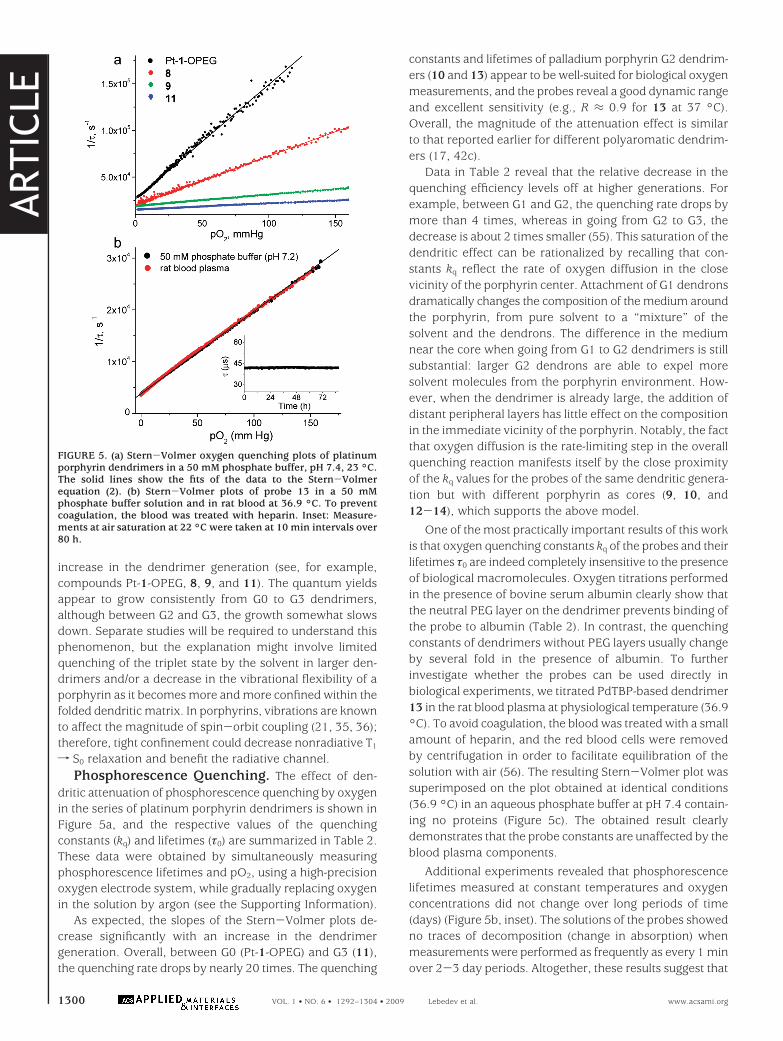
f S0 relaxation and benefit the radiative channel.Phosphorescence Quenching. The effect of den-
dritic attenuation of phosphorescence quenching by oxygenin the series of platinum porphyrin dendrimers is shown inFigure 5a, and the respective values of the quenchingconstants (kq) and lifetimes (τ0) are summarized in Table 2.These data were obtained by simultaneously measuringphosphorescence lifetimes and pO2, using a high-precisionoxygen electrode system, while gradually replacing oxygenin the solution by argon (see the Supporting Information).
As expected, the slopes of the Stern-Volmer plots de-crease significantly with an increase in the dendrimergeneration. Overall, between G0 (Pt-1-OPEG) and G3 (11),the quenching rate drops by nearly 20 times. The quenching
constants and lifetimes of palladium porphyrin G2 dendrim-ers (10 and 13) appear to be well-suited for biological oxygenmeasurements, and the probes reveal a good dynamic rangeand excellent sensitivity (e.g., R ≈ 0.9 for 13 at 37 °C).Overall, the magnitude of the attenuation effect is similarto that reported earlier for different polyaromatic dendrim-ers (17, 42c).
Data in Table 2 reveal that the relative decrease in thequenching efficiency levels off at higher generations. Forexample, between G1 and G2, the quenching rate drops bymore than 4 times, whereas in going from G2 to G3, thedecrease is about 2 times smaller (55). This saturation of thedendritic effect can be rationalized by recalling that con-stants kq reflect the rate of oxygen diffusion in the closevicinity of the porphyrin center. Attachment of G1 dendronsdramatically changes the composition of the medium aroundthe porphyrin, from pure solvent to a “mixture” of thesolvent and the dendrons. The difference in the mediumnear the core when going from G1 to G2 dendrimers is stillsubstantial: larger G2 dendrons are able to expel moresolvent molecules from the porphyrin environment. How-ever, when the dendrimer is already large, the addition ofdistant peripheral layers has little effect on the compositionin the immediate vicinity of the porphyrin. Notably, the factthat oxygen diffusion is the rate-limiting step in the overallquenching reaction manifests itself by the close proximityof the kq values for the probes of the same dendritic genera-tion but with different porphyrin as cores (9, 10, and12-14), which supports the above model.
One of the most practically important results of this workis that oxygen quenching constants kq of the probes and theirlifetimes τ0 are indeed completely insensitive to the presenceof biological macromolecules. Oxygen titrations performedin the presence of bovine serum albumin clearly show thatthe neutral PEG layer on the dendrimer prevents binding ofthe probe to albumin (Table 2). In contrast, the quenchingconstants of dendrimers without PEG layers usually changeby several fold in the presence of albumin. To furtherinvestigate whether the probes can be used directly inbiological experiments, we titrated PdTBP-based dendrimer13 in the rat blood plasma at physiological temperature (36.9°C). To avoid coagulation, the blood was treated with a smallamount of heparin, and the red blood cells were removedby centrifugation in order to facilitate equilibration of thesolution with air (56). The resulting Stern-Volmer plot wassuperimposed on the plot obtained at identical conditions(36.9 °C) in an aqueous phosphate buffer at pH 7.4 contain-ing no proteins (Figure 5c). The obtained result clearlydemonstrates that the probe constants are unaffected by theblood plasma components.
Additional experiments revealed that phosphorescencelifetimes measured at constant temperatures and oxygenconcentrations did not change over long periods of time(days) (Figure 5b, inset). The solutions of the probes showedno traces of decomposition (change in absorption) whenmeasurements were performed as frequently as every 1 minover 2-3 day periods. Altogether, these results suggest that
FIGURE 5. (a) Stern-Volmer oxygen quenching plots of platinumporphyrin dendrimers in a 50 mM phosphate buffer, pH 7.4, 23 °C.The solid lines show the fits of the data to the Stern-Volmerequation (2). (b) Stern-Volmer plots of probe 13 in a 50 mMphosphate buffer solution and in rat blood at 36.9 °C. To preventcoagulation, the blood was treated with heparin. Inset: Measure-ments at air saturation at 22 °C were taken at 10 min intervals over80 h.
ARTIC
LE
1300 VOL. 1 • NO. 6 • 1292–1304 • 2009 Lebedev et al. www.acsami.org
dendritically protected probes are superior to all previouslydescribed molecular oxygen sensors and can be directlyapplied to biological oxygen measurements and imaging.
Biological Experiments. To illustrate the perfor-mance of the probes, we performed two experiments: (1)measurements of the oxygenation of the mouse brainbecause it is affected by changes in the cerebral blood flow;(2) wide-field microscopic imaging of oxygen in intact ratbrain.
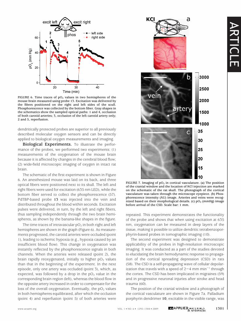
The schematic of the first experiment is shown in Figure6. An anesthesized mouse was laid on its back, and threeoptical fibers were positioned next to its skull. The left andright fibers were used for excitation (635 nm LED), while thebottom fiber served to collect the phosphorescence (57).PdTBP-based probe 13 was injected into the vein anddistributed throughout the blood within seconds. Excitationpulses were delivered, in turn, by the left and right fibers,thus sampling independently through the two brain hemi-spheres, as shown by the banana-like shapes in the figure.
The time traces of intravascular pO2 in both right and lefthemispheres are shown in the graph (Figure 6). As measure-ments progressed, the carotid arteries were occluded (point1), leading to ischemic hypoxia (e.g., hypoxia caused by aninsufficient blood flow). This change in oxygenation wasinstantly reflected by the phosphorescence signals in bothchannels. When the arteries were released (point 2), thebrain rapidly reoxygenated, initially to higher pO2 valuesthan that in the beginning of the experiment. In the nextepisode, only one artery was occluded (point 3), which, asexpected, was followed by a drop in the pO2 value in thecorresponding brain region (left), whereas the blood flow inthe opposite artery increased in order to compensate for theloss of the overall oxygenation. Eventually, the pO2 valuesin both hemispheres equilibrated, after which the occlusion(point 4) and reperfusion (point 5) of both arteries were
repeated. This experiment demonstrates the functionalityof the probe and shows that when using excitation at 635nm, oxygenation can be measured in deep layers of thetissue, making it possible to utilize dendritic tetrabenzopor-phyrin-based probes in tomographic imaging (10).
The second experiment was designed to demonstrateapplicability of the probes in high-resolution microscopicimaging. It was conducted as a part of the studies devotedto elucidating the brain hemodynamic response to propaga-tion of the cortical spreading depression (CSD) in rats(58). The CSD is a self-propagating wave of cellular depolar-ization that travels with a speed of 2-4 mm min-1 throughthe cortex. The CSD has been implicated in migraines (59)and in progressive neuronal injuries after stroke and headtrauma (60).
The position of the cranial window and a photograph ofthe cortical vasculature are shown in Figure 7a. Palladiumporphyrin dendrimer 10, excitable in the visible range, was
FIGURE 6. Time traces of pO2 values in two hemispheres of themouse brain measured using probe 13. Excitation was delivered bythe fibers positioned on the right and left sides of the scull.Phosphorescence was collected by the bottom fiber. Gray shapes inthe schematics show the sampled optical paths: 1 and 4, occlusionof both carotid arteries; 3, occlusion of the left carotid artery only;2 and 5, reperfusion.
FIGURE 7. Imaging of pO2 in cortical vasculature. (a) The positionof the cranial window and the location of KCl injection are markedon the schematic of the rat skull. The photograph of the corticalvasculature was taken through the microscope eyepiece. (b) Phos-phorescence intensity (AU) image. Arteries and veins were recog-nized based on their morphological details. (c) pO2 (mmHg) imagebefore arrival of the CSD. Scale bar: 1 mm.
ARTIC
LE
www.acsami.org VOL. 1 • NO. 6 • 1292–1304 • 2009 1301
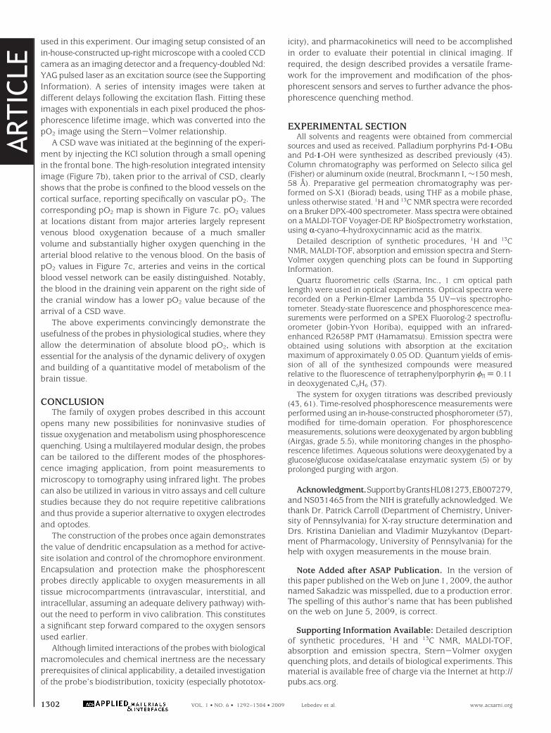
used in this experiment. Our imaging setup consisted of anin-house-constructed up-right microscope with a cooled CCDcamera as an imaging detector and a frequency-doubled Nd:YAG pulsed laser as an excitation source (see the SupportingInformation). A series of intensity images were taken atdifferent delays following the excitation flash. Fitting theseimages with exponentials in each pixel produced the phos-phorescence lifetime image, which was converted into thepO2 image using the Stern-Volmer relationship.
A CSD wave was initiated at the beginning of the experi-ment by injecting the KCl solution through a small openingin the frontal bone. The high-resolution integrated intensityimage (Figure 7b), taken prior to the arrival of CSD, clearlyshows that the probe is confined to the blood vessels on thecortical surface, reporting specifically on vascular pO2. Thecorresponding pO2 map is shown in Figure 7c. pO2 valuesat locations distant from major arteries largely representvenous blood oxygenation because of a much smallervolume and substantially higher oxygen quenching in thearterial blood relative to the venous blood. On the basis ofpO2 values in Figure 7c, arteries and veins in the corticalblood vessel network can be easily distinguished. Notably,the blood in the draining vein apparent on the right side ofthe cranial window has a lower pO2 value because of thearrival of a CSD wave.
The above experiments convincingly demonstrate theusefulness of the probes in physiological studies, where theyallow the determination of absolute blood pO2, which isessential for the analysis of the dynamic delivery of oxygenand building of a quantitative model of metabolism of thebrain tissue.
CONCLUSIONThe family of oxygen probes described in this account
opens many new possibilities for noninvasive studies oftissue oxygenation and metabolism using phosphorescencequenching. Using a multilayered modular design, the probescan be tailored to the different modes of the phosphores-cence imaging application, from point measurements tomicroscopy to tomography using infrared light. The probescan also be utilized in various in vitro assays and cell culturestudies because they do not require repetitive calibrationsand thus provide a superior alternative to oxygen electrodesand optodes.
The construction of the probes once again demonstratesthe value of dendritic encapsulation as a method for active-site isolation and control of the chromophore environment.Encapsulation and protection make the phosphorescentprobes directly applicable to oxygen measurements in alltissue microcompartments (intravascular, interstitial, andintracellular, assuming an adequate delivery pathway) with-out the need to perform in vivo calibration. This constitutesa significant step forward compared to the oxygen sensorsused earlier.
Although limited interactions of the probes with biologicalmacromolecules and chemical inertness are the necessaryprerequisites of clinical applicability, a detailed investigationof the probe’s biodistribution, toxicity (especially phototox-
icity), and pharmacokinetics will need to be accomplishedin order to evaluate their potential in clinical imaging. Ifrequired, the design described provides a versatile frame-work for the improvement and modification of the phos-phorescent sensors and serves to further advance the phos-phorescence quenching method.
EXPERIMENTAL SECTIONAll solvents and reagents were obtained from commercial
sources and used as received. Palladium porphyrins Pd-1-OBuand Pd-1-OH were synthesized as described previously (43).Column chromatography was performed on Selecto silica gel(Fisher) or aluminum oxide (neutral, Brockmann I, ∼150 mesh,58 Å). Preparative gel permeation chromatography was per-formed on S-X1 (Biorad) beads, using THF as a mobile phase,unless otherwise stated. 1H and 13C NMR spectra were recordedon a Bruker DPX-400 spectrometer. Mass spectra were obtainedon a MALDI-TOF Voyager-DE RP BioSpectrometry workstation,using R-cyano-4-hydroxycinnamic acid as the matrix.
Detailed description of synthetic procedures, 1H and 13CNMR, MALDI-TOF, absorption and emission spectra and Stern-Volmer oxygen quenching plots can be found in SupportingInformation.
Quartz fluorometric cells (Starna, Inc., 1 cm optical pathlength) were used in optical experiments. Optical spectra wererecorded on a Perkin-Elmer Lambda 35 UV-vis spectropho-tometer. Steady-state fluorescence and phosphorescence mea-surements were performed on a SPEX Fluorolog-2 spectroflu-orometer (Jobin-Yvon Horiba), equipped with an infrared-enhanced R2658P PMT (Hamamatsu). Emission spectra wereobtained using solutions with absorption at the excitationmaximum of approximately 0.05 OD. Quantum yields of emis-sion of all of the synthesized compounds were measuredrelative to the fluorescence of tetraphenylporphyrin φfl ) 0.11in deoxygenated C6H6 (37).
The system for oxygen titrations was described previously(43, 61). Time-resolved phosphorescence measurements wereperformed using an in-house-constructed phosphorometer (57),modified for time-domain operation. For phosphorescencemeasurements, solutions were deoxygenated by argon bubbling(Airgas, grade 5.5), while monitoring changes in the phospho-rescence lifetimes. Aqueous solutions were deoxygenated by aglucose/glucose oxidase/catalase enzymatic system (5) or byprolonged purging with argon.
Acknowledgment.SupportbyGrantsHL081273,EB007279,and NS031465 from the NIH is gratefully acknowledged. Wethank Dr. Patrick Carroll (Department of Chemistry, Univer-sity of Pennsylvania) for X-ray structure determination andDrs. Kristina Danielian and Vladimir Muzykantov (Depart-ment of Pharmacology, University of Pennsylvania) for thehelp with oxygen measurements in the mouse brain.
Note Added after ASAP Publication. In the version ofthis paper published on the Web on June 1, 2009, the authornamed Sakadzic was misspelled, due to a production error.The spelling of this author’s name that has been publishedon the web on June 5, 2009, is correct.
Supporting Information Available: Detailed descriptionof synthetic procedures, 1H and 13C NMR, MALDI-TOF,absorption and emission spectra, Stern-Volmer oxygenquenching plots, and details of biological experiments. Thismaterial is available free of charge via the Internet at http://pubs.acs.org.
ARTIC
LE
1302 VOL. 1 • NO. 6 • 1292–1304 • 2009 Lebedev et al. www.acsami.org
REFERENCES AND NOTES(1) Borisov, S. M.; Klimant, I. Analyst 2008, 133, 1302–1307.(2) Dendrimers and Other Dendritic Polymers; Frechet, J. M. J., Toma-
lia, D. A., Eds.; Wiley: New York, 2001.(3) (a) Gorman, C. B.; Smith, J. C. Acc. Chem. Res. 2001, 34, 60. (b)
Hecht, S.; Frechet, J. M. J. Angew. Chem., Int. Ed. 2001, 40, 74.(4) (a) Balzani, V.; Ceroni, P.; Maestri, M.; Saudan, C.; Vicinelli, V.
Top. Curr. Chem. 2003, 228, 159–191. (b) Ceroni, P.; Bergamini,G.; Marchioni, F.; Balzani, V. Prog. Polym. Sci. 2005, 30, 453–473.
(5) (a) Vanderkooi, J. M.; Maniara, G.; Green, T. J.; Wilson, D. F. J. Biol.Chem. 1987, 262, 5476–5482. (b) Wilson, D. F.; Rumsey, W. L.;Green, T. J.; Vanderkooi, J. M. J. Biol. Chem. 1988, 263, 2712–2718.
(6) Oximetry using solid-state luminescent probes or probes in whichphosphorescent chromophores are embedded in polymeric car-rier nanoparticles is a large area of research (for selected ex-amples, see refs 7 and 20). In this paper, we focus on “molecular”oxygen probes, where each sensing motif is an individual well-characterized molecule.
(7) (a) Demas, J. N.; DeGraff, B. A.; Coleman, P. B. Anal. Chem. 1999,71, 793A–800A. (b) Amao, Y. Microchim. Acta 2003, 143, 1–12.(c) Papkovsky, D. B.; O’Riordan, T. C. J. Fluores. 2005, 15, 569–584. For recent examples, see: (d) Huynh, L.; Wang, Z. U.; Yang,J.; Stoeva, V.; Lough, A.; Manners, I.; Winnik, M. A. Chem. Mater.2005, 17, 4765–4773. (e) Fernandez-Sanchez, J. F.; Roth, T.;Cannas, R.; Nazeeruddin, M. K.; Spichiger, S.; Graetzel, M.;Spichiger-Keller, U. E. Talanta 2007, 71, 242–250. (f) Borisov,S. M.; Klimant, I. Anal. Chem. 2007, 79, 7501–7509. (g) Borisov,S. M.; Nuss, G.; Klimant, I. Anal. Chem. 2008, 80, 9435–9442.
(8) (a) Pawlowski, M.; Wilson, D. F. Adv. Exp. Med. Biol. 1992, 316,179–182. (b) Rumsey, W. L.; Vanderkooi, J. M.; Wilson, D. F.Science 1988, 241, 1649–1651. (c) Shonat, R. D.; Wilson, D. F.;Riva, C. E.; Pawlowski, M. Appl. Opt. 1992, 33, 3711–3718. (d)Wilson, D. F.; Cerniglia, G. Cancer Res. 1992, 52, 3988–3993. Formore recent examples, see: (e) Kindig, C. A.; Kelley, K. M.;Howlett, R. A.; Stary, C. M.; Hogan, M. C. J. Appl. Physiol. 2003,94, 353–357. (f) Ferreira, L. F.; McDonough, P.; Behnke, B. J.;Musch, T. I.; Poole, D. C. Respir. Physiol. Neurobiol. 2006, 153,237–249. (g) Johannes, T.; Mik, E. G.; Nohe, B.; Unertl, K. E.; Ince,C. Am. J. Physiol. Ren. Physiol. 2007, 292, F796–F803. (h) Golub,A. S.; Pittman, R. N. Am. J. Physiol. Heart Circ. Physiol. 2008, 294,H2905–H2916.
(9) Finikova, O. S.; Lebedev, A. Y.; Aprelev, A.; Troxler, T.; Gao, F.;Garnacho, C.; Muro, S.; Hochstrasser, R. M.; Vinogradov, S. A.ChemPhysChem 2008, 9, 1673–1679.
(10) Apreleva, S. V.; Wilson, D. F.; Vinogradov, S. A. Appl. Opt. 2006,45, 8547–8559.
(11) Koch, C. J. Redox Cell Biology and Genetics; Elsevier: New York,2002; Vol. 352, Part A, pp 3-31.
(12) Swartz, H. M.; Clarkson, R. B. Phys. Med. Biol. 1998, 43, 1957–1975.
(13) Krishna, M. C.; English, S.; Yamada, K.; Yoo, J.; Murugesan, R.;Devasahayam, N.; Cook, J. A.; Golman, K.; Ardenkjaer-Larsen,J. H.; Subramanian, S.; Mitchell, J. B. Proc. Natl. Acad. Sci. U.S.A.2002, 99, 2216–2221.
(14) Jobsis, F. F. Science 1977, 198, 1264–1267.(15) (a) Ziemer, L. S.; Evans, S. M.; Kachur, A.; Shuman, A. L.; Cardi,
C. A.; Jenkins, W. T.; Karp, J. S.; Alavi, A.; Dolbier, W. R.; Koch,C. J. Eur. J. Nucl. Med. Mol. Imaging 2003, 30, 259–266. (b) Tatum,J. L.; Kelloff, G. J.; Gillies, R. J. Int. J. Radiat. Biol. 2006, 82, 699–757.
(16) Sibrian-Vazquez, M.; Jensen, T. J.; Vicente, M. G. H. J. Photochem.Photobiol., B 2007, 86, 9–21.
(17) (a) Vinogradov, S. A.; Wilson, D. F. Adv. Exp. Med. Biol. 1997, 428,657–662. (b) Vinogradov, S. A.; Lo, L. W.; Wilson, D. F.Chem.sEur. J. 1999, 5, 1338–1347. (c) Rietveld, I. B.; Kim, E.;Vinogradov, S. A. Tetrahedron 2003, 59, 3821–3831.
(18) (a) Dunphy, I.; Vinogradov, S. A.; Wilson, D. F. Anal. Biochem.2002, 310, 191–198. (b) Thompson, J. K.; Peterson, M. R.;Freeman, R. D. Science 2003, 299, 1070–1072. (c) Poole, D. C.;Behnke, B. J.; McDonough, P.; McAllister, R. M.; Wilson, D. F.Microcirculation 2004, 11, 317–326. (d) Pirow, R.; Baumer, C.;Paul, R. J. J. Exp. Biol. 2004, 207, 4393–4405. (e) Wilson, D. F.;Lee, W. M. F.; Makonnen, S.; Finikova, O.; Apreleva, S.; Vinogra-dov, S. A. J. Appl. Physiol. 2006, 101, 1648–1656. (f) Johannes,T.; Mik, E. G.; Ince, C. J. Appl. Physiol. 2006, 100, 1301–1310.
(19) Estrada, A. D.; Ponticorvo, A.; Ford, T. N. Opt. Lett. 2008, 33,1038–1040.
(20) (a) Xu, H.; Aylott, J. W.; Kopelman, R.; Miller, T. J.; Philbert, M. A.Anal. Chem. 2001, 73, 4124–4133. (b) Ji, J.; Rosenzweig, N.;Jones, I.; Rosenzweig, Z. Anal. Chem. 2001, 73, 3521–3527. (c)Koo, Y. E. L.; Cao, Y. F.; Kopelman, R.; Koo, S. M.; Brasuel, M.;Philbert, M. A. Anal. Chem. 2004, 76, 2498–2505. (d) Han, B. H.;Winnik, M. A.; Bourlinos, A. B.; Giannelis, E. P. Chem. Mater. 2005,17, 4001–4009. (e) Zhang, H. D.; Sun, Y. H.; Ye, K. Q.; Zhang,P.; Wang, Y. J. Mater. Chem. 2005, 15, 3181–3186. (f) Guice, K. B.;Caldorera, M. E.; McShane, M. J. J. Biomed. Opt. 2005, 10, 064031.(g) Schmalzlin, E.; van Dongen, J. T.; Klimant, I.; Marmodee, B.;Steup, M.; Fisahn, J.; Geigenberger, P.; Lohmannsroben, H. G.Biophys. J. 2005, 89, 1339–1345.
(21) Turro, N. J. Modern Molecular Photochemistry; University ScienceBooks: Sausalito, CA, 1991.
(22) At 298 K and an air pressure of 760 mmHg (oxygen fraction inthe air is 21% or 159.6 mmHg), air-equilibrated aqueous solutionsare 252 µM in O2 (Fogg, P. G. T.; Gerrard, W. Solubility of gasesin liquids; Wiley: New York, 2001).
(23) Eastwood, D.; Gouterman, M. J. Mol. Spectrosc. 1970, 35, 359–375.
(24) Kim, D. H.; Holten, D.; Gouterman, M.; Buchler, J. W. J. Am. Chem.Soc. 1984, 106, 4015–4017.
(25) Morris, K. J.; Roach, M. S.; Xu, W. Y.; Demas, J. N.; DeGraff, B. A.Anal. Chem. 2007, 79, 9310–9314.
(26) The “brightness” of the chromophore is defined as the productof the molar extinction coefficient and the emission quantumyield.
(27) DeRosa, M. C.; Mosher, P. J.; Yap, G. P. A.; Focsaneanu, K. S.;Crutchley, R. J.; Evans, C. E. B. Inorg. Chem. 2003, 42, 4864–4872.
(28) (a) Vinogradov, S. A.; Wilson, D. F. J. Chem. Soc., Perkin Trans. 21995, 103–111. (b) Vinogradov, S. A.; Lo, L.-W.; Jenkins, W. T.;Evans, S. M.; Koch, C.; Wilson, D. F. Biophys. J. 1996, 70, 1609–1617.
(29) Papkovsky, D. B.; Ponomarev, G. V.; Wolfbeis, O. S. Spectrochim.Acta, Part A 1996, 52, 1629–1638.
(30) Zhang, G.; Chen, J.; Payne, S. J.; Kooi, S. E.; Demas, J. N.; Fraser,C. L. J. Am. Chem. Soc. 2007, 129, 8942.
(31) (a) Finikova, O.; Cheprakov, A.; Beletskaya, I.; Vinogradov, S.Chem. Commun. 2001, 261–262. (b) Finikova, O. S.; Cheprakov,A. V.; Carroll, P. J.; Vinogradov, S. A. J. Org. Chem. 2003, 68, 7517–7520. (c) Finikova, O. S.; Cheprakov, A. V.; Beletskaya, I. P.;Carroll, P. J.; Vinogradov, S. A. J. Org. Chem. 2004, 69, 522–535.(d) Finikova, O. S.; Aleshchenkov, S. E.; Brinas, R. P.; Cheprakov,A. V.; Carroll, P. J.; Vinogradov, S. A. J. Org. Chem. 2005, 70, 4617–4628. (e) Finikova, O. S.; Cheprakov, A. V.; Vinogradov, S. A. J.Org. Chem. 2005, 70, 9562–9572. (f) Filatov, M. A.; Lebedev, A. Y.;Vinogradov, S. A.; Cheprakov, A. V. J. Org. Chem. 2008, 73, 4175–4185.
(32) (a) Tsvirko, M. P.; Sapunov, V. V.; Soloviyev, K. N. Opt. Spektrosk.1973, 34, 1094–1100. (b) Rozhkov, V. V.; Khajehpour, M.;Vinogradov, S. A. Inorg. Chem. 2003, 42, 4253–4255.
(33) (a) Borek, C.; Hanson, K.; Djurovich, P. I.; Thompson, M. E.;Aznavour, K.; Bau, R.; Sun, Y. R.; Forrest, S. R.; Brooks, J.;Michalski, L.; Brown, J. Angew. Chem., Int. Ed. 2007, 46, 1109–1112. (b) Sommer, J. R.; Farley, R. T.; Graham, K. R.; Yang, Y.;Reynolds, J. R.; Xue, J.; Schanze, K. S. ACS Appl. Mater. Interfaces2009, 1, 274–278.
(34) meso-Tetraarylated porphyrins, tetrabenzoporphyrins, and tet-ranaphthoporphyrins are usually abbreviated as Ar4P, Ar4TBP,and Ar4TNP, respectively, in order to distinguish them from meso-unsubstituted analogues. In the present paper, we used onlymeso-tetrarylated macrocycles, and therefore we omit the prefix“Ar4”.
(35) (a) Knyukshto, V. N.; Sagun, E. I.; Shul’ga, A. M.; Bachilo, S. M.;Starukhin, D. A.; Zen’kevich, E. I. Opt. Spectrosk. 2001, 90, 67.(b) Knyukshto, V. N.; Shul’ga, A. M.; Sagun, E. I.; Zen’kevich, E. I.Opt. Spectrosk. 2002, 92, 53. (c) Knyukshto, V. N.; Shul’ga, A. M.;Sagun, E. I.; Zen’kevich, E. I. Opt. Spectrosk. 2006, 100, 590–601.
(36) Lebedev, A. Y.; Filatov, M. A.; Cheprakov, A. V.; Vinogradov, S. A.J. Phys. Chem. A 2008, 112, 7723–7733.
(37) Seybold, P. G.; Gouterman, M. J. Mol. Spectrosc. 1969, 31, 1.(38) Taroni, P.; Pifferi, A.; Torricelli, A.; Comelli, D.; Cubeddu, R.
Photochem. Photobiol. Sci. 2003, 2, 124–129.
ARTIC
LE
www.acsami.org VOL. 1 • NO. 6 • 1292–1304 • 2009 1303
(39) Tripathy, U.; Kowalska, D.; Liu, X.; Velate, S.; Steer, R. P. J. Phys.Chem. A 2008, 112, 5824–5833.
(40) Lindsey, J. S.; Schreiman, I. C.; Hsu, H. C.; Kearney, P. C.;Marguerettaz, A. M. J. Org. Chem. 1987, 52, 827–836.
(41) (a) Arnold, D. P.; Burgess-Dean, L.; Hubbard, J.; Abdur Rahman,M. Aust. J. Chem. 1994, 47, 969–974. (b) Abel, Y.; Haake, E.;Haake, G.; Schmidt, W.; Struve, D.; Walter, A.; Montforts, F. P.Helv. Chim. Acta 1998, 81, 1978–1996.
(42) For examples, see: (a) Jin, R. H.; Aida, T.; Inoue, S. J. Chem. Soc.,Chem. Commun. 1993, 1260–1262. (b) Sadamoto, R.; Tomioka, N.;Aida, T. J. Am. Chem. Soc. 1996, 118, 3978–3979. (c) Kimura, M.;Nakada, K.; Yamaguchi, Y.; Hanabusa, K.; Shirai, H.; Kobayashi, N.Chem. Commun. 1997, 1215–1216. (d) Issberner, J.; Vogtle, F.;DeCola, L.; Balzani, V. Chem.sEur. J. 1997, 3, 706–712. (e) Vogtle,F.; Plevoets, M.; Nieger, M.; Azzellini, G. C.; Credi, A.; De Cola, L.;De Marchis, V.; Venturi, M.; Balzani, V. J. Am. Chem. Soc. 1999, 121,6290–6298. (f) Riley, J. M.; Alkan, S.; Chen, A. D.; Shapiro, M.; Khan,W. A.; Murphy, W. R.; Hanson, J. E. Macromolecules 2001, 34, 1797–1809.
(43) Rozhkov, V.; Wilson, D.; Vinogradov, S. Macromolecules 2002,35, 1991–1993.
(44) Khajehpour, M.; Rietveld, I.; Vinogradov, S.; Prabhu, N. V.; Sharp,K. A.; Vanderkooi, J. M. Proteins: Struct., Funct., Genet 2003, 53,656–666.
(45) Vinogradov, S. A. Org. Lett. 2005, 7, 1761–1764.(46) Caliceti, P.; Veronese, F. M. Adv. Drug Delivery Rev. 2003, 55,
1261–1277.(47) Gajbhiye, V.; Kumar, P. V.; Tekade, R. K.; Jain, N. K. Curr. Pharm.
Des. 2007, 13, 415–429.(48) Guillaudeu, S. J.; Fox, M. E.; Haidar, Y. M.; Dy, E. E.; Szoka, F. C.;
Frechet, J. M. J. Bioconjugate Chem. 2008, 19, 461–469.(49) Dichtel, W. R.; Baek, K. Y.; Frechet, J. M. J.; Rietveld, I. B.;
Vinogradov, S. A. J. Polym. Sci., Part A: Polym. Chem. 2006, 44,49394951.
(50) Kaminski, Z. J. Biopolymers 2000, 55, 140–164.(51) Han, S. Y.; Kim, Y. A. Tetrahedron 2004, 60, 2447–2467.(52) Biechler, S. S.; Taft, R. W. J. Am. Chem. Soc. 1957, 79, 4927–4935.(53) Dandliker, P. J.; Diederich, F.; Gross, M.; Knobler, C. B.; Louati,
A.; Sanford, E. M. Angew. Chem., Int. Ed. 1994, 33, 1739–1742.(b) Dandliker, P. J.; Diederich, F.; Gisselbrecht, J. P.; Louati, A.;Gross, M. Angew. Chem., Int. Ed. 1996, 34, 2725–2728.
(54) While this paper was in preparation, an article appeared in press(ref 33b) reporting independently on the photophysical propertiesof platinum tetraphenyltetranaphthoporphyrin.
(55) The plots of Pt-1-OPEG and 8 display lower kq values than theywould be expected to display in the absence of aggregation.
(56) In separate experiments, we have established that dendriticprobes do not permeate cellular membranes and that thepresence of erythrocytes has no effect on the calibrationconstants.
(57) Vinogradov, S. A.; Fernandez-Seara, M. A.; Dugan, B. W.; Wilson,D. F. Rev. Sci. Instrum. 2001, 72, 3396–3406.
(58) (a) Marshall, W. H. Physiol. Rev. 1959, 39, 239–279. (b) Ayata, C.;Shin, H. K.; Salomone, S.; Ozdemir-Gursoy, Y.; Boas, D. A.; Dunn,A. K.; Moskowitz, M. A. J. Cereb. Blood Flow Metab. 2004, 24, 1172–1182. (c) Sakadzic, S.; Yuan, S.; Dilekoz, E.; Ruvinskaya, S.; Vino-gradov, S. A.; Ayata, C.; Boas, D. A. Appl. Opt. 2009, in press.
(59) Hadjikhani, N.; Sanchez Del Rio, M.; Wu, O.; Schwartz, D.; Bakker,D.; Fischl, B.; Kwong, K. K.; Cutrer, F. M.; Rosen, B. R.; Tootell,R. B.; Sorensen, A. G.; Moskowitz, M. A. Proc. Natl. Acad. Sci.U.S.A. 2001, 98, 4687–4692.
(60) Nedergaard, M. Acta Neurol. Scand. 1988, 77, 81–101.(61) Khajehpour, M.; Rietveld, I.; Vinogradov, S.; Prabhu, N. V.; Sharp,
K. A.; Vanderkooi, J. M. Proteins: Struct., Funct., Genet. 2003, 53,656–666.
AM9001698
ARTIC
LE
1304 VOL. 1 • NO. 6 • 1292–1304 • 2009 Lebedev et al. www.acsami.org