Dendritic Cell mediated immune responses are shaped by ... · and select rare Ag specific T cells...
93
Dendritic Cell mediated immune responses are shaped by Interleukin-15 and Interleukin -21 Dissertation zur Erlangung des Doktorgrades der Mathematisch-Naturwissenschaftlichen Fakultät der Christian-Albrechts-Universität zu Kiel vorgelegt von Katja Brandt Kiel, den 20.12.2002
Transcript of Dendritic Cell mediated immune responses are shaped by ... · and select rare Ag specific T cells...
Dendritic Cell mediated immune responses are shaped by Interleukin-15 and Interleukin -21
Dissertation
zur Erlangung des Doktorgrades der Mathematisch-Naturwissenschaftlichen Fakultät
der Christian-Albrechts-Universität zu Kiel
vorgelegt von Katja Brandt
Kiel, den 20.12.2002
2
Cover: Dendritic cells, stained for membrane (red), IL-15 (green) and the nucleus (blue).
Referentin: Prof. Dr. Dr. Bulfone-Paus, Borstel
Koreferrent: Prof. Dr. Brocker, München
Koreferrent: Prof. Dr. Bosch, Kiel
Tag der mündlichen Prüfung: 11.02.2003
Zum Druck genehmigt: Kiel, 11.02.2003
Der Dekan

3
Für Stephanie
4
Index Index _____________________________________________________________________4
Abstract ___________________________________________________________________7
Zusammenfassung ___________________________________________________________8
Introduction ________________________________________________________________9
Dendritic Cells and their central role in T cell mediated immunity ___________________9
Interleukin-15 and its modulatory effects in immunobiology_______________________12
Interleukin-21: a new player in the cytokine network of the immune system___________14
In vivo models to analyze modulation of DC biology by IL-15 and IL-21 _____________15
Phase 1: Sensitization ___________________________________________________16
Phase 2: Elicitation _____________________________________________________17
Questions addressed ________________________________________________________19
Material and Methods _______________________________________________________20
Mice _________________________________________________________________20
Culture medium and antibodies____________________________________________20
DC preparation ________________________________________________________20
Antibodies and flow cytometric analysis_____________________________________21
Histomorphometry______________________________________________________21
RT-PCR ______________________________________________________________21
Cytokine detection______________________________________________________22
Analysis of endocytosis by flow cytometry __________________________________23
DC activation by LPS ___________________________________________________23
DC activation by various stimuli___________________________________________23
Proliferation assays _____________________________________________________23
In vitro differentiation of CD8+ T cells ______________________________________24
Confocal Microscopy ___________________________________________________24
In vivo DC migration ____________________________________________________24
Sensitization to picryl chloride (PCl) and elicitation of CHS _____________________25
Contact hypersensitivity model to FITC _____________________________________26
Irritant dermatitis to croton oil_____________________________________________26
Th1 model of DTH reaction to SRBC_______________________________________26
Homing and activation of APC ____________________________________________27
Statistical analysis ______________________________________________________27

5
Results ___________________________________________________________________28
CD11c+ DCs differentiate in the presence of IL-15 or IL-21 in vitro _________________28
IL-21 and IL-15 receptors are expressed in BM derived DCs_______________________29
IL-21 reduced MHC class II expression _______________________________________31
Increased antigen uptake by IL-21DCs contrasted by low uptake by IL-15DCs ________34
IL-21DCs keep their immature phenotype after antigen uptake and LPS stimulation ____35
IL-21DCs induce low antigen specific CD4+ T cell proliferation____________________36
IL-21DCs are unable to prime in vivo contact hypersensitivity reaction to FITC _______38
Comparable migration of all DC types to DLN _________________________________40
IL-21 inhibits LPS induced activation of normal DCs ____________________________41
Short time incubation with IL-21 inhibits DC-mediated CD8+ T cell response in vitro and
in vivo _________________________________________________________________43
IL-15 is essential for a hapten-specific CHS response, but dispensable for irritant contact
dermatitis _______________________________________________________________46
DTH footpad swelling, but not antibody production, is suppressed by IL-15 knockout or IL-
15 antagonist ____________________________________________________________49
In vivo antigen-uptake and migration/ activation of DCs are IL-15-independent________51
Bone marrow derived DCs from IL-15+/+ and IL-15-/- mice differ in their T cell
stimulatory capacity_______________________________________________________52
DC-derived IL-15 mediates differentiation of memory T cells in vitro _______________55
BMDC migration in vivo is IL-15-independent _________________________________55
DC-derived IL-15 and the IL-15Rα on DCs are essential for inducing a T cell mediated
CHS response to FITC_____________________________________________________57
DC-derived IL-15 is dispensable for B cell priming to OVA/FITC __________________58
Compared to IL-15+/+ DCs, IL-15-/- DCs produce higher levels of anti-inflammatory and
lower levels of pro-inflammatory cytokines ____________________________________59
IL-15 is internalized via IL-15Rα and co-localizes with IL-15Rα in DCs_____________60
Discussion ________________________________________________________________64

6
Effects of IL-15 and IL-21 on generation of DCs in vitro__________________________64
Effects of IL-15 and IL-21 on LPS induced DC activation in vitro __________________66
Effects of IL-15 and IL-21 on activation of DCs in vivo___________________________67
DC-derived IL-15 is essential for the induction of Th1 immune responses ____________69
Abbreviations______________________________________________________________74
References ________________________________________________________________75
Publications _______________________________________________________________88
Paper ________________________________________________________________88
Abstracts _____________________________________________________________89
Talks ________________________________________________________________90
Lebenslauf ________________________________________________________________91
Schule _______________________________________________________________91
Studium ______________________________________________________________91
Promotionsarbeit _______________________________________________________92
Preis _________________________________________________________________93

7
Abstract The immune system contains a distinct group of antigen presenting cells, called dendritic cells
(DCs), that are specialized to capture antigens and initiate T cell immunity. The complex
processes involved are critically balanced by soluble factors, such as cytokines, produced by
DCs itself or released by neighboring cells.
The goal of this study was to examine how differentiation, maturation and function of
DCs are modulated by two particular cytokines, namely IL-15 and IL-21, which possess
regulatory activities on immune responses. Here it is shown that IL-15 has stimulatory effects
on DC activation and on T cell priming. Thus IL-15 evoke highly mature DCs whereas in
contrast IL-21 mediates inhibitory effects resulting in phenotypic and functional immature
DCs with decreased T cell stimulatory capacities in vitro and in vivo.
To understand by which means DC activation is modulated by IL-15 and consequently which
T cell subsets are influenced by this cytokine, several in vivo models were investigated. On
the one hand we used genetic deficient mice for IL-15 (IL-15-/-) and its high affinity receptor
(IL-15Rα-/-) and on the other hand we treated wild type mice with an IL-15 antagonist, a
soluble IL-15Rα, to block the activity of the cytokine without affecting DC differentiation.
The studies revealed that IL-15 and IL-21, modulate DC function. DC-derived IL-15 is
essential for the initiation of Th1 type immune responses. Despite sharing a common receptor
subunit, IL-15 and IL-21 exhibit completely opposite effects on DC biology.
The dichotomous effects of IL-15 and IL-21 on DC mediated T cell activation shed new light
on the understanding of immune modulatory mechanisms. Thus, the application of agonists or
antagonists of these cytokines can be conceived as therapeutical tools to control immune
responses.

8
Zusammenfassung
Das Immunsystem beinhaltet eine besondere Gruppe von Antigen-präsentierenden Zellen, die
sogenannten Dendritischen Zellen (DZ), die darauf spezialisiert sind, Antigene aufzunehmen
und eine T Zell Immunantwort einzuleiten. Die dabei beteiligten, hoch komplexen Prozesse
werden entscheidend durch lösliche Faktoren, bezeichnet als Zytokine, beeinflusst, die von
DZ selbst oder von benachbarten Zellen sezerniert werden.
Das Ziel dieser Arbeit war, zu untersuchen, wie die Differenzierung, Reifung und
Funktion von DZ durch IL-15 und IL-21, zwei Zytokine mit Immun-regulatorischen
Eigenschaften, modulieren werden können. Dabei zeigte sich, dass IL-15 eine steigernde
Wirkung auf die DZ Aktivierung und die darauffolgende T Zell Differenzierung hatte.
Demzufolge, unterstützte IL-15 die DZ bei der Reifung, wohingegen IL-21 hemmende
Effekte vermittelte, was zur Ausbildung von phänotypisch und funktional unreifen DZ führte,
die eine geringe Kapazität zur T Zell Stimulation in vitro und in vivo hatten.
Um besser zu verstehen, wie die Aktivierung von DZ von IL-15 moduliert wird und
infolgedessen, welche T Zell Subklassen davon beeinflusst werden, haben wir uns
verschiedener in vivo Mausmodelle bedient. Wir verwendeten zum einen Gen-defiziente
Mäuse für IL-15 (IL-15-/-) und zum anderen für dessen hoch affinen Rezeptor IL-15Rα (IL-
15Rα-/-). Zusätzlich haben wir die Funktion von IL-15 in Wildtyp Mäusen durch die Gabe
eines antagonistischen sIL-15Rα geblockt ohne die DZ Differenzierung zu beeinflussen.
Diese Untersuchungen geben neue Einsichten, wie die Zytokine IL-15 und IL-21 die
Funktion von DZ modulieren. Es wurde deutlich, dass von DZ sezerniertes IL-15 essentiell ist
für die Induktion von Th1 Immunantworten. Obwohl IL-15 und IL-21 eine gemeinsame
Rezeptorkette zur Signaltransduktion nutzen, zeigen sie konträre Effekte auf die Funktionen
von DZ.
Diese Befunde werfen ein neues Licht auf das Verständnis von Immun-
modulatorischen Mechanismen. Daher ist es vorstellbar, dass Antagonisten oder Agonisten
dieser Zytokine als therapeutische Instrumente angewandt werden, um unerwünschte
Immunantworten zu kontrollieren.

Introduction
Dendritic Cells and their central role in T cell mediated immunity Host defense relies on a concerted action of both Ag nonspecific innate immunity and Ag-
specific adaptive immunity (Fearon 1996). Key features of the mammalian innate immune
system include the ability to rapidly recognize pathogen and/or tissue injury and the ability to
signal the presence of “danger” to cells of the adaptive immune system (Matzinger 1994). The
innate immune system includes phagocytic cells, natural killer cells, the complement system
and interferons. Evolutionary pressure has led to development of adaptive immunity, the key
features of which are the ability to rearrange genes of the immunoglobulin family, permitting
creation of a large diversity of Ag specific clones and immunological memory. Yet this highly
sophisticated and potent system needs to be instructed and regulated by antigen presenting
cells (APCs).
Dendritic cells (DCs) are unique APCs because they are the only ones that are able to induce
primary immune responses, thus permitting establishment of immunological memory
(Banchereau 1998). DC progenitors in the bone marrow give rise to circulating precursors that
home to tissues, where they reside as immature cells with high phagocytic capacity.
Following tissue damage, immature DCs capture Ag and subsequently migrate to the
lymphoid organs. There, DCs present Ag, bound on major histocompatibility complex (MHC)
and select rare Ag specific T cells which recognize this complex via a specific T cell receptor.
In addition, DCs express different costimulatory molecules, like CD80 and CD86 that bind to
receptors on T cells and are a prerequisite that they become activated and differentiate into
effector and memory T cells. A central aspect of the immune system is the generation of
“memory cells” after the first antigen contact, residing for many years in different locations of
the lymphoid system. These memory T cells become rapidly activated after repetitive contact
to the same antigen, resulting in clonal expansion, thereby mediating a highly efficient
secondary immune response (Janeway 2002).
This “defense system” is critically modulated by soluble factors, called cytokines, produced
by DCs itself or released by neighboring cells and providing cellular cross-talk. Cytokines
bind to specific receptors on the membrane of target cells, triggering signal transduction
pathways that ultimately alter gene expression of target cells. With this, cytokines are able to
regulate the intensity and duration of an immune response by stimulating or inhibiting

10
activation, proliferation, and/or differentiation of various cells and by controlling the secretion
of antibodies and other cytokines. As described in detail later, cytokines often use multipart
receptors, composed of different binding and signaling chains, some of them shared with
other cytokines. Specificity is guaranteed by unique, private high affinity receptor
components.
DC maturation is a continuous process initiated upon Ag encounter and/ or simulation with
inflammatory cytokines and is completed during DC-T cell interaction. While presenting Ag
to T cells, DCs pass through different functional states and are induced to enter maturation
(Banchereau 2000). Numerous factors induce and/ or regulate DC maturation, including
pathogen-related molecules like lipopolysaccharide (LPS, Rescigno 1999), bacterial DNA
(Akbari 1999; Hartmann 1999), and double stranded RNA (Cella 1999). The balance between
pro- and anti-inflammatory signals in the local microenvironment further shapes the outcome
of DC activation. The course of maturation is associated with several coordinated events (Fig.
I) such as loss of endocytic and phagocytic receptors and activity, up-regulation of co-
stimulatory molecules and MHC class II, change in morphology and cytoskeleton
reorganization, and alteration of adhesive structures to acquire high cellular motility (Winzler
1997).
Fig. I. DCs mature through at least two definable stages to become potent antigen-
presenting cells in lymphoid tissue. DCs arise from bone marrow progenitors and migrate

11
via the blood to peripheral tissues and organs, where they are highly phagocytic via receptors
such as DEC 205 and express only few co-stimulatory molecules but store much intracellular
MHC molecules in large vacuoles (left). When they pick up Ag in the peripheral tissues, they
migrate via the afferent lymphatic vessels into the regional lymph node. Here they express
high levels of T cell activating potential such as CD80 and CD86, MHC and high levels of
adhesion molecules DC-SIGN, ICAM-1, ICAM-2, LFA-1 and LFA-3 but are no longer
phagocytic (right). In addition, the phenotype changes from a relatively round cell
morphology into mature DCs with prominent protrusions.
Mature DCs exhibit aside elevated surface expression of MHCII and co-stimulatory
molecules, high T cell stimulatory capacity and thereby convert from a high efficient antigen-
capture and uptake to an antigen presenting state. Therefore DC maturation is a key
checkpoint in the initiation of immunity and is important for the overall quality of an immune
response (Liu 2001b).
The paucity and difficulties of isolating primary DC populations in vivo has limited their
study. This was circumvented by generating large amounts of these cells in vitro making cell
biologic and molecular approaches more feasible. A common progenitor for granulocytes,
macrophages, and DCs had been identified in mouse bone marrow (BM) (Inaba 1993) as a
MHC class II negative cell that can develop under the aegis of the growth factor GMCSF
(granulocytes- macrophage colony stimulating factor). Expansion of in vitro generated BM-
derived DCs was shown to be promoted by IL-4, (Sallusto 1994). However, under some
culture conditions GMCSF and IL-4 are not able to drive the full maturation of DCs, in that
the final steps are mediated by T cells itself after Ag presentation and the influence of pro-
inflammatory cytokines.
Taken together, DCs comprise a unique system of APCs designed to capture Ag at the side of
entry and then to identify cognate T cells to prime a specific immune response. Modulators of
this critical defense system would be powerful tools to better control pathogenic DC mediated
processes.

12
Interleukin-15 and its modulatory effects in immunobiology IL-15 is a pleiotropic cytokine that functions in development and/ or survival of NK-, NKT
cells respectively homeostasis and development of lymphocytes. This results in an
immunoregulatory cross-talk between natural and specific immune cells which bridges innate
and adaptive immunity as described in Fig. II.
Figure II. Multiple functions of IL-15 in the innate and adaptive immune response. IL-15
supports innate and adaptive immune cell development and homeostasis. Upon bacterial or
viral infection conserved motifs such as double-stranded RNA or LPS result in release of type
I interferons (IFNα/β) from infected host cells. Type I interferons activate DCs, stimulating
them to produce IL-15 as well as to induce the up-regulation of co-stimulatory molecules. IL-
15 selectively activates NK-, NKT-, and several T cell subclasses including CD8+ T cells
(modified after Ma 2000).

13
Although IL-15 was initially identified through its ability to mimic IL-2 induced T cell
proliferation, both cytokines exert specific functions provided by binding to unique private α-
chains that complement the IL-15Rαβγ and IL-2Rαβγ hetero-trimeric high affinity receptor
complexes (Bulfone-Paus 1997a; Bamford 1994; Wilkinson 1995). Dramatic differences exist
between these two cytokines in terms of their specific expression and the control of their
synthesis and secretion (Doherty 1996; Krause 1996; Onu 1997). For example, most tissues
express IL-15 and IL-15Rα in response to inflammatory stimuli (Ma 2000; Fehninger 2000)
including dendritic cells (DCs) (Mattei 2001), macrophages (Doherty 1996) and epithelial
cells (Rückert 2000), whereas IL-2 and IL-2Rα are expressed primarily by activated T cells
(Tagaya 1996; Grabstein 1994).
The unique roles of IL-15 became clearer with the generation of knock out mice for IL-15
(Kennedy 2000) and the IL-15Rα (Lodolce 1998), giving further evidence that IL-2 and IL-15
mediate very different functions in vivo: While IL-2-/- and IL-2Rα-/- mice spontaneously
accumulate activated T and B cells and die prematurely from autoimmune disease, IL-15-/-
and IL-15Rα-/- mice are generally healthy and specifically lack NK, NK T cells and activated
CD8+ T cells (Ku 2000; Dai 2000). The loss of these cells demonstrates that IL-15 signals via
IL-15Rα are critical for lymphoid development and/or maintenance. The underlying
mechanisms by which IL-15 supports homeostasis of memory T cells (Zhang 1998, Yajima
2002; Sprent 2001) are not yet understood. Neither is it known which cells serve as the major
source of IL-15 for T cells which express the IL-15Rα (Bulfone-Paus 1997a), nor when
exactly during the process of T cell activation IL-15 is required in vivo.
Nevertheless, it has been reported that the capacity of mature DCs to prime naïve T cells and
promote their differentiation is influenced to a high degree by the secretion of T cell
activating cytokines (reviewed by Guermonprez 2002 and Moser 2000), notably IL-15 (Li
2001). Indeed DCs produce a large array of these soluble tools used by immune cells to
modulate informative cues (Banchereau 1998). Focusing the fundamental role of IL-15 in
establishing T cell memory and activation it was conceptually attractive that DCs would
produce this cytokine to directly shape the outcome of a T cell mediated immune response.
Vice versa IL-15 acts back on DC differentiation and maturation (Mohamadzadeh 2001;
Saikh 2001) as becomes evident e.g. by the secretion of pro-inflammatory cytokines (Mattei
2001; Ohteki 1999) and increased expression of co-stimulatory molecules (Agostini 1999).
14
Therefore IL-15 may be a very important candidate for modulating DC-mediated immune
responses.
Interleukin-21: a new player in the cytokine network of the immune system
IL-21, a recently described cytokine belonging to the family of 4 α-helix bundle cytokines,
like IL-4 and IL-15, displays similar organization and very high primary sequence and
structural homology to IL-15 (Parrish-Novak 2000). Like this cytokine, IL-21 recruits the
common γ-chain as an indispensable subunit for its heterodimeric receptor complex with its
own private IL-21R chain (Parrish-Novak 2000; Ozaki 2000).
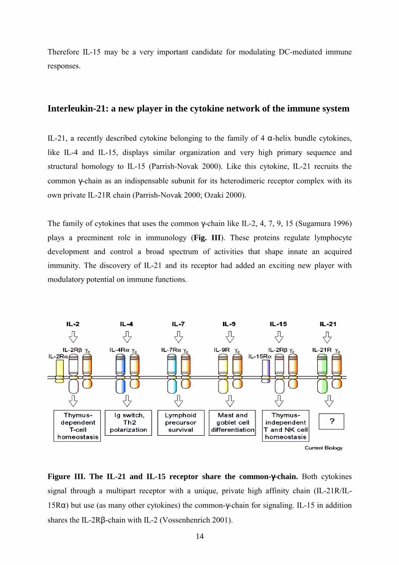
The family of cytokines that uses the common γ-chain like IL-2, 4, 7, 9, 15 (Sugamura 1996)
plays a preeminent role in immunology (Fig. III). These proteins regulate lymphocyte
development and control a broad spectrum of activities that shape innate an acquired
immunity. The discovery of IL-21 and its receptor had added an exciting new player with
modulatory potential on immune functions.
Figure III. The IL-21 and IL-15 receptor share the common-γγγγ-chain. Both cytokines
signal through a multipart receptor with a unique, private high affinity chain (IL-21R/IL-
15Rα) but use (as many other cytokines) the common-γ-chain for signaling. IL-15 in addition
shares the IL-2Rβ-chain with IL-2 (Vossenhenrich 2001).

15
IL-21 is a product of activated, but not resting, T cells (Parrish-Novak 2000), an expression
pattern which parallels that of IL-2, indicating that one of its primary functions may be
associated with T cell immune responses. However, it had been shown that IL-21 influences
not only the proliferation of T cell but also of B cells and the cytolytic activity of NK and
CD8+ T cells as well as the differentiation of NK cells out of bone-marrow stem-cells
(Parrish-Novak 2000). In addition, IL-21 synergizes with IL-2 and IL-15 in regulating these
responses (Parrish-Novak 2002). Notably, IL-21 not only provides “positive” effects on IL-15
induced NK cell expansion or B cell proliferation but mediating the opposite, hence anti-
proliferating effects, giving rise to the assumption that IL-21 has the potential not only to
support but also to abrogate immune functions at various stages, dependent on co-stimulatory
signals provided.
In general, the tissue distribution of receptors offers a strong indication of their potential sites
of action. It has been reported that IL-21R is expressed in lymphoid tissues like thymus,
lymph node, peripheral blood leukocytes as well as NK-cells (Parrish-Novak 2000),
indicating that this receptor/ligand pair could play an important role in immunomodulatory
functions. Interestingly, IL-21R expression was also found in bone marrow cells matching the
fact that IL-21 promotes the differentiation of lymphoid cells out of BM stem cells. Other γ-
chain signaling cytokines, as IL-4 and IL-15 (see above), are important for myeloid cell
development like DC generation and their further maturation. However, whether IL-21 has an
influence on DC development remains to be elucidated.
In vivo models to analyze modulation of DC biology by IL-15 and IL-21
To study DC biology in vivo under the influence of modulatory cytokines like IL-15 and IL-
21, two well-defined delayed-type hypersensitivity (DTH) reactions were explored. One of
them is a mainly CD8+ T cell dependent model of contact hypersensitivity (CHS) (Becker
1993; Hofmann 1998; Grabbe 1998; Rückert 2002) and secondly a classical DTH response to
sheep red blood cells (SRBC) was applied. This classical DTH has the advantage to not only
consider CD8+ T cell mediated effects but also allows to assess the activation of CD4+ T cells
as well as B cells including their antibody production (Kunzendorf 1996).

16
The complex process of the CHS response which is a subclass of DTH response and therefore
overlapping with mechanisms in DTH reactions can generally be divided in two phases,
which are described in the following.
Phase 1: Sensitization
Epidermal immunization, initiated by painting with soluble low molecular weight haptens,
serving as model-antigens, induce activation of epidermal Langerhans cells and DCs, which
leave the epidermis, enter the local lymph nodes (Kripke 1990, Mehling 2000), and undergo
maturation, resulting in down-regulated antigen-uptake but enhanced expression of MHC II,
CD80 and CD86 molecules. In the DLNs, where Ag is presented to naïve T cells, specific
CHS effector T cells are generated. The passive process of painting an antigen can be
mimicked by an active sensitization through direct injection of ex vivo antigen-labeled DCs,
which circumvents the Ag-uptake in the periphery but induces an equal T cell priming in
DLNs (Fig. IV).
Figure IV. Sensitization phase. DCs – in the skin most notably Langerhans cells (LC) -
reside in normal skin in a resting, immature state, characterized by high endocytotic and
antigen-processing capacity. Topical hapten application results in cytokine secretion by
keratinocytes and DCs and other cells within the skin. This launches DC activation and
17
migration of Ag-bearing DCs towards regional lymph nodes (Step 1). During this process,
DCs process the Ag and acquire a mature, functional state, associated with cytokine
secretion, enhanced expression of co-stimulatory molecules and enhanced capacity to prime
naïve T cells (step 2). In the lymph node, DCs establish contact with T cells and activate them
by sufficient expression of Ag-MHC complexes along with co-stimulatory and adhesion
molecules (step 3). As a result of Ag-specific activation, primed T cells alter their migration
pathway and begin to recirculate through peripheral tissues (step 4).
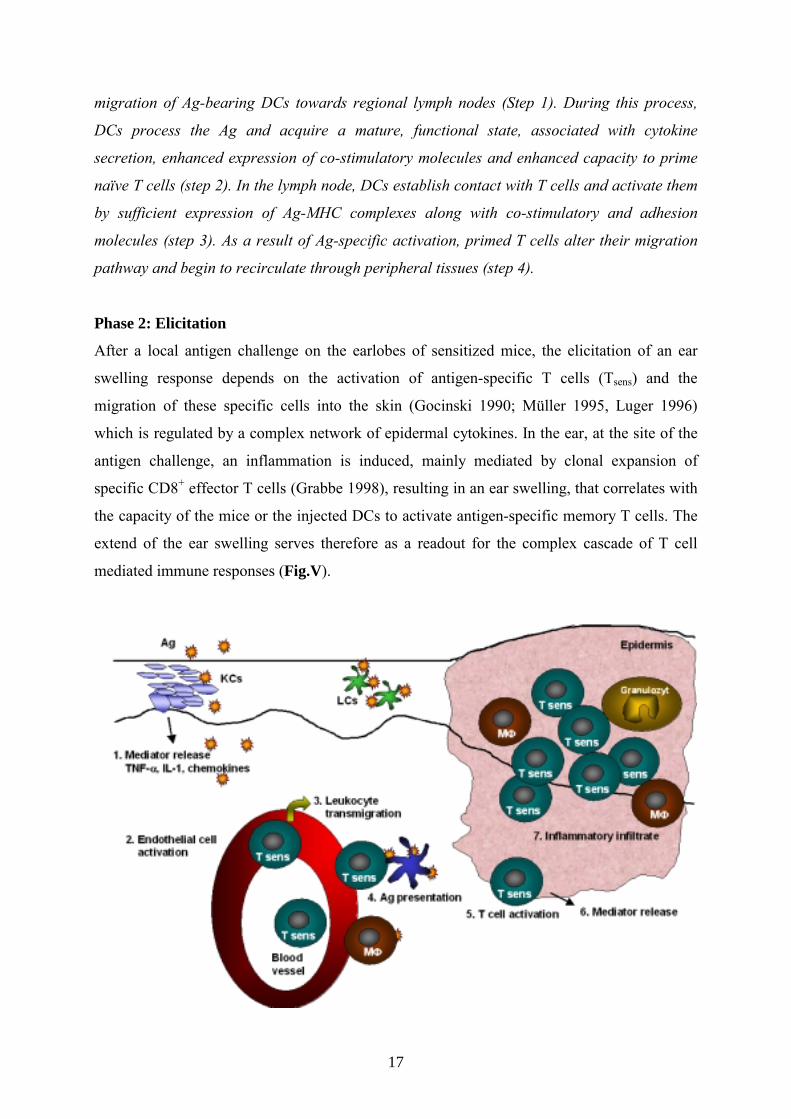
Phase 2: Elicitation
After a local antigen challenge on the earlobes of sensitized mice, the elicitation of an ear
swelling response depends on the activation of antigen-specific T cells (Tsens) and the
migration of these specific cells into the skin (Gocinski 1990; Müller 1995, Luger 1996)
which is regulated by a complex network of epidermal cytokines. In the ear, at the site of the
antigen challenge, an inflammation is induced, mainly mediated by clonal expansion of
specific CD8+ effector T cells (Grabbe 1998), resulting in an ear swelling, that correlates with
the capacity of the mice or the injected DCs to activate antigen-specific memory T cells. The
extend of the ear swelling serves therefore as a readout for the complex cascade of T cell
mediated immune responses (Fig.V).

18
Figure V. A hypothesis to describe the elicitation phase. Secondary Ag application for CHS
elicitation has the same primary effects on the skin as primary Ag contact during sensitization
(i.e. pro-inflammatory effects, DC activation). Whereas DC activation is the relevant event
during sensitization, nonspecific pro-inflammatory effects such as induction of cytokine
secretion (step 1), expression of adhesion molecules, and activation of endothelial cells (step
2) trigger elicitation of CHS, probably due to attraction of leukocytes to the site of Ag
application (step 3). Among these are primed T cells, which are activated upon Ag-
presentation by resident or infiltrating DCs and other APCs (step 4). A specific T cell
activation (step 5) induces mediator release (step 6), which amplifies the response by
generating an inflammatory process and leads to further accumulation of inflammatory cells
(step 7), resulting in clinically manifest contact dermatitis. The time between application of
antigen and elicitation of CHS is approximately 24h.
The second model used in this thesis, is the classical DTH response against SRBC. The
mechanisms of specific T cell priming by DCs in the classical DTH reaction are comparable
to the described events during CHS response. Nevertheless, the sensitization to SRBC affects
not only CD8+ but also CD4+ T cells, which provide co-stimulatory signals to B cells (“B cell
help”). Thus specific IgM-, IgG-antibody production is detectable. This gives the great
opportunity to analyze in parallel cellular and humoral immunity.
As a very important additional model system to analyze the effects of IL-15 and its high
affinity receptor, we also used IL-15- and IL-15Rα gene deficient mice. These approaches
were further supported by blocking the function of IL-15 in wildtype mice with the help of the
sIL-15Rα, which constitutes the extracellular part of the high affinity IL-15Rα (Fig. IV,
Ruchatz 1998).
Figure VI. Schematic structure of sIL-15Rα (A) and native, full length IL-15Rα (B) is shown.
Questions addressed DCs are professional antigen presenting cells that initiate T cell dependent immune responses,
and which pass through different functional states of activation, modulated by cytokines that
signal through the common γ-chain as described for IL-4. Given the structural relation
between IL-21 and the IL-15/IL-4 cytokines together with their common use of the γ-chain it
was reasonable to speculate that they influence DC development in similar ways.
This led us to investigate:
- the ability of IL-15 and IL-21, to influence differentiation, maturation and function of
bone marrow derived DCs.
- the role of DC-derived IL-15 in the induction of antigen specific T cell reaction in
vivo and in vitro.
- the mechanisms by which IL-15 influences DC-mediated T cell priming.
Such investigations will give us new insights in the development of DC mediated T cell
immune responses and propose novel strategies to prevent the immune system from
overwhelming reactions.

Material and Methods Mice
C57BL/6 IL-15-/- (Kennedy 2000) and C57BL/6 IL-15Rα-/- (Lodolce 1998) mice were bred
at the Research Center Borstel (Germany). As controls served C57BL/6 wildtype (IL-15+/+)
mice, which were purchased from Charles River (Sulzfeld, Germany). OTIItg mice, whose
transgenic CD4-TCRs specifically recognize the OVA(323-339) epitope (Robertson 2000) were
kindly provided from IFFA Credo (France). OTItg mice (Rotzschke 1991), which are
transgenic for a CD8+ T cell restricted TCR, recognizing OVA(257-264) peptide were kindly
provided by Arne von Bonin, Bernhard-Nocht-Institute (Hamburg, Germany). All mice were
used between 10-12 weeks age and maintained in specific pathogen-free conditions. All in
vivo experiments were performed in compliance with national and institutional guidelines.
Culture medium and antibodies
Cells were cultured in RPMI 1640 medium (Biochrom, Berlin, Germany) supplemented with
10% (v/v) heat-inactivated fetal calf serum (FCS) from Biochrom, 2mM L-glutamine (Life
technologies, Karlsruhe, Germany), 100U/ml penicillin and 100µg/ml streptomycin (PAA,
Linz, Austria), and 50µM β-mercaptoethanol (Life technologies) at 37°C in 5% CO2.
Unless otherwise specified, all antibodies were from Pharmingen (Heidelberg, Germany).
[3H]thymidine was purchased from Amersham Biosciences (Freiburg, Germany).
DC preparation
Bone marrow-derived DCs (BMDCs) were generated as previously described (Lutz 1999).
Briefly, bone marrow cells were flushed from the femurs and tibias of mice and erythrocytes
were lysed in 155mM NH4Cl solution for 15min at room temperature. 2x10⁶ cells were seeded
in bacterial grade, non adherent petridishes (Falcon, Becton Dickinson, Heidelberg, Germany)
in 10ml medium supplemented with 20ng/ml GMCSF (Tebu, Frankfurt, Germany) or in
addition to GMCSF plus 20ng/ml IL-15 (RnD, Wiesbaden, Germany) or 20ng/ml IL-21
which was kindly provided by D. Foster, ZymoGenetics (Seattle, U.S.A.). At day 3, another
10ml medium containing 20ng/ml GMCSF alone or plus IL-15 or IL-21 were added to the
plates. At day 6 half of the cell-free culture supernatant was exchanged, and fresh medium
containing 10ng/ml GMCSF alone or plus IL-15 or IL-21 was added. At day 8, cells were
gently removed from the culture dishes by Accutase treatment (PAA) and washed twice with

21
medium. Purity was routinely >95% as determined by flow cytometric analysis of CD11c+
expression.
Antibodies and flow cytometric analysis
The phenotype of the BMDCs was analyzed using following mAbs: CD11c (FITC or APC
labeled), MHC class II (IA/IE), CD80, CD25, CD122, γc, CD40, CD54, CD11b, CD95,
CD95L, OX40L and CD86 (Southern Biotechnology, Birmingham, USA) (all PE
conjugated). PE and FITC conjugated anti-CD3, -CD8α, -CD45/B220, -Gr1, -DX5, -NK1.1
(all from Pharmingen) and -F4/80 (Serotec, Eching, Germany) were used as a control and
found negative. We also stained for IL-15 binding on BMDCs with an IL-15-IgG2b fusion
protein (Bulfone-Paus 1997b) and secondary with biotinylated goat anti-mouse IgG2b
(Southern Biotec, Birmingham, USA) followed by streptavidin conjugated PE (Dianova,
Hamburg, Germany). Mouse IgG2b (Sigma, Taufkirchen, Germany) was used as control.
T cells were analyzed with PE and FITC conjugated antibodies specific for CD3 (145-2C11),
CD8α (53-6.7), CD25, CD44, CD62L, CD69, CD122, and CD4-APC (RM4-5). Unless
otherwise specified, all antibodies were from Pharmingen (Heidelberg, Germany). Propidium
iodide at 2µg/ml (Sigma) was added in all probes to exclude dead cells.
For staining procedure 2x10⁵ cells were incubated with 1µl mAb for 15min on ice in a total
volume of 100µl. After incubation cells were washed with PBS/EDTA/BSA. Flow cytometric
analysis was performed with a FACSCalibur, equipped with standard filters (Becton
Dickinson, Heidelberg, Germany) and CELLQuest software (Becton Dickinson).
Histomorphometry
Harvested BMDCs (5x10⁴) were transferred in 300µl fresh medium onto 8 well chamber
slides (Nunc, Wiesbaden, Germany) and incubated for 48h at 37°C, 5% CO2 to allow
adhesion and spreading of dendrites. Slides were stained with Pappenheim after drying and
pictures were taken with an Olympus Camedia digital camera adapted to an inverse Olympus
CK 40 microscope (Olympus, Hamburg, Germany).
RT-PCR
Total RNA was extracted from 5x10⁶ cells after 8 days of culture using RNAzol (Life
technologies) following the manufacturer’s instructions. cDNA was synthesized using random
hexanucleotide primers and the Superscript preamplification system II (Life technologies).
Obtained cDNA was amplified in a 20µl reaction, using 1U of AmpliTaq DNA polymerase

22
(Roche, Mannheim, Germany), 250µM of each dNTP and 2µl 10-fold PCR buffer (Roche).
The final concentration of each primer was 0.5µM. Thermal cycling conditions were 5min at
94°C and additional 30sec at 94°C. Annealing temperature was 60°C for 30sec, followed by
extension at 72°C for 30sec. 30 cycles were performed and ended with final extension at 72°C
for 10min. Reaction products were analyzed on a 1.5% agarose gel and visualized by
ethidium bromide staining. All primers used were commercially synthesized and obtained
from TIB Molbiol (Berlin, Germany) or Metabion (Martinsried, Germany). Primer sequences
were as follows: IL-21R sense: 5´-CTC AGC CAG GCA CTT CAT TCA GG-3´, IL-
21R antisense 5´-ATC ACA GGA AGG GCA TTT AGC-3´; IL-15Rα sense: 5´-AAC ATC
CAC CCT GAT TGA GTG T-3´, IL-15Rα antisense 5´-GTT TCC ATG GTT TCC ACC
TCA A-3´; IL-15 sense: 5´-GGC ATT AAG TAA TGA AAA TTT TGA-3´, IL-15 antisense
5´-CTC GCA TGC AGT CAG GAC GTG TTG-3´; IL-21 sense: CCC TTG TCT GTC TGG
TAG TCA TC-3´, IL-21 antisense 5´-ATC ACA GGA AGG GCA TTT AGC-3´; IL-
2Rα sense: 5´-GGA TCC AAG ATG GAG CCA CGC TTG CTG ACG-3´; IL-2Rα antisense
5´-AAG CTT TCA ATA CTC CAT AGT GAG CAC AAA TGT CAC C-3´; IL-2Rβ sense:
5´-GTC GAC GCT CCT CTC AGC TGT GAT GGC TAC CAT A-3´, IL-2Rβ antisense 5´-
GGA TCCCAG AAG ACG TCT ACG GGC CTC AAA TTC CAA-3´; γc sense: 5´-GTC
GAC AGA GCA AGC ACC ATG TTG AAA CTA-3´, γc antisense 5´-GGA TCC TGG GAT
CAC AAG ATT CTG TAG GTT-3´. β-actin sense 5´-GTG GGG CGC CCC AGG CAC CA-
3´, β-actin antisense 5´-CTC CTT AAT GTC ACG CAC GAT TTC-3´. To exclude
contaminations all experiments were run with a mock PCR. β-actin message expression was
used to normalize the cDNA amount. As positive controls for IL-21 and its receptor cDNA
from CD4+ T cells from C57BL/6 mice was isolated (Parrish-Novak 2000). For IL-15 and its
receptor we used cDNA from L929 fibroblasts (Bulfone-Paus 1999) and CTLL-2 cells were
the positive reference for all IL-2R chains (Tagaya 1996).
Cytokine detection
DCs (1x10⁶/ ml) in 24 well plates were stimulated for 24h with LPS (10ng/ml). Culture
supernatants were collected and analyzed for mouse cytokines using a Bio-PlexTM kit (Bio-
Rad, Munich, Germany). 50µl supernatant per sample was analyzed on the Luminex 100TM
according to manufacture’s instruction.
23
Analysis of endocytosis by flow cytometry
To quantify the endocytic activity the uptake of FITC-dextran (MW: 70.000, Molecular
Probes, Göttingen, Germany) was monitored by FACS analysis as described by Stumbles et
al (Stumbles 1998). In brief, 5x10⁵ DCs were re-suspended in 100µl RPMI/FCS containing
0.5mg/ml FITC-dextran for 30min respectively 60 or 90min at 37°C or on ice. Cells were
washed twice with ice-cold PBS/BSA and fluorescence intensity was determined by FACS.
DC activation by LPS
DCs were generated by culturing BM cells for 8 days with GMCSF as described above. These
DCs were cultured for another 24h in medium or with low dose LPS (10ng/ml) to induce DC
activation and maturation. To analyze the effects of IL-15 and IL-21 on this in vitro
maturation process, DCs were incubated with a combination of LPS and IL-15 or IL-21
(100ng/ml). Cytokine and LPS concentrations were chosen out of several titration
experiments. As control, the anti-inflammatory cytokine IL-10 (100ng/ml) was used, which
has been shown to inhibit DC maturation (Takayama 2001). After 24h, the surface phenotype
of the DCs was analyzed by FACS.
DC activation by various stimuli
DCs were generated by culturing BM cells for 8 days as described. All DC types were
cultured for another 24h with 10µg/ml OVA protein (Sigma), with 2µg/ml OVA323-339 peptide
or with low dose LPS (10ng/ml) to induce DC activation and maturation. In addition all DCs
were labeled with FITC (12,5µg/ml) as described in detail below. OVA-protein, -peptide and
LPS concentrations were chosen out of several titration experiments. As control only medium
was supplemented to the cultures. After 24h the surface phenotype of the different DCs was
analyzed by FACS.
Proliferation assays
T cells were isolated from lymph nodes (LN) of OTI and OTII TCR transgenic mice. 1x10⁵
DCs (IL-15+/+ and IL-15-/-) were aliquoted into 96-well flat-bottom culture plates (Costar
Corning, Cambridge, MA) and allowed to adhere for 2h. CD4+ cells were added at 2x10⁵ cells
per well. 0.1µM OVA(323-339) peptide (optimal concentration defined by titration experiments)
was supplemented to a final volume of 200µl. The cultures were incubated for 72h and
labeled for additional 18h with 0.2µCi/well [3H]thymidine.

24
Antigen specific CD8+ T cell proliferation assays were performed as follows: DCs were
labeled with 2µg OVA(257-264) peptide for 2h at 37°C in 500µl medium, washed twice and
plated-out into 96-well round-bottom culture plates (Greiner, Hamburg, Germany) at a density
of 1x10⁴ cells per well along with 1x10⁵ LN cells. After 48 hours, plates were pulsed for 18h
with 0.2 µCi/well [3H]thymidine. [3H]thymidine uptake was analyzed by liquid scintillation
counting (Wallac/ PerkinElmer, Freiburg, Germany). The experiments were repeated three
times with six replicates each.
In vitro differentiation of CD8+ T cells
To analyze the CD8+ T cell stimulation in vitro, 2x10⁵ DCs from IL-15+/+ and IL-15-/- mice
were labeled with OVA(257-264) peptide as described above and plated in 24 well plates
together with 2x10⁶ spleen cells from OVA CD8+ T cell transgenic OTI mice in 1ml RPMI
for 48h. Cells were harvested, and CD8+ T cells were analyzed by FACS for induction of
memory cells or apoptosis using Annexin V/Pi kit (BenderMed Systems, Vienna, Austria).
Confocal Microscopy
IL15+/+, IL15-/-, and IL-15Rα-/- DCs after 8 days culture were seeded in concentration of
5x10⁴ cells/well in 4-wells cover glass system (Lab-Tek II, Nalge Nunc Int., Naperville, USA)
the day before staining. Next day cells were fixed with 2% paraformaldehyde for 10min at
room temperature, permeabilized by 0.25% Triton X-100 for 10min and stained with 1:100
dilutions of antibodies recognizing IL-15Rα (H-109) (Santa Cruz Biotechnology, Santa Cruz,
USA) in combination with anti-IL-15 antibodies (L-20) (Santa Cruz Biotechnology) for
30min at room temperature. Alexa Fluor-488 donkey anti-goat IgG (H+L) and Alexa Fluor-
546 goat anti-rabbit IgG (H+L) (Molecular probes, Leiden, Netherlands) in dilution of 1:100
were used as second antibodies. Nuclei were stained using TOTO-3 dye (Molecular probes).
To stain cell membranes, fixed cells were incubated with wheat germ agglutinin (WGA,
Molecular Probes) for 15min at room temperature, washed and permeabilized with 0.25%
Triton X-100 followed by staining with primary and secondary antibodies. Cells were
analyzed by scanning confocal microscopy (Leica, Ruel-Malmason, France).
In vivo DC migration
In order to examine whether the differently generated DCs migrate with equal efficiency,
local draining lymph nodes (DLN) were examined. DCs were labeled with FITC as described
below and 5x10⁵ labeled DCs were injected s.c. in the hind footpad. To assess that FITC was

25
not taken up by other cells, in additional experiments, FITC labeled DCs were fixed with
0.1% glutaraldehyde and injected s.c in the hind footpad (Eggert 1999). After 24h local DLN
were removed and prepared as described (Vremec 1997). 1x10⁶ cells were stained with
αCD11c-PE Ab and 1x10⁵ cells were analyzed by flow cytometry.
Sensitization to picryl chloride (PCl) and elicitation of CHS
IL-15+/+ or IL-15-/- mice were sensitized as described (Rückert 2002) on the shaved
abdominal skin by a single painting with 50µl of 1% (w/v) picryl chloride (2,4,6-
trinitrochlorobenzene; Tokyo Kasei Co., Tokyo, Japan) in acetone/ethanol (1:3) on day 0.
Negative controls were painted with diluents. For the elicitation of the CHS, 1% PCl solved in
olive oil (20µl) was painted on both sides of one earlobe on day 5 after sensitization. Ear
thickness was measured with a spring-loaded micrometer (Mitutoyo, Elk Grove Village,
U.S.A.) before challenge and 24, 48 and 72h after challenge(Fig. A). For each mouse, the ear
swelling response was expressed as the mean increment in thickness above the basal control
value.
Where indicated, mice were injected i.p. with 50µg of soluble IL-15Rα (Ruchatz 1998) as IL-
15 antagonist in 200µl PBS, 30min before sensitization and once daily until day 5, when the
mice were challenged for the first time. A second challenge was performed at day 19 and ear
swelling was measured again 24, 48 and 72h after the second challenge.
Figure A. CHS Model to picryl chloride (PCl).
Alternatively, 72h after the first challenge mice were sacrificed and cells from DLNs were
isolated and stained for FACS as described above to estimate CD4+/ CD8+ ratios as well as
the expression of activation and memory markers on T cells.
26
Contact hypersensitivity model to FITC
To estimate the capacity of DCs to induce antigen specific T cell sensitization in vivo,
BMDCs (1x10⁶ cells/ml) from IL-15+/+, IL-15-/-, or IL-15Rα-/- mice were labeled with
12.5µg/ml of FITC (Sigma) or FITC-conjugated OVA (Harlow 1988) in PBS for 20min at
37°C as previously described (Macatonia 1986). Alternatively, IL15-/-DCs or IL-15Rα-/-DCs
were incubated for 30 min with 20ng/ml IL-15 at 37°C after FITC labeling. Short-time effects
of IL-15 and IL-21 were analyzed by incubation IL-15+/+DCs for 2h with 100ng/ml of the
cytokines before FITC labeling. Cells were washed 3 times with ice-cold RPMI/FCS medium,
resuspended and injected in one footpad with a Hamilton syringe (5x10⁵ cells in 50µl PBS).
After 5 days, mice were challenged by applying 50µl FITC (3.5mg/ml in
acetone:dibutylphtalate, 1:1, Sigma) on the dorsal and ventral sides of the right ear. As control
the left ear was painted with an equal amount of the diluent. In addition, unsensitized mice
were painted with FITC. CHS response was determined by measuring the ear swelling at 24,
48 and 72h after challenge as described above.
Irritant dermatitis to croton oil
To analyze the capacity of IL-15+/+ and IL-15-/- mice to mount an unspecific, antigen-
independent, irritant contact dermatitis (Zhang 2000), croton oil (Sigma) at 0.8% (v/v)
dissolved in acetone was painted on one ear. Painting of acetone alone served as negative
control. Ear swelling was analyzed at 24, 48, and 72h.
Th1 model of DTH reaction to SRBC
As previously described (Kunzendorf 1996), mice were sensitized by i.v. injection of 4x10⁵
sheep red blood cells (SRBC) in 100µl PBS and challenged at day 5 by injecting 2x10⁸ SRBC
in 50µl PBS into the left hind footpad. Non-immunized mice were injected with the same
dose of SRBC to determine non-specific swelling. Where indicated, mice were injected i.p.
with 50µg of sIL15Rα in diluent 30min before sensitization, and once daily until day 5, i.e.
when mice were challenged for the first time (Fig. B). 24, 48 and 72h after challenge the
footpad swelling was measured as above described. A second challenge was performed at day
19. Control groups were injected with diluent.

27
Figure B. DTH model to sheep red blood cells (SRBC).
Eight days after primary immunization, serum was collected, and hemagglutinin titers to
SRBC were measured as described (Kunzendorf 1996). In brief, the serum was incubated at
56°C for 30min and diluted using NaCl 150mM with or without 0.78% mercaptoethanol in
serial log 2 dilutions. 200µl of a 0.5% SRBC solution was added to 200µl of the serum
dilutions, incubated at 37°C for 30min, at room temperature for 2h and at 4°C for additional
12h. The serum hemagglutinin titers are the highest log 2 dilution steps in which
hemagglutination could still be determined.
Homing and activation of APC
Mice were painted on the shaved abdomen with 400µl of 0.35% FITC solution (Sigma) in
acetone: dibuthylphtalate (1:1) as previously described (Macatonia 1986). 24h later, the mice
were sacrificed and their axillary, cervical, and inguinal DLN removed, pooled and 100.000
cells were analyzed by FACS for FITC uptake. Dendritic cells (DCs) were stained with APC
labeled anti-CD11c and PE labeled anti-CD80 and -CD86 antibodies.
Statistical analysis
Results are presented as mean ± SD from pooled data of 2-3 identical experiments, (using a
total of 10-15 mice/group) all of which gave comparable results. FACS data from one
representative experiment of at least five are shown. The Student’s t-test for unpaired samples
was used for the determination of statistical differences (* p ≤ 0.05; ** p ≤ 0.01).

28
Results
CD11c+ DCs differentiate in the presence of IL-15 or IL-21 in vitro IL-21 and IL-15 are structural relatives and belong, like IL-4, to the 4 α-helix bundle cytokine
family. All three cytokines share the common γ-chain in their receptor complexes but have
their own, private receptor unit (IL-21R, IL-15Rα, IL-4Rα). Recent studies revealed that IL-
15 is critical for APC activation (Mattei 2001) and essential in the development of lymphoid
cell lineages (Kennedy 2000). IL-21 was found to act synergistic to IL-15 in the generation
and activation of lymphoid cells (Parrish-Novak 2000). On the other hand it is well accepted
that the expansion of in vitro generated bone marrow (BM)-derived DCs is highly promoted
by IL-4, (Sallusto 1994). Taken together it is evident that all these cytokines have a specific
influence on stem-cell differentiation and their further maturation.
As IL-4, IL-15 and IL-21 receptors share the common γ-chain, we analyzed whether IL-15
and IL-21, just like IL-4, may modulate the GMCSF–induced differentiation of BM stem cells
into DCs. Therefore, freshly isolated BM cells were cultured following established protocols
(Inaba 1992; Lutz 1999) with GMCSF alone (subsequently designated DCs) or GMCSF
combined with 20ng/ml IL-15 (named IL-15DCs) or 20ng/ml IL-21 (IL-21DCs). In a typical
experiment, after 8 days of culture 60-80% of the cells appear as loosely adherent clumps or
isolated floating cells with the typical dendritic cell morphology. DCs were transferred into
microchamber slides at day 8 and allowed to adhere for 48h. After Pappenheim staining the
differentially generated DCs displayed a characteristic DC phenotype such as cytoplasmic
protrusions (“dendrites”) (Fig. 1). In FACS analysis after 8 days of culture, the IL-15DCs and
IL-21DCs showed comparable size and granularity like DCs indicated by same forward-
(FSC), sideward-scatter (SSC) properties (Fig. 1). Further, the percentage of CD11c+ cells, a
specific marker for murine DCs, was similar in all conditions (Fig. 1). Moreover we assessed
by FACS, that adding IL-15 or IL-21 to BM cells in combination with GMCSF did not lead to
increased differentiation of other cell types like B cells (CD45/B220+), NK cells (NK1.1+), T
cells (CD3+), granulocytes (Gr1+), macrophages (F4/80+) or CD8α+ lymphoid DCs (not
shown). Thus, IL-15 and IL-21 neither did alter the purity of the generated DCs nor the total
yield of DCs compared to GMCSF alone (mean of 7.5x10⁶ cells per petridish out of 2x10⁶
seeded for all conditions). Given IL-15 and IL-21 alone (without GMCSF) to BM cells did
not induce differentiation of DCs (not shown).

29
Figure 1. Generation of DCs. Murine BM cells were induced to differentiate to DCs by
GMCSF (DCs), or by addition of IL-15 (IL-15DCs), or IL-21 (IL-21DCs) in combination with
GMCSF during the entire culture period of 8 days. Morphology was assessed by culturing the
DCs for 48h on micro chamber slides following Pappenheim staining. The scale bar is equal
to 100µm. Further; cells were analyzed by FACS at day 8. All cells showed no differences in
their size and granularity (2nd column, forward- and sideward-scatter, FSC, SSC).
Additionally, cells were stained for CD11c expression. The 3rd column shows that about 90%
of all cells expressed CD11c and therefore are considered to be DCs. Unstained cells served
as negative controls (bold lines). Shown is one representative out of 10 experiments.
IL-21 and IL-15 receptors are expressed in BM derived DCs The IL-15 receptor complex, which is composed of a private α-subunit, the IL-2/IL-15Rβ-
and the common γ-chain, is expressed on DCs (Mattei 2001, Fukao 2000). Using northern
blot analysis, expression of the private IL-21R subunit was previously found to be most
30
abundant in lymphoid tissues (Parrish-Novak 2000). In addition, both private receptor chains
for IL-15 and IL-21 are expressed in BM stromal cells.
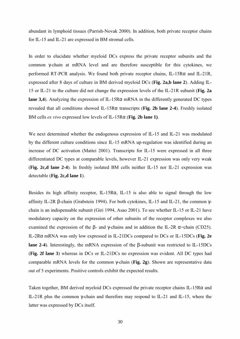
In order to elucidate whether myeloid DCs express the private receptor subunits and the
common γ-chain at mRNA level and are therefore susceptible for this cytokines, we
performed RT-PCR analysis. We found both private receptor chains, IL-15Rα and IL-21R,
expressed after 8 days of culture in BM derived myeloid DCs (Fig. 2a,b lane 2). Adding IL-
15 or IL-21 to the culture did not change the expression levels of the IL-21R subunit (Fig. 2a
lane 3,4). Analyzing the expression of IL-15Rα mRNA in the differently generated DC types
revealed that all conditions showed IL-15Rα transcripts (Fig. 2b lane 2-4). Freshly isolated
BM cells ex vivo expressed low levels of IL-15Rα (Fig. 2b lane 1).
We next determined whether the endogenous expression of IL-15 and IL-21 was modulated
by the different culture conditions since IL-15 mRNA up-regulation was identified during an
increase of DC activation (Mattei 2001). Transcripts for IL-15 were expressed in all three
differentiated DC types at comparable levels, however IL-21 expression was only very weak
(Fig. 2c,d lane 2-4). In freshly isolated BM cells neither IL-15 nor IL-21 expression was
detectable (Fig. 2c,d lane 1).
Besides its high affinity receptor, IL-15Rα, IL-15 is also able to signal through the low
affinity IL-2R β-chain (Grabstein 1994). For both cytokines, IL-15 and IL-21, the common γ-
chain is an indispensable subunit (Giri 1994, Asao 2001). To see whether IL-15 or IL-21 have
modulatory capacity on the expression of other subunits of the receptor complexes we also
examined the expression of the β- and γ-chains and in addition the IL-2R α−chain (CD25).
IL-2Rα mRNA was only low expressed in IL-21DCs compared to DCs or IL-15DCs (Fig. 2e
lane 2-4). Interestingly, the mRNA expression of the β-subunit was restricted to IL-15DCs
(Fig. 2f lane 3) whereas in DCs or IL-21DCs no expression was evident. All DC types had
comparable mRNA levels for the common γ-chain (Fig. 2g). Shown are representative data
out of 5 experiments. Positive controls exhibit the expected results.
Taken together, BM derived myeloid DCs expressed the private receptor chains IL-15Rα and
IL-21R plus the common γ-chain and therefore may respond to IL-21 and IL-15, where the
latter was expressed by DCs itself.
31
Figure 2. mRNA expression of IL-15 and IL-21 and their receptors. Freshly isolated bone
marrow (BM, lane 1) as well as DCs (lane 2), IL-15DCs (lane 3) and IL-21DCs (lane 4) were
analyzed by RT-PCR at day 8 for mRNA expression of IL-21R, IL-15Rα, IL-15, IL-21, and IL-
2R α- and β-chain as well as for the common γ-chain. β-actin message expression was used
to normalize the cDNA amount. To exclude contaminations all experiments were run with a
mock PCR and found negative (not shown). The correct size of PCR product was assessed by
a marker (M). As positive controls (C) for IL-21 and its receptor cDNA from activated CD4+
T cells from C57BL/6 mice was isolated. For IL-15 and its receptor we used cDNA from L929
fibroblasts and CTLL-2 cells were the positive reference for all IL-2R chains. Shown is one
representative experiment out of 5.
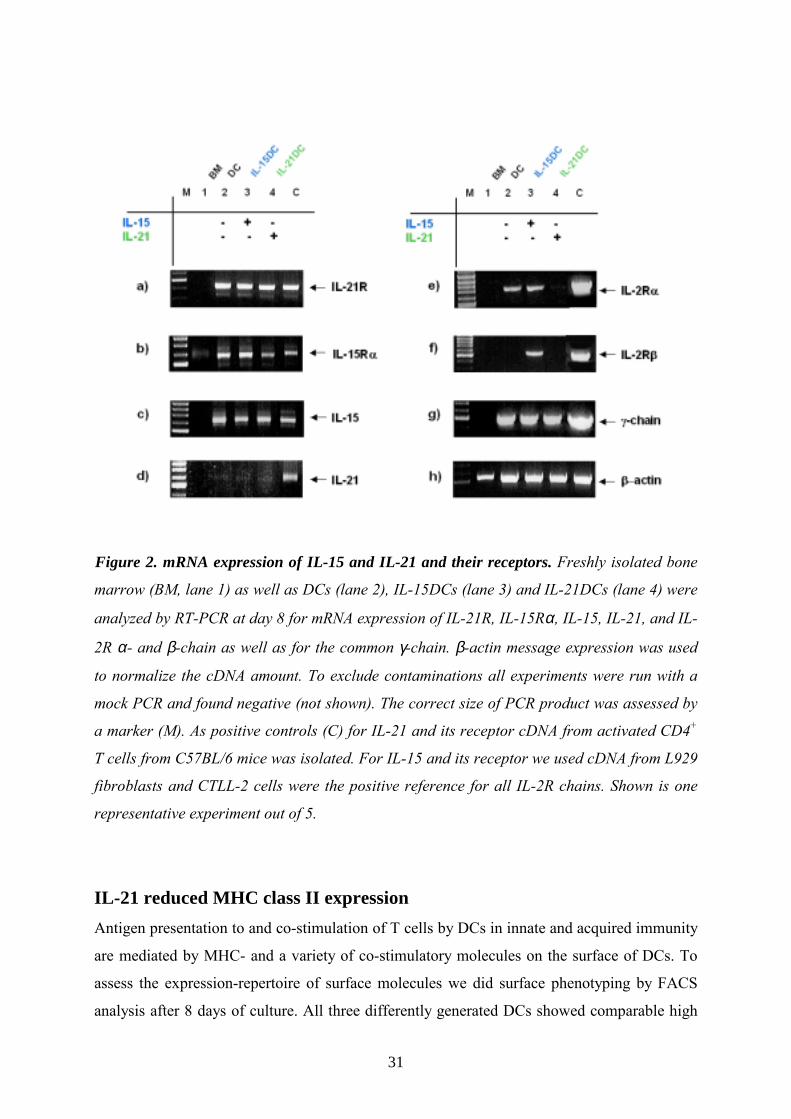
IL-21 reduced MHC class II expression Antigen presentation to and co-stimulation of T cells by DCs in innate and acquired immunity
are mediated by MHC- and a variety of co-stimulatory molecules on the surface of DCs. To
assess the expression-repertoire of surface molecules we did surface phenotyping by FACS
analysis after 8 days of culture. All three differently generated DCs showed comparable high
32
levels of CD80 whereas CD86 expression was reduced in IL-15DCs as well as in IL-21DCs
(Fig. 3a). Moreover, generation in the presence of IL-21 resulted in a significantly decreased
expression of MHC class II molecules (Fig. 3a) when compared with DCs or IL-15DCs.
a
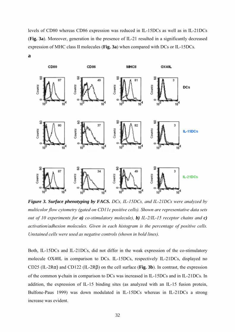
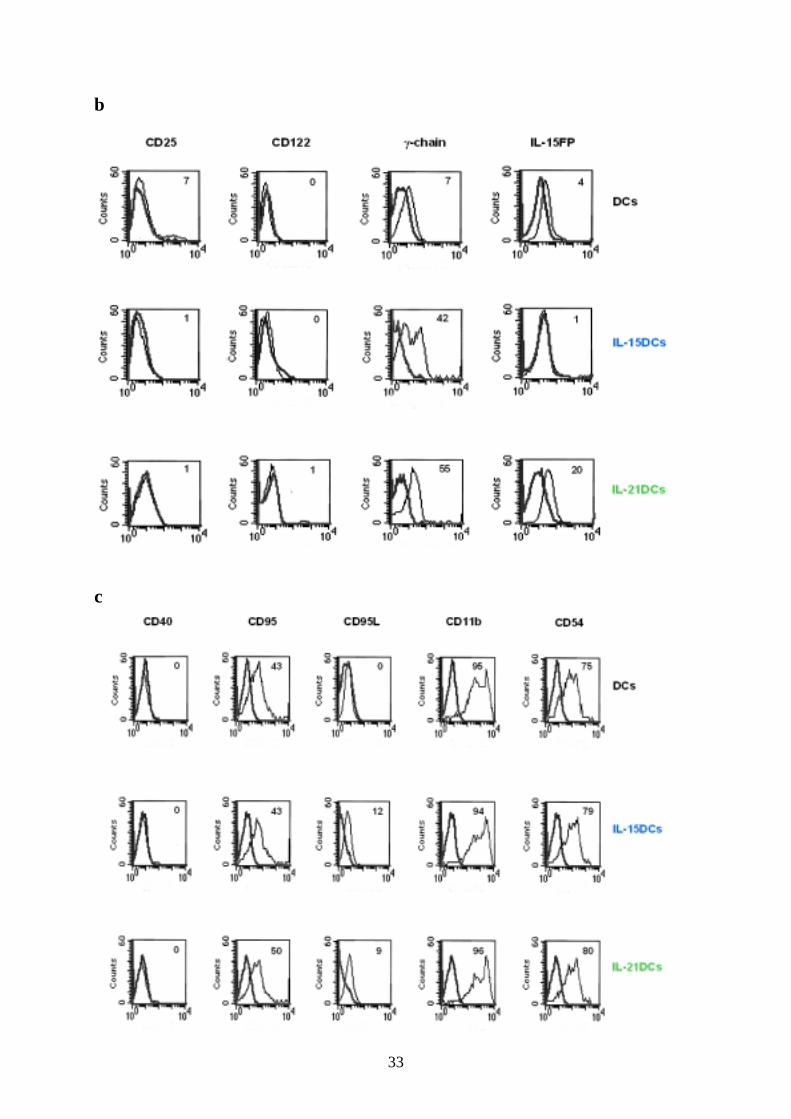
Figure 3. Surface phenotyping by FACS. DCs, IL-15DCs, and IL-21DCs were analyzed by
multicolor flow cytometry (gated on CD11c positive cells). Shown are representative data sets
out of 10 experiments for a) co-stimulatory molecule), b) IL-2/IL-15 receptor chains and c)
activation/adhesion molecules. Given in each histogram is the percentage of positive cells.
Unstained cells were used as negative controls (shown in bold lines).
Both, IL-15DCs and IL-21DCs, did not differ in the weak expression of the co-stimulatory
molecule OX40L in comparison to DCs. IL-15DCs, respectively IL-21DCs, displayed no
CD25 (IL-2Rα) and CD122 (IL-2Rβ) on the cell surface (Fig. 3b). In contrast, the expression
of the common γ-chain in comparison to DCs was increased in IL-15DCs and in IL-21DCs. In
addition, the expression of IL-15 binding sites (as analyzed with an IL-15 fusion protein,
Bulfone-Paus 1999) was down modulated in IL-15DCs whereas in IL-21DCs a strong
increase was evident.
33
b
c

34
All DC types showed similar high expression of the adhesion molecules CD54 and CD11b
and of CD95 (Fas) and had no CD40 on their surface (Fig. 3c). CD95L (FasL) was only
expressed moderately in IL-15DCs and IL-21DCs (Fig. 3c).
Although the quantity of DCs given in cell number and morphology was not altered by adding
IL-21 or IL-15 during generation, the phenotype of the DCs shown by expression of
functional relevant antigen-presenting and co-stimulatory molecules was modulated.
Increased antigen uptake by IL-21DCs contrasted by low uptake by IL-
15DCs The phenotypic changes of DCs generated in the presence of IL-21 described above,
particularly the significantly reduced expression of the MHCII molecule, point to different
maturational stages. Immature DCs are characterized by low expression of accessory
molecules especially MHCII and their increased antigen-capture activity.
To asses whether low expression of MHCII on IL-21DCs also refers to altered Ag-uptake we
studied the endocytic activity using a fluorescent model-antigen. FITC dextran uptake was
monitored after 8 days of DC culture. After 30min of incubation IL-21DCs showed a
significantly increased FITC dextran uptake at 37°C (mean fluorescence: 495) (Fig. 4).
Figure 4. Antigen uptake (endocytosis) of FITC dextran at 37°C.
35
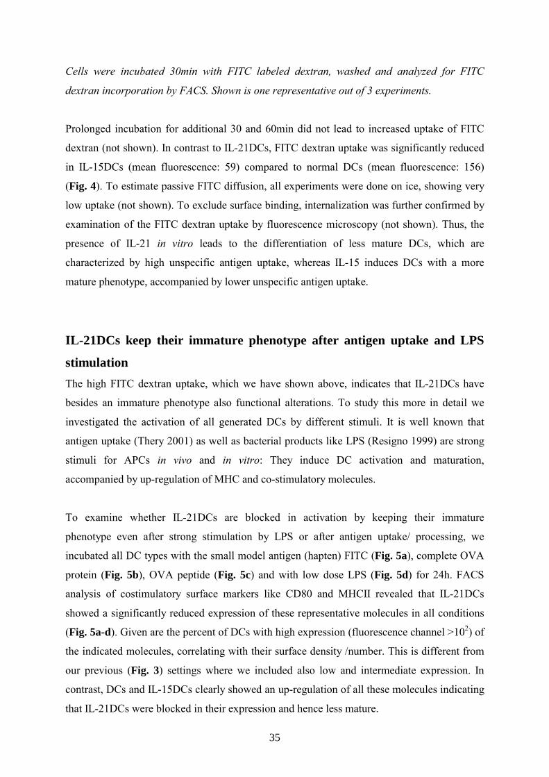
Cells were incubated 30min with FITC labeled dextran, washed and analyzed for FITC
dextran incorporation by FACS. Shown is one representative out of 3 experiments.
Prolonged incubation for additional 30 and 60min did not lead to increased uptake of FITC
dextran (not shown). In contrast to IL-21DCs, FITC dextran uptake was significantly reduced
in IL-15DCs (mean fluorescence: 59) compared to normal DCs (mean fluorescence: 156)
(Fig. 4). To estimate passive FITC diffusion, all experiments were done on ice, showing very
low uptake (not shown). To exclude surface binding, internalization was further confirmed by
examination of the FITC dextran uptake by fluorescence microscopy (not shown). Thus, the
presence of IL-21 in vitro leads to the differentiation of less mature DCs, which are
characterized by high unspecific antigen uptake, whereas IL-15 induces DCs with a more
mature phenotype, accompanied by lower unspecific antigen uptake.
IL-21DCs keep their immature phenotype after antigen uptake and LPS
stimulation The high FITC dextran uptake, which we have shown above, indicates that IL-21DCs have
besides an immature phenotype also functional alterations. To study this more in detail we
investigated the activation of all generated DCs by different stimuli. It is well known that
antigen uptake (Thery 2001) as well as bacterial products like LPS (Resigno 1999) are strong
stimuli for APCs in vivo and in vitro: They induce DC activation and maturation,
accompanied by up-regulation of MHC and co-stimulatory molecules.
To examine whether IL-21DCs are blocked in activation by keeping their immature
phenotype even after strong stimulation by LPS or after antigen uptake/ processing, we
incubated all DC types with the small model antigen (hapten) FITC (Fig. 5a), complete OVA
protein (Fig. 5b), OVA peptide (Fig. 5c) and with low dose LPS (Fig. 5d) for 24h. FACS
analysis of costimulatory surface markers like CD80 and MHCII revealed that IL-21DCs
showed a significantly reduced expression of these representative molecules in all conditions
(Fig. 5a-d). Given are the percent of DCs with high expression (fluorescence channel >102) of
the indicated molecules, correlating with their surface density /number. This is different from
our previous (Fig. 3) settings where we included also low and intermediate expression. In
contrast, DCs and IL-15DCs clearly showed an up-regulation of all these molecules indicating
that IL-21DCs were blocked in their expression and hence less mature.

36
Figure 5. Blocked maturation by various stimuli in IL-21DCs. All three generated DC types
were stimulated with a) FITC, b) OVA protein, c) OVA peptide and d) LPS for additional 24h
after 8 days of culture. Surface expression of co-stimulatory molecules CD80 and MHCII
(gated on CD11c+ cells) were analyzed by FACS. Given is the percentage of high positive
cells, defined by the marker. Shown is one representative out of two experiments.
IL-21DCs induce low antigen specific CD4+ T cell proliferation The capacity to induce specific T cell activation and proliferation is a functional in vitro (and
in vivo) hallmark of mature DCs whereas immature DCs fail to prime T cell responses. To
further characterize the immature stage of IL-21DCs and to assess the functional
consequences out of their impaired MHCII expression we set up an antigen specific T cell
proliferation assay.

37
Therefore, the different DCs were pulsed after 8 days of culture with OVA(323-339) peptide and
co-cultured for 72h with freshly isolated T cells from lymph nodes of syngenic OTIItg mice,
which have a transgenic T cell receptor for OVA(323-339) peptide. We compared the T cell
stimulatory capacity of DCs that had been cultured with GMCSF alone or in combination
with IL-15 or IL-21. As shown in Fig. 6 IL-15DCs showed a high significantly increased
ability to prime T cell proliferation (blue bars), compared to DCs (white bars), with a
maximum at OVA(323-339) peptide concentration of 0.3µM. In contrast IL-21DCs (green bars)
induced significantly lower T cell proliferation rates.
Figure 6. Co-stimulation by the different DCs. 100.000 DCs, IL-15DCs and IL-21DCs were
labeled with different OVA(323-339) peptide concentrations, washed, and incubated with
200.000 lymph-node cells from OT II mice with a transgenic CD4+ restricted T cell receptor,
which recognizes the OVA(323-339) peptide presented by MHCII. After 72h incubation cells
were labeled with 0.2µCi [3H]thymidine and analyzed 12h later. Shown is one representative
experiment out of 3. Significance compared to DCs was calculated using student’s t-test (**
p≤ 0.01).
This reduced ability of IL-21DCs to prime specific T cells could refer to the above described
reduced MHCII expression which could not be raised (even not after incubation with strong

38
stimuli like LPS) supporting the deduction that DCs generated in the presence of IL-21 are
less mature and - most important – do not acquire a mature phenotype after Ag uptake.
IL-21DCs are unable to prime in vivo contact hypersensitivity reaction to
FITC Small molecules, which act as antigens after protein binding are designated as haptens, and
induce a contact hypersensitivity (CHS) in the skin. Following primary application to skin,
epidermal DCs take up hapten-protein complex, process it and migrate towards the regional
lymph nodes, to prime an antigen-specific T cell response there. During this process, DCs
convert from an immature resting into an activated functional (mature) state (reviewed by
Grabbe 1998).
In comparison to our in vitro data, we wanted to examine whether the three types of DCs had
modulated capabilities to prime T cells in vivo and especially whether IL-21DCs were again
prevented from undergoing maturation and therefore less immunogenic. For this purpose we
labeled the different DCs in vitro with the fluorescent hapten FITC and injected them
subcutaneous in the footpad of syngenic C57BL/6 wildtype mice (sensitization phase of
CHS). After 5 days mice were challenged (elicitation phase of CHS) at the dorsal and ventral
site of one ear with FITC (the other ear with diluent only) and the ability to initiate an
antigen-specific, T cell mediated immune response was examined by measuring the FITC-
specific ear swelling 24, 48 and 72h after challenge. Mice, which had not been sensitized, but
challenged on the ear, served as negative controls for unspecific ear swelling. Mice that had
been actively sensitized with FITC labeled IL-15DCs (blue) showed a high significant
increase in the CHS ear swelling response 24h after challenge compared to mice, which had
been injected with DCs (Fig. 7a). In contrast injection of IL-21DCs (green) resulted in a high
significantly reduced CHS response at all three time points, i.e. no swelling above
unsensitized controls was observed (Fig. 7a).
To confirm that the sensitization was based on active migration of the injected, viable DCs
from the footpad to the DLN, additional control mice were injected with FITC labeled,
glutaraldehyde fixed (dead) DCs (Eggert 1999). These fixed cells failed to induce any CHS
response (not shown).
39
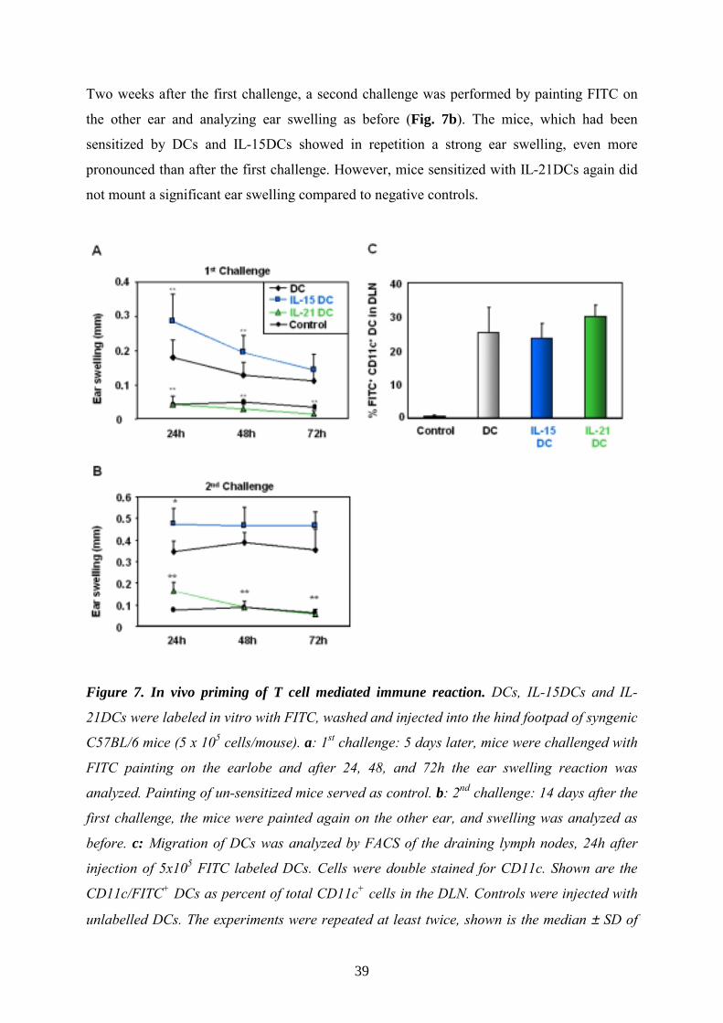
Two weeks after the first challenge, a second challenge was performed by painting FITC on
the other ear and analyzing ear swelling as before (Fig. 7b). The mice, which had been
sensitized by DCs and IL-15DCs showed in repetition a strong ear swelling, even more
pronounced than after the first challenge. However, mice sensitized with IL-21DCs again did
not mount a significant ear swelling compared to negative controls.
Figure 7. In vivo priming of T cell mediated immune reaction. DCs, IL-15DCs and IL-
21DCs were labeled in vitro with FITC, washed and injected into the hind footpad of syngenic
C57BL/6 mice (5 x 10⁵ cells/mouse). a: 1st challenge: 5 days later, mice were challenged with
FITC painting on the earlobe and after 24, 48, and 72h the ear swelling reaction was
analyzed. Painting of un-sensitized mice served as control. b: 2nd challenge: 14 days after the
first challenge, the mice were painted again on the other ear, and swelling was analyzed as
before. c: Migration of DCs was analyzed by FACS of the draining lymph nodes, 24h after
injection of 5x10⁵ FITC labeled DCs. Cells were double stained for CD11c. Shown are the
CD11c/FITC+ DCs as percent of total CD11c+ cells in the DLN. Controls were injected with
unlabelled DCs. The experiments were repeated at least twice, shown is the median ± SD of

40
12 mice. Significant differences between IL-15DCs and IL-21DCs compared to DCs were
calculated using the student’s t-test (*p≤0.05; **p≤0.01).
This clarifies that the mice sensitized with IL-21DCs were not able to establish antigen-
specific or memory T cells after the first FITC challenge in contrast to the other two groups,
which even showed a stronger recall induced inflammation with higher ear swelling after the
second challenge.
These findings are completely in line with our previous in vitro data stressing the fact that
combining GMCSF and IL-15 generates mature, “high immunogenic” DCs while IL-21
induces “functional immature” DCs, which are unable to mature to fully effective APCs after
antigen uptake neither in vitro nor in vivo.
Comparable migration of all DC types to DLN To rule out that the differences in CHS responses are due to altered migratory capacities of
the DCs, we investigated their abilities to enter draining lymph nodes (DLN) following
transfer in vivo (Lappin 1999).
For this purpose we labeled 0.5x10⁶ DCs with the fluorescent cell tracker FITC in PBS for
20min at 37°C and after washing subsequently injected the cells in the footpad. After 24h cell
suspensions from the inguinal DLNs were prepared as described (Vremec 1997), stained with
αCD11c antibody and analyzed by flow cytometry. Double positive cells (FITC+/CD11c+),
which migrated into the DLN, are given in percent of total CD11c+ DCs (Fig. 7c). As control,
unlabeled DCs were injected. The differently generated DCs showed similar migratory
capacities; 25-30% of the total lymph node CD11c+ DCs migrated within 24h from the
periphery into DLNs. This exhibits that the reduction of CHS responses by IL-21DCs in vivo
is not attributed to limited migration. In contrast, footpad-injected glutaraldehyde-fixed (dead)
FITC labeled DCs did not reach the DLN and no FITC uptake by other DCs of recipient mice
was observed, indicating that the observed migration into DLN is an active and specific
process (not shown).

41
IL-21 inhibits LPS induced activation of normal DCs Now that we found, that IL-21, when present during DC differentiation, prevent their
maturation, we subsequently wanted elucidate whether IL-21 could act also in short time
course on DC activation.
Therefore we again activated DCs, which differentiated for 8 days with GMCSF only, for
additional 24h with low dose LPS (10ng/ml) alone or in combination with 100ng/ml of IL-15
or IL-21. As a control we used the anti-inflammatory cytokine IL-10 that is known to inhibit
DC activation (Takayama 2001) by inhibiting up-regulation of co-stimulatory molecules and
blocking IL-12 expression (Corinti 2001). It was evident that the addition of IL-21 showed
similar properties like IL-10 in blocking the LPS induced up-regulation of CD80, CD86 and
IA/IE (MHCII) (Fig. 8a). This observation strongly supports the evidence that IL-21 prevents
also in vitro induced activation by LPS. Supplement of IL-15 to LPS did not further enhance
the effects on co-stimulatory molecules mediated by LPS alone. CD40 expression was
induced by LPS and not altered by the influence of any cytokine.
a
Figure 8a. Inhibition of LPS-induced DC activation.

42
Immature DCs were activated with low-dose LPS (10ng/ml) or with LPS plus IL-15, IL-21 or
IL-10 for 24h. Surface expression of co-stimulatory molecules (Fig.8a) or IL-2 / IL-15
receptors (Fig. 8b) were analyzed by FACS. Given is the percentage of high positive cells,
defined by the marker M1. Shown is one representative out of four experiments.
We further analyzed the modulation of the IL-2R subunits and the IL-15Rα by LPS (Fig. 8b).
To provide an activation marker for internal comparison, CD25 expression was studied. LPS
induced the expression of CD25 nearly twofold. Addition of IL-15 to LPS led to no further
induction of CD25. Supplemented IL-21 or IL-10 to LPS reduced CD25 expression to un-
stimulated medium levels. The IL-2R β-chain remained unaffected in all conditions, whereas
the γ-chain exhibited an increased expression especially in the presence of IL-21 and IL-10.
As expected (Kumaki 1996) IL-15R binding capacity was down-regulated with IL-15 below
un-stimulated medium values but surprisingly up-regulated on the cell surface when IL-21 or
IL-10 was added to LPS.
b
Figure 8b.

43
As a control we also checked the effects of the cytokines alone. For this purpose we
stimulated DCs with the cytokines IL-15, IL-21 and IL-10 (100ng/ml) for 24h and
subsequently did a FACS analysis. We found no pronounced up-regulation of the indicated
markers due to the stimulation with solely cytokines (not shown).
In conclusion, IL-21 not only inhibits DC maturation when present during the generation but
also the activation of DCs in the presence of high potent stimuli such as LPS.
Short time incubation with IL-21 inhibits DC-mediated CD8+ T cell
response in vitro and in vivo The first evidences we gained from our in vitro stimulation studies, revealing that IL-21
blocks also DC activation when present in parallel to LPS for a short time only, invited us to
examine the modulatory capacities of short time added IL-15 and IL-21 on DC mediated T
cell reactions in vitro and in vivo.
First we pulsed DCs for 2h with the CD8+ restricted TCR specific OVA(257-264) peptide and
added IL-15 and IL-21in parallel to peptide pulse. Subsequently, T cells from OTI mice,
which specifically recognize the pulsed OVA peptide, were given to the DCs and co-cultured
for 72h. Proliferation was assessed by [3H]thymidine incorporation. As depicted in Fig. 9a,
DCs (white bar) induced a high Ag-specific T cell proliferation, which was not further
elevated through the short time labeling with IL-15 (blue bar). Strikingly only 2h labeling
with IL-21 (green bar) high significantly suppressed the DC-mediated CD8+ T cell
proliferation.

44
Figure 9a. Short time IL-21 stimulation blocks CD8+ T cell proliferation in vitro. DCs were
incubated in parallel to peptide with IL-15 or IL-21 for 2h. Peptide specific T cells were
added and co-cultured for another 72h. T cell proliferation was estimated by [3H]thymidine
uptake. Significance was calculated using student’s t-test (**p≤0.01; ns: not significant).
This findings prompted us to test whether blocking of DC mediated, specific T cell reactions
by short time addition of IL-21 is also relevant in an in vivo situation like in CHS. For this
purpose, we incubated DCs with IL-15 (blue) and IL-21 (green) for 2h and in parallel labeled
them with FITC prior injection into the footpad. After 5 days mice were challenged with
FITC on the ear and swelling were measured at indicated time points. As shown in Fig. 9b,
IL-15 incubation for 2h could not further elevate the induction of the T cell mediated CHS
response compared to DCs. In contrast, IL-21 incubation for only 2h prior injection
completely abrogated the capacity of these DCs to induce an antigen-specific immune
response. Thus, the resulting ear swelling did not exceed the non-immunized controls.
Taken together we clearly could show that IL-21 not only influences DC differentiation by
establishing low immunogenic DCs when added during generation phase but also is able to
abrogate DC activation hence suppresses DC mediated T cell response when present only for
a short time.
45
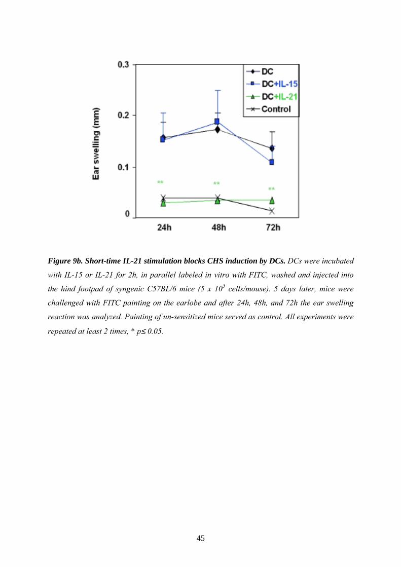
Figure 9b. Short-time IL-21 stimulation blocks CHS induction by DCs. DCs were incubated
with IL-15 or IL-21 for 2h, in parallel labeled in vitro with FITC, washed and injected into
the hind footpad of syngenic C57BL/6 mice (5 x 10⁵ cells/mouse). 5 days later, mice were
challenged with FITC painting on the earlobe and after 24h, 48h, and 72h the ear swelling
reaction was analyzed. Painting of un-sensitized mice served as control. All experiments were
repeated at least 2 times, * p≤ 0.05.

46
IL-15 is essential for a hapten-specific CHS response, but dispensable for
irritant contact dermatitis With our previous data we outlined that besides the inhibitory effects of IL-21 on DC
activation and maturation, IL-15 was able to increase immunogenic features of DCs when
present during generation. Our in vitro generated IL-15DCs could prime highest T cell
responses in vitro (Fig. 6) and in vivo (Fig. 7a). Based on these findings and with the
knowledge from literature that IL-15 is produced by DCs ion (Mattei 2001) we further asked
which role DC-derived IL-15 could play in the context of antigen specific T cell reactions in
vivo and in vitro. To clarify this we blocked the function of IL-15 in vivo by a soluble IL-
15Rα. To distinguish between effects of IL-15 and of the interplay with its specific high
affinity IL-15Rα, we extended these studies by the availability of IL-15-/- and IL-15Rα-/-
knockout mice.
To verify whether IL-15 indeed has promoting effects on DC-mediated T cell responses, we
analyzed in our first experiments, how the CHS response is established in IL-15 deficient
mice. To this end we sensitized IL-15+/+ (Wt) and IL-15-/- mice to the low molecular weight
hapten picrylchloride (PCl) by painting it on the shaved abdomen and challenged 5 days later
by painting PCl on the left ear. In addition, one group of IL-15+/+ mice was treated during the
sensitization phase with a potent and selective IL-15 antagonist, the soluble IL-
15Rα (Ruchatz 1998). Application of hapten induced contact hypersensitivity, which was
mainly CD8+ T cell mediated (Grabbe 1998). After PCl challenge resulting ear swelling
response was analyzed at 24, 48, and 72h and showed that both IL-15-/- (yellow) and sIL-
15Rα- (dark green) treated IL-15+/+ mice failed to mount an inflammatory reaction above
control levels (Fig. 10a), giving a first hind that IL-15 is essential for the development of this
type of DTH immune response.
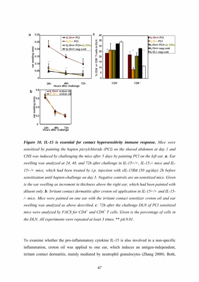
47
Figure 10. IL-15 is essential for contact hypersensitivity immune response. Mice were
sensitized by painting the hapten picrylchloride (PCl) on the shaved abdomen at day 1 and
CHS was induced by challenging the mice after 5 days by painting PCl on the left ear. a: Ear
swelling was analyzed at 24, 48, and 72h after challenge in IL-15+/+, IL-15-/- mice and IL-
15+/+ mice, which had been treated by i.p. injection with sIL-15Rα (50 µg/day) 2h before
sensitization until hapten-challenge on day 5. Negative controls are un-sensitized mice. Given
is the ear swelling as increment in thickness above the right ear, which had been painted with
diluent only. b: Irritant contact dermatitis after croton oil application in IL-15+/+ and IL-15-
/- mice. Mice were painted on one ear with the irritant contact sensitizer croton oil and ear
swelling was analyzed as above described. c: 72h after the challenge DLN of PCl sensitized
mice were analyzed by FACS for CD4+ and CD8+ T cells. Given is the percentage of cells in
the DLN. All experiments were repeated at least 3 times, ** p≤ 0.01.
To examine whether the pro-inflammatory cytokine IL-15 is also involved in a non-specific
inflammation, croton oil was applied to one ear, which induces an antigen-independent,
irritant contact dermatitis, mainly mediated by neutrophil granulocytes (Zhang 2000). Both,

48
IL-15+/+ (dark red) and IL-15-/- (yellow) mice showed a profound ear swelling, which
demonstrates that irritant contact dermatitis develops independently of IL-15 (Fig. 10b).
As already mentioned CD8+ T cells are known to be the main mediators of CHS (Watanabe
2002), we next analyzed by FACS the distribution and number of T cells in the local draining
lymph nodes (DLN). We found that the percentage as well as the total numbers of CD4+ T
cells within DLN was not significantly different between the studied groups (Fig. 10c).
However, as expected (Kennedy 2000), IL-15-/- mice (yellow bar) showed a high
significantly reduced number of CD8+ T cells. Interestingly, treatment with the sIL-
15Rα (dark green bar) did not affect the number of CD8+ T cells compared to IL-15+/+
control mice (Fig. 10c, dark red bar), although this antagonist treatment completely blocked
the CHS response.
This suggests that the role of IL-15 in CHS is not primarily related to its effect on CD8+ T cell
numbers but rather on T cell priming/activation. Therefore, besides total T cell numbers,
activation and induction of regulatory or memory T cells in DLN were assessed. No
significant induction of CD4+/CD25+, i.e. so called “regulatory T cells” (Cavani 2001) was
observed in all tested groups. However, in IL-15-/- and sIL-15Rα treated mice, CD8+ T cells
were significantly less activated (as assessed by their CD69 expression); in addition, CD8+
memory T cells (as recognized by their high expression level of CD44) were reduced in both
groups compared to untreated IL-15+/+ mice (Table 1).
IL15+/+ PCl
IL15-/- PCl
IL15+/+ PCl + sIL15Rαααα
IL15+/+ neg cont
IL15-/- neg cont
CD4+/CD25+ 6.8±1.0 6.6±0.8 7.2±0.4 5.8±0.9 5.6±1.0
CD8+/CD69+ 2.6±0.8 1.0±±±±0.3** 1.7±±±±0.2* 1.9±0.4 0.9±0.5
CD8+/ CD44hi 22±2.4 9±±±±2.3** 17±±±±4.0* 7.8±4.4 4.3±1.1
Table 1. Regulatory T cells and activated or memory CD8+ T cells in DLN. FACS analysis
of T cell activation and induction of memory T cells in the local ear-draining lymph nodes
72h after PCl challenge. Given is the percentage in relation to all CD4+ or CD8+ T cells (set
to 100%). Shown is the mean ± SD. Significance was calculated using Student’s t-test (*
p≤0.05; ** p≤0.01).

49
In conclusion, IL-15 is essential for a hapten-specific in vivo CD8+ T cell mediated CHS
reaction, including the activation as well as differentiation of memory T cells but is
dispensable for an non-specific, irritant contact dermatitis. This was further stressed by
treatment with the IL-15 antagonists, sIL-15Rα, which efficiently inhibited contact
hypersensitivity in vivo.
DTH footpad swelling, but not antibody production, is suppressed by IL-15
knockout or IL-15 antagonist In order to evaluate the role of IL-15 in both CD4+ and CD8+ T cell as well as B cell mediated
Th1 immune response, a murine DTH model of footpad swelling reaction to sheep red blood
cells (SRBC; Kunzendorf 1996) was employed. In comparison to CHS, which is mainly
mediated by CD8+ T cells, DTH to SRBC represents a both T- and B cell-dependent immune
response, which has often been used as an instructive research model for identifying immune-
regulatory factors (Kunzendorf 1996; Kalish 1999). Sensitization to SRBC induces activation
of T cells in the peripheral lymphoid tissues and subsequently specific antibody production by
B cells. After local antigen challenge in the footpad, activated T cells migrate to the site of
challenge and induce a footpad-swelling reaction, which correlates with the extent of the DTH
reaction (Kalish 1999).
Footpad swelling was analyzed 24, 48 and 72h after SRBC challenge in IL-15+/+ (dark red)
and IL-15-/- mice (yellow), or IL-15+/+ mice that had been treated with an IL-15 antagonist
(sIL-15Rα, dark green). Non-immunized mice served as negative- (open circles), IL-15+/+
mice as positive controls. As shown in Fig. 11, blocking the function of IL-15 by systemic
sIL-15Rα treatment high significantly suppressed the footpad swelling compared to the
positive control at all three time points. In comparison IL-15-/- mice also showed a
significantly reduced footpad swelling (Fig. 11). Negative controls, instead, only had a slight,
non-specific increase in footpad thickness, and no differences between IL-15+/+ (open
circles) and IL-15-/- (crosses) negative control mice were detected.
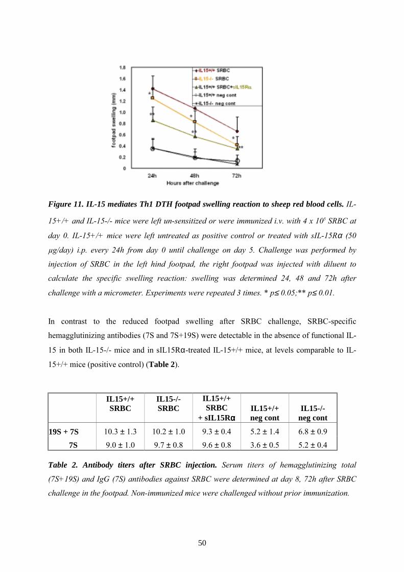
50
Figure 11. IL-15 mediates Th1 DTH footpad swelling reaction to sheep red blood cells. IL-
15+/+ and IL-15-/- mice were left un-sensitized or were immunized i.v. with 4 x 10⁵ SRBC at
day 0. IL-15+/+ mice were left untreated as positive control or treated with sIL-15Rα (50
µg/day) i.p. every 24h from day 0 until challenge on day 5. Challenge was performed by
injection of SRBC in the left hind footpad, the right footpad was injected with diluent to
calculate the specific swelling reaction: swelling was determined 24, 48 and 72h after
challenge with a micrometer. Experiments were repeated 3 times. * p≤ 0.05;** p≤ 0.01.
In contrast to the reduced footpad swelling after SRBC challenge, SRBC-specific
hemagglutinizing antibodies (7S and 7S+19S) were detectable in the absence of functional IL-
15 in both IL-15-/- mice and in sIL15Rα-treated IL-15+/+ mice, at levels comparable to IL-
15+/+ mice (positive control) (Table 2).
IL15+/+ SRBC
IL15-/- SRBC
IL15+/+ SRBC
+ sIL15Rαααα IL15+/+ neg cont
IL15-/- neg cont
19S + 7S 10.3 ± 1.3 10.2 ± 1.0 9.3 ± 0.4 5.2 ± 1.4 6.8 ± 0.9
7S 9.0 ± 1.0 9.7 ± 0.8 9.6 ± 0.8 3.6 ± 0.5 5.2 ± 0.4 Table 2. Antibody titers after SRBC injection. Serum titers of hemagglutinizing total
(7S+19S) and IgG (7S) antibodies against SRBC were determined at day 8, 72h after SRBC
challenge in the footpad. Non-immunized mice were challenged without prior immunization.

51
A second SRBC challenge on the other foot 14 days after the first SRBC injection again
revealed a significantly suppressed footpad swelling in sIL-15Rα-treated and IL-15-/- mice,
but comparable antibody titers (not shown).
These data suggest that IL-15 is essential for T cell mediated effects also in this type of DTH
model. Notably, just blocking endogenous IL-15 activity with a suitable antagonist during the
sensitization phase suffices to elicit a long-lasting DTH suppression. In contrast, our results
show that IL-15 is dispensable for B cell activation and antibody production in this example
of an in vivo-Th1 immune response.
In vivo antigen-uptake and migration/ activation of DCs are IL-15-
independent In the following, we focused on the CHS model of DTH, since it facilitates dissection of the
underlying, T cell specific mechanisms of CHS inhibition in the absence of IL-15. For this
purpose, we analyzed the antigen-uptake by epidermal DCs in the skin and their migration to
the local DLNs as well as the up-regulation of CD80 and CD86 co-stimulatory molecules on
DCs, which are critically involved in CHS reactions (Xu 1997).
In order to address which of these steps may be influenced by IL-15, we used again FITC as
model antigen, since it can easily be visualized by FACS (Macatonia 1986). As shown in
Table 3, 24h after abdominal FITC painting, the percentage of FITC+/CD11c+-DCs (i.e. cells,
which migrated in this time from the skin, where they took up Ag) in DLN was comparable in
all FITC-painted groups (the total cell number in each lymph node was also comparable in all
groups; not shown). Negative controls (without FITC painting) showed a lower amount of
DCs in the DLN (around 2.2%, compared to over 5% CD11c+ after FITC painting). Finally,
we analyzed the activation of DCs by quantifying the expression of the co-stimulatory
molecules CD80 and CD86, which play an important role in CHS responses (Katayama
1997). No significant differences in the expression of both molecules on antigen-bearing cells
that were FITC positive could be detected between all FITC painted groups. DCs from
untreated IL-15+/+ or IL-15-/- mice expressed low levels of CD80 and CD86.

52
IL15+/+ FITC
IL15-/- FITC
IL15+/+ + sIL15Rαααα
FITC
IL15+/+ Control
IL15-/- Control
CD11c+ 5.5 5.3 5.3 2.2 2.1
CD11c+/CD80+ 41.7 40.3 43.0 11.2 14.0
CD11c+/CD86+ 60.9 59.5 56.5 18.8 27.1
Table 3. IL-15 is not mandatory for cutaneous antigen uptake by and activation of DCs. IL-
15+/+ or IL-15-/- mice were painted on the abdomen with FITC or were injected 30min
before painting with sIL-15Rα (50µg) i.p. Negative controls and left untreated. 24h later, the
DLN were analyzed by FACS (100.000 cells) for total number of CD11c positive DC (1st row,
given is the total percentage of DC in lymph nodes). Furthermore, activation of the FITC+
DCs after migration from the skin was analyzed by FACS. Expression of the co-stimulatory
molecules CD80 (2nd row) and CD86 (3rd row) was estimated using PE labeled antibodies
(given is the percentage of high (fluorescence channel > 102) CD80 and CD86 positive cells
on CD11c+ FITC+ DCs in the DLN). Negative controls (without FITC painting) were
analyzed for total CD11c+ DCs in DLN.
Bone marrow derived DCs (BMDCs) from IL-15+/+ and IL-15-/- mice (see below) also
displayed no significant differences in endocytic antigen-uptake in vitro as analyzed by the
model-antigen FITC-dextran (not shown).
Taken together, DCs from IL-15+/+ and IL-15-/- mice take up antigen (in vivo and in vitro)
and migrate to local DLN with comparable efficiency, indicating that IL-15 is not essential in
modulating these important steps of CHS response.
Bone marrow derived DCs from IL-15+/+ and IL-15-/- mice differ in their
T cell stimulatory capacity In order to unravel the above described specific defects of IL-15-/- mice in T cell immunity
and memory, and to elucidate whether DC-derived IL-15 is the key in the induction of Th1
immune responses like CHS, bone marrow derived DCs (BMDCs) from IL-15+/+ or IL-15-/-
mice were employed, which had been generated 8 days in vitro by culturing with GM-CSF.
Surface phenotyping after 8 days revealed similar purity (>95%) of CD11c+ DCs. Also, the

53
co-stimulatory molecules CD80, CD86, MHC II, and CD25 were expressed at comparable
levels (Fig. 12a).
Figure 12. DC derived IL-15 mediates CD8+ T cell proliferation and memory cell
differentiation in vitro. a: Bone marrow derived DCs from IL-15+/+ and IL-15-/- mice were
analyzed for different co-stimulatory molecules on day 8 by FACS. Shown is one
representative out of 3 experiments. Given is the percentage of high positive (fluorescence
channel >102) cells except for CD11c, where all positive cells after 8 days culture are given.
b: BMDCs from IL-15+/+ and IL-15-/- mice were co-cultured with CD4+ T cells from OT II
mice with an CD4-restricted OVA transgenic T cell receptor and with OVA(323-339) peptide.
Proliferation was analyzed by 3[H]thymidine incorporation (CPM: counts per minute; n.s.:
not significant. c: Co-culture as in 12b, but with CD8+ T cells from OTI mice with a CD8-
restricted OVA transgenic T cell receptor, with OVA(257-264) peptide (*p≤0.05). d: Induction of
CD8+ memory cells and apoptosis was analyzed in the co-culture experiments with IL-15+/+
and IL-15-/- BMDCs and CD8+ OVA-TCR transgenic cells after 48h. Given are the percent
CD62Llow and CD44high/CD122+ (memory) cells as well as dead cells (positive for PI/Annexin
V). Significance was analyzed using Student’s t-test (n.s.: not significant; *p≤0.05).

54
This suggests that IL-15 is not essential for the in vitro generation of BMDCs, which is
supported by the fact that no known abnormalities in DC subtypes and number have been
noted in IL-15-/- or IL-15Rα-/- mice (Kennedy 2000; Lodolce 1998; Ohteki 2001).
DCs are specialized for antigen capture, migration and antigen-presentation to CD4+ and
CD8+ T cells (Cella 1997; Lambrecht2001). To analyze the effect of IL-15 on DC/T cell
interactions, and in order to specifically dissect the immuno-stimulatory capacity of DCs
influenced by IL-15, BMDCs from IL-15+/+ or IL-15-/- mice were studied in a co-culture
assay with syngenic T cells. The DCs were labeled with peptide fragments from ovalbumine
and were cultured with T cells in vitro. These T cells expressed a CD4+ T cell restricted,
transgenic (tg) T cell receptor (from OTII mice), which recognize the OVA(323-339) peptide
(Robertson 2000), and CD8+ transgenic T cells (from OTI mice), which recognizes the
OVA(257-264) peptide (Rotzschke 1991). As shown in Fig. 12b, peptide-labeled DCs from IL-
15+/+ (dark red bars) or IL-15-/- mice (yellow bars) induced comparable proliferation of
CD4+ tg T cells, suggesting that this T cell subpopulation proliferates independently from
DC-derived IL-15. These in vitro data correspond to the CD4+ T cell mediated production of
hemagglutinizing antibodies in vivo, which were equal in IL-15+/+ and IL-15-/- mice, as
shown above (Table 2).
In view of previous reports, indicating that IL-15 acts mainly on CD8+ T cells (Schluns 2002;
Fehniger 2001), we also studied activation of CD8+ tg T cells. Indeed, IL-15-/- BMDCs
induced significantly lower antigen-specific CD8+ T cell proliferation compared to IL-15+/+
BMDCs (Fig. 12c): CD8+ T cell proliferation was diminished when specific Ag was
presented by IL-15 deficient DCs, even though the level of MHC class I expression was
comparable on IL-15+/+ and IL-15-/- DCs (not shown). Reduced levels of CD8+ specific T
cell proliferation induced by IL-15-/- DCs could be restored by exogenous given IL-15 (not
shown).
These findings confirm that IL-15 produced by DCs is a key-player in DC mediated CD8+ T
cell priming.

55
DC-derived IL-15 mediates differentiation of memory T cells in vitro Following recognition of Ag presented by DCs, naive T cells become activated and are
polarized to differentiate into effector and subsequently memory T cells (Tuma 2002;
Weninger 2002). Studies in IL-15-/- mice revealed that IL-15 plays a role in establishing
CD8+ memory (Kennedy 2000; Lodolce 1998). However, it is not known at which stage
memory cell induction is disrupted. We next analyzed the development of these cells in vitro,
focusing on the influence of DC-derived IL-15.
These studies showed that, indeed, co-culturing of CD8+ tg T cells with BMDC from IL-
15+/+ mice induced CD8+ T cells with a memory phenotype, as evidenced by their
characteristic surface marker pattern, i.e. low expression of CD62L and CD44high/CD122+
(Fig. 12d, dark red bar). Instead, co-culture with IL-15-/- BMDCs resulted in significantly
reduced induction of the memory T cell phenotype (Fig. 12d yellow bar). By PI/Annexin
staining, only few T cells were demonstrated to have undergone apoptosis, and no significant
differences between IL-15+/+ and IL-15-/- BMDCs in this respect were detected (Fig. 12d,
right columns). This makes it very unlikely that the lack of IL-15 (a potent anti-apoptotic
agent; Bulfone-Paus 1997) only resulted in premature apoptosis of activated CD8+ cells, and
can thus not be invoked as a convincing explanation for the defect in memory T cell induction
in the absence of IL-15-secreting DCs.
In conclusion, our in vitro assays showed that the absence of DC-derived IL-15 leaves the
expression of co-stimulatory molecules on DCs unaffected, but significantly reduces the
ability of DCs to induce CD8+ T cell proliferation, and differentiation towards a memory
phenotype.
BMDC migration in vivo is IL-15-independent To elucidate whether IL-15 might affect DC functions in vivo, we applied a model of active
sensitization with FITC labeled DCs. First, we examined whether the complex migration
process of DCs in vivo depends on endogenous, DC-derived IL-15 or on exogenous IL-15
from the surrounding tissue. This was done by comparing the ability of BMDCs from IL-
15+/+ or IL-15-/- mice to enter DLN in IL-15+/+ or IL-15-/- mice following BMDC injection
in vivo (Lappin 1999). IL-15+/+ or IL-15-/- BMDCs were labeled with the fluorescent cell
tracer FITC and subsequently injected into the footpad of IL-15+/+ or IL-15-/- mice. After
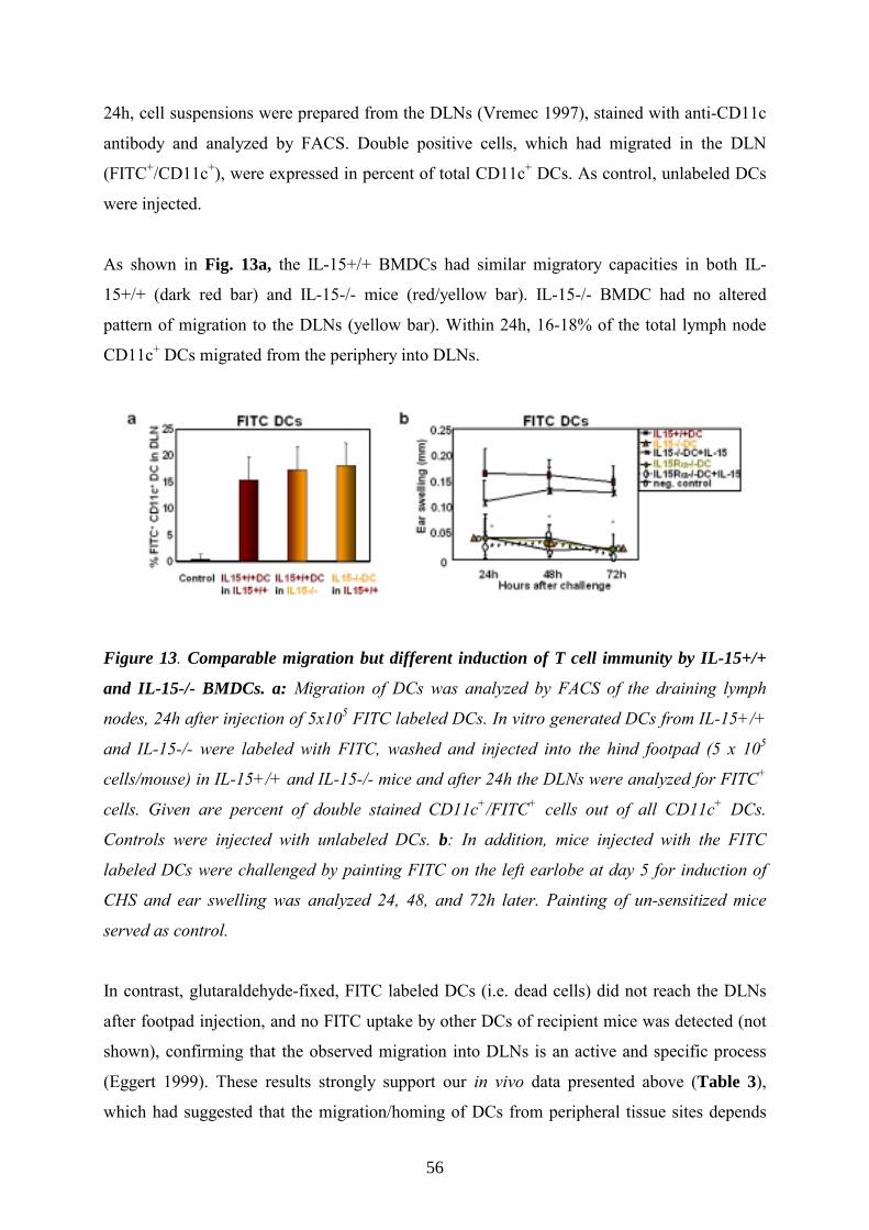
56
24h, cell suspensions were prepared from the DLNs (Vremec 1997), stained with anti-CD11c
antibody and analyzed by FACS. Double positive cells, which had migrated in the DLN
(FITC+/CD11c+), were expressed in percent of total CD11c+ DCs. As control, unlabeled DCs
were injected.
As shown in Fig. 13a, the IL-15+/+ BMDCs had similar migratory capacities in both IL-
15+/+ (dark red bar) and IL-15-/- mice (red/yellow bar). IL-15-/- BMDC had no altered
pattern of migration to the DLNs (yellow bar). Within 24h, 16-18% of the total lymph node
CD11c+ DCs migrated from the periphery into DLNs.
Figure 13. Comparable migration but different induction of T cell immunity by IL-15+/+
and IL-15-/- BMDCs. a: Migration of DCs was analyzed by FACS of the draining lymph
nodes, 24h after injection of 5x10⁵ FITC labeled DCs. In vitro generated DCs from IL-15+/+
and IL-15-/- were labeled with FITC, washed and injected into the hind footpad (5 x 10⁵
cells/mouse) in IL-15+/+ and IL-15-/- mice and after 24h the DLNs were analyzed for FITC+
cells. Given are percent of double stained CD11c+/FITC+ cells out of all CD11c+ DCs.
Controls were injected with unlabeled DCs. b: In addition, mice injected with the FITC
labeled DCs were challenged by painting FITC on the left earlobe at day 5 for induction of
CHS and ear swelling was analyzed 24, 48, and 72h later. Painting of un-sensitized mice
served as control.
In contrast, glutaraldehyde-fixed, FITC labeled DCs (i.e. dead cells) did not reach the DLNs
after footpad injection, and no FITC uptake by other DCs of recipient mice was detected (not
shown), confirming that the observed migration into DLNs is an active and specific process
(Eggert 1999). These results strongly support our in vivo data presented above (Table 3),
which had suggested that the migration/homing of DCs from peripheral tissue sites depends

57
neither on the autocrine production of IL-15 by DCs itself, nor on exogenous IL-15 secreted
by the DC microenvironment or delivered via the serum.
DC-derived IL-15 and the IL-15Rαααα on DCs are essential for inducing a T
cell mediated CHS response to FITC In order to determine directly to which extent the capacity of DCs for T cell priming and
sensitization in vivo depends on endogenous, DC-derived IL-15, IL-15+/+, IL-15-/- and IL-
15Rα-/- DCs were therefore labeled in vitro with FITC, and injected subcutaneously in the
footpad of IL-15+/+ mice. Five days after this active sensitization, mice were challenged at
the dorsal and ventral site of one ear with FITC (the other ear with diluent only) and the
ability to initiate an antigen-specific, T cell mediated CHS response was examined by
measuring the FITC-induced ear swelling 24, 48 and 72h after challenge.
Mice, which had not been sensitized, but were challenged with FITC on the ear, served as
negative controls for unspecific ear swelling (open quadrangles). Un-sensitized mice
responded with small unspecific ear swelling that was equal in IL-15+/+ and IL-15-/- (Fig.
13b, opened circles). IL-15+/+ mice that had been actively sensitized with IL-15+/+ DCs
served as positive controls (Fig. 13b, dark red quadrangles). To confirm that the
sensitization is based on active migration of the injected, viable DCs from the footpad to the
DLN, control mice were injected with FITC labeled, glutaraldehyde fixed DCs (Eggert 1999).
As expected, these fixed cells, incapable of migration failed to induce any CHS response (not
shown).
Injection of IL-15-/- DCs resulted in a highly significantly reduced CHS response that was
indistinguishable from un-sensitized controls all three time points (Fig. 13b, yellow
triangles). However, when the lack of endogenous IL-15 was substituted by incubating IL-
15-/- DCs prior to injection for only 30min with recombinant IL-15, their ability to induce T
cell sensitization and subsequent induction of a CHS response was almost restored to the level
of IL-15+/+ DCs (Fig. 13b, crosses).
Using BMDCs from IL-15Rα-/- mice, which lack the high affinity IL-15Rα chain but
produce IL-15, could not induce a CHS response above negative control values, either (Fig.
13b, dark green diamonds). As expected, loading these IL-15Rα-/- DCs prior to injection

58
with IL-15 (Fig. 13b, opened diamonds with dotted line), did not restore the capacity to
prime a T cell response.
These data clarify that endogenous IL-15, produced by DCs as well as IL-15Rα expression by
DCs, are required for the initiation of an efficient DTH response in vivo.
DC-derived IL-15 is dispensable for B cell priming to OVA/FITC To support the findings from the DTH to SRBC experiments shown above, namely that IL-15
mediates T cell activation but is not mandatory for B cell function, we used a FITC-labeled,
complete protein-antigen (Harlow 1988) to actively sensitize the mice by injecting
OVA/FITC DCs. This antigen induces CD4+ and CD8+ T as well as B cell activation.
As shown in Fig. 14a, a significant ear swelling to FITC was induced when labeled IL-15+/+
DCs were injected into IL-15+/+ mice after challenge with FITC on day 5 (as described
above). As expected, OVA/FITC labeled IL-15-/- DCs failed to induce T cell priming and
therefore CHS swelling reaction in IL-15+/+ mice (Fig. 14a, dark red diamonds). To
analyze the B cell function in these mice, the animals were bled 7 days after sensitization by
OVA/FITC labeled DCs, and anti-OVA-IgM titers were determined by ELISA. As shown in
Fig. 14b, both IL-15-/- (yellow quadrangles) and IL-15+/+ DCs were capable of inducing
anti-OVA antibody production at comparable levels.
Figure 14. DC-derived IL-15 is dispensable for B cell priming. a: DCs from IL-15+/+ and
IL-15-/- mice were labeled with OVA/FITC and injected in IL-15+/+ mice, which in addition
to T cells also activates B cells. Ear swelling was induced and analyzed as in Fig. 13b. b: B

59
cell activation was analyzed by detecting serum anti-OVA-IgM antibodies at day 8 using
ELISA. Given is the optical density (OD at 492 nm) of serial serum dilutions.
These findings are fully in line with the in vivo data presented above and underscore the
concept that DC-derived IL-15 is essential for the induction of CD8+ T cell activation and
differentiation after antigen-contact, but is dispensable for B cell priming. In addition, the
presence of the high affinity IL-15Rα chain on DC was found to be crucial in inducing CD8+
T cell response.
Compared to IL-15+/+ DCs, IL-15-/- DCs produce higher levels of anti-
inflammatory and lower levels of pro-inflammatory cytokines It remained to be determined, how IL-15/IL-15Rα mediated signaling affects the T cell
priming by DCs. It is appreciated, that cytokine production by DCs critically modulates the
outcome of immune responses during antigen-presentation to T cells (Liu 2001a) and that this
cytokine secretion pattern of DCs is modulated by many different stimuli as antigen-
processing and -presentation, DC-T cell interactions (Guermonprez 2002; Lanzavecchia
2001) and pathogens like microbial cell-wall components (Schuurhuis 2000). In response to
these signals, DCs are either activated to enter maturation, which transforms them into
specialized cytokine-secreting cells, or are kept in an immature state by anti-inflammatory
cytokines like IL-10 (Corinti 2001). Since IL-15 can alter the secretion of first-line defense
cytokines like type I interferons from DCs (Mattei 2001), it was of interest to compare our
DCs generated in vitro from IL-15+/+ and IL-15-/- mice with respect to their production of
pro-inflammatory- (IL-1β, TNF-α, IL-12) and anti-inflammatory- (IL-10) cytokines.
First, we analyzed cytokines from co-cultures of DCs and CD8+ T cells (as described above).
Pro-inflammatory, T cell derived cytokines like IL-2 and IFN-γ were found to be reduced
when Ag was presented by IL-15-/-DCs (not shown), which suggests an important role of
DC-derived IL-15 on the secretion of T cell cytokines during antigen presentation by DCs.
To distinguish between DC-derived and T cell derived cytokines we, next, also stimulated
DCs solely by low-dose LPS (Reis e Sousa 1999) and measured cytokine production. As
shown in Fig. 15, IL-15+/+ DCs (red bars) produced significantly higher amounts of IL-1β
and TNF-α than IL-15-/- DCs (yellow bars), whereas the latter produced significantly more
anti-inflammatory IL-10 in vitro (Fig. 15). The failure of IL-15-/-DCs to induce a Th1 CHS

60
response was not mediated due to reduced IL-12 production, since the levels of the Th1-
inducing cytokine IL-12 (Liu 2001a), secreted by IL-15+/+ and IL-15-/- DCs were
comparable (not shown), suggesting that the reduced CD8+ T cell activation is not indirectly
mediated by reduced IL-12 release.
Figure 15. BMDCs from IL-15+/+ mice produce more pro-inflammatory cytokines after
LPS activation. DCs from IL-15+/+ and IL-15-/- mice were either activated in vitro for 24h
with low dose LPS (10ng/ml) or were incubated with medium alone. Concentrations of pro-
and anti-inflammatory cytokines (as indicated) were detected in cell-free supernatants.
These data suggests, that DC-derived IL-15 could modulate the T cell-priming functions of
DCs in an autocrine fashion by boostering the secretion of pro-inflammatory TNF-α and IL-
1β, and by down-regulating the anti-inflammatory IL-10 release by DCs. This provides
further evidence that IL-15 is directly and critically involved in fine-tuning the DC-cytokine
network towards the mounting of an efficient, CD8+-mediated Th1 immune responses.
IL-15 is internalized via IL-15Rαααα and co-localizes with IL-15Rαααα in DCs Finally, we addressed whether membrane or intracellular IL-15-IL-15Rα interactions are
critical in this respect. As a first step towards clarifying this, IL-15/IL-15Rα (co-) localization
was analyzed by confocal microscopy. IL-15+/+, IL-15-/- and IL-15Rα-/- DCs were cultured
for 24h on micro chamber slides in medium or activated with low-dose LPS. Alternatively,
they were incubated for 30min with IL-15 or with IL-15 in the presence of a receptor-
blocking anti-IL-15Rα antibody to study which role the high-affinity receptor plays in the
uptake of IL-15.
As revealed by double staining with wheat germ agglutinin (WGA), IL-15 is expressed in
DCs mainly intracellular, but not in membrane-bound form (Fig. 16a). This expression

61
pattern differs from the one we had previously shown for monocytes (Musso 1999) and
keratinocytes (Rückert 2000). Intracellular IL-15 staining was strongly up regulated in IL-
15+/+ DCs when stimulated by LPS (Fig. 16a, green fluorescence). Exogenous IL-15 was
taken up predominantly via the IL-15Rα, (within 30min) since a blocking antibody against
IL-15Rα strongly reduced IL-15 uptake (Fig. 16a). Like IL-15, the IL-15Rα
immunoreactivity was localized more intracellular than on the membrane and its expression
there could be up regulated upon stimulation with LPS (Fig. 16). Since the IL-15Rα
immunoreactivity associated with the membrane in IL-15+/+ mice was faint, we cannot
exclude that the detection of IL-15Rα-IL-15 complexes on the membrane was below
detection-limit of our assay.
Furthermore, these studies showed that IL-15 was intracytoplasmatically co-localized with IL-
15Rα (Fig. 16a, IL-15/IL-15Rα, yellow fluorescence). This intracellular complex was also
found, when IL-15 was given exogenously to IL-15-/- DCs (Fig. 16b). This underscores the
tendency of IL-15 to form ligand/receptor complexes in DCs, and shows that ligand uptake by
the IL-15Rα is unaffected in IL-15 knockout mice. This finding also provides a convincing
explanation for the restored T cell-priming ability of IL-15-/- DCs after pre-incubation with
IL-15 in CHS (see above, Fig. 13b). Although DCs from IL-15Rα-/- mice produced and
expressed intracellular IL-15 which was up-regulated by LPS (Fig. 16c), the lack of the high-
affinity α-chain and therefore the missing interaction with its ligand, IL-15, led to the
functional deficiency in inducing T cell mediated CHS response, which could not be restored
by incubation with an excess of IL-15 (see above, Fig. 13b).

62
a
b

63
c
Figure 16. IL-15Rαααα and IL-15 are intracellular co-localized in DCs. a: DCs from IL-15+/+
b: IL-15-/- or c: IL-15Rα-/- mice were stimulated for 24h with LPS or for 30min with
100ng/ml of IL-15 or treated for 30min with anti-IL-15Rα antibodies prior to IL-15
stimulation for the next 30min. Un-stimulated cells were used as a control. Cells were fixed
with 2% paraformaldehyde and incubated with WGA (labeled with rhodamine, red
fluorescence) for 20min, washed, permeabilized and stained with anti-IL-15Rα or with anti-
IL-15 antibodies (ALEXA-488, green fluorescence). For co-localization of IL-15Rα with IL-
15, cells were fixed with 2% paraformaldehyde, permeabilized and labeled with anti-IL-15Rα
antibodies (ALEXA-564, red) in combination with anti-IL-15 antibodies (ALEXA-488, green)
and TOTO-3 for nuclei (blue). Yellow color indicates co-localization of IL-15Rα and IL-15.
Taken together our data demonstrate for the first time, that the induction of T cell immunity
and CD8+ memory depends on IL-15-IL-15Rα interaction in DCs. Hence, based on the
essential role of IL-15-IL-15Rα interactions in DCs, we suggest that the specific modulation
of this signalling loop offers a promising, novel therapeutic target for the treatment of Th1-
type autoimmune diseases, transplant rejection, and other CD8+-T cell dependent diseases and
for DC-dependent vaccination therapy.

64
Discussion Effects of IL-15 and IL-21 on generation of DCs in vitro In this study we have shown that IL-15 and its structural relative, the newly described
cytokine IL-21 (Parrish-Novak 2000) influence DC differentiation, maturation and function in
vitro and in vivo. Given IL-15 to BM stem cells in addition to GMCSF provided phenotypic
and functional mature DCs with profound MHCII expression, decreased endocytic activity
and highly increased T cell stimulatory ability in vitro and in vivo. In contrast, IL-21 mediated
completely opposite effects while inhibiting maturation of DCs and down modulated DC
induced Ag specific T cell response. With our first data, that IL-21 in combination to GMCSF
is able to support differentiation of myeloid BMDCs, we could not only corroborate results
from literature demonstrating that IL-21 influences also lymphoid cell development (Parrish-
Novak 2000), but directly report for the first time a regulatory impact of this cytokine on APC
function.
Immature DCs are characterized by constitutively sampling of their surrounding fluid by
endocytosis but rapidly lose their antigen uptake capacity upon maturation (Sallusto 1995).
Here, IL-21DCs showed highest internalization of the model antigen FITC dextran, contrasted
by very low uptake by IL-15DCs. The observed differences between IL-21DCs and IL-15DCs
point to altered maturation states, supporting the deduction that IL-21 could block DC
maturation. We extended this with data revealing that co-stimulatory molecules on IL-21DCs
remained significantly reduced even after antigen-uptake and –processing in comparison to
IL-15DCs.
Thus, IL-15 strongly promoted the differentiation of BM progenitor cells into mature DCs.
The supportive effects of IL-15 in differentiation towards mature APCs are in line with
previously published reports (Saikh 2001, Mohamadzadeh 2001). The divergent effects of
both cytokines are likely to be mediated by different signaling pathways. Even since both use
the common-γ chain, the high-affinity receptors may differ in their signal transduction after
ligand binding.
The IL-21R contains a long intracellular domain with strongly conserved motifs, including
classical Box1 and Box2 elements (Murakami 1991), which are important for signal

65
transduction, suggesting that this receptor is a signaling subunit (Parrish-Novak 2000). Even
though the private IL-21R chain is not able to transduce ligand binding signals alone or as a
homodimer (Parrish-Novak 2002) it is reasonable to speculate that it modulates γ-chain
effects likewise. It has been reported that in cooperation with the common γ-chain, the
heterodimeric IL-21R complex activates STAT (Asao 2001) and Jak (Habib 2002) pathways.
The observed inhibitory effects of IL-21 on DC generation, however, remain to be elucidated
by further analysis of signaling pathways in comparison to IL-15.
Despite the very short intracellular domain of IL-15Rα our group already demonstrated, that
TRAF2 (Bulfone-Paus 1999) as well as Syk are recruited to the intracellular receptor part
(Bulanova 2001) which strongly suggests that IL-15Rα is able to transduce a signal even in
the absence of the γ-chain. Nevertheless, IL-15 signals also via the IL-2/15Rβ chain
(Grabstein 1994), which is another important difference to IL-21 signaling. This could explain
our observation that RNA message of IL-2/15Rβ chain is predominantly expressed in the IL-
15DCs. In this regard it is worth noting that there are compelling evidences, which
demonstrated that the IL-2/15Rβ-chain co-localizes with the GMCSF receptor (Giron-Michel,
personal communication). This reinforces our results that the enhanced maturational stage of
IL-15DCs might be gained from synergistic effects of GMCSF and IL-15.
It is widely accepted that DC maturation is directly linked to T cell stimulatory capacities
(Lanzavecchia 2001). Indeed our experiments revealed that antigen-specific T cell response in
vitro was significantly reduced with IL-21DCs. IL-15DCs mediated high significantly
increased T cell proliferation rates. This is most probably due to altered antigen presenting
capacities modulated directly by IL-15 and IL-21 during generation. However, it remained to
be clarified at which stage of generation the cytokines influence stem cell differentiation in
context to GMCSF and whether this effect is reversible (“plasticity”).
That our in vitro generated DCs were susceptible for signals from IL-15 and IL-21 was
proven by message expression of the relevant receptors on mRNA level. To confirm that IL-
15 and IL-21 were taken up by DCs, we did confocal microscopy and found both cytokines
internalized.
In additional experiments, I generated BM derived murine macrophages (MF), which revealed
under the influence of IL-21 a similar, immature phenotype, since “IL-21MF” were also not

66
able to induce T cell proliferation. Therefore the here described effects of IL-21 seem to
represent a general feature of this cytokine on APC differentiation.
Effects of IL-15 and IL-21 on LPS induced DC activation in vitro Dendritic cells are able to respond directly to pathogens like microbial cell-wall components
and indirectly sensing infection through inflammatory cytokines (Rescigno 1999). In response
to these “danger” signals (Gallucci 2001), DCs are activated to enter maturation, which
transforms them into highly efficient APCs.
Our data imply that IL-15 did not further increase DC activation induced by LPS, whereas in
quite contrast IL-21 displayed inhibitory properties. Thus we showed that LPS induced up-
regulation of functional relevant molecules like MHCII, CD80, CD86 was significantly
reduced when IL-21 was given in parallel to LPS. It is known, that other cytokines, notably
from T cell released IL-10, have been implicated in impeding DC maturation, which was
associated with the retention of an “immature” phenotype (Morel 1997). Concomitant
incubation of DCs with LPS and IL-10 also blocked, like IL-21, expression of co-stimulatory
molecules, suggesting that IL-21 has comparable inhibitory effects on LPS induced DC
activation. Nevertheless, main effect of IL-10 is the suppression of IL-12 production (Corinti
2001) by DCs, which we found in ELISA not to be impaired by IL-21, suggesting that other
mechanisms are involved. Very interesting to that point was a hint that similar observations
had been found for B cells, where IL-21 prevented LPS-induced cell proliferation. However,
also in this report, the underlying mechanisms remained to be clarified (Parrish-Novak 2002).
Our findings, that IL-21 inhibits DC activation, are further supported by observations in the
human system. There, DCs were also abrogated in their maturation upon LPS activation when
IL-21 was present (Musso, manuscript submitted).
In summary, our data provide first evidences that IL-21 is an important negative regulator of
DC activation in response to LPS. In contrast IL-15 did not further enhance activation, which
was most probably due to the fact that LPS already induces a maximal response in DCs.
67
Effects of IL-15 and IL-21 on activation of DCs in vivo The CHS model provides a unique approach to assess DC induced T cell priming in vivo
(Grabbe 1998). The outcome of the T cell mediated CHS response directly reflects the
activation status of the DCs.
In accordance to our in vitro assays, we found that IL-15DCs induced a significantly
increased CHS response. The same number of IL-21DCs failed to induce any CHS above
unsensitized control mice, in line with our in vitro results. To exclude that the inability of IL-
21DCs to induce a CHS response was due to reduced migration capacity we examined the
local draining lymph nodes and found IL-21DCs homing to same extends compared to DCs
and IL-15DCs. Therefore the lack of T cell stimulatory capacity by IL-21DCs is most likely
attributed to their immature status with reduced MHCII expression and not due to a defect in
homing to DLN. Interestingly even after a second challenge two weeks later, IL-21DCs
injected mice were not able to mount a CHS response. This result argues that, IL-21DCs not
only failed to undergo maturation after antigen uptake and migration to DLN but also were
unable to establish a memory T cell response in vivo.
On the other side, IL-15 was reported to induce DC activation after short time contact (Ohteki
1999; Mattei 2001). We here present evidences that during generation of DCs, permanently
given IL-15 induced high potent APCs with regard to T cell stimulatory capacities. Since IL-
15 is produced in vivo by BM and lymph node stromal cells (Grabstein 1994), the presence of
this cytokine in the extracellular matrix may shape DC phenotype during hematopoiesis and
DC function during a variety of immune responses, which we discuss in detail below.
However, contrasting effects of IL-21 versus IL-15 on DCs are probably due to their unique
private receptor α-chains that complete the IL-15Rαβγ and IL-21R complexes and thereby
allow differential responsiveness depending on the ligand and high affinity receptor expressed
as already mentioned. Blocking of IL-21 and its private receptor and the use of IL-21/IL-21R
deficient mice could give more insight into the mechanisms, which keep DCs controlled in an
immature state. Moreover it is reasonable to speculate that T cells (either of naïve or memory
phenotype) release IL-21 during or after contact with DCs, thereby providing a negative
feedback signal keeping DCs in an immature state. Subsequently antigen presentation by
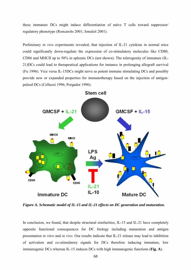
68
these immature DCs might induce differentiation of naïve T cells toward suppressor/
regulatory phenotype (Roncarolo 2001; Jonuleit 2001).
Preliminary in vivo experiments revealed, that injection of IL-21 cytokine in normal mice
could significantly down-regulate the expression of co-stimulatory molecules like CD80,
CD86 and MHCII up to 50% in spleenic DCs (not shown). The tolerogenity of immature (IL-
21)DCs could lead to therapeutical applications for instance in prolonging allograft survival
(Fu 1996). Vice versa IL-15DCs might serve as potent immune stimulating DCs and possibly
provide new or expanded properties for immunotherapy based on the injection of antigen-
pulsed DCs (Celluzzi 1996; Porgador 1996).
Figure A. Schematic model of IL-15 and IL-21 effects on DC generation and maturation.
In conclusion, we found, that despite structural similarities, IL-15 and IL-21 have completely
opposite functional consequences for DC biology including maturation and antigen
presentation in vitro and in vivo. Our results indicate that IL-21 release may lead to inhibition
of activation and co-stimulatory signals for DCs therefore inducing immature, low
immunogenic DCs whereas IL-15 induces DCs with high immunogenic functions (Fig. A).

69
DC-derived IL-15 is essential for the induction of Th1 immune responses From the above described data and in line with recent publications it became evident that IL-
15 is a critical player in the activation of DCs. Therefore the role of IL-15 in DC-mediated T
cell responses had to be focused more closely. This was done here with the use of IL-15- and
IL-15Rα- deficient mice and in parallel by blocking the function of IL-15 in wildtype mice
with an antagonistic soluble IL-15Rα (sIL-15Rα, Ruchatz 1998).
Our studies elucidate that IL-15 was absolutely required for the induction of a specific CD8+
T cell mediated CHS reaction in mice. Analyzing the DLN revealed, that the elicitation of an
inflammatory CHS response is not correlated to the absolute number of CD8+ T cells present
in the DLN, but clearly depends on their activation status. This deduction was supported by
blocking the function of IL-15 with the sIL-15Rα in wildtype mice where CHS was abrogated
despite normal CD8+ T cell numbers. Our results are consistent with the concept that one key
function of IL-15 during the initiation of a CHS response is to activate CD8+ T cells and to
establish CD8+/CD44high T memory cells (Kennedy 2000; Lodolce 1998). Vice versa,
systemic IL-15 administration, in addition to stimulating a CD8+ memory response, also
augments the primary CD8+ T cell response in vivo and drives a Th1-biased phenotype
(Rubinstein 2002). In contrast, IL-15 is not necessary for a non-specific, irritant contact
dermatitis, mediated mainly by neutrophil granulocytes (Zhang 2000). Despite the fact, that
IL-15 stimulates phagocytosis by these cells, our data indicate that tissue infiltration and –
inflammation seem to be independent of IL-15.
Nevertheless, it was not excluded that also another mechanism of the adaptive immune
response, namely the antibody production by B cells, might be affected by IL-15. To dissect
between T and B cell mediated effects of IL-15; a second DTH model to SRBC revealed that
IL-15 is essential only for T cell mediated inflammation. This is in line with findings, where
IL-15 receptor antagonists attenuated the BSA specific DTH response in mice, and decreased
T cell infiltration to the inflammation site (Kim 1998). Additionally, it is interesting to discuss
my results in the context of phenotypic properties of IL-15 deficient mice. It is known that the
lack of IL-15 does not affect the differentiation of CD4+ T cells (Kennedy 2000). This
reinforces the notion that IL-15 might be dispensable for B cell activation triggered by CD4+
T cells and antibody production in this example of DTH response.
70
In order to unravel the specific defects of IL-15-/- mice concerning T cell immunity and
memory, and to elucidate whether DC-derived IL-15 is the key in the induction of Th1
immune responses like CHS, BMDCs from IL-15+/+ or IL-15-/- mice were employed. The
failure of IL-15-/- mice to mount CHS or DTH response could be due to diminished Ag
uptake and homing of DCs in vivo. However, DCs from IL-15+/+ and IL-15-/- mice took up
antigen in equal manner (in vivo and in vitro) and migrate to local DLN with comparable
efficiency, indicating that IL-15 is not essential in modulating these important steps in
mounting a CHS response. We previously published supporting findings, describing that
injection of IL-15 in mice also had no effect on the migratory capacity of DCs (Rückert
2002). Also, previous works did not indicate a defect of DCs in Ag uptake and migration in
the absence of IL-15 or its high-affinity receptor (Kennedy 2000; Lodolce 1998).
After Ag uptake, DCs reach the DLN, present the processed Ag to naïve T cells, which then
become activated and are induced, to differentiate into memory T cells. We tested the ability
of wildtype and IL-15-/- DCs to stimulate proliferation and induce memory cell
differentiation of CD8+ T cells in vitro and found that IL-15 produced by DCs is absolutely
required for both processes. As expected, proliferation of CD4+ T cells was not altered in the
absence of IL-15.
These findings confirm that IL-15 produced by DCs is the key-player in DC mediated CD8+ T
cell priming. The underlying cause for the distinct responses of memory CD8+ (IL-15
dependent) and CD4+ (IL-15 independent) T cells on IL-15 for priming and survival are still
unknown. Since both express the IL-15R chains (Chae 1996), it is conceivable that
differences in the downstream signaling mediate these effects. It is possible however, that
responding T cells were shaped by important regulatory autocrine or paracrine acting
cytokines. To asses this directly, we analyzed cytokines from co-cultures of DCs and CD8+ T
cells. Pro-inflammatory, T cell derived cytokines like IL-2 and IFN-γ were found to be
reduced when Ag was presented by IL-15-/- DCs. Several mechanisms may contribute to this:
It is likely that DC-derived IL-15 could function in a paracrine manner either released by- or
bound to the surface of DCs and therefore directly activates T cells, similar to the paracrine
signaling properties of IL-15, which had previously been identified for monocytes (Musso
1999) and keratinocytes (Rückert 2000). If that was the case, T cells from DLN had to be
clearly more activated (assessed by surface markers) when Ag was presented by IL-15+/+DCs
than by IL-15-/-DCs which we could confirm. Alternatively, DC-derived IL-15 may act in an

71
autocrine fashion, enhancing their antigen presentation, activation and the expression of co-
stimulatory signals for T cells. This is stressed by the fact that IL-15 and its high affinity
chain are internalized, co-localized and even can enter the nucleus (Pereno 2000), where
NFκB, a central mediator for inducing cytokine encoding genes, is activated. Resulting
autocrine stimulation and cytokine production under the influence of IL-15 has already been
noted (Otheki 2001).
Taken together our data provide further evidences that IL-15 is directly and critically involved
in fine tuning the DC-cytokine network towards mounting of an efficient, CD8+-mediated Th1
immune response.
In order to directly determine to which extent the capacity of DCs for T cell priming and
sensitization in vivo depends on endogenous, DC-derived IL-15; IL-15+/+ and IL-15-/- DCs
were labeled in vitro with the fluorescent hapten FITC, and injected subcutaneously in the
footpad of IL-15+/+ mice. Injection of IL-15-/- DCs in wildtype mice completely failed to
induce a CHS response comparable to unsensitized controls. Strikingly, when the lack of IL-
15 was substituted by incubating IL-15-/- DCs prior to injection with recombinant IL-15, their
ability to induce T cell sensitization and subsequent induction of a CHS response was almost
restored to the level of IL-15+/+ DCs. Using DCs from IL-15Rα-/- mice, which lack the high
affinity IL-15Rα chain but produce IL-15, could not induce a CHS response above negative
control values, either. As expected, loading these IL-15Rα-/- DCs prior to injection with IL-
15, did not restore the capacity to prime a T cell response. These data clarify that endogenous
IL-15, produced by DCs as well as IL-15Rα expression by DCs, are both required for the
initiation of an efficient DTH response in vivo.
We speculated that IL-15 could be functional through autocrine and/or paracrine loops but
also through intracrine loops, as has previously been reported for IL-1, IL-5 and IFN-
γ (Dubois 1999). We could confirm by confocal microscopy that IL-15 indeed is internalized
in DCs by binding to its high affinity IL-15Rα chain and intracellular co-localized, therefore
might act most likely through an intracrine loop. Indeed IL-15 and its receptor α-chain were
already shown to be co-localized intracellular (Pereno 2000). Here we demonstrate for the
first time the functional consequence of this co-localization for induction of T cell
differentiation (Fig. B). Future studies will have to address, to which extent IL-15- (Meazza
1996; Onu 1997) and IL-15Rα-isoforms (Dubois 1999) show differences in ligand/receptor
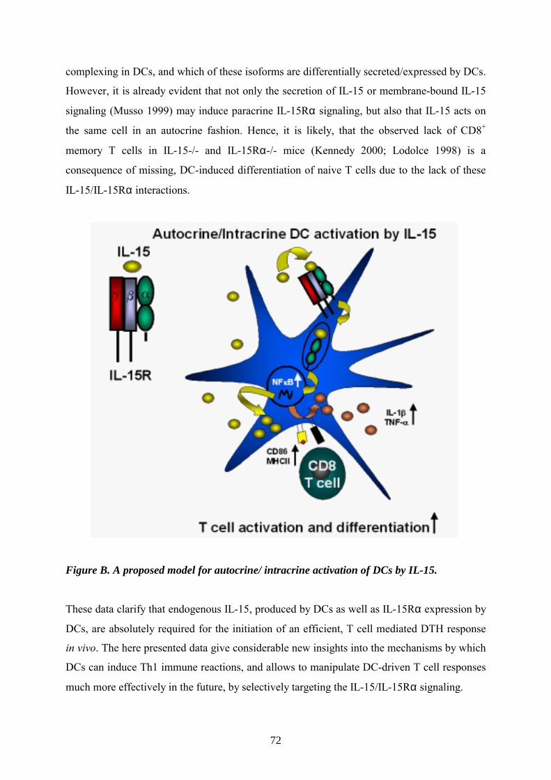
72
complexing in DCs, and which of these isoforms are differentially secreted/expressed by DCs.
However, it is already evident that not only the secretion of IL-15 or membrane-bound IL-15
signaling (Musso 1999) may induce paracrine IL-15Rα signaling, but also that IL-15 acts on
the same cell in an autocrine fashion. Hence, it is likely, that the observed lack of CD8+
memory T cells in IL-15-/- and IL-15Rα-/- mice (Kennedy 2000; Lodolce 1998) is a
consequence of missing, DC-induced differentiation of naive T cells due to the lack of these
IL-15/IL-15Rα interactions.
Figure B. A proposed model for autocrine/ intracrine activation of DCs by IL-15.
These data clarify that endogenous IL-15, produced by DCs as well as IL-15Rα expression by
DCs, are absolutely required for the initiation of an efficient, T cell mediated DTH response
in vivo. The here presented data give considerable new insights into the mechanisms by which
DCs can induce Th1 immune reactions, and allows to manipulate DC-driven T cell responses
much more effectively in the future, by selectively targeting the IL-15/IL-15Rα signaling.

73
Taken together, a complex and comprehensive analysis of the two structural related cytokines
IL-15 and IL-21 revealed very interesting, although dichotomous effects on generation,
maturation and function of dendritic cells. This series of studies offers novel, biological and
clinical important perspectives. Based on my data, we got a deeper insight in how adaptive
immune responses are shaped by the action of soluble factors on DCs, giving the opportunity
to inhibit/support DC function in health and disease. This encourage one to examine, whether
and how antagonists as well as agonists of IL-15 and IL-21 can be exploited to control
overwhelming immune reactions for therapeutical demands.

74
Abbreviations
Ag: antigen
APCs: antigen-presenting cells
BM: bone marrow
BMDC: bone marrow-derived dendritic cells
CHS: contact hypersensitivity reaction
DCs: dendritic cells (either in vivo or generated in vitro with GMCSF from BM)
DLN: draining lymph nodes
DTH: delayed type hypersensitivity
FITC: fluorescein-5-isothiocyanate
GMCSF: granulocytes-monocyte cell stimulating factor
IL: Interleukin
IL-15+/+: C57BL/6 mice
IL-15+/+ DC: dendritic cells generated with GMCSF from C57BL/6 mice
IL-15-/-: IL-15 knockout mice on C57BL/6 background
IL-15-/- DC: dendritic cells generated with GMCSF from IL-15 knock-out mice
IL-15Rα-/-: IL-15 receptor alpha knockout mice on C57BL/6 background
IL-15Rα-/- DC: dendritic cells generated with GMCSF from IL-15Rα knockout mice
IL-15DCs: dendritic cells generated with GMCSF + IL-15
IL-21DCs: dendritic cells generated with GMCSF + IL-21
MF: macrophages
PCl: picrylchloride
R: receptor
SRBC: sheep red blood cells
LPS: lipopolysaccharide

75
References
Agostini et al. CD8 T cell infiltration in extravascular tissues of patients with human immunodeficiency virus infection. Interleukin-15 upmodulates costimulatory pathways involved in the antigen-presenting cells-T cell interaction. Blood 93, 1277-1286 (1999).
Akbari et al. DNA vaccination: transfection and activation of dendritic cells as key events for immunity. J. Exp. Med. 189, 169-178 (1999).
Asao et al. Cutting edge: the common gamma-chain is an indispensable subunit of the IL-21 receptor complex. J.Immunol. 167, 1-5 (2001).
Bamford et al. The interleukin (IL) 2 receptor beta chain is shared by IL-2 and a cytokine, provisionally designated IL-T, that stimulates T cell proliferation and the induction of lymphokine-activated killer cells. Proc. Natl. Acad. Sci. U. S. A 91, 4940-4944 (1994).
Banchereau et al. Dendritic cells and the control of immunity. Nature 392, 245-252 (1998).
Banchereau et al. Immunobiology of Dendritic Cells. Annu. Rev. Immunol. 18, 767-811 (2000). Becker et al. Mechanism in allergic contact dermatitis. Exp Dermatol 2, 63-69 (1993). Blauvelt et al. Interleukin-15 mRNA is expressed by human keratinocytes, Langerhans cells, and blood-derived DCs and is downregulated by ultraviolet B radiation. J Invest Dermatol 106, 1047-1052 (1996). Bulanova et al. The IL-15R alpha chain signals through association with Syk in human B cells. J.Immunol. 167, 6292-6302 (2001). Bulfone-Paus et al. Differential regulation of human T lymphoblast functions by IL-2 and IL-15. Cytokine 9, 507-513 (1997a). Bulfone-Paus et al. Interleukin-15 protects from lethal apoptosis in vivo. Nat Med 3, 1124-1128 (1997b). Bulfone-Paus et al.. Death deflected: IL-15 inhibits TNF-alpha-mediated apoptosis in fibroblasts by TRAF2 recruitment to the IL-15Ralpha chain. FASEB J. 13, 1575-1585 (1999).

76
Cavani et al. Effector and regulatory T cells in allergic contact dermatitis. Trends Immunol. 22, 118-120 (2001).
Cella et al. Maturation, activation, and protection of dendritic cells induced by double stranded RNA. J. Exp. Med. 189, 821-29 (1999).
Celluzzi et al. Peptide-pulsed dendritic cells induce antigen-specific CTL-mediated protective tumor immunity. J Exp Med. 183, 283-7. (1996).
Chae et al. Distribution of IL-15 receptor alpha-chains on human peripheral blood mononuclear cells and effect of immunosuppressive drugs on receptor expression. J. Immunol. 157, 2813-2819 (1996).
Corinti et al. Regulatory activity of autocrine IL-10 on dendritic cell functions. J. Immunol. 166, 4312-4318 (2001).
Dai et al. The dual role of IL-2 in the genration and maintainace of CD8+ memory T cells. J. Immunol. 165, 3031-3036 (2000).
Doherty et al. Induction and regulation of IL-15 expression in murine macrophages. J. Immunol. 156, 735-741 (1996).
Dooms et al. Quiescence-inducing and antiapoptotic activities of IL-15 enhance secondary CD4+ T cell responsiveness to antigen. J Immuno.l 161, 2141-50 (1998).
Dubois et al. Natural splicing of exon 2 of human interleukin-15 receptor alpha-chain mRNA results in a shortened form with a distinct pattern of expression. J. Biol. Chem. 274, 26978-26984 (1999).
Eggert et al. Biodistribution and vaccine efficiency of murine dendritic cells are dependent on the route of administration. Cancer Res. 59, 3340-3345 (1999).
Fearon et al. The instructive role of innate immunity in the acquired immune response. Science. 272, 50-53 (1996).

77
Fehniger et al. Cutting edge: IL-15 costimulates the generalized Shwartzman reaction and innate immune interferon-gamma production in vivo. J. Immunol. 164, 1643-1647 (2000).
Fehniger et al. Interleukin 15: biology and relevance to human disease. Blood 97, 14-32 (2001).
Fukao et al. Expression of functional IL-2 receptors on mature splenic dendritic cells. Eur. J. Immnol. 30, 1453-1457 (2000).
Gallucci et al. Danger signals: SOS to the immune system. Curr.Opin.Immunol. 13, 114-119 (2001).
Giri et al. Utilization of the β and γ chains of the IL-2 receptor by the novel cytokine IL-15. EMBO J. 13, 2822-30 (1994).
Gocinski et al. Roles of CD4+ and CD8+ T cells in murine contact sensitivity revealed by in vivo monoclonal antibody depletion. J Immunol 144, 4121-4128 (1990). Gorbachev et al. Induction and regulation of T cell priming for contact hypersensitivity. Crit Rev. Immunol. 21, 451-472 (2001).
Grabbe et al. Immunoregulatory mechanisms involved in elicitation of allergic contact hypersensitivity. Immunol. Today 19, 37-44 (1998).
Grabstein et al. Cloning of a T cell growth factor that interacts with the beta chain of the interleukin-2 receptor. Science 264, 965-968 (1994).
Guermonprez et al. Antigen presentation and T cell stimulation by dendritic cells. Annu. Rev. Immunol. 20, 621-667 (2002).
Habib et al. The common γ-chain is a required signaling component of the IL-21 receptor and supports IL-21 induced proliferation via JAK3. Biochemistry 41, 8725-8731 (2002).

78
Harlow et al. Antibodies - A laboratory manual. (1988).
Hartmann et al. CpG DNA: a potent signal for growth, activation, and maturation of human dendritic cells. Proc. Natl. Acad. Sci. USA 96, 9305-10 (1999).
Hofmann et al. The anagen hair cycle induces systemic immunosuppression of contact hypersensitivity in mice. Cell Immunol 184, 65-73 (1998). Inaba et al. Granulocytes, macrophages and dendritic cells arise from a common major histocompatiblity complex calls II-negative progenitor in mouse bone marrow. Proc. Natl. Acad. Sci. USA. 90, (1993).
Janeway et al. Immunobiology. The Immune System in Health and Disease. Garland Science – New York. 5th edition, 270 (2002). Jonuleit et al. Dendritic cells as a tool to induce anergic and regulatory T cells. Trends Immunol. 7, 394-400 (2001).� Kalish et al. Molecular mechanisms of CD8+ T cell-mediated delayed hypersensitivity: implications for allergies, asthma, and autoimmunity. J. Allergy Clin. Immunol. 103, 192-199 (1999).
Katayama et al. Blockade of costimulatory molecules B7-1 (CD80) and B7-2 (CD86) down- regulates induction of contact sensitivity by haptenated epidermal cells. Br. J. Dermatol. 136, 846-852 (1997).
Kennedy et al. Reversible defects in natural killer and memory CD8 T cell lineages in interleukin 15-deficient mice. J. Exp. Med. 191, 771-780 (2000).
Kim et al. Targeting the IL-15 receptor with an antagonist IL-15 mutant/Fc gamma2a protein blocks delayed-type hypersensitivity. J. Immunol. 160, 5742-5748 (1998).
Krause et al. Genomic structure and chromosomal localization of the human IL-15 gene. Cytokine 8, 667-674 (1996).
Kripke et al. Evidence that cutaneous antigen-presenting cells migrate to regional lymph nodes during contact sensitization. J Immunol 145, 2833-2838 (1990).
79
Ku et al. Control of homeostasis of CD8+ memory T cells by opposing cytokines. Science 288, 675-678 (2000). Kumaki et al. Interleukin-15 up-regulates interleukin-2 receptor alpha chain but down-regulates its own high-affinity binding sites on human T and B cells. Eur J Immunol. 26, 1235-9 (1996). Kunzendorf et al. Suppression of cell-mediated and humoral immune responses by an interleukin-2-immunoglobulin fusion protein in mice. J. Clin. Invest 97, 1204-1210 (1996).
Lambrecht et al. Allergen uptake and presentation by dendritic cells. Curr. Opin. Allergy Clin. Immunol. 1, 51-59 (2001).
Lanzavecchia et al. The instructive role of dendritic cells on T cell responses: lineages, plasticity and kinetics. Curr. Opin. Immunol. 13, 291-298 (2001).
Lappin et al. Analysis of mouse dendritic cell migration in vivo upon subcutaneous and intravenous injection. Immunology 98, 181-188 (1999).
Li et al. IL-15 and IL-2: a matter of life and death for T cells in vivo. Nat. Med. 7, 114-118 (2001).
Liu et al. Dendritic cell lineage, plasticity and cross-regulation. Nat. Immunol. 2, 585-589 (2001a).
Liu et al. Dendritic cell subsets and lineages, and their functions in innate and adaptive immunity. Cell 106, 259-262 (2001b).
Lodolce et al. IL-15 receptor maintains lymphoid homeostasis by supporting lymphocyte homing and proliferation. Immunity. 9, 669-676 (1998).
Luger et al. Regulation of the immune response by epidermal cytokines and neurohormones. J Dermatol Sci 13, 5-10 (1996). Lutz et al. An advanced culture method for generating large quantities of highly pure dendritic cells from mouse bone marrow. J. Immunol. Methods 223, 77-92 (1999).
80
Ma et al. The pleiotropic functions of IL-15: not so IL-2 after all. J. Exp. Med. 191, 753-755 (2000).
Macatonia et al. Dendritic cells and the initiation of contact sensitivity to fluorescein isothiocyanate. Immunology 59, 509-514 (1986).
Marks-Konczalik et al. IL-2-induced activation-induced cell death is inhibited in IL-15 transgenic mice. Proc Natl Acad Sci U S A 97, 11445-50 (2000). Mattei et al. IL-15 is expressed by dendritic cells in response to type I IFN, double- stranded RNA, or lipopolysaccharide and promotes dendritic cell activation. J. Immunol. 167, 1179-1187 (2001).
Matzinger et al. Tolerance, danger, and the extended family. Annu. Rev. Immunol. 12, 991-1045 (1994).
Meazza et al. Identification of a novel interleukin-15 (IL-15) transcript isoform generated by alternative splicing in human small cell lung cancer cell lines. Oncogene 12, 2187-2192 (1996).
Mehling et al. Mycophenolate mofetil impairs the maturation and function of murine dendritic cells. J Immunol 165, 2374-2378 (2000). Mohamadzadeh et al. Interleukin 15 skews monocyte differentiation into dendritic cells with features of Langerhans cells. J. Exp. Med. 194, 1013-1020 (2001).
Morel, et al. Split activity of interleukin-10 on antigen capture and antigen presentation by human dendritic cells: definition of a maturative step. Eur.J.Immunol. 27, 26-34 (1997).
Mori et al. IL-15 promotes cytokine production of human T helper cells. J Immunol 156 2400-2405 (1996). Moser et al. Dendritic cell regulation of TH1-TH2 development. Nat. Immunol. 1, 199-205 (2000).
Müller et al. IL-12 as mediator and adjuvant for the induction of contact sensitivity in vivo. J Immunol 155, 4661-4668 (1995).

81
Murakami et al. Critical cytoplasmatic region of the interleukin 6 signal transducer gp130 is conserved in the cytokine receptor family. Proc. Natl. Akad. Sci. USA 88, 11349-11353 (1991). Musso et al. Human monocytes constitutively express membrane-bound, biologically active, and interferon-gamma-upregulated interleukin-15. Blood 93, 3531-3539 (1999).
Ohteki et al. Interleukin 12-dependent interferon gamma production by CD8alpha+ lymphoid dendritic cells. J. Exp. Med. 189, 1981-1986 (1999).
Ohteki et al. Critical role of IL-15-IL-15R for antigen-presenting cell functions in the innate immune response. Nat. Immunol. 2, 1138-1143 (2001).
Onu et al. Regulation of IL-15 secretion via the leader peptide of two IL-15 isoforms. J. Immunol. 158, 255-262 (1997).
Ozaki et al. Cloning of a type I cytokine receptor most related to the IL-2 receptor beta chain Proc. Natl. Acad. Sci USA. 97, 11439-11444 (2000).
Parrish-Novak et al. Interleukin 21 and its receptor are involved in NK cell expansion and regulation of lymphocyte function. Nature 408, 57-63 (2000).
Parrish-Novak et al. Interleukin 21 and IL-21 receptor: novel effects of NK- and T- cell responses. J. Leukoc. Biol. 72, 856-863 (2002).
Pereno et al. IL-15/IL-15Ralpha intracellular trafficking in human melanoma cells and signal transduction through the IL-15Ralpha. Oncogene 19 , 5153-5162 (2000).
Porgador et al. Induction of antitumor immunity using bone marrow-generated dendritic cells. J Immunol. 156, 2918-26 (1996).
Reis e Sousa et al. The role of dendritic cells in the induction and regulation of immunity to microbial infection. Curr. Opin. Immunol. 11, 392-399 (1999).

82
Resigno et al. Cordinatd events during bacterial-induced DC maturation. Immunol. Today 20, 200-203 (1999).
Robertson et al. DO11.10 and OT-II T cells recognize a C-terminal ovalbumin 323-339 epitope. J. Immunol. 164, 4706-4712 (2000).
Roncarolo et al. Type 1 T regulatory cells. Immunol Rev. 182, 68-79 (2001).
Rotzschke et al. Exact prediction of a natural T cell epitope. Eur. J. Immunol. 21, 2891-2894 (1991).
Rubinstein et al. Systemic Administration of IL-15 Augments the Antigen-Specific Primary CD8(+) T Cell Response Following Vaccination with Peptide-Pulsed Dendritic Cells. J. Immunol. 169, 4928-4935 (2002).
Ruchatz et al. Soluble IL-15 receptor alpha-chain administration prevents murine collagen-induced arthritis: a role for IL-15 in development of antigen- induced immunopathology. J. Immunol. 160, 5654-5660 (1998).
Rückert et al. IL-15-IgG2b fusion protein accelerates and enhances a Th2 but not a Th1 immune response in vivo, while IL-2-IgG2b fusion protein inhibits both. Eur J Immunol 28, 3312-3320 (1998). Rückert et al. Inhibition of keratinocyte apoptosis by IL-15: A new parameter in the pathogenesis of psoriasis? J Immunol 156, 2240-2250 (2000). Rückert et al. IL-2-IgG2b fusion protein suppresses murine contact hypersensitivity in vivo. J. Invest Dermatol. 119, 370-376 (2002).
Saikh et al. IL-15-induced conversion of monocytes to mature dendritic cells. Clin. Exp. Immunol. 126, 447-455 (2001).
Sallusto et al. Efficient presentation of soluble antigen by cultured human dendritic cells is maintained by granulocyte/macrophage colony-stimulating factor plus interleukin 4 and downregulated by tumor necrosis factor alpha. J.Exp.Med. 179, 1109-1118 (1994).

83
Sallusto et al. Dendritic cells use macropinocytosis and the mannose receptor to concentrate macromolecules in the major histocompatibility complex class II compartment: downregulation by cytokines and bacterial products. J.Exp.Med. 182, 389-400 (1995).
Schluns et al. Cutting edge: requirement for IL-15 in the generation of primary and memory antigen-specific CD8 T cells. J. Immunol. 168, 4827-4831 (2002).
Schuurhuis et al. Immature dendritic cells acquire CD8(+) cytotoxic T lymphocyte priming capacity upon activation by T helper cell-independent or -dependent stimuli. J. Exp. Med. 192, 145-150 (2000).
Sprent et al. Generation and maintenance of memory T cells. Curr. Opin. Immunol. 13, 248-254 (2001).
Stumbles et al. Resting respiratory tract dendritic cells preferentially stimulate T helper cell type 2 (Th2) responses and require obligatory cytokine signals for induction of Th1 immunity. J.Exp.Med. 188, 2019-2031 (1998).
Sugamura et al. The interleukin-2 receptor gamma chain: its role in the multiple cytokine receptor complexes and T cell development in XSCID. Annu. Rev. Immunol. 14, 179-205 (1996).
Tagaya et al. IL-15: A pleiotropic cytokine with diverse receptor/signaling pathways whose expression is controlled at multiple levels. Immunity 4, 329-36 (1996). Tagaya et al. Identification of a novel receptor/signal transduction pathway for IL- 15/T in mast cells. EMBO J. 15:4928-4939 (1996).
Takayama et al. Mammalian and viral IL-10 enhance C-C chemokine receptor 5 but down- regulate C-C chemokine receptor 7 expression by myeloid dendritic cells: impact on chemotactic responses and in vivo homing ability. J.Immunol. 166, 7136-7143 (2001). Thery et al. The cell biology of antigen presentation in dendritic cells. Curr. Opin. Immunol. 13, 45-51 (2001).
Tuma et al. Homeostasis of naive, effector and memory CD8 T cells. Curr. Opin. Immunol. 14, 348-353 (2002).
84
Vossenhenrich et al. Cytokines: IL-21 joins the γc-dependent network? Curr. Biol. 11, 175-177 (2001).
Vremec et al. The influence of granulocyte/macrophage colony-stimulating factor on dendritic cell levels in mouse lymphoid organs. Eur. J. Immunol. 27, 40-44 (1997).
Waldmann et al. The multifaceted regulation of interleukin-15 expression and the role of this cytokine in NK cell differentiation and host response to intracellular pathogens. Annu Rev Immunol 17, 19-49, (1999). Watanabe et al. Contact hypersensitivity: the mechanism of immune responses and T cell balance. J. Interferon Cytokine Res. 22, 407-412 (2002).
Weninger et al. Migration and differentiation of CD8+ T cells. Immunol. Rev. 186, 221-233 (2002).
Wilkinson et al. Chemoattraction of human blood T lymphocytes by interleukin-15. J. Exp. Med. 181, 1255-1259 (1995).
Winzler et al. Maturation stages of mouse dendritic cells in growth factor-dependent long-term cultures. J. Exp. Med. 185, 317-28 (1997).
Xu et al. Distinct roles for B7-1 and B7-2 determinants during priming of effector CD8+ Tc1 and regulatory CD4+ Th2 cells for contact hypersensitivity. J. Immunol. 159, 4217-4226 (1997).
Yajima et al. Overexpression of IL-15 in vivo increases antigen-driven memory CD8+ T cells following a microbe exposure. J. Immunol. 168, 1198-1203 (2002).
Zhang et al. Potent and selective stimulation of memory-phenotype CD8+ T cells in vivo by IL-15. Immunity. 8, 591-599 (1998).

85
Zhang et al. Chemical activation of innate and specific immunity in contact dermatitis. J.
Invest Dermatol. 115, 168-176 (2000).

86
Danksagung
An dieser Stelle möchte ich allen Menschen namentlich danken, die mich während meiner
Doktorarbeit unterstützt haben.
Silvia
Ich möchte Dir danken, dass Du mich sowohl spontan, als auch sehr herzlich in Deine
Laborgruppe aufgenommen hast. Deine ruhige Art, eine wissenschaftliche Vision zu
entwickeln und Deine Begabung sie sorgfältig zu vermitteln, ist faszinierend. Noch
faszinierender ist allerdings, dass Du immer die Übersicht behältst. Ich danke Dir auch für
Dein Vertrauen, dass Du in mich gesetzt hast. Die Fahrt nach Marburg wird unvergessen
bleiben.
René
Dir bin ich am meisten zu Dank verpflichtet. All die unzähligen Stunden, die wir gemeinsam
verbracht haben, um meine oder Deine wissenschaftlichen Probleme zu lösen, haben meinen
Horizont unschätzbar erweitert. Du allein weißt von der ersten Stunde an, wie viel Freude und
wie viel Frust in dieser Arbeit steckt. Du hast alles mitgetragen und dafür danke ich Dir von
Herzen.
Meinen Eltern
Euch möchte ich danken, weil Ihr mir alles gegeben habt, um jetzt hier stehen zu können.
Eurer Verständnis für meine Leidenschaft zur Wissenschaft, meine ganze Ausbildung und
nicht zuletzt Euer offenes Haus mit offenen Armen haben mir zu jederzeit Sicherheit
geschenkt.

87
Meinen Kollegen („The SBP-gang!“)
Es sei Euch von Herzen gedankt für die Vielzahl an Hilfe, die ihr mir habt zukommen lassen.
Insbesondere möchte ich André hervorheben, für seine exzellente Arbeit, seinen Teamgeist
und noch mehr für seine Beharrlichkeit, bei uns zu bleiben!
Kicki (Maria V. Odenwald)
Mit Dir ist das Labor erst ganz und gar lebendig! Dein unermüdlicher Einsatz und Dein
unwahrscheinlicher Wissensschatz hat mir an den entscheidenden Stellen geholfen. Aber auch
das „Nebenbei“, also die vielen Gespräche neben der Bench waren immer ein Augenöffner.
Jürgen
Ich möchte Dir danken, dass Du mit mir ein erstes Stück wissenschaftlichen Weges gegangen
bist. Deine flexible Art, die Daten von allen Seiten kritisch zu betrachten habe ich stets
bewundert.

88
Publications
Paper
1.) IL-2-IgG2b fusion protein suppresses murine contact hypersensitivity in vivo.
R. Rückert, K. Brandt, U. Hofmann, S. Bulfone-Paus, R. Paus. 2002 J Invest Dermatol
119:370
2.) TSST-1 induces Th1 or Th2 differentiation in naïve CD4+ T-cells in a dose and APC
dependent manner.
K. Brandt, Jürgen van der Bosch, Ralf Fliegert and Svante Gehring. 2002 Scand J
Immunol, 56:572
3.) Interleukin-21 inhibits dendritic cell activation and maturation.
K. Brandt, S. Bulfone-Paus, R. Rückert, Submitted for publication
4.) Dendritic cell-derived IL-15 is essential for the induction of Th1 immune responses
K. Brandt*, R. Rückert*, E. Bulanova, F. Mirghomizadeh, R. Paus and S. Bulfone-Paus,
(Both Authors contributed equally to this work), Submitted for publication
5.) Blocking Interleukin-15 inhibits Th2-cell differentiation, antibody production and allergic
inflammation in mice.
R. Rückert, U. Herz, T. Pohl, K. Brandt, V. Budagian, E. Bulanova, H. Dürkop, H. Renz
and S. Bulfone-Paus, Submitted for publication
6.) Interleukin-15 promotes the survival of naïve- and memory- phenotype CD8+ T cells
M. Berard, K. Brandt, S. Bulfone-Paus, and D. Tough, Submitted for publication
7.) Interleukin-21 inhibits CD8+ T cell activation and induction of contact hypersensitivity
reaction in vivo.
K. Brandt, S. Bulfone-Paus, D.C. Foster, R. Paus, R. Rückert, The Journal of Investigative
Dermatology, Submitted for publication

89
8.) IL-15 mediates activation but not generation of murine macrophages.
K. Brandt, S. Bulfone-Paus, R. Rückert, In preparation
Abstracts
1.) An in vitro model to study the influence of antigen-presenting cells on human T cell
differentiation following superantigen activation.
S. Gehring, K. Brandt, and J. van der Bosch
2. Teupitzer Colloqium. Macrophages in Inflammation. Sept. 2.-4. 1999, Teupitz, Berlin.
Germany
2.) TSST-1 induces Th1 or Th2 differentiation in naïve CD4+ T cells in a dose- and APC-
dependent manner.
K. Brandt, J. van der Bosch, and S. Gehring
32. Annual Meeting of the German Society of Immunology, Sept. 26.-29. 2001, Dresden.
Germany
Immunobiology 204: 1-2, 98, 2001
3.) Interleukin-21 inhibits dendritic cell activation and maturation.
K. Brandt, S. Bulfone-Paus, and R. Rückert
33. Anual Meeting of the German Society of Immunology. Sept. 25.-28. 2002, Marburg.
Germany.
Immunobiology 206: 1-3, 20, 2002
4.) Dendritic cell-derived interleukin-15 is essential for the induction of Th1 immune
reactions.
R. Rückert, K. Brandt, and S. Bulfone-Paus,
33. Anual Meeting of the German Society of Immunology. Sept. 25.-28. 2002, Marburg.
Germany.
Immunobiology 206: 1-3, 28, 2002

90
5.) Interleukin-21 inhibits dendritic cell activation and maturation.
K. Brandt, S. Bulfone-Paus, and R. Rückert
25. Arbeitstagung der Norddeutschen Immunologen. Nov. 15. 2002, Borstel. Germany.
Talks
1.) Dendritic cell-derived interleukin-15 is essential for the induction of Th1 immune
reactions.
K. Brandt, R. Rückert, and S. Bulfone-Paus,
33. Anual Meeting of the German Society of Immunology. Sept. 25.-28. 2002, Marburg.
Germany.
Immunobiology 206: 1-3, 28, 2002
2.) Interleukin-21 inhibits dendritic cell activation and maturation.
K. Brandt, S. Bulfone-Paus, and R. Rückert
25. Arbeitstagung der Norddeutschen Immunologen. Nov. 15. 2002, Borstel. Germany.
91
Lebenslauf
Katja Brandt Diplom-Biochemikerin
*22.08.1974 in Bremen
Carlstrasse 40
24534 Neumünster
Telefon: 04321 / 179979
e-mail: [email protected]
Schule 1980 – 1984 Grundschule in Grolland / Bremen
1984 – 1986 Orientierungsstufe in Huchting / Bremen
1986 – 1991 Gymnasium in Huchting/Bremen
1991 – 1993 Gymnasium der Holstenschule in Neumünster
Abschluss: Allgemeine Hochschulreife
09/1993 Ergänzungsprüfung zur kantonalen Maturität in der
Schweiz
Studium
10/1993 – 06/1998 Studium der Biochemie an der Universität
Zürich / Schweiz
Grosses Nebenfach: Immunologie
Semester 1994/95 Erstes Vordiplom

92
Semester 1996/97 Zweites Vordiplom
05/1997 – 12/1997 Diplomarbeit: „Gezielte Biotinylierung von
Miniantikörpern zur Herstellung von tetrameren
Komplexen mit variabler Spezifität via
Streptavidin“ im Institut für Biochemie der
Universität Zürich
06/1998 Abschluss des Studiums als Diplom Biochemikerin
Praktika und Projekte
08/1998 – 10/1998 Lehrgang als Schwesternhelferin mit Praktikum
in der Abteilung Onkologie, Hämatologie und
Nephrologie des Friedrich Ebert Krankenhauses
in Neumünster
11/1998 – 05/1999 Europäische Projektstudie: „Selection and
affinity maturation of recombinant phage
antibodies to tumor-associated HLA-
antigen/peptide complexes“ im Institut für
Pharmakologie unter Betreuung von Professor
Jan Engberg an der Pharmazeutischen
Hochschule zu Kopenhagen, Dänemark
Promotionsarbeit
07/1999 – 02/2001 Arbeit zum Thema „Die Steuerung der T-
Zelldifferenzierung durch Antigen-präsentierende
Zellen in vitro“ unter der Leitung von Dr. van der
Bosch in der Abteilung Experimentelle
Immunpharmakologie des Forschungszentrums
Borstel

93
03/2001 – 03/2003 Promotionsarbeit zum Thema „Dendritic Cell
mediated immune responses are shaped by IL-15
and IL-21” unter der Leitung von Prof. Dr. Dr.
Bulfone-Paus in der Abteilung Immunbiologie des
Forschungszentrums Borstel.
Preis 09/2002 Posterpreis 33rd. Anual Meeting of the German
Society of Immunology
Dendritic cell-derived interleukin-15 is essential for
the induction of Th1 immune reactions.
K. Brandt, R. Rückert, and S. Bulfone-Paus
Neumünster, den 20. Dezember 2002