Demyelinating Diseases Multiple sclerosis( MS ) Prof Mohammad Salah Abduljabbar.
68
Demyelinating Diseases Multiple sclerosis( MS ) Prof Mohammad Salah Abduljabbar
-
Upload
joan-hoover -
Category
Documents
-
view
213 -
download
0
Transcript of Demyelinating Diseases Multiple sclerosis( MS ) Prof Mohammad Salah Abduljabbar.

Demyelinating Diseases
Multiple sclerosis( MS )
Prof Mohammad Salah Abduljabbar

Introduction
-Demyelinating disorders of the CNS affect myelin and/or oligodendroglia
with relative sparing of axons.
-Oligodendrocytes, like Schwann cells in the peripheral nervous system, are responsible for the formation of myelin around CNS axons.
-One Schwann cell myelinates one axons but one oligodendrocyte may myelinate several contiguous axons, and the close proximity of cell to axon may not be obvious by light microscopy.
-Oligodendrocyte are present in gray matter near neural cell bodies and in white matter near axons.
-Myelin is composed of protein 20% & lipids.

Multiple Sclerosis
Chronic, progressive, degenerative
disorder of the CNS characterized by
disseminated demyelination of nerve
.fibers of the brain and spinal cord

Introduction to Multiple Sclerosis (MS)
• Chronic autoimmune disease• Progressive disease• Involves Immune System & Neurological System • Multifocal areas of demyelination• Disrupts ability of the nerve to conduct electrical
impulses• Leads to symptoms

Multiple Sclerosis (MS)
-MS referred by the British as disseminated sclerosis & by French as Sclerose en plaques.
-MS is a common demyelinating disease, characterized by focal disturbance of function and a relapsing and remitting course.
-Higher incidence of the disease found in the northern most latitude of the northern & southern hemispheres compared to southernmost latitudes.
-MS usually occur in young adults with a peak age incidence of 20-40 years.
-more female than males are affected.
-The risk of MS in relative patients increases 20 folds.

Epidemiology of MS
• Age onset 20 – 50 years old• Women are 2 times more likely to develop MS• 500,000 cases in US• Over 2.5 million people around the world• More prevalent whites of northern European
ancestry• Vitamin D3 deficiency• Genetic Influences

Multiple Sclerosis Etiology
Multiple Sclerosis Etiology
-Unknown cause
-Related to infectious, immunological, and genetic factors

Multiple Sclerosis Etiology
Possible precipitating factors include : -Infection
-Physical injury
-Emotional stress
-Excessive fatigue
-Pregnancy
-Poor state of health

Multiple SclerosisPathophysiology
Myelin sheath -Segmented lamination that wraps axons of
many nerve cells
-Increases velocity of nerve impulse conduction in the axons
-Composed of myelin, a substance with high lipid content

Multiple SclerosisPathophysiology
-Characterized by chronic inflammation, demyelination, and gliosis (scarring) in the
CNS
-Initially triggered by a virus in genetically susceptible individuals
-Subsequent antigen-antibody reaction leads to demyelination of axons

Pathogenesis of MS
Fig. 57-1

Multiple Sclerosis Pathophysiology
-Disease process consists of loss of myelin, disappearance of oligodendrocytes, and proliferation of astrocytes
-Changes result in plaque formation with plaques scattered throughout the CNS

Multiple Sclerosis Pathophysiology
-Initially the myelin sheaths of the neurons in the brain and spinal cord are attacked, but
the nerve fiber is not affected
-Patient may complain of noticeable impairment of function
-Myelin can regenerate, and symptoms disappear, resulting in a remission

Multiple Sclerosis Etiology and Pathophysiology
-Myelin can be replaced by glial scar tissue
-Without myelin, nerve impulses slow down
-With destruction of axons, impulses are totally blocked
-Results in permanent loss of nerve function

Pathology
•Scattered lesions with a greyish color.
• 1mm to several cm in size.
• Are present in the white matter of the brain and spinal cord and are referred to as plaques.
•perivenous distribution.

RECENT LESIONS LATER OLD LESION
•Myelin destruction
•Relative axon sparing
•Perivenous infiltration with MNP
•Breakdown of BBB
Astrocyte proliferation
•Relatively acellular
•More clearly demarcated.
•Bare axons are surrounded by astrocytes.
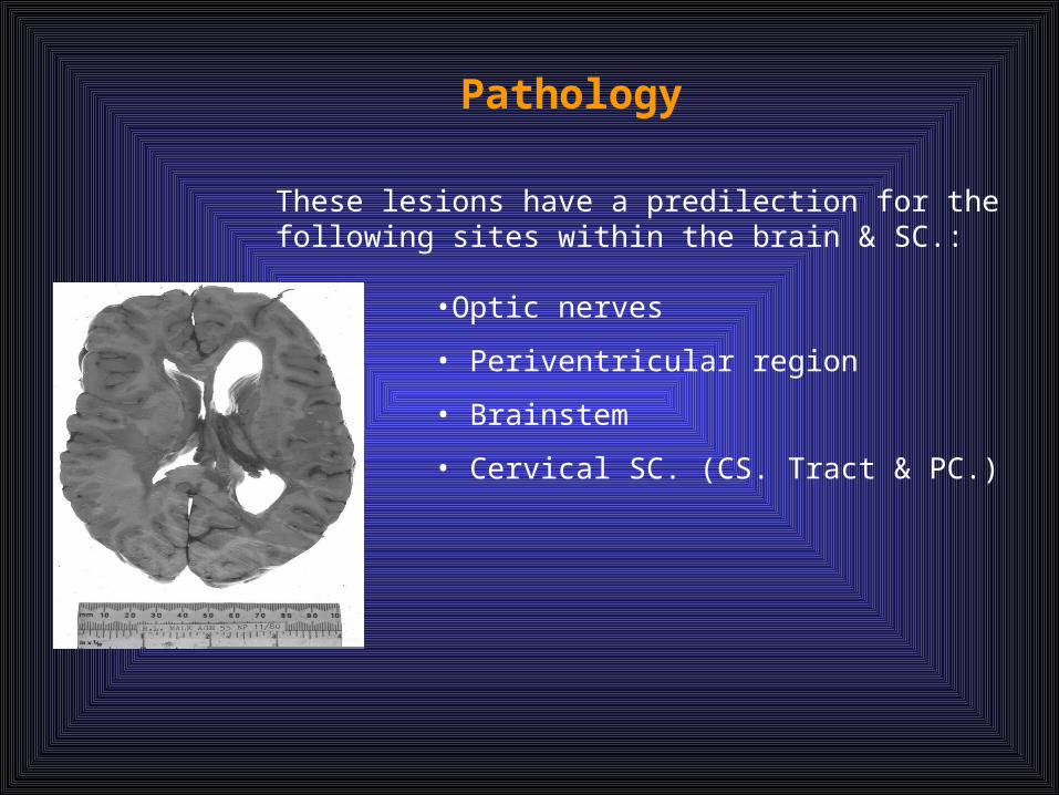
Pathology
•Optic nerves
• Periventricular region
• Brainstem
• Cervical SC. (CS. Tract & PC.)
These lesions have a predilection for the following sites within the brain & SC.:

Pathogenesis
Genetic predisposition Environmental Exposure (Virus)
Autoimmune attack by CD4 T-cell
Demyelination
Multiple Sclerosis

Role of Vitamin D in MSBackground Information
1. US cohort study found that 3.5 times more women residing in northern states were diagnosed with MS than southern states
2. Incidence of MS highest in North Temporal Climate
3. MS more prominent in areas reporting less than 2000 hours of sunshine annually
4. MS displays seasonable variability with increased activity in the Spring and lowest in the Fall.
5. A Finnish study found in MS patients lower serum vitamin D levels in the Spring.
6. A line between dietary intake of vitamin D and the incidence of MS has been suggested in Norway along the coastal areas where fatty fish, dairy products, and cereals are all fish in vitamin D consumed in higher amounts. The incidence is lower then the rest of Norway.
7. Levels of 11 25 hyroxy D3 and 1
8. Dietary information from the Nurse’s Health Study of 187,000 women showed those with a history of vitamin D supplementation as low as 400 units daily had a 40% less chance of developing MS.

Multiple SclerosisClinical Manifestations
-Vague symptoms occur intermittently over months and years
-MS may not be diagnosed until long after the onset of the first symptom
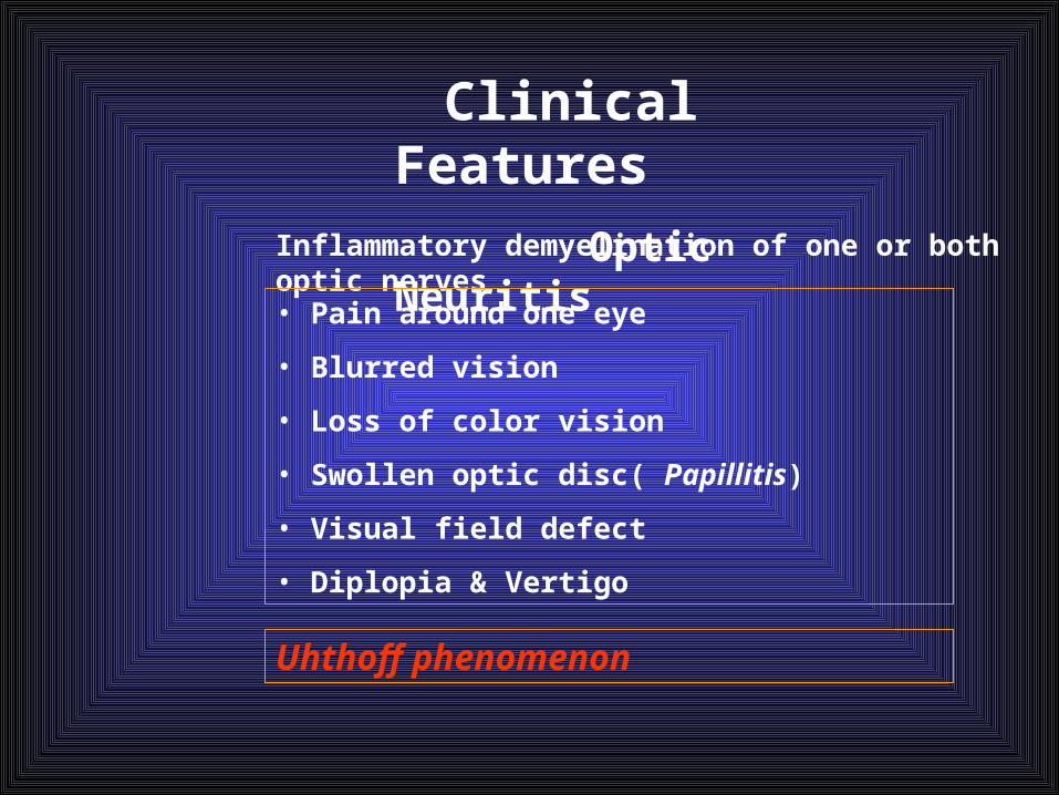
Clinical Features
Optic NeuritisInflammatory demyelination of one or both optic nerves
• Pain around one eye
• Blurred vision
• Loss of color vision
• Swollen optic disc( Papillitis)
• Visual field defect
• Diplopia & Vertigo
Uhthoff phenomenon

Multiple Sclerosis
(1 )Motor manifestations - Weakness or paralysis of limbs
- Diplopia (double vision)
- Scanning speech
- Spasticity of muscles

Clinical Features
Motor Symptoms
• Monoparesis
• Paraparesis
Signs
• Increased tone
•Hyperactive tendon reflexes
•Absent abdominal reflexes
•Pyramidal distribution weakness
Multiple Sclerosis
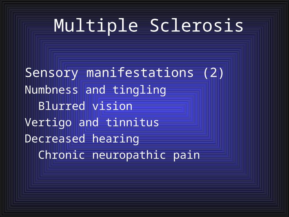
(2 )Sensory manifestations Numbness and tingling
Blurred vision
Vertigo and tinnitus
Decreased hearing
Chronic neuropathic pain

Sensory Symptoms
• Numbness & Paresthesia
• Impaired vibration & Joint position sensation
• Lhermitte’s Sign ( Shock-like sensation in the limb)
•Dysaesthesia + Sensory loss to pain & Temp.
Clinical Features

Multiple Sclerosis
(3 )Cerebellar manifestations Nystagmus
Ataxia Dysarthria Dysphagia
Multiple Sclerosis
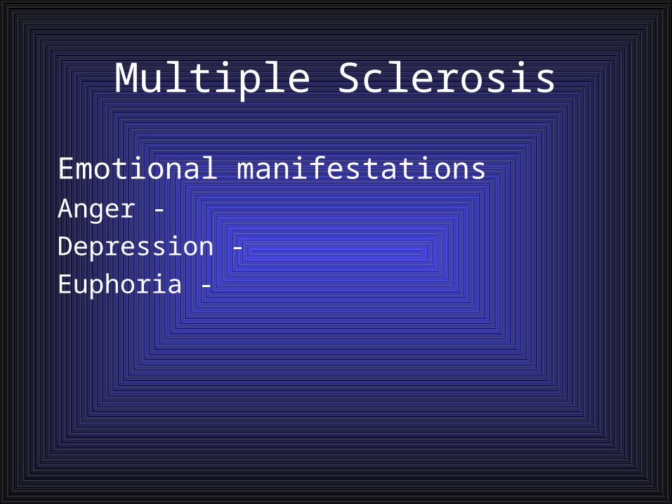
Emotional manifestations -Anger
-Depression
-Euphoria

Multiple Sclerosis
Bowel and bladder functions -Constipation
-Spastic bladder: small capacity for urine results in incontinence
-Flaccid bladder: large capacity for urine and no sensation to urinate

Multiple Sclerosis
Sexual dysfunctionErectile dysfunction
Decreased libido
Difficulty with orgasmic response
Painful intercourse
Decreased lubrication

Symptoms of MS• Vision problems• Numbness• Difficulty walking• Fatigue• Depression• Emotional changes• Vertigo & dizziness• Sexual dysfunction
• Coordination problems• Balance problems• Pain• Changes in cognitive
function• Bowel/bladder
dysfunction• Spasticity

Types of MS• Relapsing-remitting MS (RRMS)
– Affects 85% of newly diagnosed– Attacks followed by partial or complete recovery– Symptoms may be inactive for months or years
• Secondary-progressive MS (SPMS)– Occasional relapses but symptoms remain constant,
no remission– Progressive disability late in disease course

Types of MS• Primary-progressive MS (PPMS)
– Affects approximately 10% of MS population– Slow onset but continuous worsening
condition• Progressive-relapsing MS (PRMS)
– Rarest form Affects approx. 5%– Steady worsening of condition
at onset

Clinical Course
Acute MS
•Explosive onset•Death may occur in months•Dramatic recovery and prolonged remission may occur

Slowly Progressive MS•Common in older age group•No relapse/remission•Takes the form of a Progressive myelopathy
Disability
Time

Relapsing MS•Accumulating disability
Disability
Time

Benign form•Abrupt onset•Good remission•Long latent period
Time
Disability

Diseases to rule out• Viral infections• Lyme disease• CVA• Lupus• B12 deficiency• Rheumatoid arthritis• Other connective
tissue disorders
• Vasculitis• Syphilis• Tuberculosis• HIV• Sarcoidosis

Multiple SclerosisDiagnostic Studies
Diagnosis based primarily on:
-History and clinical manifestations
-Ruling out other causes of symptoms
-No definitive diagnostic test
-MRI – demonstrates presence of plaques

Investigation
No diagnostic test. Only support the clinical suspicion.Neuropsychological measurement of conduction within the CNS to detect second
asymptomatic lesion .
•Visual evoked potential(VEP)•Somatosensory evoked response
•Brain stem auditory evoked potential

• Mild pleocytosis mainly lymphocytes.
• Total protein maybe elevated
• gammaglobuline in 50%
• Electrophoresis of CSF using agar
shows discrete bands which are not
present in serum.
Oligoclonal band
normal
CSF examination

MRI
MRI is more sensitive showing white matter disease .
On T-2 weighted images, patchy area of abnormal white matter are found most commonly in cerebral hemisphere in periventricular areas; often lesions can be
present in the cerebellum , brain stem, cervical and or thoracic spinal cord
Area of demyelination in cerebral hemisphere

Demyelination in the Cervical Spinal Cord
MRI finding are not necessarily diagnostic

Multiple Sclerosis
Drug TherapyDrug Therapy
CorticosteroidsCorticosteroids-Treat acute exacerbations by reducing edema and
inflammation at the site of demyelination-Do not affect the ultimate outcome or degree of
residual neurologic impairment from exacerbation

Multiple Sclerosis
Immunosuppressive Therapy
-Because MS is considered an autoimmune disease
-Potential benefits counterbalanced against potentially serious side effects

Medications and MSTherapiesAdministrationCLASSAvonexIM 1x a weekInterferon beta-1a
BetaseronSC, every other dayInterferon beta-1b
CopaxoneSC 1x a dayGlatiramer acetate
RebifSC 3x a weekInterferon beta-1a
GilenyaOral capsule 1x dayFingolimod
TysabriIV Monthly at CenterNatalizumab

Tysabri & PML• Risk factors
– JC antibody status– Length of treatment– Prior immunosuppressant use
• Immuran (Azothrioprine)• Cytoxan• Novantrone• Methotrexate• Cellcept

Gilenya “Fingolimide”Blocks S1 Phosphate receptor keeping T & B cells in lymphoid tissue
First oral pill released by FDA three years ago for treatment of MS
Reduces relapse rate by 55-58%
Shows benefit on MRI endpoints as T2 lesion load and Gad enhancing lesions
Side effects:- Macular Edema- Heart Block- Liver Function Abnormalities- Sudden Death

Teriflunomide• Inhibits pyridine synthesis with mild lymphopenia• TEMSO Trial (Phase III) 1088 patients
– Follow for 2 years– Randomized to 7 or 14mg tablets or placebo– Results
• 31% reduction ARR• Decreased EDSS worsening by 30% (14mg)• Decreased new lesion by 39% in 7mg & 67% in 14mg
– Safety: good • Mainly diarrhea and LFT abnormal.

Side effects of MS medication
• Local injection site irritation/reactions• Flu like symptoms• Rise in liver enzymes• Decreased white cell count and platelets• Opportunistic infections• Depression• Progressive multifocal leukoencephalopathy
(PML)

Bladder problems
• Rule out UTI• Bladder training
– Strengthen pelvic muscles
• Medication• Anti-spasticity
– Vesicare– Detrol – Ditropan
• Referral to urologist for further evaluation and treatment

Depression• Selective Serotonin Reuptake
Inhibitors– Paxil– Prozac– Zoloft– Lexapro– Celexa
• Tricyclic Antidepressants– Elavil– Pamelor– Tofranil– Norpramin
• Some other medications– Desyrel– Serzone– Welbutrin– Effexor
• Referral for counseling• Psychologist• Encourage expression of
feelings will entire team and caregivers
• Work on solution together

Cognitive Changes
• Use calendar for appointment & special dates
• Use tape recorder to help remember information
• Start a diary or memory notebook
• Organize environment• Teach to make lists
• Limit noise during conversations
• Have patient repeat information and write down important points
• Encourage use of crossword puzzles and cognitive function tests
• Medications– Aricept– Namenda– Exelon patch

Fatigue
• Medications– Amantadine– Ritalin drugs– Focalin– Adderall– Provigil/Nuvigil
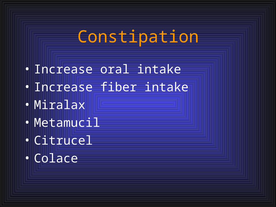
Constipation
• Increase oral intake
• Increase fiber intake
• Miralax
• Metamucil
• Citrucel
• Colace

Sexual Dysfunction
• Medications– Viagra– Cialis– Levitra

AUTOLOGOUS Stem Cell Treatment
• 10 SPMS patients• CDMS patient with HX of ON, abn VEPS, or clinical O. A. or HX of Uhthoffs
phenomenon• MRI of ON had a T2 lesion followed for 20 mo before IV of stem cells for 10 mo
afterwards• Results
• Improved V.A. and low contrast V.A.• But not in color vision or visual fields• Reduction in V.E. latency & improved amplitudes but no change OCT• Increased ON area• No change in macular volume, RFL, or MT ration• Reduction in general disability with improved in EDSS• No change in MSFC, depression , cognition• Dec. in T1 hypointense volume
• Side Effect:• Infections • Rash• Pruritus

Multiple Sclerosis
Physical therapy helps - Relieve spasticity
- Increase coordination
- Train the patient to substitute unaffected muscles for impaired ones

RIS (Radiologically Isolated Syndrome)
• White matter lesions suggestive of demyelinating disease on MRI
• Normal neurological exam• No medical history compatible with MS• Unclear whether RIS is subclinical MS or a
separated entity• About 33% of subjects with RIS develop a CIS
especially with spinal cord lesions

ACUTE DISSEMINATED ENCEPHALOMYELITIS

ADEM
• This is an inflammatory demyelinating disorder of the subcortical white matter.
• Most frequently seen in children, often evolving from antecedent infection or immunization.
• Typical presentation: encephalitic signs with non specific CSF changes and minimal or no changes on CT brain.

• Thought to be an autoimmune disease via cross reactivity of the antiviral antibodies with the myelin autoantigens.
• Viruses associated include HSV,HIV, HSV6, measles, hepatitis, influenza, EBV etc.
• There has also been an association post immunization for MMR, Influenza, BCG.

APPROPRIATE INVESTIGATIONS
• Lymphocytosis, raised CRP and ESR
• CSF- can have a raised protein but can be normal.
• CT Brain - may be normal
• MRI- gold standard for diagnosisT2 weighted images show areas of prolonged T2 in subcortical white matter, usually asymmetrical.

TREATMENT
• Empirically treated as a meningitis with cefotaxime +/- acyclovir.
• Once diagnosis is made then steroidsbecome the mainstay of management.
• Physiotherapy can also be helpful.

DIFFERENTIAL DIAGNOSIS• At first presentation, it is difficult to differentiate
between ADEM and MS.• New lesions and relapses, especially after 6/12
should alert to the possibility of MS.
• MS - no prodromal viral illness and no fever or meninigism at presentation. It presents as a monosymptomatic syndrome like optic neuritis or myelopathy and develops a relapsing remitting course.

PROGNOSIS OF ADEM
• Most make excellent progress over the following days, weeks and months with no subsequent neurological impairment.
• A minority have neurological impairment such as motor disability, visual/ cognitive or behavioral impairment.


THANK YOU