DEMONSTRATION OF NEUROSECRETORY … OF NEUROSECRETORY CELLS IN THE CEREBRAL GANGLION OF THE ABALONE,...
14
Instructions for use Title DEMONSTRATION OF NEUROSECRETORY CELLS IN THE CEREBRAL GANGLION OF THE ABALONE, NORDOTIS DISCUS REEVE Author(s) YAHATA, Takehiro Citation 北海道大學水産學部研究彙報, 22(3): 207-214 Issue Date 1971-11 Doc URL http://hdl.handle.net/2115/23452 Type bulletin File Information 22(3)_P207-214.pdf Hokkaido University Collection of Scholarly and Academic Papers : HUSCAP
Transcript of DEMONSTRATION OF NEUROSECRETORY … OF NEUROSECRETORY CELLS IN THE CEREBRAL GANGLION OF THE ABALONE,...
Instructions for use
Title DEMONSTRATION OF NEUROSECRETORY CELLS IN THE CEREBRAL GANGLION OF THE ABALONE,NORDOTIS DISCUS REEVE
Author(s) YAHATA, Takehiro
Citation 北海道大學水産學部研究彙報, 22(3): 207-214
Issue Date 1971-11
Doc URL http://hdl.handle.net/2115/23452
Type bulletin
File Information 22(3)_P207-214.pdf
Hokkaido University Collection of Scholarly and Academic Papers : HUSCAP

DEMONSTRATION OF NEUROSECRETORY CELLS IN THE CEREBRAL GANGLION OF THE ABALONE, NORDOTIS DISOUS REEVE*
Takehiro Y ARATA **
Introduction
It is well known that neurosecretion may act a greater part of hormonal control of various physiological events in invertebrates. In molluscs, since Scharrer (1935) reported the occurrence of neurosecretory cells in the opisthobranchs Aplysia limacina and Pleurobranchaea meckeri, neurosecretory phenomena have been observed in many groups of molluscs (Thomas, 1947; Lever et aI., 1961; Nolte and Kuhlman, 1964; Rohnisch, 1964; Boer, 1965).
According to Pelluet and Lane (1961) and Pelluet (1963), the brain solution of opisthobranchs . stimulated the production of eggs. And Hekstra and Lever (1960) showed that the central nervous system controls many functions in the snail. Belonging to the central nervous system, the cerebral ganglia are essential for regulating feeding activity, lung ventilation, copulation and egg laying. Moreover, in Aplysia, the bag cells of the parieto-visceral ganglia are known to affect egg laying (Coggeshall et aI., 1966; Kupfermann, 1967; Strumwasser et al., 1969; Toevs and Brackenbury, 1969).
However, much interest has been centered in the neurosecretion found in higher gastropods and comparatively little is known concerning the phenomenon in prosobranchs (cf. Scharrer, 1937; Bullock, 1965).
N ordotis, which belongs to the prosobranchs, is of great importance in fisheries. So, to clarify the mechanism controlling the gonadal maturation seems to be urgent from the point of abalone propagation.
In the present paper, the writer reports the results of histological observations on the cerebral ganglion of the abalone, N ordotis discus Reeve, sampled during the period from April 1969 to March 1970.
Before going further, the writer wishes to express his sincere thanks to Professor Kiichiro Yamamoto, under whose direction and encouragement this study has been carried out. Thanks are also due to Dr. Ryogo Yuki and Mr. Katsuo Saito, of the Hokkaido Fisheries Experimental Station, for their helpful suggestion and to the Fisheries Cooperations of Matsumae and Rebun for their friendly facilities in collecting the abalone used in the present study.
• This species is the same &I that which has hitherto been named HalioW diItuI Mn.ncK Ino •
•• Labomtory oJ Fre8k-Water Fiik-OuUure, Faculty oJ FiBkerie8, HoJckaiilo U niveraity (;ftd*~7Jca"~7Jd'~¥ •• )
-2{f1-

Bull. Fac. FiJ/h., Hokkaido Univ. [XXII,3
Material and methods
The material used in this study was the abalone, N ordotis discus Reeve, whioh were oolleoted in Southern Hokkaido (Matsumae and Hakodate faoing the Tsugaru Straits, and Rebun the Uohiura Bay) during the period from April 1969 to Maroh 1970. The animals were oarried to the laboratory, and the oerebral ganglia were dissected out and fixed with Zenker's solution, Zenker-formol solution, Masson's Formalin-Acetio acid-Aloohol (FAA) fluid Or 10% formalin diluted with sea water. The tissue was dehydrated by the usual method and embedded in paraffin. Seotions were out 6-10 p. thiok by the alternate serial seotion teohnique, resulting in the preparation of four sets of histological slides for eaoh cerebral ganglion. One of these sets was used for the paraldehyde fuohsin (AF) staining and the other three were treated each with one or other staining prooedures suoh as chrome hematoxylin-phloxine (CH-P), Masson's triohrome, Heidenhain's azan, Nissl staining or Bodian's silver stain modified by Otsuka.
Result
Nordotis discus Reeve has a pair of oerebral ganglia looated in the anterior part of the esophagus (Text-fig. 1). The ganglia are surrounded by a conneotive tissue sheath which is loaded with AF-, CH-, or phloxine positive materials (Figs. 1 and 2). The forming oells of the ganglia, being positive to the silver staining (Fig. 3), may be divided into four types on the basis of the differenoes in the morphologioal oharacteristios of the cells: (1) large cells with light nuolei; (2) oells of a medium size having also light nuolei; (3) smaller oells with dark nuclei; (4) very small and slender cells. In the following desoription, the oells of these different oategories will be designated as types A, B, C and D, respeotively. They are mostly located just below the conneotive tissue sheath, forming one to several layers of cells (Fig. 1). All of them appear to be unipolar type which sends the axon inwards. In the medulla of the ganglion, a various amount of neurosecretory material is detectable as AF or phloxine positive beaded threads or globules (Fig. 2, NSM). Between the ganglion cells, there are masses of granular bodies (Fig. 2, arrows). They originally show yellow color and are stained with AF and Nissl staining.
Type A cells The cells are fewer in number than the other cells and ooour singly or in groups composed of several oells along the surface of the oerebral ganglion. They are very large in size and are provided with light nuclei. The nucleus is spherioal or oval in shape, approximately 10 I' in size, and contains an aoentrio nucleolus. Usually chromatin bodies are present in the periphery of the nucleus. By the AF staining the nuoleoplasm reaots to light green, and a
-208-

1971] T. YAJlAT.A.: Neurosecretory cells in the abalone
T.I. T.I.
~-::::=:::::::----.l)~" 0 .•.
Text-figure 1. Diagramatic drawing of a dorsal view of the ganglion of Nordotis di8cu8 T.n.: Tentacle nerve O.n.: Optic nerve C. Pl. c.: Cerebro-pleural commissure C. Ped. c.: Cerebro-pedal commissure
Table 1. Seasonal change of the staining reaction to AF of the neurosecretory material in the nerve cells of the cerebral ganglion in Nordotis discus
MONTH APRil JUNE JULY SEPT. OCT. DEC.
+* ++ + + + ~ I +
A +++- ++*
II ± + + +1+ ++ I ++ ++ +++
B + +
+ + ~+. S ++ .... ++ .. - + ;::C
t!t-* + .... .... .... ~
-: Negative reaction ±: Weakly positive reaction +: Positive reaction *: More strongly positive reaction than + *: The most strongly positive reaction *: Sporadically observed
nucleolus and the chromatins are stained with orange G. They are dyed also with phloxine, azocarmin G and hematoxylin. The cytoplasm is stained pale green with light green, extremely faint blue with aniline blue' and weak reddish purple with Nissl staining. Mainly, in the peripheral zone of the cytoplasm appear numerous granules of secretory material (Fig. 6), and occasionally, they cover the whole of the cells.
The A type cells can be divided into subtypes A-I and A-II. The cell body of A-I cells is of cuboidal or flask shape and has a round or oval nucleus which is placed in the peripheral area (Fig. 4). The secretory granules in these cells are stainable with AF and CR. They also faintly show a positive response to
-209-

Bdl. Foe. Fi81t •• Ho/drn,ido U'IIMJ. [XXfi,3
Niss! staining. Sometimes secretory granules are observed in the axon hillock (Fig. 6, arrow). A-II cells are pear-shaped. In these cells, the nucleus with a central or acentric nucleolus is located in the basal portion of the cell (Fig. 5). The affinity of the secretory material to AF in these cells is the same as in A-I cells, nevertheless they show the affinity to phloxine instead of CR. Secretory granules are present also in the axon.
Type B cell The cells of this type are medium in size and are most numerous among the four types of cells. They are found arranged mostly in several layers below the surface of the cerebral ganglion. The cell body varies from pyriform to deformed roundness in shape. The nucleus is spherical or oval, about 7 I' in size, and occupies a large part of the cells (Fig. 7). It contains irregularly arranged chromatin masses. The nucleolus, which is spherical in shape, is not so obvious as that of the type A cells. The staining property of the cytoplasm, nucleus and nucleolus in the type B cells is similar to that in the type A cells. The secretory granules in the perikaryon are stained with AF and phloxine. The axonal transport of the granules may be well demonstrated cytologically (Fig. 8, arrow).
Type C cell Many cells lie in groups directly below the masses of B cells, and others are located singly among the latter. They are of round or oval shape, being 5 I' circa in diameter. Occasionally they occur also in the medulla of the ganglion. The nucleus of the cells is round or oval in shape and occupies a great portion of the cells. The chromatins in the form of equal sized grains are distributed in the cytoplasm. The cytoplasm is extremely thin, and rarely contains AF and phloxine positive granules. The staining property of the cells is not different from that of the aforementioned two types of cells. The axons of this type of gangion cells seemed to be obscure (Fig. 9).
Type D cell The cells are scattered almost singly throughout the ganglion. They show a cometary shape with elongated, elliptic nuclei at the proximal portion of the cells and with tail-like axons originating from the distal cytoplasm. Most of the axons extend inwards to the medulla of the ganglion (Fig. 10), but at the portion are the D cells which have axons reaching to the surface of the ganglion, fronting the connective tissue sheath (Fig. 11). The nucleus is approximately 7 p, in the long axis and has granular chromatins distributed homogeneously. The staining property of the type D cells is the same as that of the other types of cells. Phloxine positive granules are occasionally observed in the cytoplasm. The axons also reveal a distinct affinity to phloxine.
The neurosecretory material reaches a storage site situated in the connective tissue sheath (a presumed neurohemal gland) (Fig. 12).
Seasonal changes in the stainability of the AF positive neurosecretory material Seasonal changes in the staining affinity of the types A-I, A-II, Band
-210-
1971] T. YAHATA: Neurosecretory cells in the abalone
o cells to AF are shown in Table 1. In June, AF-positive granules become discernible in A-I, A-II and B cella
(Fig. 13). Henceforth, the intensity of the staining reaction gradually increases until it attains a maximum in September. At that time all cells have very abundant granules in their cytoplasm (Fig. 14). and conspicuous neurosecretory material is accumulated in the medulla of the ganglion (Fig. 15. NSM). Thereafter the staining reaction weakens gradually. In December. only A-II cells and some of the B cells have relatively large granules scattered in their cytoplasm (Fig. 16), and in March only a few cells show much weaker staining ability to AF than those in the preceding period (Fig. 17).
Discussion
Contrary to the nervous system of higher gastropods, that of prosobranoha is rather simple in anatomical constitution (Bullock, 1965). According to Crofts (1929). there are four kinds of ganglia in the head of Haliotis. that is, cerebral. buccal, pleural and pedal ganglia. The paired cerebral ganglia are situated lateral .. ly at the anterior side of the esophagus, and are widely separated from one another by, though not sharply differentiated from, the transverse commissure. The present study revealed that the cerebral ganglion is composed of four types of nerve cells (A, B, 0 and D type cells) on the basis of their cytological features. They are of unipolar neurons, each giving off a cytoplasmic process which can be identified by the presence of secretory products having a good affinity to AF, OR or phloxine. Although the cytoplasm of all cells has granules stained by AF staining. A type cells, of all cells, the richest in granules. Usually they have neurosecretory granules in the peripheral zone of the perikaryon. In invertebrate animals, it is known that neurosecretions of the neurosecretory cells are acidphilio (Scharrer, 1935, in Opisthobranchia; Boer, 1965, in Pulmonate; Hagadorn, 1966, in Polychaete; Bianchi, 1969, in Heteronemertinea). Acidphilic products are observable in the abalone, also. That is, types A-II, some of B, 0 and D cells have phloxine-positive granules for OH-P, while the product of A-I and BOrne of B cells are OH-positive. Nevertheless, these acidphilic cells exhibit AFpositive activity as noted above. This may imply that these cells might yield two kinds of secretory products of different chemical nature. As far as the author is aware such a phenomenon has not yet been reported. Therefore, further investigations with the aid of cytochemistry or electron microscopy are needed to clarify this problem.
Many workers have reported the presence of a functional relationship between the nervous system and the reproduction in invertebrates. It has been recognized that extracts of the radial nerves of starfishes are capable of inducing the release of eggs or sperm (Ohaet, 1966a, b; Kanatani, 1967; Schuetz. 1967, 1969). A
-211-
BuU. Far,. FiBh.. Hokkaiilo Univ. [XXII, 3
juvenile hormone secreted by the supra-esophagial ganglion is necessary for vitalogenesis in Nereis (Clark and Ruston, 1963) and the removal of the brain causes a decrease in rate of spermatogenesis of Hirudo (Hagadorn, 1969).
In gastropod molluscs, too, it has been evidenced that the central nervous system plays an important role in reproduction. Pelluet and Lane (1969) showed in two species of Arion that the cutting off of the tentacles causes a noticeable increase in the number of eggs, and that a homogenate of the whole brain, injected into an intact animal stimulates the production of eggs. Moreover, Pelluet (1963) suggested the presence of two distinct hormones controlling the differentiation of germ cells in Arion and Mirax; one present in the brain regulates the egg production and another in the tentacle stimulates spermatogenesis. According to Hekstra and Lever (1960), the central nervous system of the snail controls many functions. Especially, the cerebral ganglia are essential for adjusting feeding activity, lung ventilation, copulation and egg-laying. In Aplysia, the bag cells of the parieto-visceral ganglion are known to affect egg-laying (Kupfermann, 1967; Strumwasser et al., 1969) •
.As shown in Table 1, in the abalone, the nerve cells of the cerebral ganglion, except the type D cells, varies seasonally in the staining intensity to AF. In these cells, AF-positive granules which were stained weakly in June gradually increased in number to reach a maximum level in September, and then they faded afterwards . .As already reported by Yahata and Takano (1970), September is the month of prosperous breeding of this species in the places where they were collected. The rise and fall of the staining reaction of the nerve cells seems to coincide fairly with that of the breeding of the abalone. Accordingly, it may be reasonable to consider that some of the cells in the cerebral ganglia secrete a certain material or materials which may have a concern with the reproduction of the abalone. A similar observation was previously made by Menon (1966), who noticed, in Oncidium, that a conspicuous neurosecretory activity was shown by the neuronal cells of the cerebral ganglia during the breeding season.
The abalone has no prominent structure, such as the dorsal body described in pulmonates (Joosse, 1963; Lever, 1958), in the nervous system. However, the cOnnective tissue sheath enclosing the cerebral ganglia contains abundant neurosecretory materials. The connective tissue sheath in this species may be regarded as a neurohemal structure which stores and releases the neurosecretory materials. A similar case has been noted in Aplysia (Coggeshall et al., 1966; Strumwasser et al., 1969; Toevs and Brackenbury, 1969). All the while, in the medulla. of the ganglion of the abalone, neurosecretory material is accumulated. Although the direct relationship between the neurosecretory material in the medulla and that of the neurohemal glands in the connective tissue sheath is indefinite, they might suggest two possibilities that there are two kinds of neuro-
-212-
1971] T. YAHATA.: Neurosecretory cells in the abalone
hemal galnds in the ganglion of the abalone or that the neurosecretory material is accumulated in the medulla temporally and bound for the neurohemal gland in the connective tissue sheath.
To clarify a possible physiological significance of the neurosecretory material, further investigations are in progress in our laboratory.
Summary
The cerebral ganglion of the abalone, N ordotis discus Reeve, was histologically studied during the period from April, 1969 to March, 1970.
The ganglion is composed of four types of nerve cells: (A) large cells with light nuclei; (B) medium cells having also light nuclei; (C) small cells with dark nuclei; (D) very small and slender cells. The A cells are divided moreover into A-I and A-II subtypes. The types A-I and some of B cells have neurosecretory granules stained with AF and CR, whereas, the neurosecretory granules of the other cells have an affinity to AF and phloxine. The staining activity of these cells changes seasonally, reaching a maximum in September, the breeding time of the abalones. The present study has revealed that the rise and fall of the neurosecretory material coincides with the gonadal maturation.
The connective tissue sheath enclosing the ganglion may play the role of neurohemal gland.
Literature
Bianchi, S. (1969). On the neurosecretory system of OerebratuZtu marginat'IUJ (Heteronemertini). Gen. Oomp. Endocrinol. 12, 541-548.
Boer, H.H. (1965) .. A cytological and cytochemical study of neurosecretory cells in Basommatophora, with particular reference to hymnaeq, 8tagnalis L. Ach. N UrI. Zool. 16, 313-386.
Bullock, T.H. (1965). Mollusca: Gastropoda. "Structure and function in the nervous systems of invertebrates", T.H. Bullock and G.A. Horridge ads., W.H. Freeman and Company, San Francisco & London Vol. II, 1283-1386.
Chaet, A.B. (1966 a). Neurochemical control of gamete release in starfish. Bioi.. BuU. 130,43-58.
~-- (1966 b). The gamete-shedding substances of starfishes: a physiological-biochemical study. Am. Zool. 6, 263-27l.
Clark, R.B. and Ruston, R.J.G. (1963). The influence of brain extirpation on oogenesis in the polychaete Nereis diverBicolor. Gen. Oomp. Erulocrinol. 3, 529-54l.
Crofts, D.R. (1929). "The HaIiotis" J. Johnstone and R.J. Daniel ads., Univ. Press Liverpool. pp. 174.
Coggeshall, R.E., Kandel, E.R., Kupfermann, I. and Waziri, R. (1966). A morphological and functional study on a cluster of identifiable neurosecretory cells in the abdominal ganglion of ApZyBia eaZiforniea. J. Oell Bioi.. 31, 363-368.
Hagadorn, I.R. (1966). The histochemistry of the neurosecretory system in Hirudo medicinaZis. Gen. Oomp. Erulocrinol. 6, 288-294.
(1969). Hormonal control of spermatognesis in Hirudo medicinaZis. II.
-213-

Bull. Fac. FilA., BoH:aido U.,. [XXII,3
Testicular l'e8ponse to brain removal during the phase of testicular maturity. Gen. (Jomp. EndocrinoZ. 12, 469-478.
Hekstra, G.P. and Lever, J. (1960). Some effects of ganglion-extirpations in Lim'TtfU!(], Btagna1,iB. Koninlcl. Ned. Alcad. WetenBChap. Proc., Ser. C 63, 271-282.
JOOIIIe, J. (1963). The dorsal bodies and neurosecretory cells of the cerebral ganglia of Lymnaea Btagna1,i8 L. Gen. (Jomp. EndocrinoZ. 3, 709.
Kanatani, H. (1967). Neural substance responsible for maturation of oocytes and shedding of gametes in starfish. Gumma 8ymp. Erulocrirwl. 4, 65-78.
Kupfermann, I. (1967). Stimulation of egg laying: Possible neuroendocrine function of bag cells of abdominal ganglion of Aplyaia cali/arnica. Nature 216, 814-815.
Lever, J. (1958). On the relation between the medio-dorsal bodies and the cerebral ganglia in some pulmonates. Arch. Neerl. Zool., suppl., 13, 194-20l.
---, Kok, M., Meuleman, E.A. and Joosse, J. (1961). On the location of Gomoripositive neurosecretory cells in the central ganglia of Lymnaea &tagnalia. hoc. Kon. Ned. Alcad. v. WetenBCh. C 64, 640-647.
Menon, K.R. (1966). Observations on neurosecretory aotivity during reproduotive period in Oncidium vemlCulatum Cuv. (Mollusoa-Gastropoda-Pulmonata). Gen. (J(Y11/,p. Endocrirwl. 7, 186-190.
Nolte, A. and Kuhlmann, D. (1964). Histologie und Sekretion dar Cerebraldriise adulter Stylommatophoren (Gastropoda). Z. Zelljor8ch. 63, 550-567.
Pelluet, D. (1963). On the hormonal oontrol of cell differentiation in the ovotestis of slugs (Gaateropoda: Pulmonata). (Jan. J. Zool. 42, 195-199.
--- and Lane, N.J. (1961). The relation between neurosecretion and cell differentiation in the ovotestis of slugs (Gasteropoda: Pulmonata). (Jan. J. Zool. 39, 789-805.
~hnisoh, S. (1964). Untersuohungen zur Neurosekretion bei Plarwrbariua corneua L. (Basommatophora). Z. Ztll/orach. 63, 767-798.
Soharrer, B. (1935). Ueber das Hanstromsoh Organ X bei Opisthobranchiem. Pvbbl. 8taz. Zool. Napoli 15, 132-142.
--- (1937). Ober sekretorisoh titige Nervenzellen bei wirbellosen Tieren. NaturwiBaenBCha/ten Heft 9. 26, 131-138.
Schuetz, A.W. (1967). Variable sensitivity of starfish ovarian tissue to radial nerve faotor. EiqJll. (Jell Rea. 48, 183-186.
--- (1969). Chemioal properties and physiological actions of a atarfiah radial nerve factor and ovarian factor. Gen. (Jomp. Endocrinol. 12, 209-22l.
Strmnw8ll8er, F., Jacklet, J.W. and Alvarez, R.B. (1969). A 8e880nal rhythm in the neural extraot induotion of behavioral egg-laying in Aply.ria. (Jomp. Biochem. Phgaiol.29, 197-206.
Toeva, L.A. and Brackenbury, R.W. (1969). Bag cell-specific proteins and the humoral control of egg laying in Aplyaia cali/ornica. (Jomp. BiorMm. PAgIiol. 29, 207-216.
~, O.L. (1947). The oytology of the neurone8 of Beltz CI8pet".ttJ. Quart. J. Micr. Sci. 88, 445-462.
Yahata., T. and Takano. K. (1970). On the maturation of the gonad of the abalone, HaliotiB diBcua hannai. I. A comparison of the gonadal maturation of the abalones from .Ma.tsumae and Rabun in Holliido. Bull. F40. FilA., Holc1«sitlo Un; •• 21, 193-197. (in Japanese with English abstraot)
-214-

Explanation of Plates
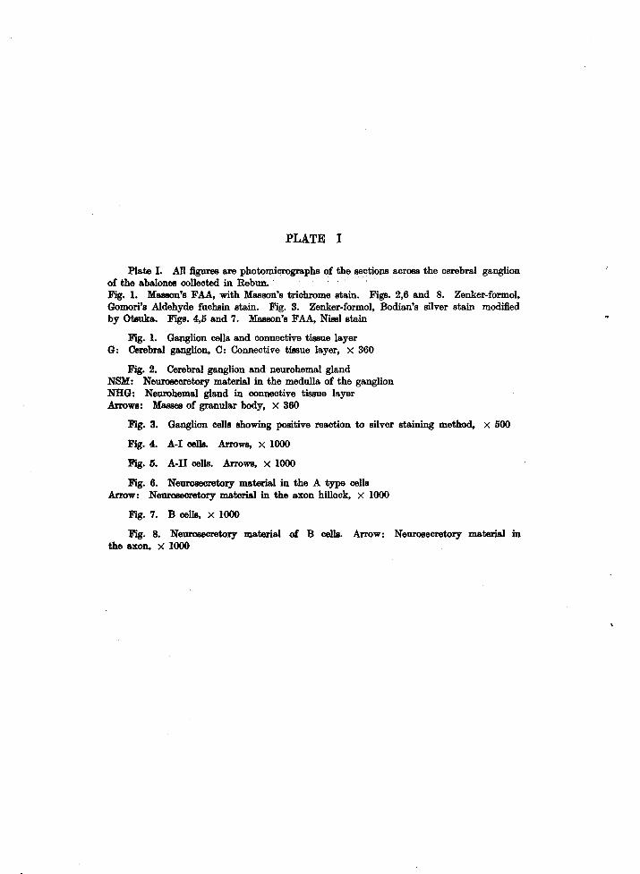
PLATE I
Plate I. All figures are photomicrographs of the sections across the cerebral ganglion of the abalones collected in Rebun. . , Fig. 1. Masson's FAA, with Masson's trichrome stain. Figs. 2,6 and 8. Zenker-formol, Gomori's Aldehyde fuchsin stain. Fig. 3. Zenker-formol, Bodian's silver stain modified by Otsuka. Figs. 4,5 and 7. Masson's FAA, Nissl stain
Fig. 1. Ganglion cells and connective tissue layer G: Cerebral ganglion, C: Connective tissue layer, X 360
Fig. 2. Cerebral ganglion and neurohemal gland NSM: Neurosecretory material in the medulla of the ganglion NHG: Neurohemal gland in connective tissue layer Arrows: Masses of granular body, X 360
Fig. 3. Ganglion cells showing positive reaction to silver staining method, X 500
Fig. 4. A-I celIs. Arrows, X 1000
Fig. 5. A-IT celIs. Arrows, X 1000
Fig. 6. Neurosecretory material in the A type celIs Arrow: Neurosecretory material in the axon hillock, X 1000
Fig. 7. B celIs, X 1000
Fig. 8. Neurosecretory material of B celIs. Arrow: Neurosecretory materiaI in the axon, X 1000

Bull. Fac. Fish., Hokkaido Univ., XXII, 3 PLATE I
T. Yahata: Neurosecretory cells in the abalone

PLATE II
Plate II. Photomicrographs of sections of the cerebral ganglion (Figs. 9-12) and seasonal variation of neurosecretory material (Figs. 13-17)
Fig. 9. Masson's FAA, with Nissl stain. Figs. 10 and 11. Zenker·formol, Gomori's chrome hematoxylin.phloxine. Figs. 12-17. Zenker·formol, Gomori's Aldehyde fuchsin
Fig. 9. C cells, X 1000
Fig. 10. D cells which extend their axons inward the ganglion, X 1000
Fig. 11. D cells which extend their axons outward the ganglion, X 1000
Fig. 12. Neurohemal gland in the connective tissue layer G: Cerebral ganglion, X 500
Fig. 13. Ganglion cells in the shell collected in June '69, X 500
Fig. 14. Ganglion cells in the shell collected in September '69, X 500
Fig. 15. Neurosecretory material (NSM) in the medulla of the ganglion preserved at the same time as Fig. 14, X 500
Fig. 16. Ganglion cells in the shell obtained in December '69, X 500
Fig. 17. Ganglion cells in the shell fixed in March '70, X 500

Bull. Fac. Fish., Hokkaido Univ., XXII, 3 PLATE II
T. Yahata: Neurosecretory cells in the abalone