Delivery of Liquid Metal to the Target Vessels as Vascular ...
21
1 Delivery of Liquid Metal to the Target Vessels as Vascular Embolic Agent to Starve Diseased Tissues or Tumors to Death Qian Wang 1,3 , Yang Yu 1,3 and Jing Liu 1,2 * 1. Department of Biomedical Engineering, School of Medicine, Tsinghua University, Beijing 100084, China. 2. Technical Institute of Physics and Chemistry, Chinese Academy of Sciences, Beijing 100190, China. 3. These authors contributed equally to this work. Correspondence should be addressed to J.L. ([email protected]) Abstract Tumor growth strongly depends on the continuous blood and nutrients supply. Theoretically, it is an ideal therapeutic way of treating tumor by vascular embolization, which also reduces the harmful effects on surrounding normal tissues. However, most of the currently available vascular embolic agents are still rather insufficient in performances to fulfill the real clinical need due to the reasons like: incomplete filling of target vasculature, being easily washed away by blood or body solution, or just producing toxicity to human body. Here from an alternative way, the body temperature liquid metal, a kind of soft and highly compliant material, was proposed for the first time as a new conceptual blood vessel embolization strategy towards tumor physical therapy. With the unique capability of easy phase transition between liquid and solid state and sub-cooling behavior, such agent can be fluently injected into and to fully fill the tiny vessels including the ending capillaries. The in vitro cytotoxicity experiments have been performed which indicated that treating localized diseased tissues through liquid metal embolic agent is acceptable. Endowed with a high density, the liquid metal-filled vessels are highly visible under the CT scan, which offers the potential of the diagnosis-treatment integration with only one single material. To further demonstrate the new liquid metal vascular embolization therapy, several typical experiments in vivo on vasculatures of rabbit ears and mouse tails have been performed to provide the evidences of destroying the targeted tissues due to liquid metal filling. To interpret the liquid metal starvation therapy effects, theoretical model has been established to simulate the tumor growth with zero, partial or complete filling of the metal agent inside the vessels. All the results support that, given appropriate administration, the liquid metal embolization is able to destruct the target regions effectively and might be able to starve the tumors to death through a
Transcript of Delivery of Liquid Metal to the Target Vessels as Vascular ...

1
Delivery of Liquid Metal to the Target Vessels as Vascular Embolic
Agent to Starve Diseased Tissues or Tumors to Death
Qian Wang 1,3
, Yang Yu 1,3
and Jing Liu 1,2*
1. Department of Biomedical Engineering, School of Medicine,
Tsinghua University, Beijing 100084, China.
2. Technical Institute of Physics and Chemistry, Chinese Academy of Sciences,
Beijing 100190, China.
3. These authors contributed equally to this work.
Correspondence should be addressed to J.L. ([email protected])
Abstract
Tumor growth strongly depends on the continuous blood and nutrients supply.
Theoretically, it is an ideal therapeutic way of treating tumor by vascular embolization,
which also reduces the harmful effects on surrounding normal tissues. However, most
of the currently available vascular embolic agents are still rather insufficient in
performances to fulfill the real clinical need due to the reasons like: incomplete filling
of target vasculature, being easily washed away by blood or body solution, or just
producing toxicity to human body. Here from an alternative way, the body
temperature liquid metal, a kind of soft and highly compliant material, was proposed
for the first time as a new conceptual blood vessel embolization strategy towards
tumor physical therapy. With the unique capability of easy phase transition between
liquid and solid state and sub-cooling behavior, such agent can be fluently injected
into and to fully fill the tiny vessels including the ending capillaries. The in vitro
cytotoxicity experiments have been performed which indicated that treating localized
diseased tissues through liquid metal embolic agent is acceptable. Endowed with a
high density, the liquid metal-filled vessels are highly visible under the CT scan,
which offers the potential of the diagnosis-treatment integration with only one single
material. To further demonstrate the new liquid metal vascular embolization therapy,
several typical experiments in vivo on vasculatures of rabbit ears and mouse tails
have been performed to provide the evidences of destroying the targeted tissues due to
liquid metal filling. To interpret the liquid metal starvation therapy effects, theoretical
model has been established to simulate the tumor growth with zero, partial or
complete filling of the metal agent inside the vessels. All the results support that,
given appropriate administration, the liquid metal embolization is able to destruct the
target regions effectively and might be able to starve the tumors to death through a

2
relatively easy and compliant way. This study lays the foundation of a promising
tumor starvation therapy with both high contrast image guidance capability and
overall treatment outcome.
Keywords: Liquid metal; Tumor treatment; Blood vessel; Embolic agent;
Starvation therapy; Diagnosis-therapy integration.
1. Introduction
Originated from somatic mutations, the tumor cells generally proliferate and
differentiate continuously under the regulation of various growth factors as well as the
oxygen and nutrients provided throughout the blood vessels [1-4]. At the early stage
when the death and the regeneration of tumor cells are nearly balanced, the existing
vascular network near the tumor tissues supplies nutrients and carries away
metabolites. Afterwards, a new tumor vascular network generates around the tumor
tissues due to the vascular endothelial growth factor secreted by the tumor cells [5].
Then, the rapidly increasing tumor cells would diffuse to other parts via the
tumor-induced angiogenesis. Clearly, the blood vessels play a significant role on the
growth and metastasis of the tumor [6].
Considering the dependency of the tumor growth on the blood vessels, the
treatment of vascular targeting therapy for starving the tumors to death has
increasingly attracted the attentions of researchers across the world [7, 8]. One of such
strategies is to prevent the tumor-induced angiogenesis by the angiogenesis inhibitors,
which was originally proposed by Folkman in the early 1970s [9]. Ever since then,
various inhibitive drugs have been developed [10-13], and such embolization therapy
has also been combined with chemotherapy or radiotherapy for better treatment [14].
However, the angiogenesis inhibitors have to be used for a long time. And some
studies found that within this period, the tumor itself would gradually generate
resistance to these drugs, which makes it not as effective as expected [15]. As another
approach focusing on blocking the existing vessels, colchicine was reported to destroy
the vessels, yet with rather high toxicity [16]. Afterwards, some other vascular
disrupting agents were studied including small molecular agents [17, 18].
Except for these drugs, there are some other physical embolization ways to offer
the possibility of starving the tissues or tumor to death by means of embolic agents,
3
such as autologous clots, coils, gelfoam, balloons, glue, nanoparticles, microspheres
embolic agents and so on [19-23]. However, the failure to embolize the vessels may
often occur. One key reason lies in that some solid embolic agents cannot conform
well to the vessels, leaving certain space at the interface for nutrients to leak in.
Another existing issue is that some embolic agents are easily rushed away by the
blood flow, such as the balloons. What’s more, the catheters or other more complex
equipments are needed in order to place some of embolic agents into the target
positions. Different from these approaches, gas embolotherapy uses gas bubbles to
occlude the blood flow [24]. The bubbles originate as small diameter liquid droplets
of dodecafluoropentane (DDFP) mixed in saline and albumin. After injected into the
vascular system, these droplets are small enough to pass through the capillary beds. At
the strategic location, the droplets are vaporized into larger bubbles through acoustic
wave to occlude flow. However, the entry of gas into the vessel system is a risk in
virtually all areas of clinical care [25].
In view of the above tumor treatment situations, this paper is dedicated to
proposing and demonstrating an alternative vascular embolization therapy, the body
temperature liquid metal, as a kind of unconventional vessel embolic agent for
starving the target tissues or tumors to death. Here, to emphasize the method itself,
although plenty of liquid metal candidates can serve as the treatment agents, we
mainly tested for the first trial the pure gallium and Ga75.5In24.5 alloy as the illustration
examples. To characterize the physical behaviors of such metal agent, the DSC
experiments on the liquid metal gallium were conducted, which revealed the thermal
properties of the gallium and verified its feasibility for the delivery of therapy. The
cytotoxicity of the gallium and indium in vitro were also evaluated with the tests of
CCK-8 and flow cytometry. As an additional advantage, the liquid metal in vessels
showed high image contrast under the X-ray scan, which indicated its great potential
of the diagnosis-treatment integration. For further investigation, we experimented on
several different tissues or transplanted tumor to demonstrate the liquid metal
embolization effect in vivo. Finally, we presented the theoretical modeling and
simulation of tumor growth affected by liquid metal enabled blockage of blood
vessels near the tumor. This study opened the way of applying the liquid metal
vascular embolization for a potential tumor starvation therapy in the coming time.
2. Principle and methods

4
2.1 Basic feature of liquid metal vascular embolic therapy
Figure 1 The principle illustration of liquid metal based tumor vascular embolization
therapy. (a) The oxygen and nutrition supplies from the vessels to tissues without any embolic
agents. (b) The incomplete occlusion. (c) The complete occlusion by the liquid metal. (d) The
physical occlusion of the blood supply to tumor. (e) The liquid metal could be injected into or
sucked out of the vessel in case of need.
As a soft, compliant and fluidic material, the liquid metal can be easily injected
and filled into the vessels and capillaries sufficiently, which stops the blood flow and
all the related physiological events thus enabled because of its high density and
fluidity. Fig. 1 illustrates the basic principle of the liquid metal embolization therapy
and surgical operation of its injection and removal. As is widely known, tissues
acquire oxygen and nutrition from the nearby feeding vessels (Fig. 1a). Though the
vascular blockage can shut down the blood supply, yet the stream is still able to pass
through the vessels interior when the embolic agent fails to completely occlude the
vessels. As a result, the reduction of blood flow does not guarantee a strong enough
effect on starving the necrotic cells (Fig. 1b) and the embolic agent can even possibly
be rushed away within a short period after it was injected. Being able to maintain in

5
liquid state at body temperature, the liquid metal agent can easily shape itself along
the vessels and fully occlude the channels due to its perfect compliance (Fig. 1c).
Compared with the normal tissues, the tumor is more dependent on the nutrients
supplies from the surrounding vessels. After injected into the vessels, the liquid metal
results in a physical occlusion and thus leads to tumor regressions (Fig. 1d). Another
merit of the current therapy still lies in that, theoretically, the liquid metal could also
be well sucked out in case of need after completing its treatment role (Fig. 1e).
2.2 Characterization of the physical properties of the liquid metal agents
We measured the thermal property of gallium (99.999%, purchased from Anhui
Rare New Materials Co. LTD, China) with the differential scanning calorimetry
device (DSC 200 F3 Maia, Netzsch Scientific Instrument Trading Co. Ltd, Germany).
The 14.63mg of gallium was put into the alumina crucible with another empty one as
the reference, which were both placed in a heating furnace. The test temperature range
included three cycles, which were set as 40°C → 60°C for 1min → -60°C for 1min →
60°C for 1min → -60°C for 1min → 60°C for 1min → -60°C for 1min → 60°C for
1min → 45°C → 70°C, and the heating and cooling rates were both prescribed at
10°C /min. The results were analyzed by the software of NETZSCH Proteus Thermal
Analysis.
2.3 Evaluation of the metal agents cytotoxicity in vitro
Two tests including the cell counting kit-8 (CCK-8) and the flow cytometry were
applied on the mouse embryonic fibroblast cells (NIH3T3) to evaluate the
cytotoxicity of the liquid metal.
A drop of the gallium was put on the six-well plates filled with culture solution,
soaking for 24 hours and 48 hours in the incubator. Meanwhile, a piece of polished
and sterilized indium and copper were soaked under the same condition.
CCK-8 method: Cells at logarithmic growth phase were treated by trypsin and
tuned into single cell suspension with a concentration of 50000/ml, then seeded in a
96-well plate with 100μL in each well. After being cultured for 24 hours, the cells
were observed under the microscope and the samples in bad state were eliminated.
The original culture solution was removed and the wells were washed with PBS
solution. Then, the soaked solution of gallium, indium, copper and the original culture

6
solution (control group) were added into the wells with 5 parallel samples in each
group. After being cultured for 24 hours, each well was washed and added with a
mixture of 100μL fresh culture solution and 10μL CCK-8. The 96-well plate were put
into the incubator for 2 hours, then optical density (OD) at 450nm wavelength were
measured with multifunctional microplate reader (EnVision, USA PerkinElmer). The
cell viability was calculated according the following equation:
Cell viability= (OD-OD0)/ (ODc-OD0)) (1)
where, ODc was the OD value of the control group and OD0 was the OD value of
black wells without any cells.
Flow cytometry method: The cells were seeded into the sterile dish of 60mm
diameter with 300000 cells each. After cultured for 24 hours in the incubator, the
original culture medium was removed and the wells were washed with PBS solution.
Then, the three soaked solutions and the original culture solution were added
separately. After cultured for another 24 hours, the cells in each dish were collected
into the centrifuge tubes. By centrifuge at 1500r/min for 5min, the supernatant was
removed and 1mL cell staining buffer and 5uL PI staining solution were added, then a
30-min ice bath followed. Finally, the cell solution were placed in the flow cytometer,
detecting the red fluorescence by flow cytometry (BD Calibur, the USA BD).
2.4 Liquid metal angiography of the tumor vessels
It was recently found that, different from other embolic agents, liquid metal has
excellent visibility under the X-ray [26]. Thus in a further study, the tumor vessels
injected with the liquid metal went through a CT scan to investigate the distribution of
the material. In order to assess this special characteristic, several representative
objects such as in vitro swine kidney, a healthy 8-week-old female CD1 mice and an
8-week-old female Barb/c nude mice (purchased by the Center of Biomedical
Analysis of Tsinghua University) with a tumor were selected to serve as the
experimental testing cases.
About 0.8mL liquid metal gallium was infused into the artery branches of the
swine kidney. Then the filled kidney was X-ray photographed by a micro-CT scanner
(XM-Tracer-130, from Institute of High Energy Physics, Chinese Academy of
Sciences).
The mouse has both arteries broad enough for liquid metal Ga75.5In24.5 alloy
infusion and thin capillaries to display the alloy’s high contrast. First, the mouse was

7
excessively anesthetized to death, then the heart was exposed by cutting open its chest.
A thin syringe needle was used to penetrate into the left ventricle of the mouse heart
and it was clamped to prevent leakage and reverse flow. With a slight cut near the
right atrium, the liquid metal alloy could finally be infused into the circulatory tunnels
and flow throughout the body. When the silvery alloy flows to the right heart, it was
supposed to finish the infusion and that the sample had been stuffed with the contrast
agent. Then the sample was taken to the imaging instrument (Super Nova CT, from
Technical Institute of Physics and Chemistry, Chinese Academy of Sciences). Images
with higher resolution were obtained from the micro-CT (XM-Tracer-130, from
Institute of High Energy Physics, Chinese Academy of Sciences).
The structures of the vessels in the tumor tissues and normal tissues may be
different. In order to offer more evidences on various tissue vasculatures, we also
managed to infuse the liquid metal Ga75.5In24.5 alloy into the tumor vessels. The mouse
breast cancer cells (EMT6, 1x10⁶cells) were injected directly into the subcutaneous at
the back of the nude mice. The tumor reached approximately a diameter of 5mm in
two weeks and the vessels were visible on the skin. After the mice was anesthetized
with intraperitoneal injection of 0.2mL 1% (g/L) sodium pentobarbital (Nembutal),
the liquid metal was injected directly into a tumor vessel via a special syringe needle
with an inner diameter of 0.3mm. Afterwards, the tumor region went under a CT scan
(Super Nova CT, from Technical Institute of Physics and Chemistry, Chinese
Academy of Sciences) for visualizing the vessels injected with liquid metal.
2.5 Liquid metal as embolic agents of blood vessels in vivo
A two-month-old male New Zealand rabbit (purchased from the Center of
Biomedical Analysis of Tsinghua University), weighted about 2.2kg, were used as the
experimental subject, because the vessels in the ears are broad and visible.
Anesthetized with intraperitoneal injection of 4% (g/L) sodium pentobarbital
(Nembutal) by the standard of 1 ml/kg, the rabbit was injected with liquid metal
gallium into the vein of one ear. It was observed that the material filled the vessels at
the rim and the tip of the ear quickly. The other ear was used for contrast. The infrared
camera (Thermovision A40, FLIR, Wilsonville, OR) was adopted to record the
temperature changes of the rabbit ear during the operational process. Afterwards, the
rabbit was observed for half an hour and then put back to the caring center. The rabbit
continued to be observed every day. Blood analysis were carried out before the

8
experiments and 1 day, 3 days, 7 days afterwards with the automatic hematology
analyzer (BM800), each time with 3 repeated trials. After the occurrence of necrosis
at the ear tip, the rabbit was sacrificed through standard procedure. Then the tissues of
both the necrotic region and the same position of the other ear were surgically cut and
immediately restored in formalin for 24 hours. The samples were then sectioned,
stained with HE methods (Hematoxylin-eosin) and observed under the microscope.
Afterwards, we injected the liquid metal gallium into the vessels of the mouse tail
and the rabbit leg in the same way. All experiments above have been approved by the
Ethics Committee of Tsinghua University, Beijing, China under contract [SYXK (Jing)
2009-0022].
2.6 Theoretical modeling and simulation of tumor growth with liquid metal
embolization
Although the liquid metal embolization of blood vessels in vivo was implemented
on the vessels of rabbit ears and legs, a case of tumor might be more illustrative. Here,
a theoretical modeling and simulation of tumor growth affected by the metal blockage
of blood vessels nearby was (see Fig. 2) carried out to interpret the starvation effect of
the current therapy.
Figure 2 Illustration of the theoretical tumor growth model. (a) Without the liquid metal in
the vessel. (b) With the liquid metal embolic agent in the vessel.
Without losing any generality, there are generally four dependent variables to
dominate the tumor growth model, including cancer cell density, oxygen density,
extracellular matrix density and matrix degrading enzymes. The functions of each
variable (further details about the functions could be seen in Alexander’s work [27])
in non-dimensional are expressed as

9
2 ( )n
nd n n f
t
(2)
2
o
od o f n o
t
(3)
fmf
t
(4)
2
m
md m n m
t
(5)
where n , o , f denote the density of cells, oxygen and extracellular matrix
respectively, and m denotes the matrix degrading enzymes.
The oxygen is supplied by the vessel and we make the assumption that the
oxygen concentration is zero at the metal blockage position. The probabilities of
movement of an individual cell are generated by discretizing the Equation (2).
Table 1 The non-dimensional parameter values used in the simulations
Parameters Value Parameters Value
nd 0.0005 1
0d (out of /in the tumors) 0.05/0.025 0
md 0.0005 0.5
0.01 0.57
50 0.025
Besides, it is assumed that the cancer cells migrate through a combination of
diffusion and haptotaxis as well as undergoing proliferation. And each cell is allowed
to migrate when the number of neighbor cells is more than one. Cell death can occur
due to lack of oxygen. In the model, we also make the assumption that each cell
produces two daughter cells when the cell has reached maturity. If no empty space
around the parent cell exists, the cell becomes quiescent until space appears. We
assume that quiescent cells only consume half amount of oxygen of the active tumor
cells. The tumor cells would not only maintain one phenotype, but also becomes more

10
unstable and more aggressive phenotypes due to mutations. In the model, we mainly
consider three phenotypes. The irreversible mutations occur at the mitosis stage.
The computation domain is divided by 200×200 grids with a space step of
0.005h and a time step of 0.0001k . Initially, 36 tumor cells are in the center. The
initial value of oxygen density, extracellular matrix density and matrix degrading
enzymes are 1, 1 and 0, respectively. The non-dimensional parameter values used in
the simulations are listed in Table 1 [27].
3. Results
3.1 Physical properties of the liquid metal
Fig. 3 depicts the DSC curves of the gallium in reciprocally reverse processes. In
either the cooling or the heating process, the measurement goes for three trials and the
results are almost identical. There is a very sharp exothermic peak over the cooling
process, and the freezing temperature is 4.5 °C, while the endothermic peak is also
steep in the heating process and the melting point is 28.3°C. The curves indicate an
easy transition between the solid and liquid phase of the gallium, which is an
excellent property for the phase control. What’s more, owing to its sub-cooling effect,
the material is able to maintain in the liquid state for a long time at the body
temperature. Thanks to its liquid phase, compliance and fluidity, the material is able to
fluently flow into and fill the tiny vessels including the capillaries whose diameter is
at a scale of micrometers.
Figure 3 The DSC curves of the gallium. (a) Cooling process. (b) Heating process.
11
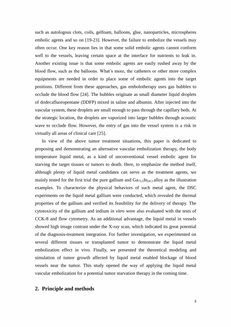
3.2 Cytotoxicity of the liquid metal in vitro
Fig. 4a shows the viability of the cells after cultured in different metal soaked
solutions with CCK-8 method. The cell viability in the copper soaked solution in 24
hours was measured as 7%, and almost all died in 48 hours. The cell viability of the
gallium soaked solution in 24 hours and 48 hours were 100.6% and 75.7%, and that of
indium were 107% and 79.7%, respectively. According to the cytotoxicity evaluation
criteria of the national standard, cell viability above 75% is qualified and safe. The
viability of the cells can also be visually demonstrated through the microscope, which
is shown in Fig. 4b. The cells of the gallium, indium soaked solution and original
culture solution were exactly of the right size and shape large, while those in the
copper soaked solution distributed sparsely and the shapes were relatively round,
which means most of them were dead.
Figure 4 Cytotoxicity in vitro. (a) The cell viability after cultured in different metal soaking
solution with CCK-8 method. (b) The cellular morphology in each experimental group
observed under the microscope. (c) The cell apoptosis with flow cytometry method. (d) The
scatter images with flow cytometry method.

12
Fig. 4c indicates cell apoptosis of each experimental group. In the PI fluorescence
intensity histograms, M1 stands for apoptosis cells while M2 represents living cells.
Compared with the control group, the relative apoptosis rates of gallium and indium
group was 10.18% and 7.9% respectively while 78.44% for the copper group. In the
scatter images in Fig. 4d, it can be further found that the strength of forward scattered
light reduced and the one side scattered light increased in the copper soaked solution,
which also indicated that there were more apoptosis and necrosis cells. However, the
gallium and indium group showed tiny difference with the control group.
These experiments have revealed that the cytotoxicity of the gallium and indium
ion are relatively low and their applications on localized severe disease treatment are
clinically acceptable, given its unique values as will be revealed in later sections.
3.3 Liquid metal angiography of the tumor vessels
Endowed with a high density, the liquid metal is an excellent contrast agent for the
vessel-like tissues under the X-ray [26], which is a unique advantage compared with
most other embolic agents.
The X-ray images of renal artery of the in vitro swine kidney are presented in Fig.
5a. Clearly, a whole branch of the renal artery network is intact and the texture of the
small vessels is rather clear. As shown in Fig. 5b, the vessels are separated with the
other tissues obviously in highlight in the CT scan reconstruction of a whole-body
infused mouse. In the image, it is easy to identify the liquid metal’s flow path
throughout the carotid, limb and tail vessels even to the end tips. Besides, the
branches in the lung, head and abdominal organs can also be distinguished with
ultra-high contrast and clarity. In order to offer more evidence of the image guidance
ability of the liquid metal alloy enhancement, Fig. 5c provides the closer X-ray
photographs of vascular network in the abdomen of the mouse which has plenty of
even tinier vessels. In the figure, the sensor focused in a square field with each side
6.6mm long, and the resolution is thus set as 13μm/pixel. Accordingly, it can be
referred to that the thinnest visible vessel is less than 30μm wide, which means a
tremendous improvement of the blood vessel visualization. Besides, the tumor vessels
filled with liquid metal are highly visible under the CT scan as expected. As can be
seen, the tumarized vasculature appeares somewhat irregular (Fig. 5d) as compared
with normal tissue vessels (Fig.5a). This clearly reflected the growth behavior of such
diseased vessels.

13
Figure 5 The images of vessels filled with the liquid metal under the X-ray. (a) The X-ray
image of renal artery of the in vitro swine kidney. (b) The top view of a liquid metal infused
mouse in a whole-body CT scan and 3D reconstruction. (c) The X-ray image of vessels in the
mouse abdomen with higher resolution. (d) 3D reconstruction image of tumor vessels.
3.4 Liquid metal embolization to starve target living tissues to death
As shown in Fig. 6a, the liquid metal has filled the rabbit ear vein at the rim and
the tip. Without abnormal reactions, the rabbit showed obvious symptoms of necrosis
only in the ear tip in three weeks, which finally come out like a dry leaf. The necrotic
changes appeared not so serious at the rim of the ear, which might be due to the
supply of certain surrounding tiny vessels. However, the very side of the ear had been
crimped, which was a reflection of the liquid metal obstruction.
As an attendant phenomenon, the temperature of the liquid metal obstructed
region would decrease due to stop of the blood flow. As a special evaluation of the
material’s influence, the temperature distributions in 0h, 24h, 48h and 72h from the
thermal infrared images are depicted in Fig. 6b. Clearly, at the moment of the
injection, the blood was immediately blocked, causing the temperature rise in the
upstream and decrease in the downstream regions. With the time went on, the area of
the low temperature region shrunk gradually, which indicated the concentration of the
material to the tip.
In order to provide a biological inspection, Fig. 6c displays the sections of both
the necrotic and normal tissues. It can be seen that near the liquid metal blocked

14
vessels, the surrounding area tends to be emptier and the cells are much smaller than
the situation in the similar region in a normal tissue. This shows the damage effect of
the liquid metal embolization treatment.
Figure 6 The effects of liquid metal embolization at the rabbit ears. (a) The symptoms of
necrosis in the ear. (b) The temperature distribution changes after 0h, 24h, 48h and 72h. (c)
Comparison between the necrosis tissue sections and control normal tissue section. (d) The
results of WBC, LYM%, MON%, GRA%, HGB, RBC and PLT* changed over time. (e)WBC,
RBC and PLT dynamic histograms distribution.
*WBC: white blood cell; LYM: lymphocyte; MON: monocyte; GRA: granulocyte; HGB:
hemoglobin; RBC: red blood cell; PLT: platelet.
Other than the morphologic analysis, blood tests were also carried out in a week
and seven parameters including WBC, LYM%, MON%, GPA%, HGB, RBC and PLT
are shown in Fig. 6d. Fig. 6e shows the WBC, RBC and PLT dynamic histograms
distribution, respectively. As can be seen from the figure, the second peak of WBC

15
distribution was lowered after one day but returned to normal after 3 days, which
indicated the reduction of the neutrophil granulocytes. These blood analysis results
demonstrated that the blood parameters would soon return to normal level after the
liquid metal gallium was injected into the blood.
Figure 7 The effects of liquid metal embolization at the mice tail. (a) The liquid metal
injected into the tail vein. (b) The regenerated vascular branches.
It should be pointed out that, not all the injections will be able to completely fill
the vessels and thus lead to damage of the target tissues. The output in fact depends on
the specific surgical injection and delivery of liquid metal. To explain this point, we
select the tail vessels of the mice as an additional test object to evaluate the blockage
effect of liquid metal embolization on the tissues in vivo. As shown in Fig. 7a, three
tail vessels of the mice were filled with liquid metal gallium. However, there was no
visible necrosis after three weeks later. Moreover, certain new small blood vessels
were even observed to regenerate (Fig.7b). The reason can be interpreted as follows.
With a single flow direction and different from former cases, it was more difficult in
current situation to fully block the femoral vessels at the tail of the mice. This led
some of the liquid metal to flow away and no visible necrosis occurs at the target
tissues over the whole experiments. Besides, these regenerated blood vessels near the
tail vessels have further contributed for the nutrition and oxygen supply. This reminds
us that, without appropriate administration of injecting the liquid metal agent, the
embolization effect as anticipated before may not always necessarily be guaranteed.
Therefore, for therapeutic purpose, careful treatment planning should be made in
16
advance to surgically deliver liquid metal into the target vessels along specific
directions. Lastly, it was also worth mentioning that these mice filled with liquid
metal were kept alive for a rather long time like several weeks. Such findings indicate
the safety and non-toxicity of the liquid metal materials staying inside the vessels on
condition that they will not cause physically physiological danger.
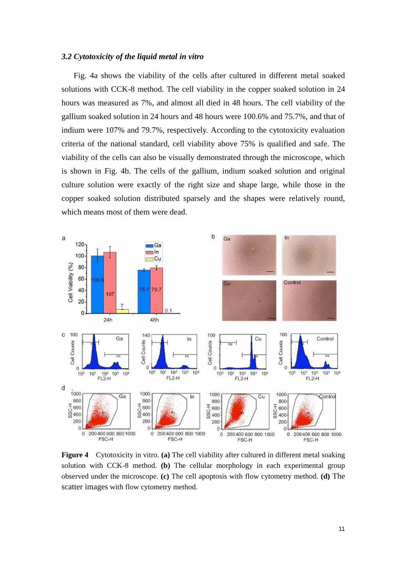
3.5 Theoretical results of tumor growth with liquid metal embolic agents
Theoretical simulations on the two cases with and without liquid metal embolic
agents in the vessels near the tumor have been implemented to interpret and evaluate
the performance of tumor growth suppression by the present method. Fig. 8 depicts
the changes of oxygen concentration near the tumor at six times (5 days, 9days,
11days, 13days, 15days and 17days later). The tumor growth consumes oxygen and
the oxygen concentration in tumor center is the lowest. In order to maintain growth,
the tumor needs adequate oxygen supply by the vessel. However, as a result of liquid
metal blockage in the vessel, the oxygen concentrations of the nearby tissues are
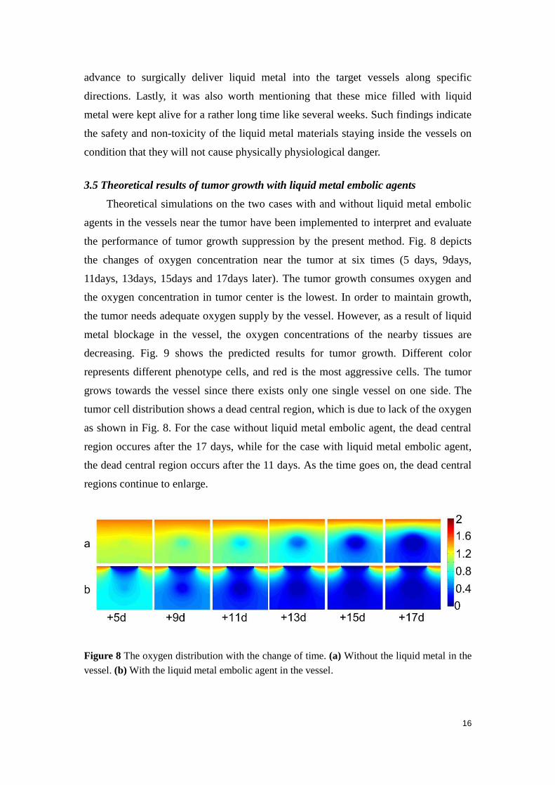
decreasing. Fig. 9 shows the predicted results for tumor growth. Different color
represents different phenotype cells, and red is the most aggressive cells. The tumor
grows towards the vessel since there exists only one single vessel on one side. The
tumor cell distribution shows a dead central region, which is due to lack of the oxygen
as shown in Fig. 8. For the case without liquid metal embolic agent, the dead central
region occures after the 17 days, while for the case with liquid metal embolic agent,
the dead central region occurs after the 11 days. As the time goes on, the dead central
regions continue to enlarge.
Figure 8 The oxygen distribution with the change of time. (a) Without the liquid metal in the
vessel. (b) With the liquid metal embolic agent in the vessel.
17
Figure 9 Spatial distribution of tumor cells with the time. (a) Without the liquid metal in
the vessel. (b) With the liquid metal embolic agent in the vessel.
4. Discussion
As is well known, the abundant vessels in the tumor tissues make it difficult for
tumor treatment. Curatie surgery can easily lead to a massive hemorrhage, and large
blood vessels would produce localized cooling in heated tissues during tumor
hyperthermia, for instance. In this study, body temperature liquid metal yields a
promising injectable tumor treatment. With its easy phase conversion, such material
owns the capability to flow into tiny vessels at liquid state and stay at the target
tissues at the solid state. Once the liquid metal was injected into the vessels, the
nearby tissues would be possibly starved to death due to cutting off the feeding route
of the nutrition and oxygen supplies.
Though the unprecedented application of the liquid metal on the tumor treatment
seems to be beyond imagination, there remains a wide range for the further utilization
of the material. In the present experiments, only gallium and Ga75.5In24.5 alloy were
tested for brief. In fact, the melting points of the liquid metal could be modified by
adding some other metals into alloys and adjusting the compounds ratio.
As a metal, the gallium is endowed with good electrical and thermal conductivity.
Hence on the base of the blocking therapy, newer treatment might still be developed
through combining the electrical and thermal stimulation together. We could even add
certain substances into the liquid metal to obtain or enhance specific properties. Ma
and Liu proposed a method to fabricate liquid metal with desired properties by
loading with nanoparticles [28]. These nanoparticles include magnetic oxides such as
Fe3O4, metallic particles and some semi-conductive particles, which further expands
the application of liquid metal as the embolic agents. What’s more, it is possible to

18
combine this approach with chemotherapy or radiotherapy by injecting mixed liquid
metal with chemotherapy and radiotherapy substances.
In this study, it is worth emphasizing the role of the liquid metal angiography,
which reveals an extra advantage of the vessel blocking therapy. The visibility of the
material under the X-ray has provided the possibility for monitoring the flow process
along the gallium injection and evaluating vascular changes and damages. The
combination of the liquid metal blocking and the X-ray imaging even opens a
promising approach to the investigation of the tumor vessel growth and distribution,
which also indicates a great potential of the diagnosis-treatment integration.
However, there still exist many issues to be solved in order to further improve the
efficiency of the liquid metal embolization. Considering the difficulties to inject the
liquid metal into the artery due to fast blood flow there and deep position in the
tissues, we almost administrated these experiments on the veins. However, as we all
know, the blood in artery flows into the capillary vessel, while that in vein blood
flows out of the capillary vessel. Therefore, it is better to inject the liquid metal into
the artery to occlude the vessel. Besides, the regeneration of the tumor vessel network
under the obstructive condition is also a common challenge in the blocking therapy.
The safety is also a non-negligible issue in this new conceptual blood vessel
embolization strategy. As mentioned above, some of the liquid metal could be washed
away from the target tissues since injection from the veins. As is shown in Fig. 10, the
liquid metal has entered the heart and lungs of the rabbit in the experiments of leg
infusion yet the rabbit is still alive. Therefore it is very important to adopt measures to
prevent this problem from happening in further studies. One possible solution is to
apply proper pressure on the proximal end to slow the blood flow velocity. Another
method to decrease the blood flow is to reduce the temperature of the blood in the
target tissues, which could also speed up the solidification of the liquid metal. It also
indicates that the in vivo application is highly sophisticated due to safety control and
thus skilled surgical operation is required as well. In this paper, we only used gallium
and Ga75.5In24.5 alloy as the embolic agents, whose melting point are both under the
37°C. Next, we will try to synthetize more other liquid metal alloys with a little higher
melting point than 37°C and test their embolization effects. In addition, a
comprehensive evaluation of long-term toxicity still requests further research.

19
Figure 10 Traces of the liquid metal in the heart and lungs of the rabbit.
5. Conclusion
In summary, the liquid metal, as a fluidic material at body temperature, has been
proposed for the first time as the blood vessel embolic agent to starve the necrotic
tissues or target tumors to death. With its merits of easy phase transition and
sub-cooling, this kind of material can be conveniently injected into the vessels with
relatively easy operations. A series of in vitro experiments on the material
cytotoxicity have been implemented to evaluate its safety and the results have
indicated that both gallium and indium irons show low toxicity on normal cell growth.
On considering its application against tumor or other severe diseases, such a
performance is quite acceptable. As an extra advantage, the liquid metal is endowed
with excellent contrast under the X-ray over the tissues, which provided a powerful
soft tool for tumor vascular research and offered great potential to realize the
diagnosis-treatment integration. Furthermore, both in vivo experiments and theoretical
model simulations have preliminarily demonstrated the performance of the liquid
metal to starve the tissues or tumors to death. Though still facing certain challenges,
the unprecedented utilization of the liquid metal agent opens a new way for
further practice in future tumor vessel blocking therapy.
Acknowledgement
This work was partially supported by the NSFC under Grant 51376102.

20
References
[1] Esther W, Michael S, and Yosef Y. Roles for growth factors in cancer progression.
Phys, 2010, 25(2): 85–101.
[2] Zhimin L, Guo J, Peter B, and Tony H. Epidermal Growth factor-Induced tumor
cell invasion and metastasis initiated by dephosphorylation and downregulation
of focal adhesion kinase. Mol Cell Biol, 2001, 21(12): 4016–4031.
[3] Wells A. Tumor invasion: Role of growth factor-induced cell motility. Adv
Cancer Res, 2000, 78:31-101.
[4] Wells A, Kassis J and Solava J, Turner T and Lauffenburger DA. Growth
factor-induced cell motility in tumor invasion. Acta Oncol, 2002, 41(2):124-30.
[5] Folkman J. Tumor angiogenesis factor. Cancer Research, 1974, 34(8): 2109-2113.
[6] Jain R K. Vascular and interstitial barriers to delivery of therapeutic agents in
tumors. Cancer Metast Re, 1990, 9(3): 253-266.
[7] Eichhorn M E, Strieth S and Dellian M. Anti-vascular tumor therapy: Recent
advances, pitfalls and clinical perspectives. Drug Resis Update, 2004, 7(2):
125-138.
[8] Thorpe P E. Vascular targeting agents as cancer therapeutics. Clin Cancer Res,
2004, 10(2): 415-427.
[9] Folkman J. What is the evidence that tumors are angiogenesis dependent? J Natl
Cancer I, 1990, 82(1): 4-7.
[10] Kerbel R S. Tumor angiogenesis: Past, present and the near future.
Carcinogenesis, 2000, 21(3): 505-515.
[11] John A and Tuszynski G. The role of matrix metalloproteinases in tumor
angiogenesis and tumor metastasis. Pathol Oncol Res, 2001, 7(1): 14-23.
[12] Kim K J, Li B, Winer J, Armanini M, Gillett N, Philips H S and Ferrara N.
Inhibition of vascular endothelial growth factor-induced angiogenesis suppresses
tumour growth in vivo. Nature, 1993, 362(6423): 841 - 844.
[13] O'Reilly M S, Boehm T, Shing Y, Fukai N, Vasios G, Lane W S, Flynn E,
Birkhead J R, Olsen B R and Folkman J. Endostatin: an endogenous inhibitor of
angiogenesis and tumor growth. Cell, 1997, 88(2): 277-285.
[14] Feldman A L and Libutti S K. Progress in antiangiogenic gene therapy of cancer.
Cancer, 2000, 89(6): 1181-1194.
[15] Quesada A R, Medina M A and Alba E. Playing only one instrument may be not
enough: Limitations and future of the antiangiogenic treatment of cancer.

21
Bioessays, 2007, 29(11): 1159-1168.
[16] Philip E T, David J C, and David C B. The first international conference on
vascular targeting: Meeting overview. Cancer Res, 2003, 63(5):1144-1147.
[17] Denekamp J. The tumour microcirculation as a target in cancer therapy: A clearer
perspective. Eur J Clin Invest, 1999, 29(9): 733-736.
[18] Hori K, Saito S and Kubota K. A novel combretastatin A-4 derivative, AC7700,
strongly stanches tumour blood flow and inhibits growth of tumours developing
in various tissues and organs. Brit J Cancer, 2002, 86(10): 1604-1614.
[19] Lovet J M and Bruix J. Systematic review of randomized trials for unresectable
hepatocellular carcinoma: Chemoembolization improves survival. Hepatology,
2003, 37(2): 429-442.
[20] Liu L X, Zhang W H and Jiang H C. Current treatment for liver metastases from
colorectal cancer. World J Gastroentero, 2003, 9(2): 193-200.
[21] Yu H, Zhu G and Xu R et al. Arterial embolization hyperthermia using As2O3
nanoparticles in VX2 carcinoma-induced liver tumors. PLoS One, 2011, 6(3):
e17926.
[22] Chen X, Lv H, Ye M, Wang S, Ni E, Zeng F, Cap C, Luo F and Yan J. Novel
superparamagnetic iron oxide nanoparticles for tumor embolization application:
Preparation, characterization and double targeting. Int J Pharm, 2012,
426(1-2):248-255.
[23] Teng M M, Chang C Y, Chiang J H, Lirng J F, Luo C B, Chen S S, Chang F C,
Guo W Y. Double-balloon technique for embolization of carotid cavernous
fistulas. Am J Neuroradiol, 2000, 21(9):1753-1756.
[24] Joseph L B. Cardiovascular bubble dynamics. Crit Rev Biomed Eng, 2005,
33(4):299–346.
[25] Claus M M and Erik S S. Gas embolism. New Engl J Med, 2000, 342(7):
476-482.
[26] Wang Q, Yu Y Pan K and Liu J. Liquid metal angiography for mega contrast
X-ray visualization of vascular network in reconstructing in-vitro organ anatomy.
IEEE Tran Biomed Eng, 2014, 61(7): 2161 - 2166.
[27] Anderson A R A. A hybrid mathematical model of solid tumour invasion: The
importance of cell adhesion. Math Med Biol, 2005, 22(2): 163-186.
[28] Ma K Q and Liu J. Nano liquid-metal fluid as ultimate coolant. Phys Lett A, 2007,
361(3): 252–256.


![Review Article Fascia and Primo Vascular Systemdownloads.hindawi.com/journals/ecam/2015/303769.pdf · vessels and vascular systems by immunostaining [ ]. ese ndings have led them](https://static.fdocuments.net/doc/165x107/5f77b86a3ee1c87ac56feae4/review-article-fascia-and-primo-vascular-vessels-and-vascular-systems-by-immunostaining.jpg)