Delivery of Anticancer Drugs Aggregation-induced Emission ...
24
Page 1/24 Redox-sensitive Polymeric Micelles With Aggregation-induced Emission for Bioimaging and Delivery of Anticancer Drugs Changzhen Sun Luzhou Medical College: Southwest Medical University Ji Lu Southwest Medical University Jun Wang Southwest Medical University Ping Hao Beijing Huimin School Chunhong Li Southwest Medical University Lu Qi Southwest Medical University Lin Yang Southwest Medical University Bin He Sichuan University - Wangjiang Campus: Sichuan University Zhirong Zhong Southwest Medical University Na Hao ( [email protected] ) Southwest University https://orcid.org/0000-0002-4527-8167 Research Keywords: Polymeric micelles, Redox-sensitive, Aggregation-induced emission, Drug delivery, Bioimaging Posted Date: September 2nd, 2020 DOI: https://doi.org/10.21203/rs.3.rs-68528/v1 License: This work is licensed under a Creative Commons Attribution 4.0 International License. Read Full License
Transcript of Delivery of Anticancer Drugs Aggregation-induced Emission ...

Page 1/24
Redox-sensitive Polymeric Micelles WithAggregation-induced Emission for Bioimaging andDelivery of Anticancer DrugsChangzhen Sun
Luzhou Medical College: Southwest Medical UniversityJi Lu
Southwest Medical UniversityJun Wang
Southwest Medical UniversityPing Hao
Beijing Huimin SchoolChunhong Li
Southwest Medical UniversityLu Qi
Southwest Medical UniversityLin Yang
Southwest Medical UniversityBin He
Sichuan University - Wangjiang Campus: Sichuan UniversityZhirong Zhong
Southwest Medical UniversityNa Hao ( [email protected] )
Southwest University https://orcid.org/0000-0002-4527-8167
Research
Keywords: Polymeric micelles, Redox-sensitive, Aggregation-induced emission, Drug delivery, Bioimaging
Posted Date: September 2nd, 2020
DOI: https://doi.org/10.21203/rs.3.rs-68528/v1
License: This work is licensed under a Creative Commons Attribution 4.0 International License. Read Full License

Page 2/24
Version of Record: A version of this preprint was published on January 7th, 2021. See the publishedversion at https://doi.org/10.1186/s12951-020-00761-9.

Page 3/24
AbstractBackground: Nano-drug delivery systems show considerable promise for effective cancer therapy.Polymeric micelles have attracted extensive attention as practical nanocarriers for target drug deliveryand controlled drug delivery system, however, the distribution of micelles and the release of the drug aredi�cult to trace in cancer cells. Therefore, the construction of a redox-sensitive multifunctional drugdelivery system for intelligent release of anticancer drugs and simultaneous diagnostic imaging andtherapy remains an attractive research subject.
Results: To construct a smart drug delivery system for simultaneous imaging and cancer chemotherapy,mPEG-ss-Tripp was prepared and self-assembled into redox-sensitive polymeric micelles with a diameterof 105 nm that were easily detected within cells using confocal laser scanning microscopy based onaggregation-induced emission. Doxorubicin-loaded micelles rapidly released the drug intracellularly whenGSH reduced the disul�de bond. The drug-loaded micelles inhibited tumor xenografts in mice, while thise�cacy was lower without the GSH-responsive disul�de bridge. These results establish an innovativemulti-functional polymeric micelle for intracellular imaging and redox-triggered drug deliver to cancercells.
Conclusions: A novel redox-sensitive drug delivery system with AIE property was constructed forsimultaneous cellular imaging and intelligent drug delivery and release. This smart drug delivery systemopens up new possibilities for multifunctional drug delivery systems.
BackgroundNanocarriers have attracted extensive attention as practical methods for target drug delivery andcontrolled drug delivery system for highly e�cient anticancer therapy.1,2 Among these carriers, polymericmicelles, self-assembling from amphiphilic block copolymers, are of great interest and importance,because of several merits such as the enhanced water solubility, improved biocompatibility, decreasedside effects, passive accumulation of the drugs in the tumor tissues and prolonged circulating time.Although considerable efforts on developing e�cient polymeric micelles have been made in recent years,especially, several polymeric micelles have been reached to clinical trials,3,4 the distribution of micellesand the release of the drug is di�cult to trace in cancer cells.5,6 Therefore, the development of theranosticnanoparticles with integrated with diagnostic imaging and therapeutic capability is urgent needed fordrug delivery systems.7-9 However, the classical methodology that encapsulates traditional �uorescentdyes into the core of nanoparticles,10-12 often suffered from �uorescence quenching of �uorescenceagents in an aggregated state because of the aggregation caused quenching (ACQ) effect, which greatlyreduces the �uorescence intensity and impedes the imaging effect, limiting their biomedicalapplications.13-15 In 2001, Tang developed a class of novel category of �uorophores with aggregation-induced emission (AIE) characteristics, which exhibit high emission e�ciencies in an aggregatedstate.16,17 Recently, the relevant AIE probes have been employed for cell imaging.18-21 This unique

Page 4/24
property makes the AIE-active polymeric micelles as the drug nanocarriers available for simultaneouscancer diagnostic and therapy.
When the drug-loaded micelles reaches the target tumor tissues, rapid release of the drug is required toimprove antitumor e�cacy and circumvent drug resistance in pathological cells. In this regard, smartpolymeric micelles with stimuli-sensitive features have been explored for target site-triggered drugsrelease in response to appropriate environment of tumor tissues.22-26 Among various stimuli-sensitiveantitumor drug nanocarriers, redox-sensitive disul�de linkage contained polymeric micelles have attractedmore and more attention for controlled delivery and intelligent release of anticancer drugs. Disul�delinkage of the polymeric micelles can be quickly cleaved by reductive substance glutathione (GSH) viathiol-disul�de exchange27 at high concentration of GSH in the intracellular environment of tumor cells(approximately 2-10 mM), while relatively stable at a low concentration of GSH in the extracellularenvironment (approximately 2-20 µM).24,28 Moreover, the GSH concentration in tumor cells is typically atleast four times higher than the values of healthy cells.29,30 In this context, the construction of a redox-sensitive multifunctional drug delivery system taking advantage of this characteristic for intelligentrelease of anticancer drugs and simultaneous cancer diagnostic and therapy remains an attractiveresearch subject.
In this study, a redox-sensitive drug delivery system with AIE property was constructed for simultaneouscellular imaging and intelligent drug delivery and release. We conjugated the AIE �uorophore Tripp tomethoxy-PEG via a redox-sensitive disul�de bond. As a control, we also prepared redox-insensitive mPEG-Tripp. We anticipated that the synthetic polymer molecule would self-assemble into micelles in whichmPEG would serve as a biocompatible shell that would circulate in the blood for a long time, and thehydrophobic AIE core would allow cellular imaging and loading of hydrophobic anticancer drug such asdoxorubicin (DOX). The disul�de bridge, in turn, would help ensure rapid release of the drug within tumorcells but not in the circulation (Scheme 1).

Page 5/24
Materials And MethodsMaterials
Methoxypoly (ethylene glycol) (mPEG; Mw 2000 g/mol), succinic anhydride (SAA), chloroform-D (CDCl3,99.8%) and deuterated dimethyl sulphoxide (DMSO-d6) were purchased from Sigma-Aldrich (Shanghai,China); 1-ethyl-(3-dimethylaminopropyl)-carbodiimide hydrochloride (EDC·HCl), methyl 4-aminobenzoateand 1-hydroxy-benzotriazole monohydrate (HOBt), from GL Biochem (Shanghai, China);tetramethylethylenediamine, N,N-diisopropyl ethylamine (DIEA) and 4-dimethylaminopyridine (DMAP),from Aladdin Chemistry (Shanghai, China); 1,3-dicyclohexylcarbodiimide (DCC) and cystaminedihydrochloride, from Asta Tech Biopharm (Chengdu, China); and doxorubicin hydrochloride (DOX·HCl),from Shanghai Yingxuan Chempharm (Shanghai, China). DOX was deprotonated as reported.31 Allsolvents were obtained from Chengdu Kelong Chemical (Chengdu, China) and puri�ed before use.Dulbecco's Modi�ed Eagle's Medium (DMEM), Roswell Park Memorial Institute (RPMI) 1640 medium,fetal bovine serum (FBS), and the cell counting kit-8 (CCK-8) were purchased from Life Technologies(Gibco, USA).
Synthesis of redox-sensitve amphiphiles mPEG-ss-Tripp
Synthesis of mPEG-SAA
Page 6/24
The mPEG (0.2 g, 0.1 mmol) and SAA (0.05 g, 0.5 mmol) were dissolved in 60 mL of anhydrous CH2Cl2with strong stirring. Pyridine (0.4 mL) was added dropwise to the mixture on an ice bath. The mixture wasstirred at room temperature for 48 h. An appropriate amount of acetic acid was added to the reactionsystem under stirring in order to neutralize salts produced in the system. The precipitate was removed by�ltration, the �ltrate was evaporated and the crude product was precipitated in a large volume of colddiethyl ether. This puri�cation procedure was repeated several times to yield mPEG-SAA.
Synthesis of mPEG-ss-NH2
The mPEG-SAA (0.81 g, 0.4 mmol), EDC·HCl (0.32 g, 1.6 mmol) and HOBT (0.22 g, 1.6 mmol) weredissolved in 60 mL of DMF and stirred at room temperature for 3 h. To the solution was added
cystamine dihydrochloride (0.14 g, 0.6 mmol), and the mixture was vigorously stirred at room temperaturefor 48 h. Then the mixture was �ltered and concentrated under reduced pressure. The dark brown residualoil was redissolved in CH2Cl2, and the organic phase was extracted three times with a saturated aqueoussolution of sodium chloride, dried with anhydrous MgSO4 overnight and concentrated. The crude productwas precipitated three times in cold anhydrous diethyl ether and dried under vacuum to obtain light-yellow solid mPEG-ss-NH2.
Synthesis of mPEG-ss-Tripp
The mPEG-ss-NH2 (0.45 g, 0.2 mmol), Tripp-COOH32 (0.21 g, 0.6 mmol), EDC·HCl (0.16 g, 0.8 mmol) andHOBT (0.11 g, 0.8 mmol) were added to 60 mL of DMF, then DIEA (0.2 mL, 1.2 mmol) was injected intothe solution, and the mixture was stirred at room temperature for 48 h. The solvent was evaporated underreduced pressure, and the residue was dissolved in CH2Cl2. The organic phase was washed three timeswith a saturated aqueous solution of sodium chloride and dried over anhydrous MgSO4. The �ltrate wasconcentrated and precipitated twice in cold diethyl ether to yield mPEG-ss-Tripp.
Characterization
Products were analyzed in DMSO-d6using 1H NMR (Ascend 400 MHz, Bruker), with tetramethylsilane asan internal reference. Micelle size and size distribution were analyzed by dynamic light scattering(Malvern ZetasizerNano ZS, UK). Micelle morphology was observed using scanning electron microscopy(S4800, Hitachi, Japan). Samples were prepared for electron microscopy by dipping a silicon pellet into asolution of freshly prepared micelles, then drying the pellet overnight at room temperature.
Preparation of drug-loaded micelles
Drug-loaded micelles DOX/mPEG-ss-Tripp were prepared using a dialysis method. Brie�y, freeze-driedamphiphilic mPEG-ss-Tripp (10 mg) and DOX (2.5 mg) were co-dissolved in 1 mL of DMSO, dispersedslowly into 10 mL of deionized water and stirred vigorously overnight. Then, the mixtures were dialyzed

Page 7/24
against deionized water at 4 °C for 12 h in dialysis tubing (Spectra/Por) with a molecular weight cut-offof 1 kDa. The solution was removed from the
dialysis tubing, centrifuged and freeze-dried. Redox-insensitive DOX/mPEG-Tripp micelles were preparedin the same way.
DOX content of drug-loaded micelles was determined by UV/vis spectroscopy (Specord 200 PLUS,Germany) at 480 nm using a standard calibration curve obtained from solutions containing differentconcentrations of DOX in DMSO. Solutions were shielded from light. Drug loading content (DLC) andencapsulation e�ciency (DLE) were calculated according to the following formulas:
DLC (%) = (weight of loaded DOX/weight of drug-loaded micelle) × 100%
DLE (%) = (weight of loaded DOX /weight of drug in feeding) × 100%
In vitro drug release
Pro�les of DOX release from micelles were investigated in phosphate-buffered saline (PBS; pH 7.4, ionicstrength = 0.01 M) alone or supplemented with 2 μM, 2 mM or 10 mM GSH. Brie�y, 1 mL of
drug-loaded micelles were transferred into dialysis tubes (Spectra/Por) with a molecular weight cut-off of1 kDa and immersed in 25 mL of release medium in vials with continuous shaking at 120 rpm and 37 °C.At predetermined intervals, 1 mL of release medium was taken out and replaced with an equal volume offresh medium. The released DOX was detected using a �uorescence spectrometer (F-7000, Hitachi,Japan) with excitation at 480 nm and emission at 550 nm. The release experiments were conducted intriplicate under sink conditions, and average values with standard deviations were presented.
In vitro cytotoxicity and antitumor e�cacy
The biocompatibility of blank micelles was evaluated using the CCK-8 assay. 293T human embryonickidney cells and 4T1 breast cancer cells were separately seeded in 96-well plates (6×10³ cells per well) in100 μL of culture medium and incubated for 24

Page 8/24
Medium was removed and replaced with 100 µL of medium containing different concentrations of blankmicelles. Then the cells were incubated for another 48 h, the medium was removed, the wells were rinsedthree times with PBS, and CCK-8 solution (100 µL, volume fraction, 10%) was added to each well. Thecells were incubated another 2 h in the dark, and absorbance was measured at 450 nm using amicroplate reader (Thermo Scienti�c MK3, USA).
Antitumor activity of drug-loaded micelles against 4T1 cells was also evaluated using the CCK-8 assay.Cells were seeded and incubated for 24 h in 96-well plates as described above, then free DOX·HCl or DOX-
Page 9/24
loaded micelles in culture medium were added at various DOX concentrations (0.001-100 μg/mL) and thecells were incubated another 48 h. After that, the cells were subjected to the CCK-8 assay as describedabove.
In vitro drug release
Pro�les of DOX release from micelles were investigated in phosphate-buffered saline (PBS; pH 7.4, ionicstrength = 0.01 M) alone or supplemented with 2 μM, 2 mM or 10 mM GSH. Brie�y, 1 mL of drug-loadedmicelles were transferred into dialysis tubes (Spectra/Por) with a molecular weight cut-off of 1 kDa andimmersed in 25 mL of release medium in vials with continuous shaking at 120 rpm and 37 °C. Atpredetermined intervals, 1 mL of release
medium was taken out and replaced with an equal volume of fresh medium. The released DOX wasdetected using a �uorescence spectrometer (F-7000, Hitachi, Japan) with excitation at 480 nm andemission at 550 nm. The release experiments were conducted in triplicate under sink conditions, andaverage values with standard deviations were presented.
In vitro cytotoxicity and antitumor e�cacy
The biocompatibility of blank micelles was evaluated using the CCK-8 assay. 293T human embryonickidney cells and 4T1 breast cancer cells were separately seeded in 96-well plates (6×10³ cells per well) in100 μL of culture medium and incubated for 24 h. Medium was removed and replaced with 100 µL ofmedium containing different concentrations of blank micelles. Then the cells were incubated for another48 h, the medium was removed, the wells were rinsed three times with PBS, and CCK-8 solution (100 µL,volume fraction, 10%) was added to each well. The cells were incubated another 2 h in the dark, andabsorbance was measured at 450 nm using a microplate reader (Thermo Scienti�c MK3, USA).
Antitumor activity of drug-loaded micelles against 4T1 cells was also evaluated using the CCK-8 assay.Cells were seeded and incubated for 24 h in 96-well plates as described above, then free DOX·HCl or
DOX-loaded micelles in culture medium were added at various DOX concentrations (0.001-100 μg/mL)and the cells were incubated another 48 h. After that, the cells were subjected to the CCK-8 assay asdescribed above.
Cellular uptake of micelles
Uptake of micelles by 4T1 cells was analyzed using confocal laser scanning microscopy and �owcytometry. Cells in logarithmic growth were seeded in 35-mm confocal dishes (2×10⁴ mL-1 cells per well)and incubated for 24 h. Then the medium was replaced with blank or DOX-loaded micelles (10 μg/mLDOX) in culture medium and cells were incubated for 1 or 3 h. The medium was removed and cells werewashed three times with cold PBS, then examined using confocal laser scanning microscopy withexcitation at 480 nm and emission at 590 nm.

Page 10/24
For the �ow cytometry tests, 4T1 cells were seeded in 6-well plates (1×10⁵ mL-1 cells per well), incubatedfor 24 h, then treated for 1 or 3 h with free DOX·HCl or DOX-loaded micelles (10 μg/mL DOX). After that,medium was removed and cells were washed three times in PBS and harvested by trypsin treatment.Finally, cells were centrifuged (1000 rpm, 5 min), resuspended in PBS, and analyzed using �ow cytometry( BD FACSCanto II, USA ).
In vivo toxicity and antitumor e�cacy
All animal experiments were performed according to institutional guidelines and the recommendations ofthe US National Institutes of Health for the care and use of research animals. Male BALB/c mice (18-20g) were purchased from West China Experimental Animal Culture Center of Sichuan University (Chengdu,China). 4T1 cells (1×10⁶) were injected subcutaneously into the right �ank, and when the inoculatedtumors reached a volume of 100-200 mm3, the animals were randomized to receive one of the followingtreatments (n = 8 per group): saline, DOX·HCl, DOX/mPEG-ss-Tripp micelles, or DOX/mPEG-Tripp micelles(DOX always at 5 mg/kg body). Treatments were delivered intravenously via the tail vein every 3 days for4 treatments. Tumor volume and body weight were monitored at prescribed time intervals, and tumorvolume was calculated using the formula: V (mm3) = 1/2 × ab2,
where a and b are the largest and smallest diameters of the tumor, respectively.
At 21 d after the fourth treatment, all mice were sacri�ced. Organs including heart, liver, spleen, lung,kidney and tumor were excised from each mouse and �xed in 4% formaldehyde. Representative tissueswere para�n-embedded, sectioned, stained with hematoxylin and eosin and examined under a lightmicroscope. Blood was assayed using standard serum tests performed by Chengdu Lilai Biotechnology(Chengdu, China) Tumor sections were immunostained with monoclonal antibody against Ki-67 toassess tumor proliferation, while sections were assayed by terminal deoxynucleotidyl transferase-mediated deoxyuridine triphosphate nick-end labeling (TUNEL) to assess the level of apoptosis.
Statistical analysis
All data are presented as mean ± standard deviation (SD). Inter-group differences were assessed forsigni�cance using Student’s t test, and p < 0.05 was de�ned as signi�cant.
Results And DiscussionCharacterization of mPEG-ss-Tripp and mPEG-Tripp
The conjugates mPEG-ss-Tripp and mPEG-Tripp were synthesized conveniently (Scheme 2), and thechemical structures of intermediates and �nal copolymers were con�rmed by 1H NMR. The protons ofmPEG-SAA showed chemical shifts at = 3.35-3.40 ppm (1), = 3.50-3.80 ppm (2, 3) and = 2.55-2.67ppm (4, 5) (Figure 1A). Conjugation with cystamine via an amido linkage gave peaks at = 2.60-3.00ppm (7, 8), which were assigned to the protons of -CH2CH2 in cystamine (Figure 1B). The appearance of

Page 11/24
peaks at = 7.70-7.80 ppm, attributed to an amide bond, con�rmed that the cystamine segment wasintroduced into mPEG-SAA. The appearance of peaks at δ = 6.50 ppm (14) and δ = 7.20-7.89 ppm (9-13),attributed to the pyrrole and benzene rings, respectively (Figure 1C), con�rmed the synthesis of theamphiphilic polymer mPEG-ss-Tripp. In mPEG-Tripp, peaks at δ = 3.35 ppm (1) and δ = 3.6 ppm (2) wereassigned, respectively, to protons in the terminal CH3 and OCH2CH2 in PEG. The other two peaks at δ =6.50 ppm (6) and δ = 7.08-7.73 ppm (3, 4, 5, 7, 8) were attributed, respectively, to the pyrrole and benzenerings (Figure S1).
Preparation of blank and DOX-loaded micelles
Based on dynamic light scattering, hydrodynamic diameters of blank micelles were 102 nm for mPEG-ss-Tripp and 99 nm for mPEG-Tripp (Figure 2A), and loading the two micelles with DOX increased theirrespective diameters to 122 and 117 nm (Figure 2B). With or without drug, micelles showed narrow sizedistributions, with a polydispersity index of 0.17-0.21. Scanning electron microscopy showed uniform,spherical sizes that were consistent with the results of dynamic light scattering. These sizes can helpnanoparticles to evade scavenging by mononuclear phagocytes as well as to target tumors passivelythrough enhanced permeability and retention.33-35
Micelles of mPEG-ss-Tripp and mPEG-Tripp showed respective DLCs of 10.5% and 10.1%, and respectiveDLEs of 71.6% and 69.5%. The relatively high DLC of micelles is due to the introduction of π-conjugatedmoieties (Tripp), which enhances π-π stacking between DOX and amphiphilic polymers. After excitationat 485 nm, free DOX·HCl emitted intense �uorescence from 500 to 700 nm, while DOX/mPEG-ss-Trippand DOX/mPEG-Tripp at the same DOX concentration (25 μg/mL) emitted much weaker signal (FigureS2). This quenching is presumably due to the π-π stacked DOX.36
AIE behavior of mPEG-ss-Tripp
Since the Tripp-COOH groups, as the hydrophobic moieties in the amphiphile mPEG-ss-Tripp, are trappedinside the micellar cores during micelle formation, the aggregation state of Tripp-COOH molecules shouldtrigger AIE. We con�rmed this in vitro using a �uorescence assay. The mPEG-ss-Tripp dissolved in DMSOshowed weak �uorescence, which increased dramatically when water was added to the DMSO solution(Figure 3A). When the water fraction reached 99% (v/v), �uorescence intensity of mPEG-ss-Tripp wasnearly 74-fold stronger than in 100% DMSO (Figure 3B). These results con�rm the AIE of mPEG-ss-Trippmicelles.
In vitro drug release
In vitro DOX release from micelles was investigated in the presence of GSH at extracellular levels (2 μM)and intracellular levels (2-10 mM). Release from mPEG-ss-Tripp micelles increased with increasing GSHconcentration (Figure 3C), suggesting that GSH
Page 12/24
triggered release. For example, after 72 h, only 40.5% of DOX was released from mPEG-ss-Tripp micellesin the presence of 2 μM GSH, compared to 37.6% of DOX in the absence of GSH. In contrast, after only 24h, 81.4% was released in the presence of 10 mM GSH and 75.2% in the presence of 2 mM GSH, and thesepercentages increased to 96.2% and 93.0% at 72 h. These results suggest that, as desired, ournanosystem delivers drug cargo selectively within cells, in response to the much higher intracellular GSHconcentration. The disul�de linkage is essential for this redox-sensitive, even after 72 h, only 35.9% ofDOX was released from mPEG-Tripp micelles in the presence of 10 mM GSH.
Change in micelle size in response to GSH
Dynamic light scattering showed that mPEG-ss-Tripp micelles swelled rapidly in the presence of GSH: inthe presence of 0.5 mM GSH, average size increased from 105 to 342 nm within 5 h; in the presence of 10mM GSH, average size increased from 342 to 531 nm in 5 h, and to >1600 nm in 12 h (Figure 4A). Incontrast, the size of mPEG-Tripp micelles changed little even after 12 h in the presence of 10 mM GSH.Consistent with GSH-triggered micelle opening and drug release, the count rate of the mPEG-ss-Trippmicelles decreased with time and with increasing GSH concentration (Figure 4B). These results suggestthat 10 mM GSH can trigger complete dissolution of mPEG-ss-Tripp micelles.
In vitro cytotoxicity of micelles
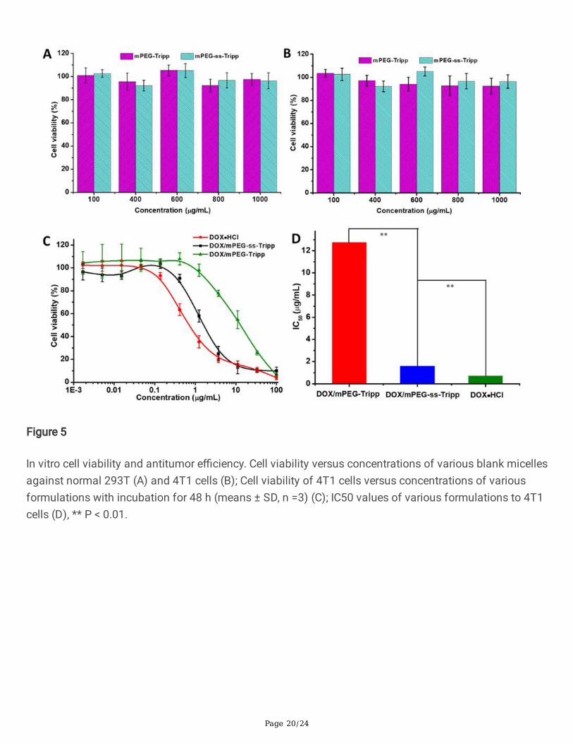
Blank micelle concentrations up to 1 mg/mL showed negligible toxicity against 293T and 4T1 cells after48 h incubation, based on the CCK-8 assay (Figure 5A and 5B). At the same time, DOX-loaded micellesreduced the viability of 4T1 cells, although their half-maximal inhibitory concentrations (IC50) were higherthan for free DOX (mPEG-ss-Tripp, 1.65 μg/mL; mPEG-Tripp, 12.74 μg/mL; free DOX·HCl, 0.71 μg/mL(Figure 5C and 5D). The greater anticancer activity of free DOX likely re�ects its ability to enter cells bypassive diffusion. The much greater anticancer activity of mPEG-ss-Tripp than mPEG-Tripp likely re�ectsthat cleavage of the disul�de bond within cells triggers faster drug release from the micelle interior thansimple diffusion.
Intracellular imaging of AIE micelles in vitro
Uptake of micelles and intracellular release of drug were analyzed using confocal �uorescencemicroscopy and �ow cytometry on the basis of AIE signal. Within 1 h of incubation with blank or DOX-loaded micelles, blue AIE �uorescence was clearly visible within the cytoplasm of 4T1 cells (Figure 6A),suggesting rapid internalization of micelles via endocytosis. Cells incubated with free DOX·HCl showedred �uorescence mainly in nuclei, re�ecting the excellent aqueous solubility of DOX and its ability to entercells and nuclei by passive diffusion. However, for the DOX-loaded micelles, most red �uorescence ofDOX was concentrated in cytoplasm;
only weak red �uorescence was observed in nuclei. Both red and blue �uorescence intensity were higherat 3 h than at 1 h, con�rming that more drug-loaded micelles were internalized into cells and sustainably

Page 13/24
released the drug intracellularly. These experiments con�rm that DOX-loaded micelles can be used forbioimaging and drug delivery.
The results of confocal microscopy were con�rmed using �ow cytometry. While control cells showed onlyauto�uorescence, cells treated with DOX·HCl showed the strongest �uorescence, followed by cells treatedwith mPEG-ss-Tripp and �nally cells treated with mPEG-Tripp (Figure 7A and 7B).
In vivo toxicity and antitumor e�cacy of micelles
Tumor-bearing BALB/c mice were treated every 3 days with saline, DOX·HCl or DOX-loaded micelles. By21 days of treatment, the tumor volume in the saline group had increased rapidly (Figure 8A), indicatingthat the saline did not have any therapeutic effect. DOX·HCl and DOX-loaded micelles, in contrast,strongly inhibited tumor growth. Notably, treatment with mPEG-ss-Tripp group showed better anticanceractivity than mPEG -Tripp group. Over the same period, animals treated with free DOX lost considerablebody weight (Figure 8B), indicating serious systemic toxicity, whereas animals treated with DOX-loadedmicelles did not. These results suggest that loading DOX into micelles can substantially reduce itstoxicity. Consistent with this idea, all mice treated with DOX-loaded micelles survived for the entire periodof 33 days, whereas 40% of mice treated with free DOX·HCl died within 18 days (Figure 8C).
Serum biochemistry assays con�rmed that free DOX·HCl was strongly toxic, which was re�ected inelevated aspartate transaminase (AST), alanine aminotransferase (ALT) and alkaline phosphatase (ALP)(Figure 8D). Indices in animals treated with DOX-loaded micelles were close to those in the saline group,indicating that the DOX-loaded micelles did not obviously harm tumor-bearing mice.
Hematoxylin-eosin staining of tumor tissue showed nearly no apoptosis or necrosis in the saline group,while it showed tumor necrosis with hemorrhage in the groups treated with free DOX·HCl or DOX-loadedmicelles (Figure 9). Staining of slices from heart, liver, spleen, lung and kidney showed numerouspathological signs in animals treated with free DOX·HCl: dissolution of myocardial cytoplasma; swelling,shrinkage or disappearance of kidney glomerular cells; white pulp atrophy and splenic corpuscledisappearance in spleen; and lobular pneumonia in lungs. No obvious pathology was observed in anytissues examined from animals treated with DOX-loaded micelles.
Immunohistochemistry and TUNEL staining
Tissue sections stained against Ki-67 antigen as an index of tumor proliferation showed that mPEG-ss-Tripp micelles suppressed proliferation more than other treatments (Figure 10). These results con�rm thatthe redox-sensitive of the drug delivery system can improve therapeutic e�cacy in vivo.
These in vivo experiments indicate that mPEG-ss-Tripp micelles lead to substantially lower toxicity thanfree DOX·HCl and greater e�cacy than redox-insensitive mPEG-Tripp micelles.
Conclusion

Page 14/24
We have designed a novel redox-sensitive amphiphilic polymer with AIE character to realize a drugdelivery system for simultaneous cellular imaging and cancer therapy. When loaded with DOX, theamphiphilic polymer can self-assemble into drug-loaded micelles, which release their therapeutic cargorapidly in response to reduction of the disul�de linkage at the high GSH concentrations inside cells. Themicelles can be tracked within cells by virtue of their AIE. Removing the redox-sensitive disul�de bridgemakes drug delivery slower and less effective against tumors. These results open up new possibilities formultifunctional drug delivery systems.
DeclarationsAcknowledgements
Not applicable.
Authors′ contributions
All authors contributed signifcantly to this work. BH, ZZ, and NH conceived and designed theexperiments. CS and JL performed the experiments. CS, JL, JW, PH, CL, LQ, LY analyzed the data. NH, BHand ZZ oversaw the design of the experiments, data analysis, and edited the manuscript. All authors readand approved the fnal manuscript.
Funding
The authors thank for the support of the collaborative fund of Luzhou Government and SouthwestMedcial University (2019LZXNYDJ16), the Transformation Project of Science and TechnologyAchievements of Southwest Medical University (2018002) and the research fund of Southwest MedicalUniversity (2017-ZRQN-037).
Availability of data and materials
All data generated or analyzed during this study are included in this published article and its additional�le.
Ethics approval and consent to participate
All of the animal experiments were carried out in accordance with the guidelines issued by SouthwestMedical University and were approved by the Ethical Committee of Southwest Medical University.
Content for publication
All authors agreed to submit this manuscript.
Competing interests
The authors declare that they have no competing interests.

Page 15/24
Author details
1 Department of Pharmaceutical Sciences, School of Pharmacy, Southwest Medical University, Luzhou646000, China. 2 A�liated Traditional Chinese Medicine Hospital, Southwest Medical University, Luzhou646000, People’s China. 3 Biological group, Beijing Huimin School, Beijing 100032, China. 4 NationalEngineering Research Center for Biomaterials, Sichuan University, Chengdu 610064, China.
References1. Hettiarachchi SD, Graham RM, Mintz KJ, Zhou Y, Vanni S, Peng Z, Leblanc RM. Triple conjugated
carbon dots as a nano-drug delivery model for glioblastoma brain tumors. Nanoscale. 2019;11:6192-6205.
2. Xu L, Zhao M, Gao W, Yang Y, Zhang J, Pu Y, He B. Polymeric nanoparticles responsive to intracellularROS for anticancer drug delivery. Colloid Surf. B-Biointerfaces. 2019;181:252-260.
3. Sun HL, Guo BN, Cheng R, Meng FH, Liu HY, Zhong ZY. Biodegradable micelles with sheddablepoly(ethylene glycol) shells for triggered intracellular release of doxorubicin. Biomaterials.2009;30:6358-6366.
4. Liu CY, Yuan J, Luo XM, Chen MH, Chen ZJ, Zhao YC, Li XH, Folate-Decorated and Reduction-Sensitive Micelles Assembled from Amphiphilic Polymer–Camptothecin Conjugates for IntracellularDrug Delivery. Mol. Pharmaceutics. 2014;11:4258-4269.
5. Wang H, Liu G, Dong S, Xiong J, Du Z, Cheng X. A pH-responsive AIE nanoprobe as a drug deliverysystem for bioimaging and cancer therapy. J. Mat. Chem. B. 2015;3:7401-7407.
�. Xue X, Zhao Y, Dai L, Zhang X, Hao X, Zhang C, Huo S, Liu J, Liu C, Kumar A, Chen W-Q, Zou G, LiangX–J. Spatiotemporal Drug Release Visualized through a Drug Delivery System with TunableAggregation‐Induced Emission. Adv. Mater. 2014;26:712-717.
7. Mura S, Nicolas J, Couvreur P. Stimuli-responsive nanocarriers for drug delivery.Nat. Mater.2013;12:991-1003.
�. Zhang C, Jin S, Li S, Xue X, Liu J, Huang Y, Jiang Y, Chen W-Q, Zou G, Liang X-J. Imaging IntracellularAnticancer Drug Delivery by Self-Assembly Micelles with Aggregation-Induced Emission (AIEMicelles). ACS Appl. Mater. Interfaces. 2014;6:5212-5220.
9. Zhang L, Wang T, Yang L, Liu C, Wang C, Liu H, Wang YA, Su Z. General Route to MultifunctionalUniform Yolk/Mesoporous Silica Shell Nanocapsules: A Platform for Simultaneous Cancer‐TargetedImaging and Magnetically Guided Drug Delivery. Chem.-Eur. J. 2012;18:12512-12521.
10. Chen Y-F, Hong J, Wu D-Y, Zhou Y-Y, D'Ortenzio M, Ding Y, Xia X-H. In vivo mapping and assay ofmatrix metalloproteases for liver tumor diagnosis. RSC Adv. 2016;6:8336-8345.
11. Ma J, Wu L, Hou Z, Song Y, Wang L, Jiang W. Visualizing the endocytosis of phenylephrine in livingcells by quantum dot-based tracking. Biomaterials. 2014;35:7042-7049.

Page 16/24
12. Peng T, Chen X, Gao L, Zhang T, Wang W, Shen J, Yang D. A rationally designed rhodamine-based�uorescent probe for molecular imaging of peroxynitrite in live cells and tissues. Chem. Sci.2016;7:5407-5413.
13. Reisch A, Klymchenko AS. Fluorescent Polymer Nanoparticles Based on Dyes: Seeking Brighter Toolsfor Bioimaging. Small. 2016;12:1968-1992.
14. Yuan Y, Kwok RTK, Tang BZ, Liu B. Targeted Theranostic Platinum(IV) Prodrug with a Built-InAggregation-Induced Emission Light-Up Apoptosis Sensor for Noninvasive Early Evaluation of ItsTherapeutic Responses in Situ. J. Am. Chem. Soc. 2014;136:2546-2554.
15. Zrazhevskiy P, Sena M, Gao X. Designing multifunctional quantum dots for bioimaging, detection,and drug delivery. Chem. Soc. Rev. 2010;39:4326-4354.
1�. Ding D, Li K, Liu B, Tang BZ. Bioprobes Based on AIE Fluorogens. Accounts Chem. Res.2013;46:2441-2453.
17. Luo J, Xie Z, Lam JWY, Cheng L, Chen H, Qiu C, Kwok HS, Zhan X, Liu Y, Zhu D, Tang BZ. Aggregation-induced emission of 1-methyl-1,2,3,4,5-pentaphenylsilole. Chem. Commun. 2001;18:1740-1741.
1�. Shi H, Kwok RTK, Liu J, Xing B, Tang BZ, Liu B. Real-Time Monitoring of Cell Apoptosis and DrugScreening Using Fluorescent Light-Up Probe with Aggregation-Induced Emission Characteristics. J.Am. Chem. Soc. 2012;134:17972-17981.
19. Singh VD, Singh RS, Paitandi RP, Dwivedi BK, Maiti B, Pandey DS. Solvent-Dependent Self-Assemblyand Aggregation-Induced Emission in Zn(II) Complexes Containing Phenothiazine-Based TerpyridineLigand and Its E�cacy in Pyrophosphate Sensing. J. Phys. Chem. C. 2018;122:5178-5187.
20. Su X, Ma B, Hu J, Yu T, Zhuang W, Yang L, Li G, Wang Y. Dual-Responsive Doxorubicin-ConjugatedPolymeric Micelles with Aggregation-Induced Emission Active Bioimaging and Charge Conversion forCancer Therapy. Bioconjugate Chem. 2018;29:4050-4061.
21. Zhang R, Sung SHP, Feng G, Zhang C-J, Kenry, Tang BZ, Liu B. Aggregation-Induced Emission Probefor Speci�c Turn-On Quanti�cation of Soluble Transferrin Receptor: An Important Disease Marker forIron De�ciency Anemia and Kidney Diseases. Anal. Chem. 2018;90:1154-116.
22. Guo X, Shi C, Wang J, Di S, Zhou S. pH-triggered intracellular release from actively targeting polymermicelles. Biomaterials 2013;34:4544-4554.
23. Hu H, Li Y, Zhou Q, Ao Y, Yu C, Wan Y, Xu H, Li Z, Yang X. Redox-Sensitive Hydroxyethyl Starch–Doxorubicin Conjugate for Tumor Targeted Drug Delivery. ACS Appl. Mater. Interfaces 2016;8:30833-30844.
24. Lee S-Y, Kim S, Tyler JY, Park K, Cheng J-X. Blood-stable, tumor-adaptable disul�de bonded mPEG-(Cys)4-PDLLA micelles for chemotherapy. Biomaterials 2013;34:552-561.
25. Luan S, Zhu Y, Wu X, Wang Y, Liang F, Song S. Hyaluronic-Acid-Based pH-Sensitive Nanogels forTumor-Targeted Drug Delivery. ACS Biomater. Sci. Eng. 2017;3:2410-2419.
2�. Xu L, Zhao M, Zhang H, Gao W, Guo Z, Zhang X, Zhang J, Cao J, Pu Y, He B. Cinnamaldehyde-BasedPoly(ester-thioacetal) To Generate Reactive Oxygen Species for Fabricating Reactive OxygenSpecies-Responsive Nanoparticles. Biomacromolecules 2018;19:4658-4667.

Page 17/24
27. Dai L, Li J, Zhang B, Liu J, Luo Z, Cai K. Redox-Responsive Nanocarrier Based on Heparin End-Capped Mesoporous Silica Nanoparticles for Targeted Tumor Therapy in Vitro and in Vivo. Langmuir2014;30:7867-7877.
2�. Yi Q, Ma J, Kang K, Gu Z. Dual cellular stimuli-responsive hydrogel nanocapsules for delivery ofanticancer drugs. J. Mat. Chem. B 2016;4:4922-4933.
29. Liu Z, Shen N, Tang Z, Zhang D, Ma L, Yang C, Chen X. An eximious and affordable GSH stimulus-responsive poly(α-lipoic acid) nanocarrier bonding combretastatin A4 for tumor therapy. Biomater.Sci. 2019;7:2803-2811.
30. Zhang T-T, Xu C-H, Zhao W, Gu Y, Li X-L, Xu J-J, Chen H-Y. A redox-activated theranostic nanoagent:toward multi-mode imaging guided chemo-photothermal therapy. Chem. Sci. 2018;9:6749-6757.
31. Shuai X, Ai H, Nasongkla N, Kim S, Gao J. Micellar carriers based on block copolymers of poly(ε-caprolactone) and poly(ethylene glycol) for doxorubicin delivery. J. Control. Release 2004;98:415-426.
32. Hao N, Sun C, Wu Z, Xu L, Gao W, Cao J, Li L, He B. Fabrication of Polymeric Micelles withAggregation-Induced Emission and Forster Resonance Energy Transfer for Anticancer Drug Delivery.Bioconjugate Chem. 2017;28:1944-1954.
33. Jiang Y, Lu H, Khine YY, Dag A, Stenzel MH. Polyion Complex Micelle Based on Albumin–PolymerConjugates: Multifunctional Oligonucleotide Transfection Vectors for Anticancer Chemotherapeutics.Biomacromolecules 2014;15:4195-4205.
34. Maeda H, Wu J, Sawa T, Matsumura Y, Hori K. Tumor vascular permeability and the EPR effect inmacromolecular therapeutics: a review. J. Control. Release 2000; 65:271-284.
35. Wang H, He J, Cao D, Zhang M, Li F, Tam KC, Ni P. Synthesis of an acid-labile polymeric prodrug DOX-acetal-PEG-acetal-DOX with high drug loading content for pH-triggered intracellular drug release.Polym. Chem. 2015;6:4809-4818.
3�. Liang Y, Deng X, Zhang L, Peng X, Gao W, Cao J, Gu Z, He B. Terminal modi�cation of polymericmicelles with π-conjugated moieties for e�cient anticancer drug delivery. Biomaterials 2015:71:1-10.
Figures

Page 18/24
Figure 1
1H NMR spectra of mPEG-SAA (CDCl3) (A); mPEG-ss-NH2 (DMSO-d6) (B) and mPEG-ss-Tripp (DMSO-d6)(C).
Figure 2

Page 19/24
DLS and SEM images of blank micelles (A); DOX-loaded micelles (B).
Figure 3
The �uorescent spectra of mPEG-ss-Tripp in DMSO/water mixtures with different fractions of water (fw),insert image showing �uorescence of mPEG-ss-Tripp solutions dissolved in pure DMSO and 99% water(A); plot of �uorescent intensity of mPEG-ss-Tripp versus water content of DMSO/water mixture, I =�uorescent intensity of mPEG-ss-Tripp in mixed solution, I0 = �uorescent intensity of mPEG-ss-Tripp inpure DMSO (λex= 330 nm) (B); the release pro�les of DOX-loaded micelles at pH 7.4 with or without GSH,means ± SD (n =3) (C).
Figure 4
The size changes of mPEG-Tripp and mPEG-ss-Tripp micells in different concentration of GSH solution(A); the size changes and the count rate of mPEG-ss-Tripp micells over time with or without 10 mM GSH(B), ** P < 0.01.
Page 20/24
Figure 5
In vitro cell viability and antitumor e�ciency. Cell viability versus concentrations of various blank micellesagainst normal 293T (A) and 4T1 cells (B); Cell viability of 4T1 cells versus concentrations of variousformulations with incubation for 48 h (means ± SD, n =3) (C); IC50 values of various formulations to 4T1cells (D), ** P < 0.01.

Page 21/24
Figure 6
The confocal laser scanning microscopy images of 4T1 cells incubated with DOX∙HCl, DOX/mPEG-Trippmicells, and DOX/mPEG-ss-Tripp micelles for 1 h and 3 h, the scale bar was 20 μm.
Figure 7
The �ow cytometry histogram pro�les of 4T1 cells incubated with DOX∙HCl, DOX/mPEG-Tripp micellsand DOX/mPEG-ss-Tripp micelles for 1 h (A) and 3 h (B), the DOX concentration was 10 μg/mL.

Page 22/24
Figure 8
The in vivo anticancer activity of DOX-loaded micelles. The volumes of tumors treated with DOX-loadedmicelles (A); body weights of the mice treated with DOX-loaded micelles (B); survival rate of tumor-bearing mice (C); histogram of serum biochemical test of DOX- loaded micelles (D). Data was presentedas means ± SD (n=8), * P < 0.05, ** P < 0.01.

Page 23/24
Figure 9
Histological analysis of the main organs of tumor bearing mice treated with different methods by H&Estaining (×400). Each group of BALB/c mice (male, n = 8) was intravenously administered four times at athree-day interval at a dose of 5 mg/kg (DOX).

Page 24/24
Figure 10
The TUNEL immunohistochemical (IHC) staining and the Ki-67 antigen staining of tumor tissues (×400).Data was presented as mean ± SD (n = 6), ** P < 0.01. The dose of DOX was 5 mg/kg.
Supplementary Files
This is a list of supplementary �les associated with this preprint. Click to download.
SupportingInformationHao.docx