Deletion of cytoplasmic sequences of the nerve growth factor ...
5
Communication THE JOURNAL OF B~LOGICAL CHEMISTRY Vol. 265, No. 17. Issue of June 15, pp. 9595-9598,199O 0 1990 by The American Society for Biochemistry and Molecular Biology, Inc. Printed in U.S. A. Deletion of Cytoplasmic Sequences of the Nerve Growth Factor Receptor Leads to Loss of High Affinity Ligand Binding* (Received for publication, November 20, 1989) Barbara L. Hempstead, Nila Pati& Bonnie Thiel, and Moses V. Chao$ From the Hematology/Oncology Division Department of Medicine and the $Department of Cell Biology and Anatomy, Cornell Uniuersity Medical College, New York. New York 10021 The nerve growth factor (NGF) receptor is a glyco- sylated transmemhrane protein present on the cell sur- face as both high and low affinity forms, but biological responsiveness requires interactions of NGF with the high affinity site. We have tested the effects of muta- tions in the intracellular domain of the receptor upon its cell surface expression and equilibrium binding of ‘26’I-NGF. Although mutant receptors lacking the en- tire cytoplasmic domain are processed and expressed at the cell surface and are capable of binding to NGF, the absence of cytoplasmic sequences leads to a loss of high affinity binding and to a lack of an appropriate cross-linking pattern as assessed by N-hydroxysucci- nimidyl I-azidobenzoate photoaffinity cross-linking. These results, taken together with the highly con- served nature of these cytoplasmic sequences, implies that the interaction of the receptor with an accessory molecule is necessary to form the high affinity recep- tor. NGF,’ the target-derived neurotrophic factor responsible for the differentiation and survival of selective neuronal pop- ulations, initiates its effects by interacting with a cell surface receptor. NGF binds to both high and low affinity receptors (l-3). In many responsive cells, such as neurons or rat pheo- chromocytoma cells (PC12), approximately lo-15% of the receptors display high affinity binding with a Kd of 10-l’ M with the remainder of the receptors possessing a Kd of lo-’ M. Experimental evidence strongly suggests that biological re- sponsiveness to NGF is dependent upon interactions with the high affinity receptor. The molecular characteristics which determine high affinity * This work was supported by National Cancer Institute Clinical Investigator Award K08CA01367, a March of Dimes basic research award, and a Andrew W. Mellon teacher scientist award (all to B. L. H.) and by grants from the National Institutes of Health (NS-21072). the Hirschl/Caulier Trust, and the special group of donors to the Dorothy Rodbell Cohen Foundation (to M. V. C.). The costs of publication of this article were defrayed in part by the payment of page charges. This article must therefore be hereby marked “adver- tisement” in accordance with 18 U.S.C. Section 1734 solely to indicate this fact. Materials-Materials were obtained from sources described previ- ously (16). PC12, A875, and Hs294 cells were maintained as described (9). NGF was obtained from Bioproducts for Science, rabbit anti- mouse NGF antiserum from Collaborative Research, and HSAB and EDC from Pierce Chemical Co. ‘?-NGF was nrenared as described previously (5, 16). ‘The abbreviations used are: NGF, nerve growth factor; PC12, cultured rat pheochromocytoma cells; NR18, cultured mutagenized Constructs-The full-length human NGF receptor cDNA, El (lo), was subcloned into pBR322, digested with SacI, and religated, result- rat pheochromocytoma cells lacking NGF receptors; HSAB, N-hy- droxysuccinimidyl 4-azidobenzoate; EDC, l-ethyl 3-(3-dimethylami- ing in a receptor cDNA with an intact coding region and poliadenyl- nopropyl)carbodiimide hydrochloride. ation signal but lacking 1759 base pairs of the 3’-untranslated region (pSL). For the mutant PS, pSL was partially digested with PvuII and from low affinity receptors are not well defined. Affinity cross- linking studies using the PC12 cell line, lz51-NGF, and the reagent HSAB suggest that the high affinity NGF-NGF recep- tor complex is a 158-kDa species, whereas the low affinity NGF-NGF receptor complex is a lOO-kDa species (4). How- ever, cross-linking studies using EDC demonstrate that both the high and low affinity NGF-NGF receptor complexes cor- respond to a single 103-kDa species (5). Additional evidence that the two kinetic forms of the receptor are related comes from similar peptide maps of the two receptor complexes (6) and the interconversion of the low and high affinity forms of the receptor during receptor internalization (7). Molecular cloning of the human NGF receptor indicates the receptor gene is a single copy gene (8) and is comprised of six exons spanning over 23 kilobases (9). The sequence of the human (lo), rat (ll), and chicken (12) NGF receptors predicts a structure containing a signal sequence, an extra- cellular domain rich in cysteine residues, a single transmem- brane domain, and a 155-amino acid cytoplasmic domain. The NGF receptor lacks significant homology with other receptors, growth factors, or oncogenes. It does share, however, limited homology in the alignment of extracellular cysteine residues with the CD40 molecule (13). A single receptor mRNA species of 3.8 kilobases has been detected in cells expressing both high and low affinity receptors (8, 10, 11). No evidence has been found for related NGF receptor genes or differential mRNA processing (14). Introduction of receptor cDNA clones in fibroblasts and melanoma cells generates cells that express only the low affinity form of the receptor (10, 11). When the human receptor cDNA was introduced into the NR18 cell line, a PC12 receptor-negative variant (15), the transformant dis- played (a) both high and low affinity receptors detectable by receptor binding and (b) biological responsiveness to NGF as assayed by induction of c-fos transcription (16). Therefore, the receptor gene can give rise to both affinity forms. These findings support the hypothesis that a single binding protein is common to both forms of the NGF receptor and suggest that an additional molecule is required to produce the high affinity form of the NGF receptor. In this report we have utilized the cDNA clone of the receptor to generate deletions in the cytoplasmic domain of the receptor. Examination of receptor cDNA sequences from several species including human, rat, and chicken (10-12) indicates that the most conserved region of the receptor molecule extends from the membrane-spanning segment into the cytoplasmic domain. Our results indicate that the gener- ation of the high affinity form of the receptor requires the presence of the cytoplasmic domain of the receptor. EXPERIMENTAL PROCEDURES 9595 by guest on March 14, 2018 http://www.jbc.org/ Downloaded from
-
Upload
nguyenhanh -
Category
Documents
-
view
216 -
download
0
Transcript of Deletion of cytoplasmic sequences of the nerve growth factor ...

Communication THE JOURNAL OF B~LOGICAL CHEMISTRY Vol. 265, No. 17. Issue of June 15, pp. 9595-9598,199O
0 1990 by The American Society for Biochemistry and Molecular Biology, Inc. Printed in U.S. A.
Deletion of Cytoplasmic Sequences of the Nerve Growth Factor Receptor Leads to Loss of High Affinity Ligand Binding*
(Received for publication, November 20, 1989)
Barbara L. Hempstead, Nila Pati& Bonnie Thiel, and Moses V. Chao$
From the Hematology/Oncology Division Department of Medicine and the $Department of Cell Biology and Anatomy, Cornell Uniuersity Medical College, New York. New York 10021
The nerve growth factor (NGF) receptor is a glyco- sylated transmemhrane protein present on the cell sur- face as both high and low affinity forms, but biological responsiveness requires interactions of NGF with the high affinity site. We have tested the effects of muta- tions in the intracellular domain of the receptor upon its cell surface expression and equilibrium binding of ‘26’I-NGF. Although mutant receptors lacking the en- tire cytoplasmic domain are processed and expressed at the cell surface and are capable of binding to NGF, the absence of cytoplasmic sequences leads to a loss of high affinity binding and to a lack of an appropriate cross-linking pattern as assessed by N-hydroxysucci- nimidyl I-azidobenzoate photoaffinity cross-linking. These results, taken together with the highly con- served nature of these cytoplasmic sequences, implies that the interaction of the receptor with an accessory molecule is necessary to form the high affinity recep- tor.
NGF,’ the target-derived neurotrophic factor responsible for the differentiation and survival of selective neuronal pop- ulations, initiates its effects by interacting with a cell surface receptor. NGF binds to both high and low affinity receptors (l-3). In many responsive cells, such as neurons or rat pheo- chromocytoma cells (PC12), approximately lo-15% of the receptors display high affinity binding with a Kd of 10-l’ M with the remainder of the receptors possessing a Kd of lo-’ M.
Experimental evidence strongly suggests that biological re- sponsiveness to NGF is dependent upon interactions with the high affinity receptor.
The molecular characteristics which determine high affinity
* This work was supported by National Cancer Institute Clinical Investigator Award K08CA01367, a March of Dimes basic research award, and a Andrew W. Mellon teacher scientist award (all to B. L. H.) and by grants from the National Institutes of Health (NS-21072). the Hirschl/Caulier Trust, and the special group of donors to the Dorothy Rodbell Cohen Foundation (to M. V. C.). The costs of publication of this article were defrayed in part by the payment of page charges. This article must therefore be hereby marked “adver- tisement” in accordance with 18 U.S.C. Section 1734 solely to indicate this fact.
Materials-Materials were obtained from sources described previ- ously (16). PC12, A875, and Hs294 cells were maintained as described (9). NGF was obtained from Bioproducts for Science, rabbit anti- mouse NGF antiserum from Collaborative Research, and HSAB and EDC from Pierce Chemical Co. ‘?-NGF was nrenared as described previously (5, 16).
‘The abbreviations used are: NGF, nerve growth factor; PC12, cultured rat pheochromocytoma cells; NR18, cultured mutagenized
Constructs-The full-length human NGF receptor cDNA, El (lo), was subcloned into pBR322, digested with SacI, and religated, result-
rat pheochromocytoma cells lacking NGF receptors; HSAB, N-hy- droxysuccinimidyl 4-azidobenzoate; EDC, l-ethyl 3-(3-dimethylami-
ing in a receptor cDNA with an intact coding region and poliadenyl-
nopropyl)carbodiimide hydrochloride. ation signal but lacking 1759 base pairs of the 3’-untranslated region (pSL). For the mutant PS, pSL was partially digested with PvuII and
from low affinity receptors are not well defined. Affinity cross- linking studies using the PC12 cell line, lz51-NGF, and the reagent HSAB suggest that the high affinity NGF-NGF recep- tor complex is a 158-kDa species, whereas the low affinity NGF-NGF receptor complex is a lOO-kDa species (4). How- ever, cross-linking studies using EDC demonstrate that both the high and low affinity NGF-NGF receptor complexes cor- respond to a single 103-kDa species (5). Additional evidence that the two kinetic forms of the receptor are related comes from similar peptide maps of the two receptor complexes (6) and the interconversion of the low and high affinity forms of the receptor during receptor internalization (7).
Molecular cloning of the human NGF receptor indicates the receptor gene is a single copy gene (8) and is comprised of six exons spanning over 23 kilobases (9). The sequence of the human (lo), rat (ll), and chicken (12) NGF receptors predicts a structure containing a signal sequence, an extra- cellular domain rich in cysteine residues, a single transmem- brane domain, and a 155-amino acid cytoplasmic domain. The NGF receptor lacks significant homology with other receptors, growth factors, or oncogenes. It does share, however, limited homology in the alignment of extracellular cysteine residues with the CD40 molecule (13). A single receptor mRNA species of 3.8 kilobases has been detected in cells expressing both high and low affinity receptors (8, 10, 11). No evidence has been found for related NGF receptor genes or differential mRNA processing (14).
Introduction of receptor cDNA clones in fibroblasts and melanoma cells generates cells that express only the low affinity form of the receptor (10, 11). When the human receptor cDNA was introduced into the NR18 cell line, a PC12 receptor-negative variant (15), the transformant dis- played (a) both high and low affinity receptors detectable by receptor binding and (b) biological responsiveness to NGF as assayed by induction of c-fos transcription (16). Therefore, the receptor gene can give rise to both affinity forms. These findings support the hypothesis that a single binding protein is common to both forms of the NGF receptor and suggest that an additional molecule is required to produce the high affinity form of the NGF receptor.
In this report we have utilized the cDNA clone of the receptor to generate deletions in the cytoplasmic domain of the receptor. Examination of receptor cDNA sequences from several species including human, rat, and chicken (10-12) indicates that the most conserved region of the receptor molecule extends from the membrane-spanning segment into the cytoplasmic domain. Our results indicate that the gener- ation of the high affinity form of the receptor requires the presence of the cytoplasmic domain of the receptor.
EXPERIMENTAL PROCEDURES
9595
by guest on March 14, 2018
http://ww
w.jbc.org/
Dow
nloaded from
9596 NGF Receptor Cytoplasmic Mutants
Stul, religated, and a plasmid lacking the PruII (940 nucleotides)- Stul (1111 nucleotides) fragment was isolated. Following EcoRI diges- tion, the 1.7.kilohase PS receptor cDNA was subcloned into the E:coRI site of the expression vector pMV7. The Xba mutant was obtained by linearization ofpSL at 940 nucleotides with partial PuuII digestion. Universal termination Xba linkers (Biolahs) were ligated to the linearized plasmid, cut with XbaI, and religated to generate a TAG termination codon at the PcuII site. The 2.0-kilobase fragment containing the receptor cDNA was subcloned into the EcoRI site of pMV7. Mutated sequences were verified by DNA sequencing accord- ing to Maxam-Gilbert (17).
Retrouiral &ne Transfer-Helper-free virus stocks were generated in the PA317 packaging cell line using the above constructs (19). Stable transfectants were isolated after neomycin selection and pu- rified by rosetting with the monoclonal antibody ME20.4 (ATCC) and rabbit anti-mouse IgG-coupled human erythrocytes (8). Culture media from the PAX17 transfectants was used to infect NR18 cells. Stable transfectants were again isolated following neomycin selection by rosette analysis.
Affinity Cross-linking and Immunoprecipitation of NGF Recep- tars-NGF receptors were labeled by cross-linking to ““I-NGF as previously described (5, 6, 16). Cells were washed in phosphate- buffered saline containing 1 mg/ml each of glucose and bovine serum albumin, then resuspended at 2 x lO”cells/ml. For EDC cross-linking, a 1.Wml sample was incubated with 1 nM ““I-NGF in the presence or absence of 5 pM unlabeled NGF for 1 h at 22 “C. EDC at 4 mM was added and incubated at 22 “C for 15 min. For HSAB cross- linking, a 1.0.ml sample of cells was incubated with 0.5 nM ““I-NGF for 2 h at 4 “C in the absence or presence of 5 pM NGF. HSAB (50 pM) was added and the reaction exposed to a Spectroline 0.6-A long wave ultraviolet lamp for 10 min. After extensive washing in phos- phate-buffered saline containing 50 mM lysine, cells were lysed in 10 mM Tris (pH 7.4), 66 mM EDTA, 1% Nonidet P-40, 0.4% deoxycho- late in the presence of 1 mM phenylmethylsulfonyl fluoride, 1 mg/ml aprotinin, 1 mg/ml leupeptin, and 1 pg/ml DNase. Receptor com- plexes cross-linked by HSAB were further purified using immunopre- cipitation with rabbit anti-mouse NGF antisera and protein A-Seph- arose. After extensive washing with 150 mM NaCl, 10 mM Tris (pH 7.4), 1% Nonidet P-40, 0.4% deoxycholate, 0.1% sodium dodecyl sulfate, immunoprecipitates were solubilized in sodium dodecyl sul- fate sample buffer and gel electrophoresis, and autoradiography was performed as described (16).
Equilibrium Binding Ana/>nsis-Binding of ““I-NGF was assessed to crude membrane preparations as described (16). Each binding reaction was carried out in triplicate in the presence and absence of excess unlabeled NGF. Only binding values above 50% specific bind- ing were used in analysis. The LIGAND program was utilized to analyze the data.
RESULTS
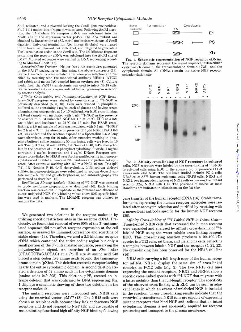
We generated two deletions in the receptor molecule by utilizing specific restriction sites in the receptor cDNA. Pre- viously, we found that removal of over 90% of the 3’-untrans- lated sequence did not affect receptor expression at the cell surface, as assayed by immunofluorescence and rosetting of stable clones (14). Therefore, we used a 2.3-kilobase receptor cDNA which contained the entire coding region but only a small portion of the 3’-untranslated sequence, preserving the polyadenylation signal. An insertion of an Xba linker (CTAGTCTAGACTAG) at a PuuII site at amino acid 248 placed a st.op codon five amino acids beyond the transmem- brane domain (pXba). This deletion created a receptor lacking nearly the entire cytoplasmic domain. A second deletion cre- ated a deletion of 57 amino acids in the cytoplasmic domain (amino acids 249-305). This deletion, pPS, created an in- frame deletion that was confirmed by DNA sequencing. Fig. 1 displays a schematic drawing of these two deletions in the receptor molecule.
The mutant receptors were introduced into NR18 cells using the retroviral vector, pMV7 (18). The NR18 cells were chosen as recipient cells because they lack endogenous NGF receptors and do not respond to NGF (15) and are capable of reconstituting functional high affinity NGF binding following
Sll]&ll Extracel lular Cytoplasmlc sequerw TM
El
b-- ps Xba
FIG. 1. Schematic representation of NGF receptor cDNAs. The receptor domains represent the signal sequence, extracellular cysteine-rich domain, the transmemhrane domain (TM), and the cytoplasmic domain. All cDNAs contain the native NGF receptor polyadenylation site.
200-
97-
68-
FIG. 2. Affinity cross-linking of NGF receptors in cultured cells. NGF receptors were labeled by the cross-linking of ““I-NGF to cultured cells using EDC in the absence (-) or presence (+) of excess unlabeled NGF. The cell lines studied include: PC12 cells; NR18 cells; A875 human melanoma cells; NRPS cells; NRXl and NRX2, two independent isolates of NR18 cells expressing the mutant receptor Xba; NRl-1 cells (16). The positions of molecular mass standards are indicated in kilodaltons on the left side.
gene transfer of the human receptor cDNA (16). Stable trans- formants expressing the human receptor molecules were iso- lated after neomycin selection and purified by rosetting with a monoclonal antibody specific for the human NGF receptor (20).
Affinity Cross-linking of ‘2iZ-Labled NGF to Intact Cells- Transformed NR18 cells that expressed the human receptor were expanded and analyzed by affinity cross-linking of InnI- labeled NGF using the water-soluble cross-linking reagent, EDC. This cross-linking reaction produces a 90-100-kDa species in PC12 cells, rat brain, and melanoma cells, reflecting a complex between labeled NGF and the receptor (5, 21, 22). No cross-linking has been observed for the NR18 cell line (16).
NR18 cells carrying a full-length copy of the human recep- tor mRNA, NRl-1, display the same size of cross-linked complex as PC12 cells (Fig. 2). The two NR18 cell lines expressing the mutant receptors, NRX2 and NRPS, show a specific cross-linked species with lL”I-NGF that migrates with a faster mobility than the full-length receptor. The specificity of the observed cross-linking with EDC can be seen in adja- cent lanes in which an excess of unlabeled NGF is included in the reaction. These cross-linking results indicate that the retrovirally transformed NR18 cells are capable of expressing mutant receptors that bind NGF and indicate that an intact cytoplasmic domain is not apparently required for receptor processing and transport to the plasma membrane.
by guest on March 14, 2018
http://ww
w.jbc.org/
Dow
nloaded from

NGF Receptor Cytophmic Mutants
FIG. 3. Equilibrium binding analysis of NGF receptors in cell membrane preparations. Binding of ““I-NGF was assessed in crude mem- brane preparations by filter binding (16). Membranes were analyzed from NRl-1 cells (A), NRPS cells (B), and NRXba cells (C).
Hs294 PC12 NRl - + - + +
220-
FIG. 4. HSAB affinity cross-linking of NGF receptors in cultured cells. NGF receptors were labeled by the cross-linking of ““I-NGF to cells using HSAB in the absence (-) or presence (+) of excess unlabeled NGF. The cell lines studied are PC12 cells (PC12), NRl-1 cells (NRZ), NRPS cells (PS), NRXba cells (XI), and Hs294 melanoma cells (Ifs294). Autoradiographic exposure times at -70 “C were 5 days for PC12 cells, 2 days for Hs294 cells, and 21 days for the NRl, PS, and Xl cells. The positions of molecular mass standards are indicated in kilodaltons on the left side. The arrow signifies the position of the 160-kDa species.
Scatchurd Plot Analysis-Since the high and low affinity NGF receptors can be distinguished by equilibrium binding, Scatchard plot analysis was carried out by means of a mem- brane binding assay. Crude membranes were prepared from cells and assayed by binding to ‘*“I-NGF. Membranes ob- tained from NRl-1 cells, containing the intact receptor, dis- played both high affinity receptors and low affinity receptors (Fig. 3) similar to the equilibrium binding pattern obtained with PC12 membranes (16).
However, cell lines expressing the pPS and pXba constructs display a linear Scatchard plot, with a & of 2.6 nM for pPS and a Kd of 8 nM for pXba. Several independently derived cell lines expressing these truncated receptors gave the same equilibrium binding data. The number of receptors ranged from 10,000 to 40,00O/cell. Equilibrium binding characteristic of the high affinity class was not detected. Hence, NR18 cells expressing truncated NGF receptors displayed only low affin- ity binding sites, implying that an intact cytoplasmic domain is required for reconstitution of the high affinity binding site.
HSAB Cross-linking-Several previous studies with the lipophilic photoaffinity agent HSAB have demonstrated, in addition to the 90-lOO-kDa ‘*“I-NGF-NGF receptor complex, a higher molecular species of 143 (23) or 158 kDa (4). The higher molecular weight product has been thought to repre- sent the slow dissociating high affinity receptor species (4). The smaller complex has been ascribed to the low affinity species (4, 6, 23). Using HSAB, we determined whether the higher molecular weight species could be correlated with the state of the introduced receptors in NR18 cells.
L 1 1 50 100
PS Xl - + +
1 150
I 1 1 100 200 300
Bound (fmole/mg)
1 1 I loo 200 300
Cross-linking of PC12 cells, Hs294 melanoma cells, and NRl-1 cells with HSAB yields a major observed species of 100,000 daltons. However, in PC12 and NRl-1 cell lines, a distinctive cross-linked species at 150-160 kDa can be ob- served (Fig. 4). This higher molecular weight species was not due to artifactual binding to a nonspecific protein, since pretreatment with excess unlabeled NGF eliminated the cross-linking pattern. In addition, Hs294 melanoma cells, which express only low affinity binding sites, lack the 160- kDa cross-linked receptor complex. The NRl-1 cells display the two affinity classes by equilibrium binding (Fig. 3), and HSAB cross-linking confirms that both forms of the receptor are present. The reduced intensity of the cross-linked species in NRl-1 cells (Fig. 4) reflects the significantly lower number of receptors in this line (16).
In contrast to the NRl-1 cell line, the NRX2 and NRPS line display only a cross-linked receptor species consistent with a complex of the truncated receptor with iz51-NGF (Fig. 3). The intact receptor can be cross-linked to NGF in NR18 cells to form a higher molecular weight species; however, truncated versions have lost the ability to form this cross- linked product. The absence of cytoplasmic sequences in the receptor can therefore be correlated with the loss of high affinity binding and the lack of cross-linking to an accessory protein.
DISCUSSION
We have generated mutations within the cytoplasmic do- main of the NGF receptor which affect the binding properties of the extracellular domain of the receptor to its ligand. These mutant receptors bind NGF with a Kd consistent with the low affinity binding site and are unable to generate the molecular complex characteristic of the high affinity receptor following cross-linking with a lipophilic cross-linking reagent HSAB. The data presented here strongly support the model in which high affinity binding requires the interaction of the cyto- plasmic portion of the receptor with an accessory molecule(s). The possibility that the molecular complex characteristic of the high affinity receptor (160 kDa) is a dimer of receptor molecules has been excluded by identification of a 200-kDa species as the receptor dimer by peptide mapping (24).
The most highly conserved region of the NGF receptor is a membrane-associated domain, consisting of the 22-amino acid membrane-spanning domain and flanking sequences (19 amino acids of the extracellular region and 42 residues of the cytoplasmic region). This region is 95% identical among receptors of human, rat, and chicken origin (12) and bears some resemblance to the a-helical membrane-spanning do-
by guest on March 14, 2018
http://ww
w.jbc.org/
Dow
nloaded from

9598 NGF Receptor Cytoplasmic Ivfutants
main of the (Y subunit of the high affinity rat IgE Fc receptor. Of note, the functional expression of the Fcr receptor (Y subunit is dependent upon interaction with two additional subunits, /3 and y (25, 26). Similarly, the formation of the high affinity NGF receptor complex may occur by coupling of membrane-associated molecules to the highly conserved por- tion of the receptor.
The mutant NGF receptors lack (a) all but the initial five amino acids in the cytoplasmic domain or (b) 58 amino acids in the proximal portion of the cytoplasmic domain. Both truncated receptors disrupt the integrity of the highly con- served membrane-flanking region of the receptor. Unlike the full-length receptor, which is capable of reconstituting both high and low affinity states in appropriate recipient cells, the mutant receptors display only low affinity sites. These results are similar to those reported with the epidermal growth factor receptor where truncations in the cytoplasmic domain of the receptor abolished high affinity binding sites for epidermal growth factor (27, 28).
The function of the cytoplasmic domain of the NGF recep- tor and its role in signal transduction is unknown. Numerous models can account for the absence of high affinity receptor formation upon perturbation of the membrane-flanking cy- toplasmic domain. First, this domain could be required for interaction with an accessory protein necessary for high affin- ity receptor complexes. This accessory protein could bind to NGF directly, or bind only to the cytoplasmic portion of the receptor, or interact with a conformationally altered NGF- NGF receptor complex, similar to the altered platelet-derived growth factor-platelet-derived growth factor receptor complex detected following ligand-receptor interaction (29). Alterna- tively, the cytoplasmic domain may be modified by phos- phorylation. Although the receptor does not contain an ATP binding site or an intrinsic kinase activity, phosphorylation at serine and threonine residues has been demonstrated in uiuo (30, 31). The mutants described here lack numerous serine and threonine residues and therefore may display al- tered levels of phosphorylation.
Although the primary molecular events associated with NGF-mediated receptor signaling remain unclear, recent re- ports have implicated a role of glycosyl phosphatidylinositol hydrolysis (32), protein kinase C activation (33, 34), and tyrosine kinase activation (35) following NGF binding. The cytoplasmic mutations in the NGF receptor should prove useful both in dissecting the molecular events involved in signal transduction and in elucidating the biochemical struc- ture of the high affinity receptor complex.
REFERENCES 1. Sutter, A., Riopelle, R. J., Harris-Warrick, R. M., and Shooter,
E. M. (1979) J. Biol. Chem. 254,5972-5982 2. Landreth, G. E., and Shooter, E. M. (1980) Proc. Natl. Acad. Sci.
U. S. A. 77,4751-4755 3. Schechter, A. L., and Bothwell, M. A. (1981) Cell 24,867-874
4.
5.
6.
7. 8.
9.
10.
11.
12.
13.
14.
15.
16.
17.
18.
19.
20.
21.
22.
23.
24.
25. 26.
27. 28.
29.
30.
31.
32.
33.
34.
35.
Hosang, M., and Shooter, E. M. (1985) J. Biol. Chem. 260,655- 662
Green, S. H., and Greene, L. A. (1986) J. Biol. Chem. 261,15316- 15326
Kouchalakos, R. N., and Bradshaw, R. A. (1986) J. Biol. C&m. 261, 16054-16059
Eveleth, D. D.. and Bradshaw. R. A. (1988) Neuron 1. 929-936 Chao, M. V.,‘Bothwell, M. A., Ross, A. H., Koprowski, H.,
Lanahan, A. A., Buck, C. R., and Sehgal, A. (1986) Science 232.518-521
Sehrral. A.. Patil. N.. and Chao. M. (1988) Mol. Cell. Biol. 8.3160- 3167'
Johnson, D., Lanahan, A., Buck, C. R., Sehgal, A., Morgan, C., Mercer, E., Bothwell, M., and Chao, M. (1986) Cell 47, 545- 554
Radeke, M. J., Misko, T. P., Hsu, C., Herzenberg, L. A., and Shooter, E. M. (1987) Nature 325,593-597
Large, T. H., Weskamp, G., Helder, J. C., Radeke, M. J., Misko, T. P., Shooter, E. M., and Reichardt, L. F. (1989) Neuron 2, 1123-1134
Stamenkovic, I., Clark, E. A., and Seed, B. (1989) EMBO J. 8, 1403-1410
Hempstead, B., Patil, N., Olson, K., and Chao, M. (1988) Cold Surinx Harbor SymD. Quunt. Biol. 53,477-485
Bot‘hweil, M. A., Schechtkr, A. L., and Vaughn, K. M. (1980) Cell 21,857-866
Hempstead, B., Schleifer, L. S., and Chao, M. V. (1989) Science 243.373-375
Maxam, A., and Gilbert, W. (1980) Methods Enzymol. 65, 499- 560
Kirschmeier, P., Housey, G. M., Johnson, M. D., Perkins, A. S., and Weinstein. I. B. (1988) DNA 7. 219-225
Miller, A. D., and Buttimord, C. (1986) Mol. Cell. Btil. 6, 2895- 2902
Ross, A. H., Grob, P., Bothwell, M., Elder, D. E., Ernst, C. S., Marano. N.. Ghrist. B. F. D.. Slemn. C. C.. Herlvn. M.. Atkin- son, B., ‘and Koprowski, H. (1984) Proc. gad. Ai&. Sk. U. S. A. 81,6681-6685
Grob, P., Berlot, C. H., and Bothwell, M. A. (1983) Proc. Natl. Acad. Sci. U. S. A. 80,6819-6823
Taniuchi, M., Schweitzer, J. B., and Johnson, E. M. (1986) Proc. Natl. Acad. Sci. U. S. A. 83, 1950-1954
Massague, J., Guillette, B. J., Czech, M. P., Morgan, C. J., and Bradshaw, R. A. (1981) J. Biol. Chem. 256,9419-9424
Buxser, S., Puma, P., and Johnson, G. L. (1985) J. Biot. Chem. 260,1917-1926
Kinet. J. P. (1989) CeZZ 57, 351-354 Blank, U., Ra, C., Miller, L., White, K., Metzger, H., and Kinet,
J. P. (1989) Nature 337,187-189 Schlessinger, J. (1986) J. Cell BioZ. 103, 2067-2072 Prywes, R., Livneh, E., Ullrich, A., and Schlessinger, J. (1986)
EMBO J. 5,2179-2190 Keating, M. T., Escobedo, J. A., and Williams, L. T. (1988) J.
Biol. Chem. 263,12805-12808 Grob, P. M., Ross, A. H., Koprowski, H., and Bothwell, M. (1985)
J. Biol. Chem. 260,8044-8049 Taniuchi, M., Johnson, E. M., Roach, P. J., and Lawrence, J. C.
(1986) J. Biol. Chem. 261, 13342-13349 Chan, B. L., Chao, M. V., and Saltiel, A. L. (1989) Proc. Natl.
Acad. Sci. U. S. A. 86, 1756-1760 Hama, T., Huang, K. P., and Guroff, G. (1986) Proc. Natl. Acad.
Sci. U. S. A. 83,2353-2357 Hall, F. L., Fernyhough, P., Ishii, D. N., and Vulliet, P. R. (1988)
J. Biol. Chem. 263,4460-4466 Maher, P. (1988) Proc. Natl. Acad. Sci. U. S. A. 85,6788-6791
by guest on March 14, 2018
http://ww
w.jbc.org/
Dow
nloaded from
B L Hempstead, N Patil, B Thiel and M V Chaoof high affinity ligand binding.
Deletion of cytoplasmic sequences of the nerve growth factor receptor leads to loss
1990, 265:9595-9598.J. Biol. Chem.
http://www.jbc.org/content/265/17/9595Access the most updated version of this article at
Alerts:
When a correction for this article is posted•
When this article is cited•
to choose from all of JBC's e-mail alertsClick here
http://www.jbc.org/content/265/17/9595.full.html#ref-list-1
This article cites 0 references, 0 of which can be accessed free at
by guest on March 14, 2018
http://ww
w.jbc.org/
Dow
nloaded from