DEHP and DINP Induce Tissue- and Gender-Specific ... · DEHP and DINP Induce Tissue- and...
11
DEHP and DINP Induce Tissue- and Gender-Specific Disturbances in Fatty Acid and Lipidomic Profiles in Neonatal Mice: A Comparative Study Yichao Huang, Fengjiang Sun, Hongli Tan, Yongfeng Deng, Zhiqiang Sun, Hexia Chen, Jing Li, and Da Chen* School of Environment and Guangdong Key Laboratory of Environmental Pollution and Health, Jinan University, Guangzhou 510632, China * S Supporting Information ABSTRACT: Di-isononyl phthalate (DINP) is considered one of the main industrial alternatives to di(2-ethylhexyl)phthalate (DEHP), a well- known chemical with various toxic effects including the disruption with lipid metabolism. However, the potential effects of DINP on lipid metabolism have rarely been investigated in mammals. Our study demonstrated that exposure of neonatal mice to DEHP and DINP at a daily dose of 0.048 or 4.8 mg/kg from postnatal day 0 (PND0) to PND21 caused nonmonotonic as well as tissue- and gender-specific alterations of total fatty acid (FA) compositions in plasma, heart, and adipose tissues. However, the patterns of disruption differed between DEHP- and DINP-treated groups. On the basis of targeted lipidomic analyses, we further identified gender-specific alterations of eight lipid classes in plasma following DEHP or DINP exposure. At the higher dose, DEHP induced decreases in total phosphatidylcholines and phosphatidylinositol (PI) in females and increases in phosphatidylethanolamines (PEs) and triglycerides in males. By contrast, DINP at the higher dose caused alterations of PEs, PIs, phosphatidylserines, and cholesterols exclusively in male mice, but no changes were observed in female pups. Although the most significant dysregulation of lipid metabolism was often observed for the higher dose, the lower one could also disrupt lipid profiles and sometimes its effects may even be more significant than those induced by the higher dose. Our study for the first time identified tissue- and gender-specific disruptions of FA compositions and lipidomic profiles in mice neonatally exposed to DINP. These findings question the suitability of DINP as a safe DEHP substitute and lay a solid foundation for further elucidation of its effects on lipid metabolism and underlying mechanisms. ■ INTRODUCTION As one of the major groups of environmental contaminants with global occurrences, phthalates have been ubiquitously found in human urine and blood. 1 Di(2-ethylhexyl)phthalate (DEHP) represents one of the most studied phthalates, mainly because of its high occurrence rate and reproductive and endocrine disrupting toxicities. 2 This had led to the use of alternative chemicals as DEHP substitutes, among which di- isononyl phthalate (DINP) represents a major replacement. The annual global production of DINP was estimated to be 1.5 million tons in 2013, 3 reaching approximately 75% of annual DEHP production volume. 4 Although DINP has been suggested with decreased carcinogenic toxicity compared with DEHP, previous work has demonstrated that DINP can cause disturbances to the immune system 5,6 and liver and kidney damage at a daily dose of 20−200 mg/kg body weight. 7 However, compared with numerous DEHP studies, DINP’s toxicities have been subjected to much less investigations. In particular, DINP’s chronic effects in mammals, such as the interference with lipid synthesis and metabolism, remain largely uninvestigated. Lipids, either endogenously synthesized through lipogenesis or originated from diet, are composed of saturated fatty acids (SFAs), monounsaturated fatty acids (Monos), trans-fatty acids (Trans), and polyunsaturated fatty acids (PUFAs). Fatty acids (FAs) not only provide sources of energy but also constitute signi ficant components of cell membranes. 8 Metabolized lipids also serve as hormonal mediators, which regulate multiple functions including inflammation responses. 9 A large number of studies have demonstrated that some lipid molecules (e.g., plasma phospholipid FAs) can serve as important biomarkers in the development of chronic metabolic conditions at various disease stages. 10−12 Some studies have reported that DEHP and its metabolite mono-(2-ethylhexyl)phthalate (MEHP) can disrupt lipid metabolism by altering total triglycerides (TG), 13,14 mainly through activation of the nuclear peroxisome proliferator- Received: July 21, 2019 Revised: September 30, 2019 Accepted: October 2, 2019 Published: October 2, 2019 Article pubs.acs.org/est Cite This: Environ. Sci. Technol. XXXX, XXX, XXX-XXX © XXXX American Chemical Society A DOI: 10.1021/acs.est.9b04369 Environ. Sci. Technol. XXXX, XXX, XXX−XXX Downloaded via JINAN UNIV on October 29, 2019 at 06:18:06 (UTC). See https://pubs.acs.org/sharingguidelines for options on how to legitimately share published articles.
Transcript of DEHP and DINP Induce Tissue- and Gender-Specific ... · DEHP and DINP Induce Tissue- and...

DEHP and DINP Induce Tissue- and Gender-Specific Disturbances inFatty Acid and Lipidomic Profiles in Neonatal Mice: A ComparativeStudyYichao Huang, Fengjiang Sun, Hongli Tan, Yongfeng Deng, Zhiqiang Sun, Hexia Chen, Jing Li,and Da Chen*
School of Environment and Guangdong Key Laboratory of Environmental Pollution and Health, Jinan University, Guangzhou510632, China
*S Supporting Information
ABSTRACT: Di-isononyl phthalate (DINP) is considered one of themain industrial alternatives to di(2-ethylhexyl)phthalate (DEHP), a well-known chemical with various toxic effects including the disruption withlipid metabolism. However, the potential effects of DINP on lipidmetabolism have rarely been investigated in mammals. Our studydemonstrated that exposure of neonatal mice to DEHP and DINP at adaily dose of 0.048 or 4.8 mg/kg from postnatal day 0 (PND0) toPND21 caused nonmonotonic as well as tissue- and gender-specificalterations of total fatty acid (FA) compositions in plasma, heart, andadipose tissues. However, the patterns of disruption differed betweenDEHP- and DINP-treated groups. On the basis of targeted lipidomicanalyses, we further identified gender-specific alterations of eight lipid classes in plasma following DEHP or DINP exposure. Atthe higher dose, DEHP induced decreases in total phosphatidylcholines and phosphatidylinositol (PI) in females and increasesin phosphatidylethanolamines (PEs) and triglycerides in males. By contrast, DINP at the higher dose caused alterations of PEs,PIs, phosphatidylserines, and cholesterols exclusively in male mice, but no changes were observed in female pups. Although themost significant dysregulation of lipid metabolism was often observed for the higher dose, the lower one could also disrupt lipidprofiles and sometimes its effects may even be more significant than those induced by the higher dose. Our study for the firsttime identified tissue- and gender-specific disruptions of FA compositions and lipidomic profiles in mice neonatally exposed toDINP. These findings question the suitability of DINP as a safe DEHP substitute and lay a solid foundation for furtherelucidation of its effects on lipid metabolism and underlying mechanisms.
■ INTRODUCTION
As one of the major groups of environmental contaminantswith global occurrences, phthalates have been ubiquitouslyfound in human urine and blood.1 Di(2-ethylhexyl)phthalate(DEHP) represents one of the most studied phthalates, mainlybecause of its high occurrence rate and reproductive andendocrine disrupting toxicities.2 This had led to the use ofalternative chemicals as DEHP substitutes, among which di-isononyl phthalate (DINP) represents a major replacement.The annual global production of DINP was estimated to be 1.5million tons in 2013,3 reaching approximately 75% of annualDEHP production volume.4
Although DINP has been suggested with decreasedcarcinogenic toxicity compared with DEHP, previous workhas demonstrated that DINP can cause disturbances to theimmune system5,6 and liver and kidney damage at a daily doseof 20−200 mg/kg body weight.7 However, compared withnumerous DEHP studies, DINP’s toxicities have beensubjected to much less investigations. In particular, DINP’schronic effects in mammals, such as the interference with lipidsynthesis and metabolism, remain largely uninvestigated.
Lipids, either endogenously synthesized through lipogenesisor originated from diet, are composed of saturated fatty acids(SFAs), monounsaturated fatty acids (Monos), trans-fattyacids (Trans), and polyunsaturated fatty acids (PUFAs). Fattyacids (FAs) not only provide sources of energy but alsoconstitute significant components of cell membranes.8
Metabolized lipids also serve as hormonal mediators, whichregulate multiple functions including inflammation responses.9
A large number of studies have demonstrated that some lipidmolecules (e.g., plasma phospholipid FAs) can serve asimportant biomarkers in the development of chronic metabolicconditions at various disease stages.10−12
Some studies have reported that DEHP and its metabolitemono-(2-ethylhexyl)phthalate (MEHP) can disrupt lipidmetabolism by altering total triglycerides (TG),13,14 mainlythrough activation of the nuclear peroxisome proliferator-
Received: July 21, 2019Revised: September 30, 2019Accepted: October 2, 2019Published: October 2, 2019
Article
pubs.acs.org/estCite This: Environ. Sci. Technol. XXXX, XXX, XXX−XXX
© XXXX American Chemical Society A DOI: 10.1021/acs.est.9b04369Environ. Sci. Technol. XXXX, XXX, XXX−XXX
Dow
nloa
ded
via
JIN
AN
UN
IV o
n O
ctob
er 2
9, 2
019
at 0
6:18
:06
(UT
C).
See
http
s://p
ubs.
acs.
org/
shar
ingg
uide
lines
for
opt
ions
on
how
to le
gitim
atel
y sh
are
publ
ishe
d ar
ticle
s.

activated receptors (PPARs).15−18 Transcriptomics and non-targeted global metabolomics both demonstrated the alterationof lipid metabolism as a consequence of DEHP exposure.13
However, as a major DEHP substitute, DINP has receivedmuch less attention with regard to its potential impacts on lipidmetabolism, particularly in mammals. To the best of ourknowledge, the only data available were reported in aquaticorganisms including zebrafish and seabream studies, whichindicated that exposure to DINP causes disturbances in lipid-related metabolism, as well as the induction of oxidative stressand activation of immune responses.19−22
Consequently, the main goal of this study was to investigateand compare the disturbances of FA and lipidomic profiles inneonatal mice following exposure to DEHP and DINP. As apilot study of a larger-scale project aiming to elucidate thepotential effects of DEHP substitutes on lipid synthesis andmetabolism, the specific objectives of the present work were toexplore differential effects of DEHP and DINP on (1) thedistributions of FA compositions in different tissues andgenders and (2) gender-specific changes of lipidomic profilesin plasma. Findings from the present study will lay a solidfoundation for further elucidation of the impacts of DINP onlipid metabolism homeostasis, underlying mechanisms, andassociated biological consequences. The findings also contrib-ute to a better elucidation of whether DINP is a safe substituteto DEHP.
■ MATERIALS AND METHODSChemicals and Reagents. Reference standards of DINP
and DEHP were purchased from Sigma (St. Louis, USA). FAmethyl ester (FAME) reference standard mixture (GLC-463)was obtained from NuChek (Prep Inc., USA). The internalstandard mixture for lipidomic assay, containing bis-(monoacylglycero)phosphate 14:0/14:0; ceramide, Cer 18:1/17:0; phosphatidylcholine, PC 14:0/14:0; phosphatidyletha-nolamine, PE 17:0/17:0; phosphatidylglycerol, PG 14:0/14:0;and phosphatidylserines, PS 17:0/17:0, was purchased fromSigma (St. Louis, USA). Dihexosylceramide 18:1/16:0 (d3);glucosylceramide, GC 18:1/16:0 (d3); phosphatidylinositol, PI16:0/16:0; and trihexosylceramide 18:1/17:0 were purchasedfrom Matreya LLC (Pleasant Gap, USA). All solvents usedwere of high-performance liquid chromatography grade (FisherScientific, New Hampshire, USA), except for methanol andwater (Optima, Fisher Scientific, USA).Animal Experiment. Six male and 18 female-specific
pathogen-free Kunming mice (8−10 weeks old) were obtainedfrom Guangdong Medical Laboratory Animal Center (Guangz-hou, China). Animals were acclimated for 10 days prior toexperiments and were maintained under a 12 h light/12 h darkcycle at a room temperature of 25 °C. Glass water bottles andpolypropylene cages were used in this study. After acclimation,each cage contained one male and two female mice at 7 pm formating. Once confirmed pregnant, each female mouse was keptin individual cage until birth. All mice were fed ad libitum on astandard chow.Dams were allowed to give birth naturally at term. Newborn
mice were recorded as postnatal day 0 (PND0), and theyremained with their natural mothers in the same cage untilPND21. Each cage was randomly assigned to a treatment(DEHP or DINP) or control group. Each group containedthree replicate cages. Both male and female pups were exposedto vehicle control (corn oil), DEHP (high dose 4.8 mg/kg bw/day or low dose 0.048 mg/kg bw/day), or DINP (high dose
4.8 mg/kg bw/day or low dose 0.048 mg/kg bw/day) throughsubcutaneous injection at 10−12 am daily from PND0 toPND21. The high dose chosen corresponds to the no-observed-adverse-effect level (NOAEL)23 suggested for DEHPand DINP, while the low dose is slightly lower than thetolerable daily intake (TDI) dose of DEHP (0.05 mg/kg bw/day) and DINP (0.15 mg/kg bw/day) according to theEuropean EU Risk Assessment Reports (EU RAR).23 Dosageswere adjusted daily for body weight change, and such aninjection approach was chosen in order to maintain aconsistent dosage, as well as considering its practicality forneonates. Pups were kept with their dams until PND21 andwere fasted for 6 h prior to sampling on PND22. The exposureperiod of PND0−PND21 was employed because this is animportant developmental stage as well as a sensitive window oftime within which the pups are susceptible to environmentalstresses.24
At PND22, 10 male and 10 female pups were randomlyselected from three replicate cages per treatment group (threeeach from two cages and another four from the third cage).These pups were selected for phthalate metabolite residue,biochemical, and FA/lipidomic analyses. Urine samples werecollected using the stainless metabolic cage method, and urinefrom two random pups was pooled to obtain sufficient volumefor chemical analyses. Basic phenotype measurements,including body weight, abdominal circumference, and bodylength, were conducted before tissue collection. Blood sampleswere collected via cardiac puncture into an ethylenediaminete-traacetic acid-treated vacutainer and kept on ice prior toseparation of plasma and erythrocytes at 2800g centrifugation.Brain, heart, kidney, liver, and reproductive white adiposetissues were subsequently sampled from each pup. Urine,plasma, and tissue samples for biochemical and oil red Ostaining were all snap frozen in liquid nitrogen andsubsequently stored in −80 °C. Samples for mRNA analysiswere placed in RNAlater solution (QIAGEN, Germany) andthen stored in −80 °C. Urine samples for phthalate metaboliteanalysis were stored in −20 °C.Plasma total glucose and liver total bile acid (TBA) were
determined using a commercial kit (Nanjing JianchengBioengineering Institute, China), performed according to themanufacturer’s instructions. Frozen liver sections were stainedwith oil red O staining to examine if there was histopatho-logical hepatic steatosis (see the Supporting Information fordetails). Animal experiments in this study were approved bythe Laboratory Animal Ethics Committee of Jinan University(Guangzhou, China).
FA Analysis. An aliquot of 50 μL of plasma or ∼0.1 g ofhomogenized tissue samples was extracted using the Folchmethod with a mixture of methanol and chloroform (1:2, v/v).25 Lipids were then transmethylated to FAMEs and thenextracted with heptane. The final extracts were determined bygas chromatography−mass spectrometry. Detailed informationon sample preparation and instrumental analysis is summarizedin the Supporting Information. Quality assurance and control(QA/QC) procedures included the process of a solvent blankand a commercial plasma sample (BestBio, Shanghai) alongwith every batch of samples. The latter serves as QC samplesto determine intra- and interassay coefficient of variations(CVs). Results were presented as weight percentages of totalFAs. Our in-house QC data demonstrated intra- and interassayCVs of less than 8.5 and 9.8%, respectively, for major FAs (i.e.,concentration > 0.1%).
Environmental Science & Technology Article
DOI: 10.1021/acs.est.9b04369Environ. Sci. Technol. XXXX, XXX, XXX−XXX
B
Lipidomic Analysis. An aliquot of 50 μL of plasma wasspiked with internal standard mixture (400 pmol each) prior tothe Folch extraction.25 After drying, the lipid extract wasdissolved in 500 μL of a mixture of methanol, tetrahydrofuran,and water (5:2:3, v/v/v) and subjected to instrumental analysiswhich employed an ExionLC AD ultraperformance LC(UPLC, AB Sciex, Toronto, Canada) equipped with anAgilent Eclipse Plus C18 column (Agilent Technologies) andcoupled to an AB Sciex Q TRAP 5500 mass spectrometer.Details on sample preparation and instrumental determinationare given in the Supporting Information. QA/QC proceduresincluded the process of solvent blanks, solvent spiked withinternal standards, matrix blanks without spiking internalstandards, and commercial plasma samples serving as QCsamples to determine intra- and interassay CVs. Concen-trations were calculated by relating peak areas of the analytesto peak areas of their corresponding internal standard. Our in-house QC data demonstrated intra- and interassay CVs of lessthan 14.7 and 16.8% for analytes above quantitation limits (i.e.,1.8−17.2 nmol/L), respectively.Urinary Phthalate Metabolite Analyses. Urinary
metabolites of DEHP and DINP were determined to confirminternal exposure in the pups following the method describedby Asimakopoulos et al. with modifications.26 In brief, 500 μLof urine was transferred into polypropylene tubes and spikedwith 10 μL of surrogate standard (mono-benzyl phthalate-d4).The mixture was then hydrolyzed with β-glucuronidase/sulfatase (Sigma-Aldrich, Munich, Germany), extracted witha mixture of methyl tert-butyl ether and ethyl acetate (4:1; v/v). The extract was dried under gentle nitrogen stream and
then reconstituted with a mixture of acetonitrile and water(6:4; v/v) and spiked with 13C12-bisphenol A. Instrumentalanalysis employed an ExionLC AD UPLC equipped with anAgilent Extend-C18 column (Narrow Bore RR 2.1 × 100 mm,3.5 μm, 80 Å; Agilent Technologies) and coupled to an ABSciex Q TRAP 5500 MS. The limit of quantification (LOQ),defined as an analyte response 10 times the standard deviationof the noise, was determined to be 0.5 ng/mL for MEHP,mono(2-ethyl-5-oxohexyl) phthalate (MEOHP), and mono(2-ethyl-5-carboxypentyl)phthalate (MECPP) and 1 ng/mL formonoisononyl phthalate (MINP) and mono carboxyoctylphthalate (MCOP). Details on sample preparation, instru-mental determination, and QA/QC practices are given in theSupporting Information.
Lipid Metabolism-Related Gene Expression Analysis.The expression of genes related to lipid metabolism27
including peroxisome proliferator-activated receptors (Pparα,Pparβ, and Pparγ), lipoprotein lipase (Lpl), acetyl CoAcarboxylase-1 (Acc1), FA synthase (Fas), stearoyl CoAdesaturase-1 (Scd1), FA elongase-6 (Elovl6), Acyl-coenzymeA oxidase-1, palmitoyl (Acox-1), FA elongase-2 (Elovl2), andFA desaturase (Fads1 and Fads2) were analyzed byquantitative real-time polymerase chain reaction (qRT-PCR)assay. Detailed information on qRT-PCR is described in theSupporting Information.
Statistical Analysis. All data are presented as mean ±standard deviation unless otherwise stated. FA percentage datawere arc-sin converted prior to statistical analyses. For eachtype of control or treatments, we performed intragroupcomparisons between replicate cages using nonparametric
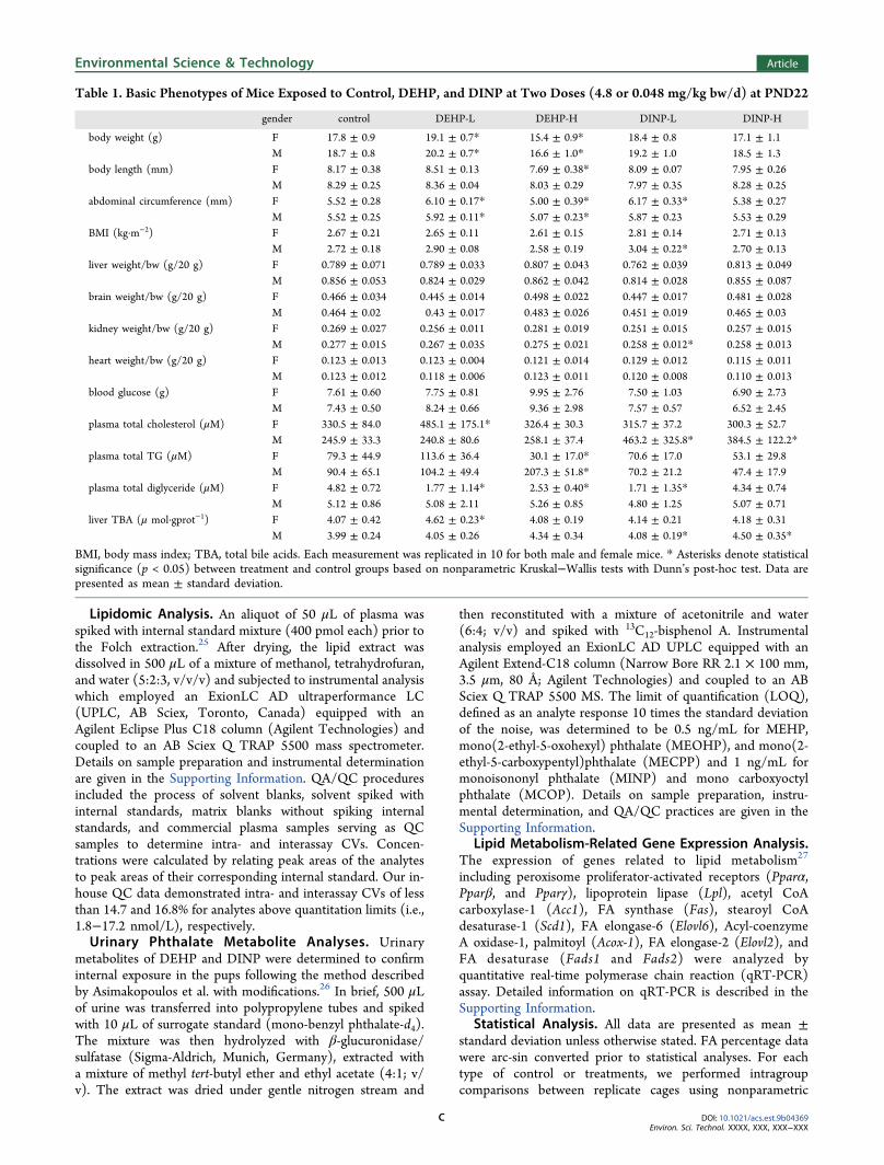
Table 1. Basic Phenotypes of Mice Exposed to Control, DEHP, and DINP at Two Doses (4.8 or 0.048 mg/kg bw/d) at PND22
gender control DEHP-L DEHP-H DINP-L DINP-H
body weight (g) F 17.8 ± 0.9 19.1 ± 0.7* 15.4 ± 0.9* 18.4 ± 0.8 17.1 ± 1.1M 18.7 ± 0.8 20.2 ± 0.7* 16.6 ± 1.0* 19.2 ± 1.0 18.5 ± 1.3
body length (mm) F 8.17 ± 0.38 8.51 ± 0.13 7.69 ± 0.38* 8.09 ± 0.07 7.95 ± 0.26M 8.29 ± 0.25 8.36 ± 0.04 8.03 ± 0.29 7.97 ± 0.35 8.28 ± 0.25
abdominal circumference (mm) F 5.52 ± 0.28 6.10 ± 0.17* 5.00 ± 0.39* 6.17 ± 0.33* 5.38 ± 0.27M 5.52 ± 0.25 5.92 ± 0.11* 5.07 ± 0.23* 5.87 ± 0.23 5.53 ± 0.29
BMI (kg·m−2) F 2.67 ± 0.21 2.65 ± 0.11 2.61 ± 0.15 2.81 ± 0.14 2.71 ± 0.13M 2.72 ± 0.18 2.90 ± 0.08 2.58 ± 0.19 3.04 ± 0.22* 2.70 ± 0.13
liver weight/bw (g/20 g) F 0.789 ± 0.071 0.789 ± 0.033 0.807 ± 0.043 0.762 ± 0.039 0.813 ± 0.049M 0.856 ± 0.053 0.824 ± 0.029 0.862 ± 0.042 0.814 ± 0.028 0.855 ± 0.087
brain weight/bw (g/20 g) F 0.466 ± 0.034 0.445 ± 0.014 0.498 ± 0.022 0.447 ± 0.017 0.481 ± 0.028M 0.464 ± 0.02 0.43 ± 0.017 0.483 ± 0.026 0.451 ± 0.019 0.465 ± 0.03
kidney weight/bw (g/20 g) F 0.269 ± 0.027 0.256 ± 0.011 0.281 ± 0.019 0.251 ± 0.015 0.257 ± 0.015M 0.277 ± 0.015 0.267 ± 0.035 0.275 ± 0.021 0.258 ± 0.012* 0.258 ± 0.013
heart weight/bw (g/20 g) F 0.123 ± 0.013 0.123 ± 0.004 0.121 ± 0.014 0.129 ± 0.012 0.115 ± 0.011M 0.123 ± 0.012 0.118 ± 0.006 0.123 ± 0.011 0.120 ± 0.008 0.110 ± 0.013
blood glucose (g) F 7.61 ± 0.60 7.75 ± 0.81 9.95 ± 2.76 7.50 ± 1.03 6.90 ± 2.73M 7.43 ± 0.50 8.24 ± 0.66 9.36 ± 2.98 7.57 ± 0.57 6.52 ± 2.45
plasma total cholesterol (μM) F 330.5 ± 84.0 485.1 ± 175.1* 326.4 ± 30.3 315.7 ± 37.2 300.3 ± 52.7M 245.9 ± 33.3 240.8 ± 80.6 258.1 ± 37.4 463.2 ± 325.8* 384.5 ± 122.2*
plasma total TG (μM) F 79.3 ± 44.9 113.6 ± 36.4 30.1 ± 17.0* 70.6 ± 17.0 53.1 ± 29.8M 90.4 ± 65.1 104.2 ± 49.4 207.3 ± 51.8* 70.2 ± 21.2 47.4 ± 17.9
plasma total diglyceride (μM) F 4.82 ± 0.72 1.77 ± 1.14* 2.53 ± 0.40* 1.71 ± 1.35* 4.34 ± 0.74M 5.12 ± 0.86 5.08 ± 2.11 5.26 ± 0.85 4.80 ± 1.25 5.07 ± 0.71
liver TBA (μ mol·gprot−1) F 4.07 ± 0.42 4.62 ± 0.23* 4.08 ± 0.19 4.14 ± 0.21 4.18 ± 0.31M 3.99 ± 0.24 4.05 ± 0.26 4.34 ± 0.34 4.08 ± 0.19* 4.50 ± 0.35*
BMI, body mass index; TBA, total bile acids. Each measurement was replicated in 10 for both male and female mice. * Asterisks denote statisticalsignificance (p < 0.05) between treatment and control groups based on nonparametric Kruskal−Wallis tests with Dunn’s post-hoc test. Data arepresented as mean ± standard deviation.
Environmental Science & Technology Article
DOI: 10.1021/acs.est.9b04369Environ. Sci. Technol. XXXX, XXX, XXX−XXX
C

Kruskal−Wallis test followed by Dunn’s post-hoc test andfound no significant differences in body weight and thecompositions of major FA groups (SFAs, Monos, and PUFAs).This demonstrated minimal within-group variations. There-fore, each pup was considered as an individual sample forstatistical analyses. Comparisons among control and treatmentgroups were conducted via Kruskal−Wallis with the Dunn’spost-hoc test. Nonparametric Wilcoxon−Mann−Whitney testwas performed to compare the differences between control andany treatment group. We also performed the analysis ofvariance (ANOVA) and heat map analysis for the illustrationof lipidomic alterations using the online MetaboAnalyst4.0platform containing R packages.28 Other statistical analysesand figure illustrations were performed with GraphPad Prismv8.0 (GraphPad, USA). The level of significance was set at α =0.05 throughout the study.
■ RESULTS AND DISCUSSION
Major Phenotype Changes Following Exposure. Miceexposed to the low and high doses of DEHP exhibited urinaryMEHP concentrations of 2.9 ± 0.6 and 123 ± 133 ng/mL atPND22, respectively, whereas urinary concentrations werebelow LOQ in control groups. In addition to MEHP, DEHP’ssecondary metabolites, including MECPP, MEOHP, andMEHHP, were also detected with a combined concentrationof 69.7 ± 23.6 and 3426 ± 1140 ng/mL in mice exposed to thelow and high doses of DEHP, respectively. In DINP-treatedmice, concentrations of urinary MINP (a major metabolite ofDINP) and MCOP (secondary metabolite) were detected tobe 36.4 ± 5.4 and 3570 ± 196 ng/mL in the low- and high-dose groups, respectively. No significant differences wereobserved in the concentrations of any metabolites betweengenders. These results validated the occurrence of internalexposure.Neonatal exposure to DEHP or DINP appeared to alter a
number of phenotypes in mice, while the impacts differedbetween chemicals and dosages (Table 1). At PND22, bothmale and female pups exposed to the high dose of DEHP(referred to as DEHP-H) exhibited lower body weightscompared with control groups, although the body mass index(BMI) was not changed. By contrast, exposure to the high dose
of DINP (DINP-H) did not significantly change body weightsor BMI of pups. Interestingly, exposure to low-dose DEHP(DHEP-L) increased body weight and abdominal circum-ferences in both genders, exhibiting obesogenic effects.Although no significant changes in body weight or bodylength were observed, exposure to low-dose DINP (DINP-L)did result in an increase of female abdominal circumferencesand male BMI. These data suggest obesogenic effects ofDEHP-L and DINP-L exposure, agreeing with the findingsfrom previous studies.14,29−31 Therefore, distinct mechanismsof actions could exist at different doses. The nonmonotonicdose−response curves of phthalates have been reported inboth animal studies32 and human cohort studies.33 Exposure atrelatively high levels could cause damages to cell differentiationand growth, thus hindering normal growth,29,31 whereas low-level exposure could disrupt the endocrine system bydisrupting hormonal functions.14,29
Exposure resulted in little changes in liver weight amongmost groups except for the DINP-L treatment which causedsignificant liver weight reduction in male mice. DEHP-Hdecreased kidney, heart, and brain weights exclusively in themales, while DINP-H treatments diminished male heart weightcompared to the control pups. Biochemical assays showedinconsistent trends of changes in plasma total cholesterol, totalTGs, diglycerides, and liver TBA in male and female pups(Table 1). Taken together, the above results clearly suggestgender- and/or tissue-specific effects, as well as the differencesin biochemical consequences between DEHP and DINPexposure. Therefore, subsequent analyses were conducted toexplore gender- and tissue-specific effects of DEHP and DINPon lipid metabolism.
Tissue- and Gender-Specific Effects on FA Composi-tion. DEHP is known to interrupt FA metabolic pathwaysthrough PPAR regulation,13 while little has been done toexplore the potential interference of DINP with FAmetabolism homeostasis in mammals and the underlyingmechanisms. In this study, we observed for the first time thatboth DEHP-H and DINP-H could cause major alterations inFA compositions (i.e., more than 12 out of 33 FAs affected) inplasma (Figure 1), white adipose tissue, and heart (Tables S4and S5) and some lesser alterations (i.e., 3−11 out of 33 FAs
Figure 1. Plasma total FA compositions in mice exposed to vehicle (corn oil), DEHP-L (0.048 mg/kg bw/day), DEHP-H (4.8 mg/kg bw/day),DINP-L (0.048 mg/kg bw/day), or DINP-H (4.8 mg/kg bw/day) during PND0−PND21 (N = 10 for each group). (a−e) Plasma FAcompositions in female mice; (f−j) FA compositions in male mice. Data are presented as mean with standard error of the mean. Statistical analyseswere conducted using the Kruskal−Wallis test, followed by the Dunn’s post-hoc test. *p < 0.05, **p < 0.01, ***p < 0.001, and ****p < 0.0001.
Environmental Science & Technology Article
DOI: 10.1021/acs.est.9b04369Environ. Sci. Technol. XXXX, XXX, XXX−XXX
D

Table
2.Plasm
aTotal
FACom
position
sin
MiceTreated
withCon
trol,DEHP,or
DIN
Pat
TwoDoses
(0.048
or4.8mg/kg
bw/d)a
female
male
metabolite
control
DEH
P-Lb
DEH
P-Hc
DIN
P-Lb
DIN
P-Hc
control
DEH
P-L
DEH
P-H
DIN
P-L
DIN
P-H
C14_0
1.39
±0.27
0.91
±0.55
1.29
±0.05
1.11
±0.26*
2.33
±0.71**
1.04
±0.22
0.86
±0.23
1.46
±0.20****
0.92
±0.12
2.33
±0.21****
C15_0
0.22
±0.03
0.14
±0.03***
0.21
±0.01
0.14
±0.03***
0.19
±0.03*
0.18
±0.03
0.13
±0.03**
0.21
±0.01**
0.13
±0.02**
0.20
±0.02
C16_0
26.2
±0.5
25.0
±0.8**
23.6
±0.9****
25.5
±1.4
26.2
±1.1
24.6
±0.74
24.4
±0.8
26.1
±1.2***
25.0
±0.8
26.6
±1.4**
C16_1
/n-7
1.32
±0.18
0.82
±0.22****
1.13
±0.08**
0.77
±0.20****
1.33
±0.48
1.19
±0.21
0.86
±0.22**
1.14
±0.13
0.76
±0.14****
1.43
±0.35*
C18_0
11.0
±1.1
8.83
±0.95**
10.7
±0.5
10.0
±0.6*
11.0
±1.2
9.76
±0.99
8.57
±0.65**
11.5
±0.8***
9.33
±0.47
12.4
±0.8****
t18_
1/n-7
0.27
±0.16
0.10
±0.11**
0.18
±0.03
0.09
±0.087**
0.12
±0.12**
0.18
±0.08
0.08
±0.05**
0.70
±0.41**
0.08
±0.05**
0.05
±0.01****
t18_
1/n-9
0.16
±0.15
0.06
±0.08*
0.07
±0.01
0.05
±0.07*
0.07
±0.11
0.18
±0.11
0.06
±0.04***
0.33
±0.13**
0.09
±0.08*
0.05
±0.02***
C18_1
/n-9
9.81
±1.95
14.0
±2.1***
11.5
±0.8*
10.9
±0.9
9.81
±0.73
12.8
±1.6
14.9
±1.2**
13.3
±2.1
11.7
±0.9*
8.34
±0.58****
C18_1
/n-7
1.47
±0.16
1.85
±0.22**
1.56
±0.06*
1.56
±0.12*
1.41
±0.11
1.70
±0.19
1.84
±0.15*
1.63
±0.19
1.55
±0.12*
1.30
±0.04****
C18_2
/n-6
27.4
±3.0
32.5
±2.3***
32.2
±1.0***
30.9
±2.7*
27.5
±1.8
32.2
±2.4
33.1
±1.9
31.9
±1.3
33.8
±1.5
25.2
±1.2****
C18_3
/n-6
0.35
±0.07
0.20
±0.06****
0.31
±0.08
0.23
±0.05***
0.41
±0.19
0.24
±0.05
0.20
±0.03*
0.28
±0.07
0.21
±0.02*
0.44
±0.16**
C20_0
0.18
±0.04
0.10
±0.03**
0.27
±0.05***
0.10
±0.03****
0.14
±0.07*
0.24
±0.07
0.13
±0.06***
0.26
±0.05
0.13
±0.05***
0.17
±0.03***
C18_3
/n-3
0.59
±0.19
0.37
±0.13**
0.94
±0.09***
0.34
±0.12**
0.55
±0.10
0.91
±0.28
0.50
±0.19***
0.92
±0.12
0.48
±0.22***
0.61
±0.04***
C20_1
/n-9
0.31
±0.08
0.24
±0.05*
0.52
±0.12***
0.23
±0.06*
0.25
±0.10*
0.49
±0.17
0.30
±0.12**
0.25
±0.06****
0.29
±0.09**
0.24
±0.05****
C20_2
/n-6
0.26
±0.03
0.23
±0.02*
0.36
±0.01****
0.29
±0.02*
0.39
±0.06****
0.25
±0.04
0.21
±0.03**
0.30
±0.06*
0.24
±0.03
0.44
±0.01****
C20_3
/n-6
0.91
±0.20
0.65
±0.14**
0.84
±0.06
0.68
±0.13**
0.91
±0.22
0.68
±0.09
0.59
±0.06**
0.66
±0.15
0.62
±0.05
1.10
±0.26****
C22_0
0.15
±0.05
0.06
±0.04**
0.10
±0.01*
0.07
±0.04***
0.09
±0.02**
0.09
±0.04
0.04
±0.01***
0.11
±0.02
0.06
±0.03*
0.11
±0.02*
C20_4
/n-6
11.9
±2.1
10.1
±1.3*
9.16
±0.52**
12.6
±1.5
11.3
±0.9
9.09
±1.94
9.82
±1.49
6.19
±2.27**
11.4
±1.5**
11.4
±0.7***
C22_1
/n-9
1.06
±0.52
0.27
±0.35**
0.52
±0.11*
0.33
±0.35**
0.18
±0.08***
0.43
±0.16
0.18
±0.14**
0.25
±0.10**
0.16
±0.09****
0.16
±0.02****
C20_5
/n-3
0.50
±0.10
0.25
±0.09***
0.60
±0.04*
0.30
±0.08***
0.57
±0.16
0.48
±0.13
0.29
±0.10***
0.55
±0.20
0.32
±0.05***
0.84
±0.08****
C24_0
0.11
±0.04
0.05
±0.03***
0.08
±0.01*
0.06
±0.02**
0.08
±0.02*
0.07
±0.03
0.04
±0.02*
0.13
±0.07*
0.05
±0.02*
0.07
±0.02
C22_4
/n-6
0.21
±0.07
0.05
±0.06***
0.23
±0.02
0.05
±0.06***
0.20
±0.07
0.17
±0.06
0.06
±0.06**
0.12
±0.05*
0.05
±0.04****
0.33
±0.05****
C24_1
/n-9
0.06
±0.01
0.05
±0.01
0.04
±0.00***
0.06
±0.01
0.04
±0.01**
0.04
±0.01
0.04
±0.01
0.03
±0.01
0.05
±0.01*
0.03
±0.01
C22_5
/n-3
0.68
±0.21
0.34
±0.14**
0.73
±0.08
0.48
±0.12*
0.97
±0.21*
0.43
±0.16
0.31
±0.09*
0.41
±0.37
0.36
±0.05
1.16
±0.11****
C22_6
/n-3
3.40
±0.76
2.81
±0.44*
2.77
±0.16*
3.07
±0.43
3.90
±0.64
2.49
±0.92
2.56
±0.36
1.19
±1.01**
2.68
±0.45
4.86
±0.35****
totalSF
As
39.3
±1.9
35.1
±1.9****
36.3
±1.4***
36.97±
1.41**
40.1
±1.6
36.0
±1.6
34.1
±1.2**
39.8
±1.9****
35.6
±0.7
42.0
±0.9****
totalMonos
14.0
±1.7
17.2
±1.9****
15.3
±0.9*
13.9
±0.6
13.0
±0.7
16.7
±1.7
18.1
±1.2*
16.6
±2.3
14.5
±1.2**
11.5
±0.5****
totalPU
FAs
46.2
±1.8
47.5
±1.9
48.1
±0.7**
49.0
±1.35**
46.7
±1.3
47.0
±1.3
47.6
±1.7
42.5
±3.3**
49.8
±1.6***
46.4
±0.5
totalTrans
0.43
±0.31
0.16
±0.19*
0.25
±0.04
0.14
±0.16**
0.19
±0.23**
0.36
±0.18
0.14
±0.09***
1.03
±0.50**
0.17
±0.13**
0.10
±0.03****
totalN-3
5.17
±0.96
3.76
±0.64**
5.04
±0.26
4.19
±0.58*
6.00
±0.99
4.31
±1.04
3.66
±0.54
3.07
±1.37*
3.83
±0.33
7.47
±0.34****
totalN-6
41.0
±2.0
43.7
±1.9*
43.1
±0.5*
44.8
±1.7**
40.7
±1.8
42.6
±1.3
43.9
±2.0
39.4
±2.1****
45.9
±1.5****
39.0
±0.8****
N6/N3
8.24
±1.75
11.9
±1.9***
8.58
±0.39
10.9
±1.8**
7.45
±1.89
10.5
±2.8
12.3
±2.1
15.3
±6.6
12.1
±1.1
5.23
±0.36****
DHA+EP
A4.08
±0.96
3.15
±0.55*
3.50
±0.20
3.55
±0.54
4.87
±0.81
2.92
±1.07
2.87
±0.41
1.60
±1.38*
3.03
±0.49
6.03
±0.42****
aNonparametricWilcoxon−Mann−
Whitney
testwas
performed
tocompare
thedifferencesbetweencontroland
treatm
entgroups.*p<0.05,**p
<0.01,***p<0.001,and****
p<0.0001.D
ataare
presentedas
mean±standard
deviation.SF
As,saturatedFA
s;Monos,m
onounsaturated
FAs;PU
FAs,polyunsaturatedFA
;Trans,trans
FAs.bLo
wdose
=0.048mg/kg
bw/d.cHighdose
=4.8mg/kg
bw/d.
Environmental Science & Technology Article
DOI: 10.1021/acs.est.9b04369Environ. Sci. Technol. XXXX, XXX, XXX−XXX
E

affected) in liver (Table S6), whereas no significant changeswere observed in kidney and brain (Figure S1). The alterationsalso exhibited a dose- and gender-specific manner which differsbetween chemicals.Plasma. DEHP and DINP induced significant disturbances
in plasma total FA compositions at both the high and lowdoses, but the effects differed between genders and doses(Figure 1). DEHP-H significantly decreased SFA compositionbut increased the PUFA content in female pups, while itsignificantly decreased the PUFA content and increased theTrans level in males. By contrast, DINP-H did not change FAcompositions in female pups with the exception for Trans butsignificantly altered the compositions of SFAs, PUFAs, andTrans in male pups. In particular, DINP-H significantly alteredthe N-6/N-3 ratios and DHA + EPA (long-chain N-3)percentages in male pups only, whereas no change wasobserved in pups of either sex during DEHP-H treatments. Ithas been clear that the long-chain N-3 FAs are protective incardiovascular diseases, cancers (including breast, colorectal,prostate, and others), asthma, and inflammatory diseases in thebowel and joints.34 Maintaining a balanced plasma ratio oftotal N-6/N-3 has been demonstrated to be essential inmodulating inflammatory activities, cancers, and rheumatoidarthritis.35 Although increased N-6 can be beneficial inreducing low-density lipoprotein cholesterol (LDL-C, amajor risk factor for coronary heart diseases),36 the elevationin N-6 would compete with N-3 for essential enzymesincluding elongase and desaturase and consequently result indiminished N-3. Our results clearly demonstrate that malemice could be more effectively impacted by DINP than femaleswhen exposed to a high dose, whereas both genders could beaffected by DEHP with respect to FA compositions. Gender-specific alteration of FA compositions, particularly the moreselective effects on males following DINP-H exposure, impliesthat DINP could potentially cause stronger antiandrogenic orestrogenic effect on the endocrine system than the commonlyrecognized endocrine disrupting chemical DEHP.Effects on FA compositions are also dose-dependent for
each chemical (Table 2). Unlike DINP-H which appeared toexhibit more selective effects on the males, DINP-L altered FAcomposition in both genders. DINP-L altered the composi-tions of Monos, PUFAs, and Trans in males and SFAs, PUFAs,and Trans in female pups. Similarly, DEHP-L also exhibitedimpacts on both genders, but the alteration pattern differedbetween the high and low doses. Compared with DEHP-H,DEHP-L caused more significant changes in SFAs, Monos, andTrans, but no change was observed in PUFAs in female pups.In particular, the N-6/N-3 ratios were significantly increasedby DEHP-L, suggesting that following exposure the high levelsof N-6 out-competed N-3 for essential enzymes required forPUFA elongation and desaturation,35 whereas such analteration was not observed under DEHP-H exposure. Inmale pups, DEHP-L and DEHP-H even caused oppositealterations of PUFAs (i.e., increase under the low dose butdecrease under the high-dose exposure). The differentsensitivities to low and high doses of DINP have also beenreported previously in zebrafish models.21,22 For example,Forner-Piquer et al. reported that exposure to the low andmedium doses of DINP (0.42 and 4.2 μg/L, respectively)significantly disrupted the expression of selected genes (e.g.,Fasn and Agpat-4) related to lipid metabolism, whereas thehigh dose (42 μg/L) did not influence their expressionscompared with the controls.21 For other genes (e.g., Acat-2,
Hnf4a, and Lepr) in the same experiments, their expressionswere downregulated by all three doses.21 These findings, alongwith ours, indicated that DINP and DEHP could inducenonmonotonic disruption of lipid metabolism homeostasis indifferent model species.Plasma FA composition alteration is typically attributed to
the change of dietary intake,37 as well as lipid transportation ormetabolic processes.38 Because diet was controlled under thesame conditions in our experiments, the change of plasma totalFA composition was more likely due to the alteration of FA’sincorporation into lipoproteins and FA metabolism. Changesin plasma FA compositions could have important clinicalimplications.39,40 Increased levels of SFAs and decreased levelsof Monos and/or PUFAs in plasma, as observed as a result ofDINP-H and DEHP-H exposure in males, have beendemonstrated to be associated with heightened risks of manymetabolic syndromes, including obesity, type II diabetes, andCHDs.9,40 Elevation in Monos and PUFAs, however, has beenlinked to protective effects including improved cardiovascularand metabolic functions.9 Increase in the N-6/N-3 ratio, asobserved following DEHP-L or DINP-L exposure in femalepups, has been well demonstrated to be a risk factor for obesitythrough the mechanism of adipogenesis, brain−gut−adiposetissue axis, and overall inflammation.35 Indeed, increases inbody weights or abdominal circumferences have been observedin female pups following exposure to DEHP-L or DINP-L inthe present study.
Heart and White Adipose Tissues. In the heart tissue,DEHP-H caused increase in total unsaturated FAs in femalepups but decrease in males, whereas DINP-H inducedelevation in total unsaturated FAs in both genders. DEHP-Lshowed the same trend of desaturation as that of DEHP-H infemales but had little influence on male pups. Compared withDEHP-L, DINP-L posed a more potent effect on the majorityof FA, inducing FA desaturation in both genders (Table S5).Increases in PUFAs and decreases in SFAs are associated withan elevation of LDL-C but reduction in the high-densitylipoprotein cholesterol (HDL-C, the antiatherogenic lip-oprotein).40 Long-term stress with elevated LDL-C andreduced HDL-C constitutes a major risk factor forcardiovascular events.41 Therefore, the long-term effect ofearly-life stage exposure to DEHP and DINP on cardiovascularrisks merits further evaluations.DINP exposure failed to induce major alterations of FA
composition in reproductive white adipose tissues of male pupsunder both doses but caused FA desaturation in females,although at a lesser extent than that observed in the heart. Thisis opposite to the more selective effects on males occurring inplasma. Pups exposed to DEHP-H contained significantlyreduced levels of SFAs but heightened Monos and/or PUFAs,primarily N-6 FAs, in white adipose tissues of both genders. Bycontrast, DEHP-L did not cause changes in major FAcompositions in both genders (Table S4). FA composition inthe white adipose tissue is dominantly affected by diet and alsoaffected by endogenous synthesis of SFAs and Monos, as wellas the catabolism of PUFAs at a lesser degree.42 These FAchanges could result in the promotion of adipose tissue fatstorage and suppression of insulin-mediated fat mobilization,leading to increased energy storage and risks of insulinresistence.43
Liver, Kidney, and Brain. DINP and DEHP exhibited verylittle impact on liver’s major FA compositions in both gendersunder the two investigated doses, except that DEHP-H
Environmental Science & Technology Article
DOI: 10.1021/acs.est.9b04369Environ. Sci. Technol. XXXX, XXX, XXX−XXX
F

induced minor but significant changes of SFAs, Monos, andPUFAs in female pup liver. This is in agreement with our oilred O stained histological analysis in liver tissues which did notexhibit hepatic steatosis (Figure S4). Moreover, liver mRNAexpression data indicated that transcription factors and keyrate-limiting enzymes regulating metabolism, including Pparα,Pparβ, Pparγ, Lpl, Fas, Scd1, Elovl6, Acox-1, Elovl2, Fads1, andFads2, were altered and the alteration varied betweenchemicals or doses (Figure S5). Specifically, the upregulationof Pparα and Acox-1 in both DEHP-H and DINP-Htreatments agreed with our observation on decreased bodyweights, as both enzymes could promote lipid catabolismincluding β-oxidation, resulting in increased energy expendi-ture. Interestingly, a downregulation of Pparγ expression, atranscription factor regulating cell differentiation and adipo-genesis,44 was observed in the DINP-L treatments, while noalteration was observed in any other treatments. Thenonactivation of Pparγ may partially explain the lack ofhepatic steatosis as observed in the oil red O staining analysisbecause no cell differentiation or adipogenesis was upregulated.The downregulation of the expressions of Fas and Elovl6 is inline with the reduction in Monos observed in DINP-H pups(Figure 1), while the upregulation of Fads1 and Fads2expressions was seen in DEHP-treated pups where an elevationof PUFA compositions was observed. These data providefurther evidence on the alterations in FA compositionsdetermined in treated mice.Additionally, we found no changes in major FAs in kidney
and brain (Figure S1). Previous studies reported kidney andliver damage in Kunming mice following DINP exposure, butthe histological toxicities were only observed under muchhigher daily doses (i.e., 20−200 mg/kg bw/d) than our study.7
The study also reported increases in oxidative stress, whichresulted in lipid perioxidation in both liver and kidney tissues,suggesting that exposure to high doses of DINP could causeincreased FA oxidation. This is in agreement with the reporton zebrafish that hepatic Pparα expression, not Pparγ, was
disrupted as a result of DINP exposure.21 Therefore, it isdeduced that DINP could cause FA composition alterationthrough catabolism but only at relatively high doses.Although minor but significant reductions in male brain
weight was observed during DEHP-H treatment, there were nosignificant differences in brain FA compositions followingDEHP or DINP exposure (Figure S1). This suggests thatDEHP or DINP unlikely affects FA metabolism or uptake inbrain under the investigated doses. However, one should notoverextrapolate the null effect on neurodevelopment orcognitive function. Previous data suggested that gestationalexposure to DEHP caused impaired cognitive function inneonatal Sprague Dawley rats at a dose twice the high doseused in our study.45 More work is needed to understand thecausations of these phthalate chemicals, if any, on neuro-degeneration.
Gender-Specific Effects on Plasma Lipidome. DifferentFAs would compete for the incorporation in individual plasmalipid classes, lipoproteins, and red blood cells in blood, afeature that determines some of the characteristics of FAdistribution in lipid pools.46 Understanding such lipidomicprofiles has important biological significance as each of theselipid classes is involved in different biological functions andpathogenesis of diseases. For example, PE plays an importantphysiological role in mediating multiple enzymes, includingethanolamine-phosphate cytidylyltransferase47 and phosphati-dylethanolamine N-methyltransferase,48 while imbalances ofPE can cause mitochondrial malfunctions and nonalcoholicfatty liver diseases (NAFLD).11 PS are also important in thepathogenesis of NAFLD, while disruption in ceramide (Cer)and glucosylceramide equilibrium could induce insulinresistance and NAFLD.49,50 Moreover, it has been demon-strated that TG and cholesterols (CE) are the majorcontributors to dysfunctional lipid metabolism, which maylead to many metabolic disorders.11
Given that both DEHP and DINP could induce gender-specific changes in plasma total FA composition, we further
Figure 2. Changes in major plasma lipid classes in male and female mice neonatally exposed to low (0.048 mg/kg bw/day) or high (4.8 mg/kg bw/day) doses of DEHP or DINP compared with the control groups (N = 10 for each group). Data are presented as mean with standard error of themean. The vertical axis shows the concentration fold changes to the mean values of control groups for their respective gender. Statistical analyseswere conducted using the Kruskal−Wallis test, followed by the Dun’s post-hoc test to compare each with its control. Asterisks denote statisticalsignificance: *p < 0.05, **p < 0.01, ***p < 0.001, and ****p < 0.0001. Abbreviations for lipid classes: phosphatidylcholine (PC), lyso PC (LPC),phosphatidylinositol (PI), phosphatidylserines (PS), triglycerides (TG), and cholesterols (CE).
Environmental Science & Technology Article
DOI: 10.1021/acs.est.9b04369Environ. Sci. Technol. XXXX, XXX, XXX−XXX
G

explored plasma lipidomic alterations using the targetedmetabolomic approach, which measured 24 lipid classes (andsubclasses) with a total of 363 lipid metabolites (details aregiven in Figure S2 and Tables S2 and S3). These lipid classesincluded Cer, sphingomyelin, PC, PC alkyl ether (PC-(O)),PC plasmalogen (PC-(P)), lyso PC (LPC), PE, lyso PE, PI,PS, PG, diglyceride, TG, and CE, as well as other groups.Both DEHP and DINP caused sexually dimorphic changes
in a significant number of lipid species but with differentpatterns (Figures 2 and 3, see Table S7 for detailedcomparisons). DEHP-H exposure induced decreases in PC,PC-(O), PC-(P), LPC, and PIs in female pups, whereas noeffects on these lipid classes were observed in males.Conversely, there were significant increases in PE and TG inmales but none in female pups. By contrast, DINP-H causedalterations of PE, PI, PS, and CE only in male mice, but nochanges were observed in their female counterparts. This resultis in good agreement with the above plasma FA compositiondata, which showed that at the higher dose DINP selectivelyinduced significant FA composition change in male mice butlesser effects on the females. In low-dose treatment groups,DEHP-L prompted lipid changes predominantly in TG andCE for female pups only, whereas DINP-L induced PSelevation only in female pups and higher concentrations of CEexclusively in male mice. The increases in TG and CE provideadditional evidence for the obesogenic effect of DEHP-L andDINP-L observed in our work along with other studies.14,29−31
This is because dyslipidemia, including increased plasma TG,particularly the very-low-density lipoprotein TG (VLDL-TG),is associated with the change of plasma LDL and HDL lipidcomposition, which further leads to abdominal obesity.14,30,31
Elevated plasma LDL-C and reduced HDL-C can also belinked to the aggregation of atherogenic remnants andoverabundance of liver apolipoprotein B containing lip-oproteins. This could further provoke proinflammatoryresponses, insulin resistance, and subsequent metabolicdiseases.51−53
The increased ratios of PE/PC, as observed followingDEHP-H and DINP-H exposure, are important features ofNAFLD,54 while changes in PS are linked to mitochondrialdysfunctions.47 Although we did not observe NAFLD in bothtreatment groups, DINP-treated mice did show heightenedhepatic TBA concentrations in the males (Table 1), furtherindicating possible occurrence of metabolic dysfunctions.In our endeavor to identify important biomarkers responsive
to phthalate exposure, we identified a number of specific lipidsby partial least squares-discriminant analysis (PLS-DA), amongwhich five lipid species had variable importance in projectionscores greater than five. LPC 18:0 and TG 18:2/18:2/18:2were responsive to both DEHP and DINP, and higher dosestend to cause more potent alterations. The CE 20:1demonstrated responses only in low-dose groups, whereasCE 18:1 and PI 38:4 showed phthalate-specific alterations forDINP and DEHP, respectively. Following the same rationale,we also identified other lipid molecules with the biomarkerpotential, including PUFAs, DHA + EPA, total N-3, andC22_6/n-3 in adipose tissues and Monos, SFAs, C14_0, andN6/N3 in heart tissues. These lipid species could potentiallyserve as biomarkers for investigating lipid-related diseasesfollowing exposure to DEHP, DINP, or other relatedendocrine disrupting chemicals. However, additional studiesare needed to verify their biomarker roles.We performed two-way ANOVA for FA data in order to
investigate the interactions between gender and treatmentgroups. These results suggested that gender could play someroles in the effects of DEHP and DINP on plasma FAcompositions. In addition to the effect of different treatmentson FAs, gender alone seemed to have effect on some plasmaFAs including Monos and PUFAs but not on SFAs. Suchanalyses provide further indication that the disruptions in FAsinduced by different phthalate treatments may also be affectedby gender. It remains unclear on the mechanisms underlyingthe differences observed in lipidomic profile changes betweenmale and female pups following neonatal exposure. It ispossible that the gender-specific effects may be a consequence
Figure 3. Heat map of plasma lipidomic metabolites in the groups treated with control, low (0.048 mg/kg bw/day), or high (4.8 mg/kg bw/day)doses of DEHP or DINP based on the z scores determined in both female (a) and male pups (b). Abbreviations for lipid classes: ceramide (Cer),sphingomyelin (SM), phosphatidylcholine (PC), phosphatidylcholine alkyl ether (PC-(O)), phosphatidylcholine plasmalogen (PC-(P)), lyso PC(LPC), phosphatidylethanolamines (PE), lyso PE (LPE), phosphatidylinositol (PI), phosphatidylserines (PS), diglyceride (DG), triglycerides(TG), and cholesterols (CE).
Environmental Science & Technology Article
DOI: 10.1021/acs.est.9b04369Environ. Sci. Technol. XXXX, XXX, XXX−XXX
H

of disruption in estrogen receptor α (ERα) and proliferator-activated receptor gamma coactivator 1 alpha (PGC-1α). Bothreceptors have been demonstrated to be essential part of thehypothalamic-signaling network.55 ERα, as a nuclear receptoractivated by the sex hormone estrogen, is critical for theregulation of lipid metabolism in males through themodulation of acetyl-CoA carboxylase (Acc1) and FA synthase(Fas) and the activation of ESR1 binding to Pck-1, G6Pase, Fas,and Acc1 promoters.56 It is plausible to speculate that theabove-mentioned receptors and promoters could be disruptedbecause of the strong estrogenic effects of the investigatedphthalates on male mice over a wide range of doses.2 This canfurther result in dysregulated gene expression on lipidmetabolism and subsequently the observed discrepancies inlipidomic perturbation predominantly in male pups. Furtherwork is needed to elucidate the dysregulation of key hormonalpathways, particularly in relation to lipid metabolism.We would also like to point out the limitations of the present
study. First, although urinary analysis revealed no difference inmetabolite concentrations between genders, the potentialinfluence of gender-specific accumulation, distribution, metab-olism, and elimination (ADME) patterns on gender- andtissue-specific lipid metabolism cannot be completely excluded.The ADME should be taken into consideration for futureelucidation of lipid metabolism imbalance following exposure.Second, only two doses were considered in our study, whereaslower but more environmentally relevant doses should beconsidered in future studies, particularly for the exploration oflong-term exposure. Third, only plasma was investigated forlipidomic analysis. Other tissues should also be utilized inorder to reveal more useful information and potentialbiomarkers.Health Implications. Lipids play essential roles in the
etiology of most, if not all, of the metabolic diseases. Byconducting detailed assessment of FA composition in differenttissues and plasma lipidomic profiles, our study provides animportant basis in the understanding of the long-term toxicityof DEHP and DINP through the modulation of lipidbiosynthesis and metabolism. Several important implicationsfrom the present study are summarized below.In this study, we have identified that DEHP and DINP
exposure could cause nonmonotonic dysregulation of lipidmetabolism in a gender-specific and tissue-specific pattern. Thenonmonotonic action indicates that low doses could stilldisrupt lipid profiles, and sometimes, the effects may be moreharmful in certain tissues than those induced by high doses.Our findings on the gender- and tissue-specific patterns arealso significant in that previous investigations have mainlyfocused on the reproductive function aspect of genderspecificity, whereas other organs and tissues have been deemedof having unisexual functions. Our data revealed the gender-specific alteration of FA compositions in plasma, heart, andwhite adipose tissues following DEHP or DINP exposure,indicating gender-dependent health risks. In particular, ourfindings on the gender-specific alteration of lipidomic profilesupon DINP exposure suggest that compared with the females,males are subjected to greater risks of lipid metabolismperturbation following early-life stage exposure to DINP andincreased chances of developing health conditions, such asinsulin resistance, type II diabetes, obesity, and cardiovascularevents, in later lives.40 Therefore, future studies should takegender and tissue into consideration in order to better
characterize the risks associated with lipid metabolismdysregulation following exposure to anthropogenic chemicals.Our findings also reveal that neonatal exposure to DEHP or
DINP at a dose lower than TDI could still exert adverse effectson lipid metabolism. This can be significant because theseeffects on lipid dysregulation could become risk factors fordeveloping chronic metabolic diseases including obesity (asevidenced in our study), type II diabetes, and cardiovasculardiseases. Indeed, the high occurrence of phthalate exposure isconcurrent with the growing epidemic of chronic metabolicsyndromes.33,57 Further investigations are critically needed tobetter elucidate the long-term health effects of low-dosephthalate exposure and underlying mechanisms via bothanimal and human cohort studies.Thorough lipidomic analyses may facilitate the identification
of certain lipid species potentially as biomarkers of exposure toendocrine disrupting chemicals. Given that some of these lipidbiomarkers may be tightly associated with many diseases suchas metabolic syndromes, changes in these biomarkers can beused to predict potential risks of developing these diseases.Additionally, our data raise questions on the suitability of
using DINP as an industrial substitute to DEHP. Similar toDEHP, DINP can also disrupt FA composition and lipidomicprofiles following neonatal exposure, although the twochemicals differ in disruption patterns. Compared withDEHP, DINP also exhibits more selective and strongerimpacts on male pups than on the females, indicating themore susceptibility of males to DINP exposure. Thus,compared with DEHP, DINP exposure may induce com-parable or even greater risks associated with lipid metabolismin males, such as hormonal dysregulation, cell signaling andmitochondrial disruption, and even metabolic diseases. Thesefindings indicate that DINP is not a safe substitute for DEHP.Its nonmonotonic action, as well as tissue- and gender-specificeffects, on lipid regulation provides an important message toboth consumers and decision-makers when evaluating itsenvironmental and human health risks. Future studies areneeded to explore the underlying mechanisms of lipidmetabolism disruption from DINP exposure and potentiallong-term effects on metabolic syndromes following theinterruption of lipid metabolism homeostasis.
■ ASSOCIATED CONTENT*S Supporting InformationThe Supporting Information is available free of charge on theACS Publications website at DOI: 10.1021/acs.est.9b04369.
Detailed method description for FA and lipidomicanalyses (PDF)
■ AUTHOR INFORMATIONCorresponding Author*E-mail: [email protected] Huang: 0000-0003-1186-1300Da Chen: 0000-0001-5563-0091NotesThe authors declare no competing financial interest.
■ ACKNOWLEDGMENTSThe authors would like to acknowledge Weijie Su (School ofEnvironment, Jinan University, China) for technical support
Environmental Science & Technology Article
DOI: 10.1021/acs.est.9b04369Environ. Sci. Technol. XXXX, XXX, XXX−XXX
I

for instrument analysis. The authors would also like to thankDr. Pan Yang from the same research team for assistingphthalate metabolite analyses. This work was financiallysupported by the Natural Science Foundation of China (no.21777059) and Guangdong Innovative and EntrepreneurialResearch Team Program (no. 2016ZT06N258).
■ REFERENCES(1) Hogberg, J.; Hanberg, A.; Berglund, M.; Skerfving, S.;Remberger, M.; Calafat, A. M.; Filipsson, A. F.; Jansson, B.;Johansson, N.; Appelgren, M.; Håkansson, H. Phthalate diesters andtheir metabolites in human breast milk, blood or serum, and urine asbiomarkers of exposure in vulnerable populations. Environ. HealthPerspect. 2008, 116, 334−339.(2) ECHA ECA. Dissemination SiteSubstance Information: Bis(2-ethylhexyl) Phthalate; European Chemical Agency: 2017.(3) Tomar, R. S.; Budroe, J. D.; Cendak, R. Evidence on theCarcinogenicity of Diisononyl Phthalate (DINP); Office of Environ-mental Health Hazard Assessment, Oct, 2013.(4) Shelby, M. D. NTP-CERHR monograph on the potential humanreproductive and developmental effects of di (2-ethylhexyl) phthalate(DEHP). Ntp. Cerhr. Mon. 2006, 18, 1−64.(5) Hwang, Y.-H.; Paik, M.-J.; Yee, S.-T. Diisononyl phthalateinduces asthma via modulation of Th1/Th2 equilibrium. Toxicol. Lett.2017, 272, 49−59.(6) Koike, E.; Yanagisawa, R.; Sadakane, K.; Inoue, K.-i.; Ichinose,T.; Takano, H. Effects of diisononyl phthalate on atopic dermatitis invivo and immunologic responses in vitro. Environ. Health Perspect.2010, 118, 472−478.(7) Ma, P.; Yan, B.; Zeng, Q.; Liu, X.; Wu, Y.; Jiao, M.; Liu, C.; Wu,J.; Yang, X. Oral exposure of Kunming mice to diisononyl phthalateinduces hepatic and renal tissue injury through the accumulation ofROS. Protective effect of melatonin. Food Chem. Toxicol. 2014, 68,247−256.(8) den Besten, G.; van Eunen, K.; Groen, A. K.; Venema, K.;Reijngoud, D.-J.; Bakker, B. M. The role of short-chain fatty acids inthe interplay between diet, gut microbiota, and host energymetabolism. J. Lipid Res. 2013, 54, 2325−2340.(9) Klein-Platat, C.; Drai, J.; Oujaa, M.; Schlienger, J.-L.; Simon, C.Plasma fatty acid composition is associated with the metabolicsyndrome and low-grade inflammation in overweight adolescents. Am.J. Clin. Nutr. 2005, 82, 1178−1184.(10) Matthan, N. R.; Ooi, E. M.; Van Horn, L.; Neuhouser, M. L.;Woodman, R.; Lichtenstein, A. H. Plasma phospholipid fatty acidbiomarkers of dietary fat quality and endogenous metabolism predictcoronary heart disease risk: a nested case-control study within theWomen’s Health Initiative observational study. J. Am. Heart Assoc.2014, 3, No. e000764.(11) Meikle, P. J.; Summers, S. A. Sphingolipids and phospholipidsin insulin resistance and related metabolic disorders. Nat. Rev.Endocrinol. 2017, 13, 79−91.(12) Huang, Y.; Clements, P. R.; Gibson, R. A. Robust measurementof vitamin A status in plasma and blood dried on paper.Prostaglandins, Leukotrienes Essent. Fatty Acids 2015, 102−103, 31−36.(13) Hao, C.; Cheng, X.; Guo, J.; Xia, H.; Ma, X. Perinatal exposureto diethyl-hexyl-phthalate induces obesity in mice. Front. Biosci. 2013,E5, 725−733.(14) Hao, C.; Cheng, X.; Xia, H.; Ma, X. The endocrine disruptormono-(2-ethylhexyl) phthalate promotes adipocyte differentiationand induces obesity in mice. Biosci. Rep. 2012, 32, 619−629.(15) Hurst, C. H.; Waxman, D. J. Activation of PPAR and PPAR byEnvironmental Phthalate Monoesters. Toxicol. Sci. 2003, 74, 297−308.(16) Xu, Y.; Cook, T. J.; Knipp, G. T. Effects of di-(2-ethylhexyl)-phthalate (DEHP) and its metabolites on fatty acid homeostasisregulating proteins in rat placental HRP-1 trophoblast cells. Toxicol.Sci. 2005, 84, 287−300.
(17) Corton, J. C.; Lapinskas, P. J. Peroxisome proliferator-activatedreceptors: mediators of phthalate ester-induced effects in the malereproductive tract? Toxicol. Sci. 2005, 83, 4−17.(18) Peraza, M. A.; Burdick, A. D.; Marin, H. E.; Gonzalez, F. J.;Peters, J. M. The toxicology of ligands for peroxisome proliferator-activated receptors (PPAR). Toxicol. Sci. 2006, 90, 269−295.(19) Forner-Piquer, I.; Mylonas, C. C.; Calduch-Giner, J.;Maradonna, F.; Gioacchini, G.; Allara, M.; Piscitelli, F.; Di Marzo,V.; Perez-Sanchez, J.; Carnevali, O. Endocrine disruptors in the diet ofmale Sparus aurata: Modulation of the endocannabinoid system at thehepatic and central level by Di-isononyl phthalate and Bisphenol A.Environ. Int. 2018, 119, 54−65.(20) Forner-Piquer, I.; Santangeli, S.; Maradonna, F.; Rabbito, A.;Piscitelli, F.; Habibi, H. R.; Di Marzo, V.; Carnevali, O. Disruption ofthe gonadal endocannabinoid system in zebrafish exposed todiisononyl phthalate. Environ. Pollut. 2018, 241, 1−8.(21) Forner-Piquer, I.; Maradonna, F.; Gioacchini, G.; Santangeli, S.;Allara, M.; Piscitelli, F.; Habibi, H. R.; Di Marzo, V.; Carnevali, O.Dose-Specific Effects of Di-Isononyl Phthalate on the Endocannabi-noid System and on Liver of Female Zebrafish. Endocrinology 2017,158, 3462−3476.(22) Carnevali, O.; Santobuono, M.; Forner-Piquer, I.; Randazzo, B.;Mylonas, C. C.; Ancillai, D.; Giorgini, E.; Maradonna, F. Dietarydiisononylphthalate contamination induces hepatic stress: a multi-disciplinary investigation in gilthead seabream (Sparus aurata) liver.Arch. Toxicol. 2019, 93, 2361−2373.(23) ECHA, ECA. Review of New Available Information for Bis (2-ethylhexyl) Phthalate (DEHP); European Chemical Agency, 2010.(24) Foulds, C. E.; Trevino, L. S.; York, B.; Walker, C. L. Endocrine-disrupting chemicals and fatty liver disease. Nat. Rev. Endocrinol.2017, 13, 445−457.(25) Folch, J.; Lees, M.; Sloane Stanley, G. H. A simple method forthe isolation and purification of total lipides from animal tissues. J.Biol. Chem. 1957, 226, 497−509.(26) Asimakopoulos, A. G.; Xue, J.; De Carvalho, B. P.; Iyer, A.;Abualnaja, K. O.; Yaghmoor, S. S.; Kumosani, T. A.; Kannan, K.Urinary biomarkers of exposure to 57 xenobiotics and its associationwith oxidative stress in a population in Jeddah, Saudi Arabia. Environ.Res. 2016, 150, 573−581.(27) Jump, D. B. Fatty acid regulation of hepatic lipid metabolism.Curr. Opin. Clin. Nutr. Metab. Care 2011, 14, 115−120.(28) Chong, J.; Xia, J. MetaboAnalystR: an R package for flexibleand reproducible analysis of metabolomics data. Bioinformatics 2018,34, 4313−4314.(29) Niermann, S.; Rattan, S.; Brehm, E.; Flaws, J. A. Prenatalexposure to di-(2-ethylhexyl) phthalate (DEHP) affects reproductiveoutcomes in female mice. Reprod. Toxicol. 2015, 53, 23−32.(30) Wei, Z.; Song, L.; Wei, J.; Chen, T.; Chen, J.; Lin, Y.; Xia, W.;Xu, B.; Li, X.; Chen, X.; Li, Y.; Xu, S. Maternal exposure to di-(2-ethylhexyl)phthalate alters kidney development through the renin-angiotensin system in offspring. Toxicol. Lett. 2012, 212, 212−221.(31) Grande, S. W.; Andrade, A. J. M.; Talsness, C. E.; Grote, K.;Golombiewski, A.; Sterner-Kock, A.; Chahoud, I. A dose-responsestudy following in utero and lactational exposure to di-(2-ethylhexyl)phthalate (DEHP): reproductive effects on adult female offspring rats.Toxicology 2007, 229, 114−122.(32) Myers, J. P.; Zoeller, R. T.; vom Saal, F. S. A clash of old andnew scientific concepts in toxicity, with important implications forpublic health. Environ. Health Perspect. 2009, 117, 1652−1655.(33) James-Todd, T.; Stahlhut, R.; Meeker, J. D.; Powell, S.-G.;Hauser, R.; Huang, T.; Rich-Edwards, J. Urinary phthalate metaboliteconcentrations and diabetes among women in the National Healthand Nutrition Examination Survey (NHANES) 2001-2008. Environ.Health Perspect. 2012, 120, 1307−1313.(34) Gomez Candela, C.; Bermejo Lopez, L. M.; Loria Kohen, V.Importance of a balanced omega 6/omega 3 ratio for the maintenanceof health: nutritional recommendations. Nutr. Hosp. 2011, 26, 323−329.
Environmental Science & Technology Article
DOI: 10.1021/acs.est.9b04369Environ. Sci. Technol. XXXX, XXX, XXX−XXX
J

(35) Simopoulos, A. An Increase in the Omega-6/Omega-3 FattyAcid Ratio Increases the Risk for Obesity. Nutrients 2016, 8, 128.(36) Harris, W. S.; Mozaffarian, D.; Rimm, E.; Kris-Etherton, P.;Rudel, L. L.; Appel, L. J.; Engler, M. M.; Engler, M. B.; Sacks, F.Omega-6 fatty acids and risk for cardiovascular disease: a scienceadvisory from the American Heart Association Nutrition Subcommit-tee of the Council on Nutrition, Physical Activity, and Metabolism;Council on Cardiovascular Nursing; and Council on Epidemiologyand Prevention. Circulation 2009, 119, 902−907.(37) Meyer, B. J.; Mann, N. J.; Lewis, J. L.; Milligan, G. C.; Sinclair,A. J.; Howe, P. R. C. Dietary intakes and food sources of omega-6 andomega-3 polyunsaturated fatty acids. Lipids 2003, 38, 391−398.(38) Burdge, G. α-Linolenic acid metabolism in men and women:nutritional and biological implications. Curr. Opin. Clin. Nutr. Metab.Care 2004, 7, 137−144.(39) Wang, L.; Folsom, A. R.; Zheng, Z. J.; Pankow, J. S.; Eckfeldt, J.H. Plasma fatty acid composition and incidence of diabetes in middle-aged adults: the Atherosclerosis Risk in Communities (ARIC) Study.Am J Clin Nutr 2003, 78, 91−98.(40) Vessby, B. Dietary fat, fatty acid composition in plasma and themetabolic syndrome. Curr. Opin. Lipidol. 2003, 14, 15−19.(41) Marín-García, J.; Goldenthal, M. J. Fatty acid metabolism incardiac failure: biochemical, genetic and cellular analysis. Cardiovasc.Res. 2002, 54, 516−527.(42) Garaulet, M.; Perez-Llamas, F.; Perez-Ayala, M.; Martínez, P.;de Medina, F. S.; Tebar, F. J.; Zamora, S. Site-specific differences inthe fatty acid composition of abdominal adipose tissue in an obesepopulation from a Mediterranean area: relation with dietary fattyacids, plasma lipid profile, serum insulin, and central obesity. Am. J.Clin. Nutr. 2001, 74, 585−591.(43) Frayn, K. N.; Arner, P.; Yki-Jarvinen, H. Fatty acid metabolismin adipose tissue, muscle and liver in health and disease. EssaysBiochem. 2006, 42, 89−103.(44) Kersten, S.; Desvergne, B.; Wahli, W. Roles of PPARs in healthand disease. Nature 2000, 405, 421−424.(45) Lin, H.; Yuan, K.; Li, L.; Liu, S.; Li, S.; Hu, G.; Lian, Q.-Q.; Ge,R.-S. In Utero Exposure to Diethylhexyl Phthalate Affects Rat BrainDevelopment: A Behavioral and Genomic Approach. Int. J. Environ.Res. Public Health 2015, 12, 13696−13710.(46) Rise, P.; Eligini, S.; Ghezzi, S.; Colli, S.; Galli, C. Fatty acidcomposition of plasma, blood cells and whole blood: relevance for theassessment of the fatty acid status in humans. Prostaglandins,Leukotrienes Essent. Fatty Acids 2007, 76, 363−369.(47) Selathurai, A.; Kowalski, G. M.; Burch, M. L.; Sepulveda, P.;Risis, S.; Lee-Young, R. S.; Lamon, S.; Meikle, P. J.; Genders, A. J.;McGee, S. L.; Watt, M. J.; Russell, A. P.; Frank, M.; Jackowski, S.;Febbraio, M. A.; Bruce, C. R. The CDP-Ethanolamine PathwayRegulates Skeletal Muscle Diacylglycerol Content and MitochondrialBiogenesis without Altering Insulin Sensitivity. Cell Metab. 2015, 21,718−730.(48) Fu, S.; Yang, L.; Li, P.; Hofmann, O.; Dicker, L.; Hide, W.; Lin,X.; Watkins, S. M.; Ivanov, A. R.; Hotamisligil, G. S. Aberrant lipidmetabolism disrupts calcium homeostasis causing liver endoplasmicreticulum stress in obesity. Nature 2011, 473, 528−531.(49) Cano, A.; Buque, X.; Martínez-Una, M.; Aurrekoetxea, I.;Menor, A.; García-Rodríguez, J. L.; Lu, S. C.; Martínez-Chantar, M.L.; Mato, J. M.; Ochoa, B.; Aspichueta, P. Methionine adenosyl-transferase 1A gene deletion disrupts hepatic very low-densitylipoprotein assembly in mice. Hepatology 2011, 54, 1975−1986.(50) Aerts, J. M.; Ottenhoff, R.; Powlson, A. S.; Grefhorst, A.; vanEijk, M.; Dubbelhuis, P. F.; Aten, J.; Kuipers, F.; Serlie, M. J.;Wennekes, T.; Sethi, J. K.; O’Rahilly, S.; Overkleeft, H. S.Pharmacological inhibition of glucosylceramide synthase enhancesinsulin sensitivity. Diabetes 2007, 56, 1341−1349.(51) Klop, B.; Elte, J.; Cabezas, M. Dyslipidemia in obesity:mechanisms and potential targets. Nutrients 2013, 5, 1218−1240.(52) Despres, J. P.; Moorjani, S.; Tremblay, A.; Ferland, M.; Lupien,P. J.; Nadeau, A.; Bouchard, C. Relation of high plasma triglyceridelevels associated with obesity and regional adipose tissue distribution
to plasma lipoprotein-lipid composition in premenopausal women.Clin. Invest. Med. 1989, 12, 374−380.(53) Makhoul, Z.; Kristal, A. R.; Gulati, R.; Luick, B.; Bersamin, A.;O’Brien, D.; Hopkins, S. E.; Stephensen, C. B.; Stanhope, K. L.;Havel, P. J.; Boyer, B. Associations of obesity with triglycerides and C-reactive protein are attenuated in adults with high red blood celleicosapentaenoic and docosahexaenoic acids. Eur. J. Clin. Nutr. 2011,65, 808−817.(54) Arendt, B. M.; Ma, D. W. L.; Simons, B.; Noureldin, S. A.;Therapondos, G.; Guindi, M.; Sherman, M.; Allard, J. P. Nonalcoholicfatty liver disease is associated with lower hepatic and erythrocyteratios of phosphatidylcholine to phosphatidylethanolamine. Appl.Physiol., Nutr., Metab. 2012, 38, 334−340.(55) Morselli, E.; Frank, A. P.; Palmer, B. F.; Rodriguez-Navas, C.;Criollo, A.; Clegg, D. J. A sexually dimorphic hypothalamic responseto chronic high-fat diet consumption. Int. J. Obes. 2016, 40, 206−209.(56) Qiu, S.; Vazquez, J. T.; Boulger, E.; Liu, H.; Xue, P.; Hussain,M. A.; Wolfe, A. Hepatic estrogen receptor alpha is critical forregulation of gluconeogenesis and lipid metabolism in males. Sci. Rep.2017, 7, 1661.(57) James-Todd, T. M.; Huang, T.; Seely, E. W.; Saxena, A. R. Theassociation between phthalates and metabolic syndrome: the NationalHealth and Nutrition Examination Survey. 2001-2010. Environ. Health2016, 15, 52.
Environmental Science & Technology Article
DOI: 10.1021/acs.est.9b04369Environ. Sci. Technol. XXXX, XXX, XXX−XXX
K