Degradation and adhesive/cohesive strengths of … homepage: Available online at Research Paper...
8
journal homepage: www.elsevier.com/locate/jmbbm Available online at www.sciencedirect.com Research Paper Degradation and adhesive/cohesive strengths of a reservoir-based drug eluting stent W.L. Shan a,b,c,n , J. Du a,b , E.P. Hampp a,b , H. Li c , M. Johnson c , G. Papandreou d , C.A. Maryanoff c , W.O. Soboyejo a,b a The Department of Mechanical and Aerospace Engineering, Princeton University, Princeton, NJ 08544, USA b Princeton Institute for the Science and Technology of Materials, Princeton University, Princeton, NJ 08544, USA c Cordis Corporation, a Johnson & Johnson Company, Spring House, PA 19477, USA d Cordis Corporation, a Johnson & Johnson Company, Warren, NJ 07059, USA article info Article history: Received 21 February 2012 Accepted 9 April 2012 Available online 21 April 2012 Keywords: Drug eluting stents Degradation Adhesive/cohesive strengths Hydrolysis Morphology Molecular weight abstract This paper presents the results of loss of mechanical strengths due to the degradation that occurs in a model reservoir-based coronary stent, the NEVO TM Sirolimus-eluting Stent (NEVO TM SES). The adhesion of the formulation to the reservoir and cohesion within the formulation in the time course of hydrolysis were determined using a micro-testing system that was developed specifically for the measurements of the adhesive and cohesive strengths of suspended polymeric films. The strengths were measured after hydration, during degradation with gentle agitation, as well as degradation with pulsatile mechanical loading. The morphol- ogy and molecular weight changes in the time course of NEVO TM SES formulation degradation were also studied using Scanning Electron Microscopy (SEM) and Gel Permeation Chromato- graphy (GPC) techniques. Morphological changes, such as pore formation, lagged behind the decrease in the molecular weight of the formulation. In contrast, the adhesion/cohesion strengths showed that the mechanical integrity of the stents dropped significantly within a few hours of hydration, before reaching a plateau. Despite the significant molecular weight decrease and morphological changes, the plateau mechanical strengths reached were essentially the same during degradation, under both, mechanically unloaded and loaded conditions. & 2012 Elsevier Ltd. All rights reserved. 1. Introduction Drug-eluting stents (DES) have demonstrated efficacy in the treatment of coronary artery disease in combating the re- narrowing of the stented artery, known as restenosis (Fattori and Piva, 2003; Price et al., 2009; Li et al., 2011). The first generation DES are typically composed of metallic scaffolds, conformally coated with a mixture of cytotoxic or cytostatic drugs and durable polymers. The adhesion of the coatings on DES systems have also been studied using microscopy (Strickler et al., 2010), nano-scratch techniques (Tang et al.), nano-indentation methods (Burke et al., 2008), atomic force microscopy (AFM) (Wolf et al., 2008). These studies have shown that the surface energy of the coatings to the usually metallic substrate ð0:2 Jm 2 Þ is on the same order as the cohesive surface energy of plastics (Burke et al., 2008; Wolf et al., 2008). This explains why these coatings do not dela- minate from their substrates after manufacturing. All the 1751-6161/$ - see front matter & 2012 Elsevier Ltd. All rights reserved. http://dx.doi.org/10.1016/j.jmbbm.2012.04.008 n Corresponding author: Tel.: þ1 732 208 8800. E-mail address: [email protected] (W.L. Shan). journal of the mechanical behavior of biomedical materials14 (2012) 208–215
-
Upload
duongkhanh -
Category
Documents
-
view
214 -
download
0
Transcript of Degradation and adhesive/cohesive strengths of … homepage: Available online at Research Paper...

Available online at www.sciencedirect.com
journal homepage: www.elsevier.com/locate/jmbbm
j o u r n a l o f t h e m e c h a n i c a l b e h a v i o r o f b i o m e d i c a l m a t e r i a l s 1 4 ( 2 0 1 2 ) 2 0 8 – 2 1 5
1751-6161/$ - see frohttp://dx.doi.org/10
nCorresponding aE-mail address:
Research Paper
Degradation and adhesive/cohesive strengths of areservoir-based drug eluting stent
W.L. Shana,b,c,n, J. Dua,b, E.P. Hamppa,b, H. Lic, M. Johnsonc, G. Papandreoud,C.A. Maryanoff c, W.O. Soboyejoa,b
aThe Department of Mechanical and Aerospace Engineering, Princeton University, Princeton, NJ 08544, USAbPrinceton Institute for the Science and Technology of Materials, Princeton University, Princeton, NJ 08544, USAcCordis Corporation, a Johnson & Johnson Company, Spring House, PA 19477, USAdCordis Corporation, a Johnson & Johnson Company, Warren, NJ 07059, USA
a r t i c l e i n f o
Article history:
Received 21 February 2012
Accepted 9 April 2012
Available online 21 April 2012
Keywords:
Drug eluting stents
Degradation
Adhesive/cohesive strengths
Hydrolysis
Morphology
Molecular weight
nt matter & 2012 Elsevie.1016/j.jmbbm.2012.04.00
uthor: Tel.: þ1 732 208 [email protected]
a b s t r a c t
This paper presents the results of loss of mechanical strengths due to the degradation that
occurs in a model reservoir-based coronary stent, the NEVOTM Sirolimus-eluting Stent (NEVOTM
SES). The adhesion of the formulation to the reservoir and cohesion within the formulation in
the time course of hydrolysis were determined using a micro-testing system that was
developed specifically for the measurements of the adhesive and cohesive strengths of
suspended polymeric films. The strengths were measured after hydration, during degradation
with gentle agitation, as well as degradation with pulsatile mechanical loading. The morphol-
ogy and molecular weight changes in the time course of NEVOTM SES formulation degradation
were also studied using Scanning Electron Microscopy (SEM) and Gel Permeation Chromato-
graphy (GPC) techniques. Morphological changes, such as pore formation, lagged behind the
decrease in the molecular weight of the formulation. In contrast, the adhesion/cohesion
strengths showed that the mechanical integrity of the stents dropped significantly within a few
hours of hydration, before reaching a plateau. Despite the significant molecular weight decrease
and morphological changes, the plateau mechanical strengths reached were essentially the
same during degradation, under both, mechanically unloaded and loaded conditions.
& 2012 Elsevier Ltd. All rights reserved.
1. Introduction
Drug-eluting stents (DES) have demonstrated efficacy in the
treatment of coronary artery disease in combating the re-
narrowing of the stented artery, known as restenosis (Fattori
and Piva, 2003; Price et al., 2009; Li et al., 2011). The first
generation DES are typically composed of metallic scaffolds,
conformally coated with a mixture of cytotoxic or cytostatic
drugs and durable polymers. The adhesion of the coatings on
r Ltd. All rights reserved8
00.n.edu (W.L. Shan).
DES systems have also been studied using microscopy
(Strickler et al., 2010), nano-scratch techniques (Tang et al.),
nano-indentation methods (Burke et al., 2008), atomic force
microscopy (AFM) (Wolf et al., 2008). These studies have
shown that the surface energy of the coatings to the usually
metallic substrate ð�0:2 Jm�2Þ is on the same order as the
cohesive surface energy of plastics (Burke et al., 2008; Wolf
et al., 2008). This explains why these coatings do not dela-
minate from their substrates after manufacturing. All the
.

j o u r n a l o f t h e m e c h a n i c a l b e h a v i o r o f b i o m e d i c a l m a t e r i a l s 1 4 ( 2 0 1 2 ) 2 0 8 – 2 1 5 209
prior work has been done with stents fresh-out-of-package
that have not been subjected to simulated use environment.
However, recent concerns about the long-term safety of
lifetime implants have emerged (Li et al., 2011). Several years
after implantation, a small number of patients have devel-
oped long-term thrombosis, which is thought to be due to the
residual effects of the durable coatings that were used in the
first generation of the DES (Li et al., 2011). Hence, to avoid the
use of durable coatings, degradable systems have been
studied recently (Price et al., 2009; Colombo and Karvouni,
2000; Soares et al., 2010), either as coatings (Price et al., 2009),
or as entire stents (Colombo and Karvouni, 2000; Soares et al.,
2010). One common family of degradable polymers for stents
is (poly-lactide-co-glycolide) (PLGA) (Colombo and Karvouni,
2000; Soares et al., 2010; Park, 1995).
PLGA polymers hydrolyze upon implantation to release car-
boxylate and alcohol containing oligomers and monomers. This
hydrolysis generates polymer chains of lower molecular weight,
which are initially trapped in the bulk of the implant. When very
low molecular weight fragments are generated, they dissolve in
water, forming pores in the bulk of the implant. Eventually, the
entire implant dissolves, and the mechanism of degradation is
described as bulk erosion (Park, 1995; Lu et al., 2000; Engineer
et al., 2011). It has been demonstrated that PLGA systems
degrade, generating water-soluble oligomers and monomers
(Park, 1995; Lu et al., 2000; Engineer et al., 2011).
In the case of controlled release formulations containing
PLGA polymers and drugs, several parameters influence the
reaction described above, such as the dimensions and loca-
tion of the implant. Hydrolysis can also occur, with or with-
out the application of mechanical load, which may influence
the degradation. For example, Fan et al. (2008) have shown
that degradation in the presence of compressive and/or
tensile load differs from degradation under unloaded condi-
tions. In addition, medical devices are terminally sterilized
commonly using e-beam or g-sterilization. The interaction of
a high-energy beam with the polymer leads to chain scission,
further impacting the mechanism of hydrolysis.
One such prototype stent, the NEVOTM Sirolimus-eluting
Coronary Stent (NEVOTM SES) (Fig. 1) by Cordis Corporation, a
Johnson & Johnson company, comprises of an L605 Co-Cr
Fig. 1 – Optical microscopy image of NEVOTM SES.
alloy scaffold with hundreds of micron-scale reservoirs that
contain a mixture of sirolimus and (poly-D, L-lactide-co-
glycolide). Porcine safety studies have shown that the
NEVOTM SES formulation fully degraded in about 90 days
(Price et al., 2009).
In an effort to evaluate the adhesion of the formulation to
the surrounding reservoir, and the cohesion within the
formulation inlay, a micron-scale push-out testing technique
was developed (Shan et al., 2012). This was used to study the
cohesive and adhesive strengths in freshly manufactured
NEVOTM SES. Prior work has shown that small probes induce
cohesive failure, whereas larger probes induce adhesive
failures (Shan et al., 2012).
This paper presents the results of the first ever study of
adhesion and cohesion as a function of degradation for the
NEVOTM SES. Hydrolysis studies were performed without and
with the application of cyclic load. The work focused on both,
the failure between the metallic reservoir and the formula-
tion, and the failure within the formulation. In addition to the
adhesion/cohesion measurements, hydrolyzed samples were
evaluated for their morphology and molecular weight during
the time course of degradation of NEVOTM SES. The results
show a correlation between adhesion/cohesion strengths and
degradation.
2. Material and methods
2.1. Micro scale push-out tests on NEVOTM SES
The NEVOTM SES samples (Fig. 1) that were used in this study
were provided by Cordis Corporation, Spring House, PA. They
were tested within 2–3 months of the date of manufacturing.
The testing system and method have been described in detail
in Shan et al. (2012). Essentially a tungsten probe was driven
by a piezo-transducer to apply loads to the suspended
formulation within the reservoirs of NEVOTM SES (Fig. 2).
Under in situ microscopic imaging, the critical load for failure
was recorded. In the scope of the current work, both a small
probe (10 mm� 20 mm� 50 mm, Fig. 3a) and a large probe
(45 mm� 90 mm� 120 mm, Fig. 3b) were used. The suitability
of the testing system was also verified daily using a standard
Fig. 2 – Simplification of NEVOTM SES mechanical testing
system.

Fig. 3 – (a) Small tungsten probe with cross-section
10 lm� 20 lm; and (b) large tungsten probe with cross-
section 45 lm� 90 lm.
j o u r n a l o f t h e m e c h a n i c a l b e h a v i o r o f b i o m e d i c a l m a t e r i a l s 1 4 ( 2 0 1 2 ) 2 0 8 – 2 1 5210
polycarbonate thin film, prior to stent testing (Shan et al.,
2012). Each set of tests, including the daily suitability check,
was conducted five times.
2.2. Sample preparation for hydrolyzed stents
The stents were tested after various exposures to water, as
follows:
(1) Exposure to high relative humidity: The stents were
placed in a sealed chamber containing a saturated sodium
chloride solution, which maintained 75% relative humidity.
After 3, 24 and 72 hours of exposure, the stents were tested
for adhesion/cohesion strengths with large and small probes.
(2) Short term hydrolysis: The stents were placed in
deionized water for 1, 3, 12, 24 and 72 hours, respectively.
The samples were then air-dried for 30 min before testing to
determine the adhesion/cohesion strengths with large and
small probes.
(3) Long term hydrolysis in test tubes: The stents were
exposed to simulated physiological conditions that were
based on the ASTM F1635-04a Code entitled, ‘‘Standard Test
Method for In Vitro Degradation Testing of Hydrolytically
Degradable Polymer Resins and Fabricated Forms for Surgical
Implants.’’ All the NEVOTM SES were deployed into individual
vials. Phosphate buffered saline (PBS) (pH 7.4, 4 mL) was
added to each vial before conducting in vitro degradation
studies under simulated physiological conditions in an orbi-
tal shaker maintained at 3772 1C with gentle agitation at 65
revolutions per minute (rpm).
At regular intervals, samples were removed for analysis.
Sink conditions and adequate buffering capacity were main-
tained by removing the media and adding fresh PBS at weekly
or biweekly time intervals. The samples were degraded for 1,
3, 8, 14, 30, 60 days, respectively. At each time point, the
samples were removed from the media, rinsed with deio-
nized water, dried with nitrogen, and stored at �20 1C. To
measure adhesion/cohesion strengths, the samples were
thawed to room temperature and tested.
(4) Long term hydrolysis in fatigue tester: To evaluate the
degradation of the NEVOTM SES under mechanically loaded
conditions, a Cordis’ Next Generation Coating Durability
Tester (NGCDT) was used. This tester performed fatigue
conditioning with the general test parameters identified in
ASTM F2477-06 code entitled, ‘‘Standard Test Methods for
in vitro Pulsatile Durability Testing of Vascular Stents.’’ The
testing conditions included the following conditions: (1)
physiological pulse rate cycling at 1.2 Hz; (2) physiological
temperature of 37 1C, (3) physiological pressure and HPLC
grade water solution with biological growth inhibitors.
Water was utilized instead of phosphate-buffered saline to
prevent corrosion of the metal fatigue equipment, which may
interact with the NEVOTM SES inlays. The tester fatigued 30
stent pairs at a time. To simulate a clinical environment, a
pair of stent was deployed under simulated use conditions:
each stent was tracked through a tortuous track that simu-
lated aggressive coronary anatomy, and deployed in a silicone
mock artery. A second stent was then deployed in the same
manner with a �5 mm overlap. Each stent was then over-
expanded using a balloon catheter to the maximum allowed
diameter for the design. After all mock artery modules were
installed on the NGCDT, pulsatile loading at 1.2 Hz was
initiated. Periodic distension and flow measurements were
conducted to ensure the proper operation of the equipment.
Stent pairs were degraded for 21, 42, 63, and 84 days,
respectively. They were removed from the mock artery
modules for analysis.
To preserve the hydrolyzed samples, after removal from the
mock arteries, the stents were rinsed with deionized water,
dried with a stream of nitrogen, and stored in the freezer at
�20 1C. To measure adhesion/cohesion, the samples were
thawed to room temperature and tested.
(5) Freeze–thaw cyclic thermal loading: The stents that
went through one or more freeze-thaw cycles were tested

j o u r n a l o f t h e m e c h a n i c a l b e h a v i o r o f b i o m e d i c a l m a t e r i a l s 1 4 ( 2 0 1 2 ) 2 0 8 – 2 1 5 211
to evaluate the effect of sample storage on the mechanical
properties of the formulation. The stents were first dipped
into deionized water for 1 h, which was followed by one or
more freeze–thaw cycles. One freeze–thaw cycle involved
keeping the sample into a freezer at �20 1C for 24 h, and
taking it out into lab air at 25 1C for 1 h of thawing. The stents
were then tested for adhesion strength with a large probe.
2.3. Molecular weight changes during the lifetime of theNEVOTM SES
At each time point, three stents were recovered from the
degradation media, gently washed with distilled water, dried
under a flow of nitrogen and dissolved in tetrahydrofuran,
THF (400 m L). The solution was analyzed by Gel Permeation
Chromatography (GPC). Molecular weights were calculated
using polystyrene standards (Mw from 580 to 377,400,
Polymer Labs).
2.4. Morphology changes during the lifetime of theNEVOTM SES
Scanning electron microscopy (SEM) was performed on a
Zeiss EVO SEM (Carl Zeiss SMT Inc, USA) to characterize the
appearance of the formulation inlays inside the reservoirs.
Samples for hydrolysis in the fatigue tester at 21, 42, 63, 84, 98
and 126 days were evaluated, in order to capture the mor-
phological changes in the time course of degradation.
3. Theory
3.1. Finite element modeling (FEM)
FEM was performed to study the effects of probe tip sizes on
the failure modes. The AbaqusTM finite element software
package (Dassault Systemes Simulia Corp., Providence, RI,
USA) was used to compute the stress distributions associated
with different probe tip sizes. An axisymmetric model was
used as a simplification of the three-dimensional geometry
(Shan et al., 2012).
In this model, the probe was approximated as a cylinder,
while the reservoir was approximated as a hollow cylinder
that contains the polymer film. The thicknesses of the
surrounding metal and the geometry of the suspended film
were based on simplified geometries of NEVOTM SES. The
length of the probe tip was 80 mm. It was assumed that all the
materials exhibited isotropic elastic behavior. The axisym-
metric boundary condition was applied at the symmetry axis.
Also, the outside edge of the stent was fixed to have no
displacements and rotations. Vertical displacement was
applied to the top of the probe. The adhesive interactions
allowed no relative displacements between the probe and the
formulation inlay at the contact interface (Shan et al., 2012).
Two simulations, one with a relatively small probe and one
with a relatively large probe were performed. The principal
stress distribution and the Von Mises stress distribution were
both calculated. In both sets of stresses, the stresses are
highest underneath the probe. However, when a large probe
is used, the stresses near the interface of formulation inlay
and the stent are also high. As the probe displacement
increases, the stresses at the formulation inlay and stent
interface increase, until they are sufficient to promote inter-
facial failure and inlay push-out. For the same displacement,
the large probe applied larger force on the inlay than the
small probe. Also, the stress concentrations beneath the
probe decreased with the increasing probe tip size, while
the stresses along the formulation inlay and stent interface
increased. This suggests that the larger probe tip is more
likely to promote adhesive failure along the formulation inlay
and stent interface, than cohesive failure within the formula-
tion inlay (Shan et al., 2012).
3.2. Theoretical modeling for failure modes transition inpush-out test
Consider the general case of a force applied to a circular
probe that was used to push against a suspended film, as
shown in Fig. 2. In the case where interfacial stresses are
sufficient to induce failure between the side walls and the
polymeric film, the interfacial shear strength ti, is given by:
ti ¼ Fi=ð2pRHÞ ð1Þ
where Fi is the force required to induce interfacial failure, R is
the radius of the polymeric film and H is the depth of the
interface (Fig. 2). Similarly, for smaller probe sizes, cohesive
failure may occur when a cohesive circumferential crack is
induced around the probe tip and pushed through the film
thickness. Under such scenarios, a lower bound estimate of
the cohesive strength is given by the condition in which the
crack has propagated completely through the thickness. This
gives:
tc ¼ Fc=ð2prhÞ ð2Þ
where Fc is the force required to induce cohesive failure
occurs, r is the probe tip radius and h is the thickness of the
film in the middle section. Since ti and tc may be considered
as measurements of strength, then the transition from
cohesive failure to adhesive failure may be determined from
the critical condition at which the force required for inter-
facial failure Fi becomes equal to that required for cohesive
failure Fc. From Eqs. (1) and (2), this critical condition, i.e.
Fi ¼ Fc, gives:
rtrans ¼ RHti=ðtchÞ ð3Þ
where rtrans denotes the probe size when the transition
occurs. When rortrans, we have FcoFi and the suspended film
undergoes cohesive failure when the applied external force F
reaches Fc first; when r4rtrans, we have Fc4Fi and the
suspended film undergoes adhesive failure when the applied
external force F reaches Fi first. Hence, a transition should
occur from cohesive failure at small probe tip radius to
interfacial failure at larger probe tip radius, with a critical
probe size rtrans given by Eq. (3), as shown schematically in
Fig. 4. Thus, the cohesive strengths can be determined from
the cohesive failures induced by a small probe, while adhe-
sive strengths from adhesive failures induced by a large
enough probe.

j o u r n a l o f t h e m e c h a n i c a l b e h a v i o r o f b i o m e d i c a l m a t e r i a l s 1 4 ( 2 0 1 2 ) 2 0 8 – 2 1 5212
4. Results and discussion
4.1. Exposure of NEVOTM SES to high relative humidity
The stents were exposed to 75% relatively humidity for 3, 24
and 72 hours, respectively. Water uptake was expected
because PLGA is a hygroscopic polymer. However, since the
exposure was brief, no hydrolysis was expected. Indeed, GPC
analysis showed that the molecular weight of the polymeric
formulation did not change during this brief exposure to
moisture (Fig. 5). Also, SEM analysis indicated no detectable
morphological changes, which were only observed after 21
days of degradation (Fig. 6).
In contrast, the critical forces for failure gradually decreased
(Fig. 7). Specifically, when evaluating adhesion by a large probe,
the forces to failure decreased from a level of 80 mN, for
NEVOTM SES fresh out of the package, to a plateau of approxi-
mately 20 mN after 24 h. The cohesive forces to failure, mea-
sured using the small probe, only decreased slightly, from 15 mN
to a plateau of approximately 10 mN after a 3-h exposure. The
small probe induced cohesive failures for all tests, while the
large probe induced adhesive failures for all tests.
4.2. Exposure of NEVOTM SES to Water
The stents that were directly exposed to deionized water for
up to 72 h (to further probe the impact of water uptake on
Fig. 4 – Failure modes transition.
Fig. 5 – Molecular weight chan
adhesion and cohesion) did not exhibit any significant
changes in morphology or molecular weight during such
exposure. Hence, the results of the prolonged exposure were
similar to those of the exposure to moisture, where the
molecular weight and morphology of the NEVOTM SES for-
mulation did not change.
The critical forces for failure are shown in Fig. 8. The
adhesive forces decreased sharply from approximately
80 mN, for fresh out of package NEVOTM SES samples, to
approximately 15 mN. The cohesive forces decreased from
approximately 15 mN to a plateau of approximately 10 mN,
after 1 h of hydration. Again, the small probe induced
cohesive failures for all tests, while the large probe induced
adhesive failures for all tests.
4.3. Exposure of NEVOTM SES long-term hydrolysis in testtubes
The results that were obtained from the stents that were
hydrolyzed at pH 7.4 in vials containing phosphate-buffered
saline for up to 60 days are presented in Figs. 5 and 9.
Samples hydrolyzed longer than 60 days did not contain
adequate amount of formulation in the reservoirs for adhe-
sion tests. In any case, the molecular weight decreased
gradually from 60 kDa to 30 kDa in the 2-months of hydro-
lysis (Fig. 5). The critical forces by the large probe are shown
in Fig. 9. The critical forces decreased from approximately
100 mN to 5 mN after degradation for 60 days.
The failure modes were clearly adhesive for the samples in
test tubes up to 30 days, but cohesive for samples degraded
further. The reason for this could be that the polymer has
degraded to an extent that not much material was left within
the formulation inlay. Thus, the adhesive and cohesive
strengths of the formulation have changed drastically from
those under fresh conditions. Thus the large probe used did
not induce adhesive failure anymore, but cohesive failure
instead, for the samples degraded for long time periods.
4.4. Long term hydrolysis in fatigue tester
The results obtained from the stents that were subjected to
cyclic loading in a fatigue tester (where hydrolysis occurred
ges with degradation time.
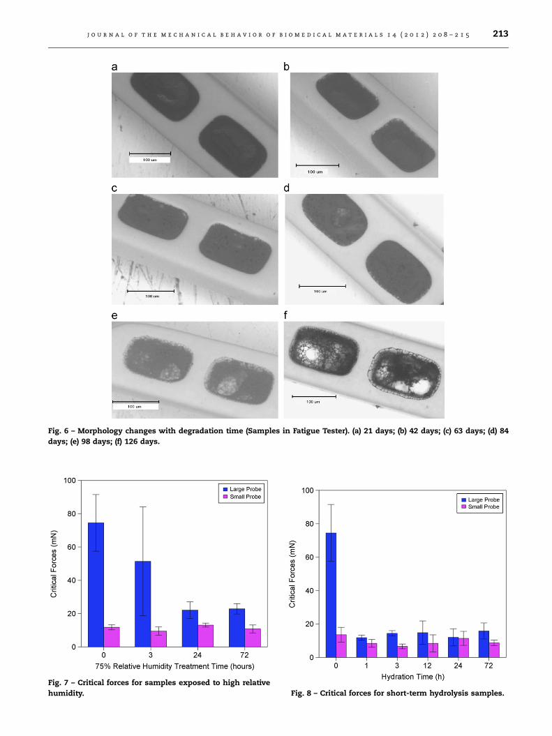
Fig. 6 – Morphology changes with degradation time (Samples in Fatigue Tester). (a) 21 days; (b) 42 days; (c) 63 days; (d) 84
days; (e) 98 days; (f) 126 days.
Fig. 7 – Critical forces for samples exposed to high relative
humidity. Fig. 8 – Critical forces for short-term hydrolysis samples.
j o u r n a l o f t h e m e c h a n i c a l b e h a v i o r o f b i o m e d i c a l m a t e r i a l s 1 4 ( 2 0 1 2 ) 2 0 8 – 2 1 5 213

Fig. 9 – Critical forces for long-term hydrolysis samples in
test tubes.
Fig. 10 – Critical forces for long-term hydrolysis samples in
fatigue tester.
Fig. 11 – Critical force for samples undergone freeze–thaw
cycles.
j o u r n a l o f t h e m e c h a n i c a l b e h a v i o r o f b i o m e d i c a l m a t e r i a l s 1 4 ( 2 0 1 2 ) 2 0 8 – 2 1 5214
under mechanically loaded conditions for up to 126 days) are
presented in Figs. 5, 6 and 10. Samples fatigued for longer than
84 days exhibited significant porosity in the reservoirs, generat-
ing adhesion/cohesion data below the detection limit of the
tester. GPC results (Fig. 5) demonstrated that the molecular
weight decreased slightly faster than the vial samples, which
confirmed results of earlier studies (Fan et al., 2008).
SEM analysis showed that, during the initial 21 days, no
detectable changes were observed in the morphology of the
formulation (Fig. 6a). However, after 42 days, progressive pore
formation and thinning in the center of the formulation were
detected (Fig. 6b–f). The lag between molecular weight and
morphological changes has been extensively reported in the
literature (Engineer et al., 2011; Fan et al., 2008). The critical
forces to failure using the large probe are plotted in Fig. 10.
The critical forces decreased rapidly from approximately
100 mN when fresh, to about 18 mN after 21 days, and to
1 mN after degradation for 84 days, when the formulation
inlay exhibited significant porosity (Fig. 6d).
Similar to the test tube samples, the failure modes were
clearly adhesive for the samples in the fatigue tester up to 42
days, but cohesive for samples degraded further. As shown by
the SEM images (Fig. 6), the polymer has degraded to an extent
that not much material was left within the formulation inlay.
4.5. Freeze–thaw cyclic thermal loading
The force measurements from both the vial and fatigue tester
studies showed profound effects of water uptake on the
adhesion of the formulation to the metallic reservoir in
NEVOTM SES. All samples from these two studies were pre-
served using nitrogen drying and freezing at �20 1C. It was
important to demonstrate that these storage conditions did not
impact the results. The samples were, therefore, subjected to
water, and then dried with nitrogen and stored in a freezer.
To simulate extreme sample handling procedures, stents
experienced up to four freeze–thaw cycles and then were
tested for adhesion. The critical forces for failure measured
using the large probe were plotted in Fig. 11. Hydration of
fresh stents reduced the adhesion forces from 100 mN to
approximately 15 mN, confirming previous results.
For samples after one freeze–thaw cycle, the forces measured
were approximately 18 mN. This value was not statistically
different than the value for samples that have not undergone
storage. Similar to samples exposed to brief water submersion,
the small probe induced cohesive failures for all tests, while the
large probe induced adhesive failures for all tests. Furthermore,
samples exposed to additional freeze–thaw cycles were ana-
lyzed. No practical difference was observed, as a result of
additional handling. These excluded the effects of storage factor
in the hydrolyzed samples from vials and the fatigue tester.
4.6. Implications
In this study we ensured that sample handling did not impact
the adhesion/cohesion measurements. The results demon-
strated that an initial exposure to water led to a significant
decrease in cohesive and adhesive strengths of the NEVOTM

j o u r n a l o f t h e m e c h a n i c a l b e h a v i o r o f b i o m e d i c a l m a t e r i a l s 1 4 ( 2 0 1 2 ) 2 0 8 – 2 1 5 215
SES. Further exposure to water, which resulted in formulation
degradation, reduced adhesive and cohesive strengths at a
much slower rate. In the end, mass loss and porosity resulted
in measurements below the limit of detection. The same
trends were observed in stents subjected to degradation
under mechanical/thermal cycling conditions. In the pre-
sence and absence of mechanical/thermal loading, compar-
able adhesion/cohesion results were observed.
The results showed that the decrease in mechanical
strengths was primarily due to the initial exposure to water.
Based on the data, it appears that water absorption had
the greatest impact. Remarkably, the subsequent greater
progress of ester hydrolysis, which was accompanied by dra-
matic morphological changes, as seen by SEM, did not seem to
have a significant impact on the cohesive and adhesive
strengths.
Furthermore, the application of mechanical load seemed to
have little or no effect on the adhesive and cohesive strengths
of the stents, even though the molecular weight decrease
occurred at a faster rate. So we conclude that water absorp-
tion is the most important phenomenon.
Hence, the current results suggest that the water absorbed
by the interface (between the bulk of the formulation and the
metallic reservoir) played a major role in the decrease of
mechanical strengths of the NEVOTM SES. This layer of water
seems to bind irreversibly to the interface, as our attempts to
remove the water did not result in further changes in
cohesive or adhesive strengths.
5. Summary and concluding remarks
This paper presents the results of a mechanical (cohesive and
adhesive) strength characterization of NEVOTM SES during the
time course of formulation degradation. A micron-scale push-
out testing method was used to measure the mechanical
strengths. SES exposed to humidity, brief water submersion
and long-term hydrolysis (with and without mechanical loading)
were tested for mechanical strengths. Molecular weight and
morphology changes in long-term hydrolysis were also investi-
gated. Despite the ever-changing molecular weight and mor-
phology, the mechanical strength loss occurred largely during
the initial hours of exposure to water. The mechanical strengths
then reached a plateau before decreasing ultimately to zero, as
degradation of the formulation proceeded to completion.
r e f e r e n c e s
Burke, M., Clarke, B., Rochev, Y., Gorelov, A., Carroll, W., 2008.Estimation of the strength of adhesion between a thermo-responsive polymer coating and nitinol wire. Journal of Mate-rials Science: Materials in Medicine 19, 1971–1979.
Colombo, A., Karvouni, E., 2000. Biodegradable stents: fulfillingthe mission and stepping away. Circulation 102 (4), 371–373.
Engineer, C., Parikh, J., Raval, A., 2011. Effect of copolymer ratioon hydrolytic degradation of poly(lactide-co-glycolide) fromdrug eluting coronary stents. Chemical Engineering Researchand Design 89, 328–334.
Fan, Y.B., Li, P., Zeng, L., Huang, X.J., 2008. Effects of mechanicalload on the degradation of poly(D,L-lactic acid) foam. PolymerDegradation and Stability 93, 677–683.
Fattori, R., Piva, T., 2003. Drug-eluting stents in vascular inter-vention. Lancet 361, 247–249.
Li, Y., Bhindi, R., Khachigian, L.M., 2011. Recent developments indrug-eluting stents. Journal of Molecular Medicine 89,545–553.
Lu, L., Peter, S.J., Lyman, M.D., Lai, H.L., Leite, S.M., Tamada, J.,Uyama, S., Vacanti, J.P., Langer, R., Mikos, A.G., 2000. In vitroand in vivo degradation of porous poly(DL-lactic-co-glycolicacid) foams. Biomaterials 21, 1837–1845.
Park, T.G., 1995. Degradation of poly(lactic-co-glycolic acid)microspheres: effect of copolymer composition. Biomaterials16, 1123–1130.
Price, G.S., Lapointe, J.M., Luk, A., Guy, G.L., SteeseBradley, G.,Dooley, J., Rogers, C., 2009. Vascular safety of overlappingsirolimus-eluting reservoir technology stents in the porcinecoronary artery model. Journal of the American College ofCardiology 53 (10) A74–A74.
Shan, W.L., Du, J., Hampp, E.P., Li, H., Papandreou, G., Maryanoff,C.A., Soboyejo, W.O., 2012. Adhesion and cohesion of struc-tures containing suspended microscopic polymeric films. ActaBiomaterialia 8 (4), 1469–1480.
Soares, J.S., Moore, J.E., Rajagopal, K.R., 2010. Modeling of defor-mation-accelerated breakdown of polylactic acid biodegrad-able stents. Journal of Medical Devices 4 (4), 041007.
Strickler, F., Richard, R., McFadden, S., Lindquist, J., Schwarz, M.C.,Faust, R., Wilson, G.J., Boden, M., 2010. In vivo and in vitrocharacterization of poly(styrene-b-isobutylene-b-styrene)copolymer stent coatings for biostability, vascular compat-ibility and mechanical integrity. Journal of Biomedical Materi-als Research A 92 (2), 773–782.
Tang, F., Ding, N., Pacetti, S. Nano-scratch adhesion evaluation ofthe XIENCE VTM drug eluting stent coating. Society forBiomaterials 2008 Annual Meeting.
Wolf, K.V., Zong, Z., Meng, J., Orana, A., Rahbar, N., Balss, K.M.,Papandreou, G., Maryanoff, C.A., Soboyejo, W.O., 2008. Aninvestigation of adhesion in drug-eluting stent layers. Journalof Biomedical Materials Research 87A, 272–281.