Defining functional areas in individual human brains...
13
Defining functional areas in individual human brains using resting functional connectivity MRI ☆ Alexander L. Cohen, a, ⁎ Damien A. Fair, a Nico U.F. Dosenbach, b Francis M. Miezin, a,b Donna Dierker, c David C. Van Essen, c Bradley L. Schlaggar, a,b,c,d and Steven E. Petersen a,b,c,e, ⁎ a Department of Neurology, Washington University School of Medicine, St. Louis, MO 63110, USA b Department of Radiology, Washington University School of Medicine, St. Louis, MO 63110, USA c Department of Anatomy and Neurobiology, Washington University School of Medicine, St. Louis, MO 63110, USA d Department of Pediatrics, Washington University School of Medicine, St. Louis, MO 63110, USA e Department of Psychology, Washington University School of Medicine, St. Louis, MO 63130, USA Received 24 September 2007; revised 8 December 2007; accepted 24 January 2008 Available online 25 March 2008 The cerebral cortex is anatomically organized at many physical scales starting at the level of single neurons and extending up to functional systems. Current functional magnetic resonance imaging (fMRI) studies often focus at the level of areas, networks, and systems. Except in restricted domains, (e.g., topographically-organized sensory re- gions), it is difficult to determine area boundaries in the human brain using fMRI. The ability to delineate functional areas non-invasively would enhance the quality of many experimental analyses allowing more accurate across-subject comparisons of independently identified functional areas. Correlations in spontaneous BOLD activity, often referred to as resting state functional connectivity (rs-fcMRI), are especially promising as a way to accurately localize differences in patterns of activity across large expanses of cortex. In the current report, we applied a novel set of image analysis tools to explore the utility of rs-fcMRI for defining wide-ranging functional area boundaries. We find that rs-fcMRI patterns show sharp transitions in correlation patterns and that these putative areal boundaries can be reliably detected in individual subjects as well as in group data. Additionally, combining surface-based analysis techniques with image processing algorithms allows automated mapping of putative areal boundaries across large expanses of cortex without the need for prior information about a region's function or topography. Our approach reliably produces maps of bounded regions appropriate in size and number for putative functional areas. These findings will hopefully stimulate further methodological refinements and validations. © 2008 Elsevier Inc. All rights reserved. Introduction In Churchland and Sejnowski's famous diagram showing the levels of neuroanatomical organization, a level labeled “maps” is interposed between networks (i.e., columns) and systems (Church- land and Sejnowski, 1991). This level was called “maps” because many of the most accurately defined entities at this scale (~1 cm) are topographically organized cortical areas. Classic examples include the multiple retinotopic maps of primary and extrastriate visual cortex, where each map constitutes a separate representation of the visual field and contains neurons with a distinctive collection of functional characteristics. Organization at this scale is not limited to visual cortical regions, nor to topographically organized somatosensory (Clark et al., 1988) or auditory (Langers et al., 2007) maps. Distinct subregions have been reported throughout the cortex, including motor (Strick, 1988), and orbitofrontal (Carmichael and Price, 1994) cortex as well as the complete hemispheric partitioning schemes of Brodmann (1909) and other classical anatomists. Although topography is not seen in every region, it can be combined with other attributes, as suggested by Passingham et al. (2002) for frontal and motor regions, to provide a distinct “fingerprint” that can be used for the identification of individual regions. For the remainder of this report, regions that represent separable functional domains of cortex will be referred to as “functional areas”. This is in distinction to a more general term “region” or “region of interest”, which may encompass all or part of several functional areas. Because functional areas possess unique combinations of inputs, outputs, and internal structure, each functional area is thought to make a distinct contribution to information processing. Thus, the study of each area's normal function, developmental trajectory, and modified responses following loss or injury, would be greatly aided by the ability to accurately and reliably define the location and boundaries of functional areas in individual living humans. www.elsevier.com/locate/ynimg NeuroImage 41 (2008) 45 – 57 ☆ Sentence summary: Resting state functional connectivity can be used in a semi-automated fashion to delineate putative functional area borders across the human cortical surface. ⁎ Corresponding authors. Washington University School of Medicine, Department of Neurology-Campus Box 8111, 660 S. Euclid, St. Louis, MO 63110, USA. Fax: +1 314 362 6110. E-mail addresses: [email protected], [email protected] (A.L. Cohen), [email protected] (S.E. Petersen). Available online on ScienceDirect (www.sciencedirect.com). 1053-8119/$ - see front matter © 2008 Elsevier Inc. All rights reserved. doi:10.1016/j.neuroimage.2008.01.066
Transcript of Defining functional areas in individual human brains...

www.elsevier.com/locate/ynimg
NeuroImage 41 (2008) 45–57Defining functional areas in individual human brains using restingfunctional connectivity MRI☆
Alexander L. Cohen,a,⁎ Damien A. Fair,a Nico U.F. Dosenbach,b Francis M. Miezin,a,b
Donna Dierker,c David C. Van Essen,c Bradley L. Schlaggar,a,b,c,d and Steven E. Petersena,b,c,e,⁎
aDepartment of Neurology, Washington University School of Medicine, St. Louis, MO 63110, USAbDepartment of Radiology, Washington University School of Medicine, St. Louis, MO 63110, USAcDepartment of Anatomy and Neurobiology, Washington University School of Medicine, St. Louis, MO 63110, USAdDepartment of Pediatrics, Washington University School of Medicine, St. Louis, MO 63110, USAeDepartment of Psychology, Washington University School of Medicine, St. Louis, MO 63130, USA
Received 24 September 2007; revised 8 December 2007; accepted 24 January 2008Available online 25 March 2008
The cerebral cortex is anatomically organized at many physical scalesstarting at the level of single neurons and extending up to functionalsystems. Current functional magnetic resonance imaging (fMRI)studies often focus at the level of areas, networks, and systems. Exceptin restricted domains, (e.g., topographically-organized sensory re-gions), it is difficult to determine area boundaries in the human brainusing fMRI. The ability to delineate functional areas non-invasivelywould enhance the quality of many experimental analyses allowingmore accurate across-subject comparisons of independently identifiedfunctional areas. Correlations in spontaneous BOLD activity, oftenreferred to as resting state functional connectivity (rs-fcMRI), areespecially promising as a way to accurately localize differences inpatterns of activity across large expanses of cortex. In the currentreport, we applied a novel set of image analysis tools to explore theutility of rs-fcMRI for defining wide-ranging functional areaboundaries. We find that rs-fcMRI patterns show sharp transitionsin correlation patterns and that these putative areal boundaries can bereliably detected in individual subjects as well as in group data.Additionally, combining surface-based analysis techniques with imageprocessing algorithms allows automated mapping of putative arealboundaries across large expanses of cortex without the need for priorinformation about a region's function or topography. Our approachreliably produces maps of bounded regions appropriate in size andnumber for putative functional areas. These findings will hopefullystimulate further methodological refinements and validations.© 2008 Elsevier Inc. All rights reserved.
☆ Sentence summary: Resting state functional connectivity can be used ina semi-automated fashion to delineate putative functional area borders acrossthe human cortical surface.⁎ Corresponding authors. Washington University School of Medicine,
Department of Neurology-Campus Box 8111, 660 S. Euclid, St. Louis, MO63110, USA. Fax: +1 314 362 6110.
E-mail addresses: [email protected], [email protected](A.L. Cohen), [email protected] (S.E. Petersen).
Available online on ScienceDirect (www.sciencedirect.com).
1053-8119/$ - see front matter © 2008 Elsevier Inc. All rights reserved.doi:10.1016/j.neuroimage.2008.01.066
Introduction
In Churchland and Sejnowski's famous diagram showing thelevels of neuroanatomical organization, a level labeled “maps” isinterposed between networks (i.e., columns) and systems (Church-land and Sejnowski, 1991). This level was called “maps” becausemany of the most accurately defined entities at this scale (~1 cm) aretopographically organized cortical areas. Classic examples includethe multiple retinotopic maps of primary and extrastriate visualcortex, where each map constitutes a separate representation of thevisual field and contains neurons with a distinctive collection offunctional characteristics.
Organization at this scale is not limited to visual cortical regions,nor to topographically organized somatosensory (Clark et al., 1988)or auditory (Langers et al., 2007) maps. Distinct subregions havebeen reported throughout the cortex, including motor (Strick, 1988),and orbitofrontal (Carmichael and Price, 1994) cortex as well as thecomplete hemispheric partitioning schemes of Brodmann (1909)and other classical anatomists. Although topography is not seen inevery region, it can be combined with other attributes, as suggestedby Passingham et al. (2002) for frontal and motor regions, to providea distinct “fingerprint” that can be used for the identification ofindividual regions.
For the remainder of this report, regions that represent separablefunctional domains of cortex will be referred to as “functional areas”.This is in distinction to a more general term “region” or “region ofinterest”, which may encompass all or part of several functionalareas. Because functional areas possess unique combinations ofinputs, outputs, and internal structure, each functional area is thoughtto make a distinct contribution to information processing. Thus, thestudy of each area's normal function, developmental trajectory, andmodified responses following loss or injury, would be greatly aidedby the ability to accurately and reliably define the location andboundaries of functional areas in individual living humans.
46 A.L. Cohen et al. / NeuroImage 41 (2008) 45–57
Four criteria have been proposed for defining cortical areas,based mainly on studies of non-human primates (Felleman and VanEssen, 1991; Van Essen, 1985): Function (as defined by lesion-behavior or having neurons whose functional properties are distinctfrom neighboring cortex), Architectonics (having unique arrange-ments of cells, myelin density, and/or combinations of chemicalmarkers, etc.), Connections (having a different combination ofinputs and outputs from neighboring cortex), and Topography(having a topographic map that can be used to define boundaries, forexample between primary visual cortex and V2).
Unfortunately, the current ability to define functional areas bythese criteria in human cerebral cortex is inadequate. fMRI andlesion studies provide some localization ability, but their precision isrelatively low. Historic architectonic partitioning schemes, such asthe rendition of areas introduced by Korbinian Brodmann, unfor-tunately often contain incorrect boundaries and do not addressindividual variation of location and extent (see Van Essen andDierker, 2007). Promising methodologies such as architectonicmeasurements in living humans (Scheperjans et al., 2007) andconnectional studies using DTI (Klein et al., 2007) are currently stilllimited to a small set of areas. Mapping of topographic organizationis mostly restricted to sensory and motor areas.
Recently,measures of correlation between resting brain regions (so-called resting-state functional connectivity, or rs-fcMRI) have demon-strated promise in describing boundaries between functional areas inlimited regions of cortex (Margulies et al., 2007). Resting statefunctional connectivity is a method for evaluating regional interactionsthat occur when a subject is not performing an explicit task (Achardet al., 2006; Beckmann et al., 2005; Biswal et al., 1995; Damoiseauxet al., 2006; Dosenbach et al., 2007; Fair et al., 2007a,b; Fox et al.,2005; Greicius et al., 2003; Lowe et al., 1998; Nir et al., 2006; Salvadoret al., 2005). It is based on the discovery that low-frequency (b~0.1Hz)BOLD fluctuations in distant, but apparently functionally related greymatter regions, show strong correlations at rest (Biswal et al., 1995;Damoiseaux et al., 2006; Lowe et al., 1998; Nir et al., 2006). These lowfrequency BOLD fluctuations are presumed to relate to spontaneousneural activity (Biswal et al., 1995; Leopold et al., 2003; Nir et al.,2006). Additionally, since rs-fcMRI does not require active engage-ment in a behavioral task, it unburdens experimental design, subjectcompliance, and training demands, making it attractive for studies ofdevelopment, aging, and clinical populations (Bokde et al., 2006;Castellanos et al., 2008; Greicius et al., 2007; Greicius et al., 2004;Rombouts and Scheltens, 2005; Tian et al., 2006;Whalley et al., 2005).
Cross-correlating the time series of a particular brain region (seedregion) with all other voxels in the brain can illuminate which voxelsare “functionally connected” with the seed region, in that their timecourses are highly correlated. For example, a seed region in the leftprimary motor cortex shows “functional connections” with the rightprimary motor cortex as well as supplementary motor area (SMA)(Biswal et al., 1995) and other regions. These voxel-wise correlationmaps can be generated for individual subjects (Fox et al., 2006). Thespecific mechanisms relating neural activity to these very slow(N10 s) fluctuations in the BOLD response are not known, but thespatial patterns are similar to those revealed by the functionalactivation data: regions that co-activate tend to have correlated rs-fcMRI signals while regions that become negative when a seedregions activates tend to be negatively correlated with the seedregion (Fox et al., 2006; Greicius et al., 2003).
Because “seeds” can be placed in any cortical or subcorticallocation, development of methods for analyzing these correlationmaps could provide a basis for delineating the location and
boundaries of a large number of functional areas in single individuals.The analyses presented belowprovide evidence that rs-fcMRI can aidin the delineation of a large number of functional area boundaries inindividual human cortex. They show that (i) changes in correlationmaps occur abruptly as the seed locationmoves systematically acrossthe cortical sheet, suggesting the presence of a boundary rather than asmooth gradation; (ii) these transitions occur in many locations inindividual subjects; (iii) customized image processing techniques canbe used to identify putative boundaries and bounded regions acrosslarge expanses of cortex; (iv) the boundaries appear reliable whenassessed with independent measurements; (v) the overall map has theappropriate granularity to reflect area-level cortical parcellation.
Methods
Overview
The aim of the analysis stream presented here is to identifylocations on the cortex where the pattern of rs-fcMRI changesrapidly, potentially representing boundaries between functionalareas. Our approach utilizes established fcMRI voxel-wise correla-tion methods, coupled to several novel analysis techniques that areperformed on a surface representation of the cortex. These includeestablished edge detection and image segmentation algorithms usedin computer vision and image analysis programs. These surface-based operations treat the brain as a 2D sheet, while volume-basedanalyses treat the brain as a 3D volume.
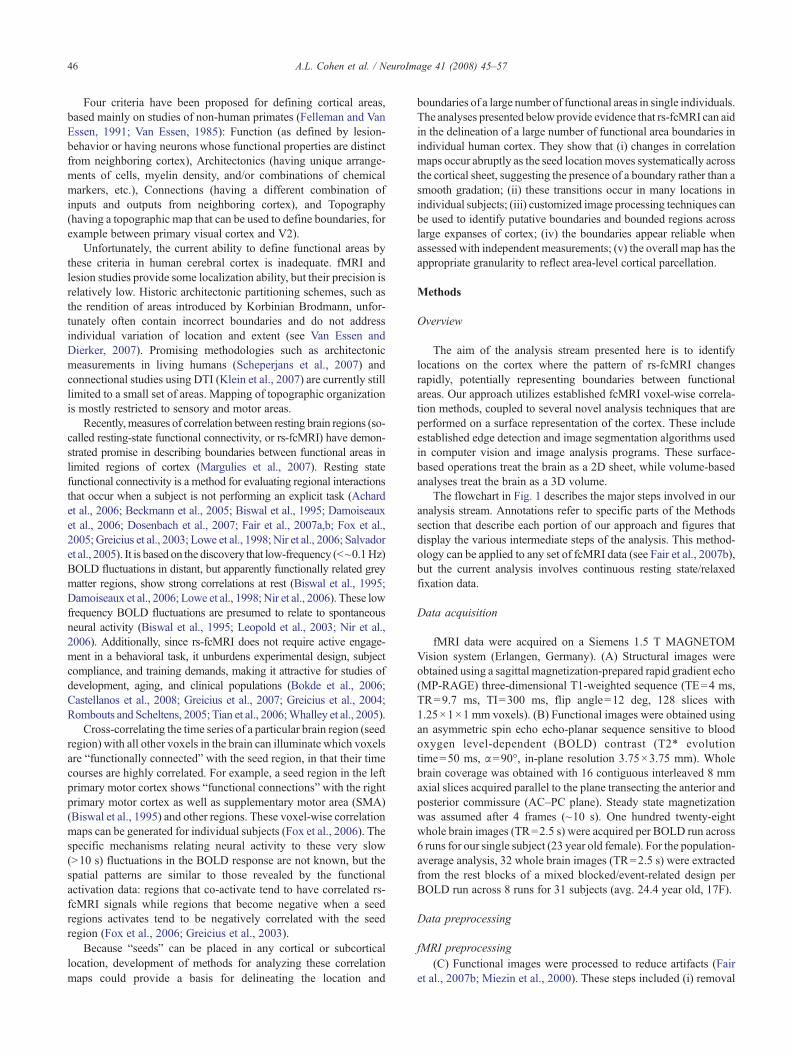
The flowchart in Fig. 1 describes the major steps involved in ouranalysis stream. Annotations refer to specific parts of the Methodssection that describe each portion of our approach and figures thatdisplay the various intermediate steps of the analysis. This method-ology can be applied to any set of fcMRI data (see Fair et al., 2007b),but the current analysis involves continuous resting state/relaxedfixation data.
Data acquisition
fMRI data were acquired on a Siemens 1.5 T MAGNETOMVision system (Erlangen, Germany). (A) Structural images wereobtained using a sagittal magnetization-prepared rapid gradient echo(MP-RAGE) three-dimensional T1-weighted sequence (TE=4 ms,TR=9.7 ms, TI=300 ms, flip angle=12 deg, 128 slices with1.25×1×1 mm voxels). (B) Functional images were obtained usingan asymmetric spin echo echo-planar sequence sensitive to bloodoxygen level-dependent (BOLD) contrast (T2* evolutiontime=50 ms, α=90°, in-plane resolution 3.75×3.75 mm). Wholebrain coverage was obtained with 16 contiguous interleaved 8 mmaxial slices acquired parallel to the plane transecting the anterior andposterior commissure (AC–PC plane). Steady state magnetizationwas assumed after 4 frames (~10 s). One hundred twenty-eightwhole brain images (TR=2.5 s) were acquired per BOLD run across6 runs for our single subject (23 year old female). For the population-average analysis, 32 whole brain images (TR=2.5 s) were extractedfrom the rest blocks of a mixed blocked/event-related design perBOLD run across 8 runs for 31 subjects (avg. 24.4 year old, 17F).
Data preprocessing
fMRI preprocessing(C) Functional images were processed to reduce artifacts (Fair
et al., 2007b; Miezin et al., 2000). These steps included (i) removal

Fig. 1. Flowchart outlining the analysis stream presented here and the techniques involved. Bolded letters refer to specific portions of the Methods section thatdescribe each procedural operation. Examples of several steps in the procedure are denoted by the figure where they can be found.
47A.L. Cohen et al. / NeuroImage 41 (2008) 45–57
of a central spike caused by MR signal offset, (ii) correction of oddvs. even slice intensity differences attributable to interleavedacquisition without gaps, (iii) correction for head movement withinand across runs, and (iv) across-run intensity normalization to awhole brain mode value of 1000.
Several of the temporal nuisance signals that need to beregressed out to examine the resting state signal are related toanatomical structures, such as white matter and the ventricles (seers-fcMRI preprocessing below). This entails registering the data foreach subject to an atlas space, so that common imaging masks andfcMRI seeds can be used to define these nuisance signals in eachsubject. This transformation of the functional data to atlas space, inthis case, 711-2B (Buckner et al., 2004; Talairach and Tournoux,1988; Ojemann et al., 1997), was computed for each individual viathe MP-RAGE scan. Each run then was resampled in atlas space(Talairach and Tournoux, 1988) on an isotropic grid (3 mm voxels)combining movement correction and atlas transformation in oneinterpolation (Lancaster et al., 1995; Snyder, 1996). This singleinterpolation procedure eliminates blurring that would be intro-duced by multiple interpolations. All subsequent operations wereperformed on the atlas-transformed volumetric time series.
rs-fcMRI preprocessing(D) Pre-processing for functional connectivity analyses was
performed on the fMRI data, as in Fox et al. (2005), to optimize the
time-series data and remove spurious variance. These steps includeremoval of the linear trend, temporal band-pass filtering (0.009 Hzbfb0.08 Hz), spatial smoothing (6 mm full width at half maximum(FWHM)), as well as regression of several “nuisance” signals andtheir time-based first order derivatives, including six motionparameters, and whole brain, ventricular, and white matter signals.
Surface-based analysis(E) The structural MRI volume for the single-subject analysis
was spatially normalized to the 711-2B volumetric MRI atlas(Lancaster et al., 1995; Snyder, 1996) and resampled to 1 mmvoxels. Segmentation of the cortical mid-thickness as well as surfacereconstruction was done using Caret 5.3 software (Van Essen et al.,2001) (http://brainmap.wustl.edu/caret/). Surface flattening wasaccomplished by making cuts along five standardized trajectoriesthat help minimize distortions (see Fig. 2 in Van Essen, 2005). Thisflattened surface was then used to generate a grid of seed points, orpatch, which was then used to generate rs-fcMRI correlationmaps asdescribed below.
rs-fcMRI boundary generation
Cortical seed and rs-fcMRI correlation map generation(F) On the flat map of the subject's cortex, a Cartesian 3 mm point
grid was created using Caret to define a set of seeds representing a

Fig. 2. Shown are transverse sections and lateral hemispheric views, mapped to the PALS human cortical atlas (Van Essen, 2005), showing the functionalconnectivity patterns of regions of interest in the angular (upper slice and lateral view) and supramarginal gyrus (lower slice and lateral view). Highlighted(circles) are a few of the salient differences. Seed regions are indicated with filled dark blue circles. The strength of positive and negative correlations are shownin warm and cool colors, respectively.
48 A.L. Cohen et al. / NeuroImage 41 (2008) 45–57
patch of the cortical surface that respects cortical folding patterns (asshown in Fig. 5A). This allowed us to treat the cortex as a 2D structureand use standard 2D image processing techniques. (G) Thecorresponding 3D stereotactic coordinates for each Cartesian gridpoint were used to generate 3 mm diameter spherical regions ofinterest around each volumetric seed voxel. This sampling densityprovides a fine-grained map without excessive oversampling of thefMRI data (3 mm voxel size, but with 6 mm FWHM smoothing).While each seed point is 3 mm apart on the flattened representation,the folding pattern of the brain results in some seeds being further than3 mm apart in the underlying volume, while others are closer than3 mm in the underlying volume. (H) For each seed, volumetriccorrelation maps were generated as in Fox et al. (2005) and Fair et al.(2007b) by correlating the time course of this region of interest withthe time courses of all other voxels over the entire volume of the brain.This creates a volumetric correlation map for each seed, where theintensity at each voxel is the Fisher Z-transformed correlation (r)between that voxel and the seed region for the volume.
eta2 matrix creation for each seed(I) To compute the similarity between seed locations, an eta2
coefficient was calculated for every seed pair. eta2 is equal to thefraction of the variance in one signal accounted for by variance in asecond signal where comparisons are done on a point by pointbasis. The more similar two signals, or in this case, images, are thehigher the eta2 coefficient between them. eta2 can vary in valuefrom 0 (no similarity) to 1 (identical). To determine the similarityor difference between the large-scale correlational structure of twoseed locations, eta2 is calculated between the two volumetric
correlation maps (a and b) generated from these two seed locationsand is equal to:
eta2 ¼ 1� SSWithin
SSTotal¼ 1�
Pni¼1
ai � mið Þ2þ bi � mið Þ2h i
Pni¼1
ai � PM
� �2þ bi � PM
� �2h i ð1Þ
where ai and bi represent the values at position i in maps a and b,respectively.mi is the mean value of the two images at position i, (ai+bi) /2, and M-bar is the grand mean value across the mean image, m,or across all locations in both correlation maps. eta2 thus measures thedifference in the values at corresponding points in the two images, notstrictly whether the points vary in similar ways, and can detectdifferences and similarities in the volumetric correlation maps usinginformation from all of the voxels of the entire volume. The eta2
coefficients are stored in a series of matrices (the same size and shapeas the patch of seeds) such that each seed has a corresponding matrixrepresenting the eta2 coefficients of that seed's volumetric correlationmap compared to the volumetric correlation maps of all other seeds inthe patch (as shown in Fig. 5B).
We use eta2 to compare images instead of correlation, r, becauseour goal is to quantify the difference or similarity of the two images,not the correlational relationship between them. While correlation isoften used for similarity description, there are instances where thecorrelation coefficient between images is unaffected by changes inthe two images which make them more or less similar from eachother. Two examples where this is readily apparent are scaling andoffset; if the value of each voxel in one map is exactly double thevalue of another, they will have a correlation coefficient of 1, but are

49A.L. Cohen et al. / NeuroImage 41 (2008) 45–57
still different from one another at every point and will have eta2
values that may be much less than unity. Similarly, if the value ofeach voxel in one map is 100 units greater than another, they willagain have a correlation coefficient of 1, even though every voxel isdifferent. In fact, the correlation coefficient will not change from 1 ifthe scaling factor increases or decreases in the first case, or if theoffset factor increases or decreases in the second case. In both cases,however, eta2 will measure and detect these differences andwill varyas a scaling factor, offset, or another form of variation changes thedifference between the two maps. The eta2 coefficient will onlyequal 1 if they are, in fact, identical at every point.
Edge detection algorithms(J) Since the aforementioned matrix of eta2 coefficients (i.e., eta2
profile) is a 2D array of values across the cortical surface, it can betreated as flat image data. To find salient edges in these arrays, theCanny edge detection algorithm (Canny, 1986), as implemented inthe Image Processing Toolbox (v7.2) of the MATLAB softwaresuite, was applied to each seed's eta2 profile ‘image’. The Cannymethod smoothes the image with a Gaussian filter to reduce noise,and then creates a gradient image that locates regions with highspatial derivatives. High gradient values represent locations wherethe original image was rapidly changing (i.e., peaks in the firstderivative). After eliminating pixels in the 2D array that are not localmaxima in the gradient image, the algorithm tracks along thehighlighted regions of the image and, using high and low thresholds,categorizes each location as an edge or not. To prevent hysteresis, ifthe magnitude of the pixel is below the low threshold, it is set to zero,
Fig. 3. Panels A–C show the locations of the angular (blue) and supramarginal (red)shows some of the connectivity maps derived from the series of seed regions. Epatterns. Panel D (lower panel) represents the eta2 values derived when comparingmaps (last red line), and so forth for all maps.
while if themagnitude is above the high threshold, it is considered anedge. If the magnitude of the pixel is between the two thresholds,then the location is only considered an edge if there is a neighboringpixel that itself has a gradient above the high threshold.
Our current implementation uses the defaultMATLAB algorithmto generate these two thresholds such that the high threshold iscalculated to be the lowest value at which no more than 30% of thepixels are detected as edges, and the low threshold is defined as 40%of the high threshold. The use of edge detection here is purely to findthe gradient peaks that are spatially stable across short stretches ofeach of the eta2 profile ‘images’. Thus, the specific thresholds for theedge detection algorithm do not have to be manually set each time.The primary goal is to identify and differentiate locations withstrong, spatially coherent peaks as being different from locations thatare relatively smooth or have incoherent gradient peaks, across someor most of the eta2 profiles. The present adaptive threshold algorithmevidently performs this function adequately.
The result of processing the eta2 profile set with an edge detectionalgorithm is a set of binary images representing the locations of rapidchanges in each grid point's eta2 profile (blue overlay in Fig. 5C).
Putative areal boundary map generation(K) Since the edge determination is binary, averaging across the
entire set of seed matrix images at each location gives the relativelikelihood that a particular location was determined to be an edgeacross the set of seed matrices. This gives a probabilistic orputative edge location map in which the intensity represents howlikely a location is actually a functional border (Fig. 5D).
regions. Black dots in C indicate the seed regions used. Panel D (upper panel)ncircled are particular differences that highlight the changing connectivitythe AG map with all other maps (first blue line), the SMG map with all other

50 A.L. Cohen et al. / NeuroImage 41 (2008) 45–57
Putative functional area identification(L) Since the intensity of the putative edge map represents the
likelihood that a given location is not a member of a functional area,our data can be transformed into regions of interest that representputative functional areas using the morphology-based watershedtransform (Vincent and Soille, 1991). This method treats each lowintensity (low probability of being an edge) region as a ‘valley’ that
Fig. 4. Panel A displays the eta2 coefficients between each seed point's correlationPanel B shows the location of the line of seed points on the left hemisphere, as well aThe medial wall hole is shown in orange, with ‘a’ and ‘p’ designating anterior andpurple on both the inflated medial view and the flattened view. Panel C shows the
progressively fills until reaching ambiguous locations between regions(in this case putative edges in the overall edge map) (Fig. 7).
Results
We first demonstrate that rs-fcMRI patterns can be strikinglydifferent in a population-average data set even when the seed regions
maps, as in Fig. 3D. Triangle and circle designate locations of rapid change.s the nearby artificial ‘cuts’ created during the process of flattening the cortex.posterior ends of the anterior medial wall cut. The cingulate cut is shown inresults of hierarchical clustering the eta2 profiles shown in panel A.

51A.L. Cohen et al. / NeuroImage 41 (2008) 45–57
are relatively close (2.08 cm).We then explore the ramifications of thisobservation more systematically in single-subject data, expanding onthe implications this has for defining functional areas across the cortex.
rs-fcMRI of closely apposed seed regions shows putative arealboundaries where map patterns change abruptly
Voxel-wise correlations were performed on interleaved restingstate fMRI data (see Fair et al., 2007b) acquired from 31 healthyadult subjects using functionally defined seed regions (12 mmdiameter spheres surrounding the peaks of activation) placed in thenearby supramarginal (−52, −42, +24) and angular (−49, −62,+29) gyri. These regions of interest, whose centers are separatedby 2.08 cm (vector distance in 3D), were derived from a studyinvestigating the development of lexical processing that showedthese nearby regions to have similar but dissociable developmentalprofiles for a set of lexical tasks (Church et al., 2008).
The angular and supramarginal gyri seed regions (small bluespheres in left hemispheres in Fig. 2) show markedly differentfunctional connectivity profiles, indicating that rs-fcMRI can beremarkably different between nearby functional areas. Regions havingpositive correlations (warm colors) with the angular gyrus seed (toprows of volume and surface views) show very little overlap withregions showing positive correlations with the supramarginal gyrusseed (bottom rows of volume and surface views). Derived from groupdata, these results are consistent with previous studies demonstratingthat correlation patterns are reliable across subjects and investigators.
Fig. 5. Panel A shows the 2D patch of seed regions on this subject’s flattened cortin panels A and D for comparison). The eta2 profile for one of the seeds (blue circautomated edge-detection algorithm that generates borders, blue overlay in panel Cresults in the putative edge map shown in panel D, where intensity of each locatedge.
Although correlation maps from nearby seeds can be quitedifferent, delineating boundaries necessitates that these differencesdo not progress smoothly across the brain, but rather show abruptlocal changes, similar to those seen in connectional anatomy andfunctional properties (e.g., Felleman and Van Essen, 1991; Maunselland Van Essen, 1983). To test for such a transition, a series ofspherical seed regions (3 mm diameter) were generated between thecenters of the supramarginal and angular gyri regions from group data(Figs. 3A and B). The locations for these intermediate regions weredelineated on the PALS atlas flat map and projected to the volume viathe PALS ‘average fiducial surface’ (Van Essen, 2005).
We analyzed the connectivity images for the series of seed regionsby visual inspection and by computing the similarity of eachconnectivity image to each of the other connectivity images using aneta2 coefficient. While a correlation coefficient measures therelationship between changes in two images, the eta2 coefficientprovides a better measure of the overall similarity or differencebetween images (see Methods for details).
As shown in Fig. 3D, the eta2 coefficients demonstrate a transitionbetween the locations of seeds R7, R8, and R9, as the profile of eta2
coefficients drastically change within 1 cm (3 map-mm separationbetween seeds, Van Essen and Drury, 1997). Thus, the eta2 profilescan be divided into three groups: AG-R6 (blue), R10-SMG (red), andan intermediate zone R7-R9 (grey) that is a putative transition regionbetween the other two groups. The transition region found here iswider than the typical transition between architectonically defined
ex (Note: The line of points and boundary locations from Fig. 4 are plottedle) is shown in panel B. Each of these eta2 maps are then analyzed with an. Averaging all of the detected edge maps (binary blue overlay in panel C)ion reflects the fraction of maps in which that location was considered an

Fig. 6. Panel A shows the original patch of the left cingulate cortex shown inFig. 5. Panel B demonstrates that edge locations identified in theneighboring posterior patch align with those in the original patch, eventhough the two data sets do not share seed points or correlation maps. PanelC shows that the independently analyzed overlapping patch is consistentwith the matching regions of A and B.
52 A.L. Cohen et al. / NeuroImage 41 (2008) 45–57
areas (Zilles et al., 2002), but this may reflect individual variability inthe location of areal boundaries in the contributing population.
rs-fcMRI in a single subject can delineate multiple putative arealboundaries simultaneously
Feasibility using single subject data is important, as the groupdata are inherently blurred by imperfect registration across subjects.Thus, a surface-based “fiducial” representation of the cortical mid-thickness was obtained for a single subject using the SureFit corticalsegmentation algorithm available in the CARET software package(Van Essen et al., 2001). Correlation (volume) maps were generatedfor a set of 3 mm diameter spherical seed regions along the leftcingulate sulcus and adjacent medial cortex (3 map-mm separationalong a line on the flat map). For each of the 25 seed locations, itscorrelation map was compared to the correlation maps for the 24other seed locations along this line, yielding a linear profile thatpeaked at unity for the given seed location.
To objectively separate the eta2 profiles into groups, as done inFig. 3, hierarchical clustering analysis was performed on the eta2
profiles to find any strong divisions among the set (Cordes et al., 2002;Dosenbach et al., 2007; Salvador et al., 2005). A ‘1-eta2’ calculationwas used as a distance measure between the profiles. The commonlychosen UPGMA (Unweighted Paired GroupMethod with Arithmeticmean) hierarchical clustering method (Eisen et al., 1998; Handl et al.,2005) sorted the eta2 profiles for each seed's correlationmap into threemain groups (Fig. 4C), which recapitulates the anatomical ordering aswell as the distinct profile shape differences seen in Fig. 4A (bluecurves vs. green curves vs. red curves). Further inspection of theclusters reveals two abrupt changes in the green curves (triangle andcircle locations), which occurred at similar locations for the othercurves, indicating functional transitions that are candidates for arealboundaries. While there is a general decrease in eta2 coefficient withdistance from the comparison point, abrupt changes are concentratedat specific locations along the line regardless of which initialcomparison points are chosen. Thus, rs-fcMRI functional transitionscan be derived from individual subject data, and multiple transitionsare evident even in this relatively limited view.
Sharp transition zones, or “edges”, can be mapped across the 2Dcortical surface using automated image processing techniques
The line-based approach above provides proof of principle thatrs-fcMRI measures can delineate putative cortical boundaries inindividual subjects. However, mapping functional transitionsthroughout the cortex using this approach would be highly time-consuming and inefficient. Therefore, it was important to develop acomputational approach that takes advantage of the information inrs-fcMRI data more efficiently.
Using a 2D grid of seed regions (i.e., a “patch”) on the corticalsurface (Fig. 5A), eta2 coefficients were computed for all pairs ofseed regions within the patch, yielding a 2D eta2 profile map for eachof the seed regions comprising the patch. Each 2D eta2 profile map(Fig. 5B) was processed with a Canny edge detection algorithm(Canny, 1986), a method for rapid automated discrimination ofstrong gradients (edges), creating a binary “edge map” for each seed(Fig. 5C, blue overlay). These binary maps were then averaged togenerate an ‘edge consistency map’ for the grid, where intensity ateach location represents the fraction of maps in which that locationwas considered an edge (Fig. 5D). The line-based and edge-detection-based approaches are consistent with one another. The
transitions obtained in the line analysis described above (circles andtriangles) spatially align with the putative edges determined by theedge consistency analysis, as indicated by the bright red pixelscrossing the line of seed points.
As seen in Fig. 5D, the set of candidate boundaries derived usingthis method extend over regions of the flat map that are appropriatein size and trajectory for cortical area boundaries. Thus, processinglarge portions of the brain can be done much more efficiently usingan automated computational approach. In addition, because theanalysis is sensitive to gradients in any direction along the corticalsheet, it is inherently more appropriate for systematic identificationof cortical area boundaries.
Several putative areas in Fig. 5D are only partially enclosed bythe presently detected boundaries. This might in part reflect a non-

Fig. 7. Panel A shows the rs-fcMRI derived boundaries generated above.Applying a watershed image segmentation algorithm parses the patch intocontiguous non-overlapping regions least likely to be edges (i.e., most likelyto be areas) shown in panel B, which can then be individually identified andlabeled for investigation and validation as shown in panel C.
53A.L. Cohen et al. / NeuroImage 41 (2008) 45–57
optimal thresholding strategy in the current algorithm but it may alsorepresent a lack of differentiation between functional areas based onresting functional connectivity. If two neighboring functional areashave similar connectivity profiles, the eta2 coefficient between mapsin the two areas will be high, and such boundaries may not bedetected by our current methods. Thus, convergence across methodsand a combination of different approaches may be needed toelucidate the entire set of cortical functional areas.
Boundaries generated from adjacent cortical surface patches yieldconsistent results
Since the edge consistency map is derived from the correlationmaps for a particular set of cortical loci, it is conceivable that theresultant pattern is specific to the chosen patch and is unrelated tocortical areal boundaries. We assessed this possibility by analyzingtwo additional patches, or sets of cortical seed points. First, a patch ofdorso-medial cortex adjacent to that used above (blue box, Figs. 6Aand B) was analyzed to test whether an independent data set wouldshow continuity with the pattern of edge locations seen previously.Second, an overlapping patch corresponding to half of the originaldata set and half of the new independent data set (green box, Figs. 6Aand C) was used. The same edge detection analyses were applied toboth new sets to find putative edge locations.
As seen in Fig. 6B, edge consistency maps generated using acompletely separate but adjacent set of seed point sets revealconsistent edges that align with one another. When superimposingthe edge maps from two independent or overlapping patches ontothe same surface, considerable consistency is noted, including thecontinuous boundaries marked with arrows in Figs. 6B and C.These results provide qualitative evidence that our approach canconsistently identify boundary contours across the cortex in asingle human subject.
Generating boundaries allows automatic definition of putativefunctional areas
Since the edge consistency maps show continuity acrossextended regions of cortex, it should be possible to group contiguousseed points surrounded by putative edges into putative functionalareas, using existing image segmentation algorithms. A watershedsegmentation algorithm (Vincent and Soille, 1991) was applied tothe edge consistency map. Fig. 7 demonstrates the progression fromedges (panel A) to bounded and labeled “areas” (panel C).
Using a putative edge map, a patch of cortex can be segmentedinto several bounded and partially bounded areas by a watershedalgorithm. This suggests that rs-fcMRI derived putative edges andstandard imaging segmentation methods should allow parcellationof an individual's cortical surface into putative functional areas.While these bounded areas may in some cases represent only a partof one or more than one functional area, it allows for the generationof ROIs that can be validated using complementary methods.
Discussion
Imaging and functional areas
Since the mid 1980s, functional neuroimaging has facilitatedprogress in cognitive neuroscience—the study of neural substratesunderlying mental processes and behavior. Typically, functional

54 A.L. Cohen et al. / NeuroImage 41 (2008) 45–57
neuroimaging identifies brain regions that are differentially activatedby different task states or affected by specified behavioral events. Weuse the terms “regions” and “regions of interest” advisedly wheneverwe are unsure whether functional imaging has identified differentialactivation in awhole and distinct functional area.We recommend thatthe term “area” be reserved for “functional areas” and that “region” beused for a collection of voxels or otherwise defined region of interest.
One of the overarching goals of functional neuroimaging is to usedifferential activity between conditions to identify specific informa-tion processing operations reflected in separate functional areas (e.g.,Posner et al., 1988). Ascertaining a large-scale collection of func-tional areas in any mammal, let alone humans, is not straightforwardand currently incomplete (Levitt, 2003; Van Essen, 2004a,b).
A fundamental problem in trying to identify functional areas inhumans is that many of the methods used to generate the relativelyprecise definitions available in non-human animals are notavailable for studies in living humans, as noted above. Recently,the use of areal connections to define areas has been employed inhumans. Diffusion tensor imaging (DTI) tractography, whichmeasures the directional diffusion of water within a voxel, canreveal local anisotropic differences in fiber bundles in neighboringregions of cortex in living humans, and was recently used todelineate some human cortical areas (Behrens et al., 2006; Croxsonet al., 2005; Johansen-Berg et al., 2004; Johansen-Berg et al.,2005; Klein et al., 2007). The use of probabilistic fiber bundledifferences in diffusion tractography is in some respects analogousto the use of blunt dissection to identify major fiber bundles inhumans. However, the challenge of accurately dissociating cross-ing bundles of fibers with DTI speaks to the need for a convergingmethod of areal definition.
Since rs-fcMRI measures correlated activity, it might in principlereflect mainly direct (monosynaptic) anatomical connections.Empirically, though, the linkage between highly correlated regionscan evidently be indirect, through one or more intermediate regionsor common external input (Vincent et al., 2007). Even if functionalconnectivity is not directly equivalent to monosynaptic anatomicalconnections, a functional area's history of interaction with otherareas is likely to be consistent across its extent, and distinct betweenseparate areas. Thus, this method may be well suited for delineatingthe location and boundaries of a large number of functional areas.
Overcoming individual variation
Individual variation must be considered when comparing func-tional areas across subjects or populations of subjects (i.e., in cross-sectional development and aging studies, or between patientpopulations). To address this problem, investigators have imple-mented neuroimaging approaches that rely upon improved registra-tion techniques (volumetric or surface-based), presuming thatalignment of anatomical features will improve the alignment offunctional areas. The most advanced registration methods availableattempt to compensate for individual variation in brain surfaceshape, size, and folding pattern (Lyttelton et al., 2007; Van Essen,2005; Van Essen and Dierker, 2007). However, this approach doesnot provide an ideal solution because the location and extent of eachfunctional area vary substantially from person to person, irrespectiveof anatomical landmarks (Amunts et al., 2000; Amunts et al., 1999;Andrews et al., 1997; Uylings et al., 2005; Van Essen et al., 1984).Our current work is performed on a within subject basis, but futureacross subject comparisons will be made through the PALS B12atlas (Van Essen and Dierker, 2007) using CARET which can
account for more individual differences than volumetric averaging.We hope to then directly examine inter-subject variation of areallocation and how this can be used to additionally refine registration.
To overcome the difficulties with regard to individual variability,many studies use a large number of subjects such that, afteralignment, the activated brain region common to the majority ofsubjects will emerge as the active focal point (e.g., Dosenbach et al.,2006). This gives a “best guess” approximation of the centroid of thecommon activated brain region, presumably located within an actualfunctional area (Lancaster et al., 1995), but “blurs” the variability inlocation and extent of areas across individuals.
In some cases, a lack of specificity can lead to regions as farapart as 4 cm in stereotactic space, one quarter the anterior–posterior length of the average brain, being referred to by the samename, and considered as part of the same functional entity (e.g., theapplication of the name “dorsolateral prefrontal cortex” in Kerns,2006, and Luks et al., 2007). However, without the availability ofmore precise regional definitions, this common practice isunavoidable in order to have a common descriptive language.
Another method utilized to overcome individual variability is touse the average activation of many repetitions of a region-specific“localizer” task in individuals (e.g., Swallow et al., 2003). Analysescan then be performed on a subject-by-subject basis using theactivation-delineated peak. However, such localizer tasks exist forrelatively few locations in the brain, and the considerable similarityin the functional properties of many neighboring functional areasoften makes it difficult to differentiate areas based solely on theiractivation to a particular task or stimulus set (e.g., Swallow et al.,2003).
Therefore, functional activations and fcMRI seeds are currentlymost commonly referred to by their stereotactic coordinates oranatomic (gyral and sulcal) locations. The generation of cortex-wide maps of functional areas for individuals would allow for moreaccurate and functionally meaningful labels to be applied withoutrelying on stereotaxis with its added concerns about individualareal variation.
rs-fcMRI functional area definition
We have demonstrated that rs-fcMRI patterns can abruptlychange between putative functional areas and that this signal isstrong enough to be detected in individual subjects as well as ingroup data. Additionally, combining surface-based analysis techni-ques with image processing algorithms allows for the simultaneousdelineation of candidate/putative area borders across expanses ofcortex in automated fashion without the need for prior informationabout a region's function or topography. We have also shown thatputative borders generated from independent data from a separateportion of cortex yield similar and consistent results with our initialdata set. Finally, defining borders with these methods providesusable and biologically plausible putative areas for use as regionmasks for functional studies or as seeds for use in functionalconnectivity studies.
Our approach combines several disparate methods that aid thecurrently used analyses: (i) Due to the ease of acquisition, rs-fcMRIcan be accurately acquired from typical and atypical populations.(ii) The use of widespread surface-equidistant seed regions removesthe need for prior stipulations about the location of specificfunctional areas. Surface-based definition of areas also greatlydecreases the amount of processing needed, while still definingsalient and meaningful functional boundaries and areas across the
55A.L. Cohen et al. / NeuroImage 41 (2008) 45–57
cortex. (iii) eta2 is a useful similarity index for comparing fcMRImaps, as it captures the difference or similarity between two imagesas distinct from the correlation between them (Pearson's r statistic)and allows for rapid analysis of differences. (iv) The inclusion ofautomated image processing techniques allows for hypothesis-independent generation of functional areas across wide expanses ofcortex in rapid fashion. Specifically, the Canny edge detectionalgorithm permits the detection of continuous yet near-thresholdborders, while excluding spurious noise. (v) Regions identified inindividual subjects can be easily labeled and transferred to standardfMRI region generation programs used for analysis of functionaldata from task paradigms, presumably increasing the signal-to-noiseratio over group-average defined regions. Additionally, thecombination of existing methods used in this study can also beextended to other efforts in brain mapping. The methods describedhere and elsewhere (Margulies et al., 2007) may also provide a basisfor comparisons between species (Buckner and Vincent, 2007;Vincent et al., 2007). Clearly, more work remains to refine themethods presented here.
Future directions/caveats
Functional validationWhile our rs-fcMRI derived boundaries and areas are within a
plausible range in size for known cortical areas (Van Essen andDierker, 2007) and have a biologically plausible distribution,validation against functional data is essential and is currently inprogress. Test–retest validation needs to be performed by scanningthe same subjects in different sessions separated by several days orweeks. Since the location of functional areas should not change overtime, even if their fcMRI patterns change, we anticipate being able todetect the same borders across multiple scans separated after monthsor years. The borders generated using rs-fcMRI can be directlycompared to functional activations in the same individual for areaswhere there is topographic organization. For example, the borders ofearly visual areas can be activated using visual meridia stimuli forretinotopy. Additionally, robust localizers, such as eye-movementfor the frontal eye fields (FEF), should generate peaks of activity thatlocalize within putative rs-fcMRI functional areas and not acrossdetected boundaries. Finally, comparison across subjects for theabove validations can be performed to determine if the fidelity ofdetecting borders is variable across subjects.
Method refinementWhile we have currently performed our analysis on the cortical
surface, these algorithms could potentially be expanded to work in3 dimensions for parcellation of deep brain nuclei. We focus hereon surface-based definition of areas as it greatly decreases theamount of processing needed, while providing salient boundariesand areas across the cortex.
Using flat maps to delineate seed locations is problematic wherethere are artificial cuts (discontinuities) such as that in the cingulatesulcus (Fig. 4B, purple line) or near the natural boundary of themedial wall, which exists even when closed topologies are used.This can be resolved in future analyses using overlapping patchesdefined on closed topologies (e.g., defined on a spherical or veryinflated surface). Also, the watershed algorithm, by design, willalways produce closed boundaries; however, as clear from Fig. 6,some of these putative areas extend beyond a single patch. Thus,while demonstrated here on a local scale, watershed segmentationshould preferably be performed on the entire cortical surface at once.
The methods used in this report describe biologically plausiblefunctional areas, but alternative analysis methods may enhance therobustness of the results. For instance, edge detection on the initialeta2 profiles is currently performed by the Canny algorithm, butmany other edge detection techniques are also available, includingthe Roberts-Cross, Prewitt, Sobel, Marr-Hildreth, zero-crossings ofthe 2nd derivative method, and the Rothwell method (Lim, 1990;Parker, 1997). Additionally, it may prove advantageous to utilizethe entire range of the underlying gradient magnitudes to produce aprobabilistic boundary map, retaining much of the information thatis discarded when creating binary edge maps of the initial eta2
maps for each location. Therefore, methods that do not requireedge detection will be explored as well.
Converging methods in the field
In addition to our own efforts at defining areal borders, severalother groups are working on converging methods that should allowcross-modality validation and increased confidence in the bordersthat overlap across methodologies. Johansen-Berg et al. (2004,2005) and Rushworth et al. (2006) have used DTI to separatespecific functional areas based on the underlying tractography.Margulies et al. (2007) have recently shown regional differences inresting state connectivity across large sections of the anteriorcingulate gyrus.
Thematuration of the abovemethods (Johansen-Berg et al., 2004,2005; Margulies et al., 2007; Nir et al., 2006) and those used in thismanuscript could radically change the way functional neuroimagingdata are analyzed in basic, translational, and clinical settings. Iffunctional areas could be reliably identified within individualsubjects, spatially normalizing individual brains using probabilisticatlases could be supplanted by the individual's own functional arealocations as constraints on the registration process. The ability todelineate an individual's functional areas would greatly improve theutility of fMRI for clinical diagnosis and prognosis. Such capabilitieswould herald a new era of non-invasive investigation of brain areafunction.
Acknowledgments
The authors thank the participants in this study, as well as JessicaA. Church and StevenM.Nelson for logistical and editing assistanceand John Harwell for assistance with CARET. This work wassupported in part by a NSF/IGERT Program Fellowship (Cognitive,Computational, and Systems Neuroscience Pathway) to AlexanderCohen and a Washington University Chancellor's Fellowship andUNCF/Merck Graduate Science Research Dissertation Fellowshipto Damien Fair; and by NIH NSADA (B.L.S.), NS32979 (S.E.P.),NS41255 (S.E.P.), and NS46424 (S.E.P.), The McDonnell Centerfor Higher Brain Function (S.E.P., B.L.S.), The Burroughs Well-come Fund (B.L.S.), and The Charles A. Dana Foundation (B.L.S.).
References
Achard, S., Salvador, R., Whitcher, B., Suckling, J., Bullmore, E., 2006. Aresilient, low-frequency, small-world human brain functional networkwith highly connected association cortical hubs. J. Neurosci. 26,63–72.
Amunts, K., Schleicher, A., Burgel, U., Mohlberg, H., Uylings, H.B., Zilles,K., 1999. Broca's region revisited: cytoarchitecture and intersubjectvariability. J. Comp. Neurol. 412, 319–341.

56 A.L. Cohen et al. / NeuroImage 41 (2008) 45–57
Amunts, K., Malikovic, A., Mohlberg, H., Schormann, T., Zilles, K., 2000.Brodmann's areas 17 and 18 brought into stereotaxic space-where andhow variable? Neuroimage 11, 66–84.
Andrews, T.J., Halpern, S.D., Purves, D., 1997. Correlated size variations inhumanvisual cortex, lateral geniculate nucleus, and optic tract. J. Neurosci.17, 2859–2868.
Beckmann, C.F., DeLuca, M., Devlin, J.T., Smith, S.M., 2005. Investiga-tions into resting-state connectivity using independent componentanalysis. Philos. Trans. R. Soc. Lond., B Biol. Sci. 360, 1001–1013.
Behrens, T.E., Jenkinson,M., Robson,M.D., Smith, S.M., Johansen-Berg, H.,2006. A consistent relationship between local white matter architectureand functional specialisation in medial frontal cortex. Neuroimage 30,220–227.
Biswal, B., Yetkin, F.Z., Haughton, V.M., Hyde, J.S., 1995. Functionalconnectivity in the motor cortex of resting human brain using echo-planar MRI. Magn. Reson. Med. 34, 537–541.
Bokde, A.L., Lopez-Bayo, P., Meindl, T., Pechler, S., Born, C., Faltraco, F.,Teipel, S.J., Moller, H.J., Hampel, H., 2006. Functional connectivity ofthe fusiform gyrus during a face-matching task in subjects with mildcognitive impairment. Brain 129, 1113–1124.
Brodmann, K., 1909. Vergleichende lokalisationslehre der grosshirnrinde inihren prinzipien dargestellt auf grund des zellenbaues. J. A. Barth, Leipzig.
Buckner, R.L., Head, D., Parker, J., Fotenos, A.F., Marcus, D., Morris, J.C.,Snyder, A.Z., 2004. A unified approach for morphometric and functionaldata analysis in young, old, and demented adults using automated atlas-based head size normalization: reliability and validation against manualmeasurement of total intracranial volume. Neuroimage 23, 724–738.
Buckner, R.L., Vincent, J.L., 2007. Unrest at rest: default activity andspontaneous network correlations. Neuroimage 37, 1091–1096.
Canny, J., 1986. A computational approach to edge detection. IEEE. Trans.Pattern Anal. Mach. Intell. PAMI-8, 679–698.
Carmichael, S.T., Price, J.L., 1994. Architectonic subdivision of the orbitaland medial prefrontal cortex in the macaque monkey. J. Comp. Neurol.346, 366–402.
Castellanos, F.X., Margulies, D.S., Kelly, A.M.C., Uddin, L.Q., Ghaffari,M., Kirsch, A., Shaw, D., Shehzad, Z., Di Martino, A., Biswal, B.,Sonuga-Barke, E.J.S., Rotrosen, J., Adler, L.A., Milham, M.P., 2008.Cingulate-precuneus interactions: a new locus of dysfunction in adultattention-deficit/hyperactivity disorder. Biol. Psychiatry 63, 332–337.
Church, J.A., Coalson, R.S., Lugar, H.M., Petersen, S.E., Schlaggar, B.L.,2008. A Developmental fMRI study of reading and repetition revealschanges in phonological and visual mechanisms over age. Cereb. Cortex.(Electronic publication ahead of print).
Churchland, P.S., Sejnowski, T.J., 1991. Perspectives on cognitiveneuroscience. In: Lister, R.G., Weingartner, H.J. (Eds.), Perspectiveson Cognitive Neuroscience. Oxford University Press, Oxford.
Clark, S.A., Allard, T., Jenkins, W.M., Merzenich, M.M., 1988. Receptivefields in the body-surface map in adult cortex defined by temporallycorrelated inputs. Nature 332, 444–445.
Cordes, D., Haughton, V., Carew, J.D., Arfanakis, K., Maravilla, K., 2002.Hierarchical clustering to measure connectivity in fMRI resting-statedata. Magn. Reson. Imaging. 20, 305–317.
Croxson, P.L., Johansen-Berg, H., Behrens, T.E., Robson, M.D., Pinsk, M.A.,Gross, C.G., Richter, W., Richter, M.C., Kastner, S., Rushworth, M.F.,2005. Quantitative investigation of connections of the prefrontal cortexin the human and macaque using probabilistic diffusion tractography.J. Neurosci. 25, 8854–8866.
Damoiseaux, J.S., Rombouts, S.A., Barkhof, F., Scheltens, P., Stam, C.J.,Smith, S.M., Beckmann, C.F., 2006. Consistent resting-state networksacross healthy subjects. Proc. Natl. Acad. Sci. U. S. A. 103,13848–13853.
Dosenbach, N.U., Visscher, K.M., Palmer, E.D., Miezin, F.M., Wenger, K.K.,Kang, H.C., Burgund, E.D., Grimes, A.L., Schlaggar, B.L., Petersen, S.E.,2006. A core system for the implementation of task sets. Neuron 50,799–812.
Dosenbach, N.U., Fair, D.A., Miezin, F.M., Cohen, A.L., Wenger, K.K.,Dosenbach,R.A.T., Fox,M.D., Snyder, A.Z., Vincent, J.L., Raichle,M.E.,
Schlaggar, B.L., Petersen, S.E., 2007. Distinct brain networks for adaptiveand stable task control in humans. Proc. Natl. Acad. Sci. U. S. A. 104,11073–11078.
Eisen, M.B., Spellman, P.T., Brown, P.O., Botstein, D., 1998. Clusteranalysis and display of genome-wide expression patterns. Proc. Natl.Acad. Sci. U. S. A. 95, 14863–14868.
Fair, D.A., Dosenbach, N.U.F., Church, J.A., Cohen, A.L., Brahmbhatt, S.,Miezin, F.M., Barch, D.M., Raichle,M.E., Petersen, S.E., Schlaggar, B.L.,2007a. Development of distinct control networks through segregation andintegration. Proc. Natl. Acad. Sci. U. S. A. 104, 13507–13512.
Fair, D.A., Schlaggar, B.L., Cohen, A.L., Miezin, F.M., Dosenbach, N.U.,Wenger, K.K., Fox, M.D., Snyder, A.Z., Raichle, M.E., Petersen, S.E.,2007b. A method for using blocked and event-related fMRI data to study“resting state” functional connectivity. Neuroimage 35, 396–405.
Felleman, D.J., Van Essen, D.C., 1991. Distributed hierarchical processingin the primate cerebral cortex. Cereb. Cortex 1, 1–47.
Fox,M.D., Snyder, A.Z., Vincent, J.L., Corbetta,M., VanEssen, D.C., Raichle,M.E., 2005. The human brain is intrinsically organized into dynamic,anticorrelated functional networks. Proc. Natl. Acad. Sci. U. S. A. 102,9673–9678.
Fox, M.D., Snyder, A.Z., Zacks, J.M., Raichle, M.E., 2006. Coherentspontaneous activity accounts for trial-to-trial variability in humanevoked brain responses. Nat. Neurosci. 9, 23–25.
Greicius, M.D., Krasnow, B., Reiss, A.L., Menon, V., 2003. Functionalconnectivity in the resting brain: a network analysis of the default modehypothesis. Proc. Natl. Acad. Sci. U. S. A. 100, 253–258.
Greicius, M.D., Srivastava, G., Reiss, A.L., Menon, V., 2004. Default-modenetwork activity distinguishes Alzheimer's disease from healthy aging:evidence from functional MRI. Proc. Natl. Acad. Sci. U. S. A. 101,4637–4642.
Greicius, M.D., Flores, B.H., Menon, V., Glover, G.H., Solvason, H.B.,Kenna, H., Reiss, A.L., Schatzberg, A.F., 2007. Resting-state functionalconnectivity in major depression: abnormally increased contributionsfrom subgenual cingulate cortex and thalamus. Biol. Psychiatry 62,429–437.
Handl, J., Knowles, J., Kell, D.B., 2005. Computational cluster validation inpost-genomic data analysis. Bioinformatics 21, 3201–3212.
Johansen-Berg, H., Behrens, T.E., Robson, M.D., Drobnjak, I., Rushworth,M.F., Brady, J.M., Smith, S.M., Higham, D.J., Matthews, P.M., 2004.Changes in connectivity profiles define functionally distinct regions inhuman medial frontal cortex. Proc. Natl. Acad. Sci. U. S. A. 101,13335–13340.
Johansen-Berg, H., Behrens, T.E., Sillery, E., Ciccarelli, O., Thompson, A.J.,Smith, S.M., Matthews, P.M., 2005. Functional–anatomical validationand individual variation of diffusion tractography-based segmentation ofthe human thalamus. Cereb. Cortex. 15, 31–39.
Kerns, J.G., 2006. Anterior cingulate and prefrontal cortex activity in anFMRI study of trial-to-trial adjustments on the Simon task. Neuroimage33, 399–405.
Klein, J.C., Behrens, T.E., Robson, M.D., Mackay, C.E., Higham, D.J.,Johansen-Berg, H., 2007. Connectivity-based parcellation of humancortex using diffusion MRI: establishing reproducibility, validity andobserver independence in BA 44/45 and SMA/pre-SMA. Neuroimage34, 204–211.
Lancaster, J.L., Glass, T.G., Lankipalli, B.R., Downs, H., Mayberg, H., Fox,P.T., 1995. A modality-independent approach to spatial normalization oftomographic images of the human brain. Hum. Brain Mapp. 3, 209–223.
Langers, D.R., Backes, W.H., van Dijk, P., 2007. Representation oflateralization and tonotopy in primary versus secondary human auditorycortex. Neuroimage 34, 264–273.
Leopold, D.A., Murayama, Y., Logothetis, N.K., 2003. Very slow activityfluctuations in monkey visual cortex: implications for functional brainimaging. Cereb. Cortex 13, 422–433.
Levitt, P., 2003. Structural and functional maturation of the developingprimate brain. J. Pediatr. 143, S35–S45.
Lim, J.S., 1990. Two-Dimension Signal and Image Processing. PrenticeHall, Englewood Cliffs, NJ.

57A.L. Cohen et al. / NeuroImage 41 (2008) 45–57
Lowe, M.J., Mock, B.J., Sorenson, J.A., 1998. Functional connectivity insingle and multislice echoplanar imaging using resting-state fluctuations.Neuroimage 7, 119–132.
Luks, T.L., Simpson, G.V., Dale, C.L., Hough, M.G., 2007. Preparatoryallocation of attention and adjustments in conflict processing. Neuro-image 35, 949–958.
Lyttelton, O., Boucher, M., Robbins, S., Evans, A., 2007. An unbiasediterative group registration template for cortical surface analysis.Neuroimage 34, 1535–1544.
Margulies, D.S., Kelly, A.M., Uddin, L.Q., Biswal, B.B., Castellanos, F.X.,Milham, M.P., 2007. Mapping the functional connectivity of anteriorcingulate cortex. Neuroimage 37, 579–588.
Maunsell, J.H.R., Van Essen, D.C., 1983. The connections of the middletemporal visual area (MT) and their relationship to a cortical hierarchy inthe macaque monkey. J. Neurosci. 3, 2563–2586.
Miezin, F., Maccotta, L., Ollinger, J., Petersen, S., Buckner, R., 2000.Characterizing the hemodynamic response: effects of presentation rate,sampling procedure, and the possibility of ordering brain activity basedon relative timing. NeuroImage 11, 735–759.
Nir, Y., Hasson, U., Levy, I., Yeshurun, Y., Malach, R., 2006. Widespreadfunctional connectivity and fMRI fluctuations in human visual cortex inthe absence of visual stimulation. Neuroimage 30, 1313–1324.
Ojemann, J.G., Akbudak, E., Snyder, A.Z., McKinstry, R.C., Raichle, M.E.,Conturo, T.E., 1997. Anatomic localization and quantitative analysis ofgradient refocused echo-planar fMRI susceptibility artifacts. NeuroImage 6,156–167.
Parker, J.R., 1997. Algorithms for Image Processing and Computer Vision.John Wiley & Sons, Inc., New York.
Passingham, R.E., Stephan, K.E., Kotter, R., 2002. The anatomical basis offunctional localization in the cortex. Nat. Rev. Neurosci. 3, 606–616.
Posner, M.I., Petersen, S.E., Fox, P.T., Raichle, M.E., 1988. Localization ofcognitive operations in the human brain. Science 240, 1627–1631.
Rombouts, S., Scheltens, P., 2005. Functional connectivity in elderlycontrols and AD patients using resting state fMRI: a pilot study. Curr.Alzheimer Res. 2, 115–116.
Rushworth, M.F., Behrens, T.E., Johansen-Berg, H., 2006. Connectionpatterns distinguish 3 regions of human parietal cortex. Cereb. Cortex16, 1418–1430.
Salvador, R., Suckling, J., Schwarzbauer, C., Bullmore, E., 2005. Undirectedgraphs of frequency-dependent functional connectivity in whole brainnetworks. Philos. Trans. R. Soc. Lond., B. Biol. Sci. 360, 937–946.
Scheperjans, F., Hermann, K., Eickhoff, S.B., Amunts, K., Schleicher, A.,Zilles, K., 2007. Observer-independent cytoarchitectonic mapping of thehuman superior parietal cortex. Cereb. Cortex. (Electronic publicationahead of print).
Snyder, A.Z., 1996. Difference image vs. ratio image error function forms inPET–PET realignment. In: Myer, R., Cunningham, V.J., Bailey, D.L.,Jones, T. (Eds.), Quantification of Brain Function Using PET. AcademicPress, San Diego, CA, pp. 131–137.
Strick, P.L., 1988. Anatomical organization of multiple motor areas in thefrontal lobe: Implications for recovery of function. In: Waxman, S.G.(Ed.), Advances in Neurology. Raven Press, New York, pp. 293–312.
Swallow, K.M., Braver, T.S., Snyder, A.Z., Speer, N.K., Zacks, J.M., 2003.Reliability of functional localization using fMRI. Neuroimage 20,1561–1577.
Talairach, J., Tournoux, P., 1988. Co-Planar Stereotaxic Atlas of the HumanBrain. Thieme Medical Publishers, Inc., New York.
Tian, L., Jiang, T., Wang, Y., Zang, Y., He, Y., Liang, M., Sui, M., Cao, Q.,Hu, S., Peng, M., Zhuo, Y., 2006. Altered resting-state functionalconnectivity patterns of anterior cingulate cortex in adolescents withattention deficit hyperactivity disorder. Neurosci. Lett. 400, 39–43.
Uylings, H.B., Rajkowska, G., Sanz-Arigita, E., Amunts, K., Zilles, K.,2005. Consequences of large interindividual variability for human brainatlases: converging macroscopical imaging and microscopical neuroa-natomy. Anat. Embryol. (Berl) 210, 423–431.
Van Essen, D.C., 1985. Functional organization of primate visual cortex. In:Peters, A., Jones, E.G. (Eds.), Cerebral Cortex. Plenum Press, NewYork,pp. 259–329.
Van Essen, D.C., 2004a. Origin of visual areas in macaque and humancerebral cortex. In: Chalupa, L.M., Werner, J.S. (Eds.), VisualNeuroscience. MIT Press, Cambridge, MA, pp. 507–521.
Van Essen, D.C., 2004b. Surface-based approaches to spatial localizationand registration in primate cerebral cortex. Neuroimage 23 (Suppl. 1),S97–S107.
Van Essen, D.C., 2005. A population-average, landmark- and surface-based(PALS) atlas of human cerebral cortex. Neuroimage 28, 635–662.
Van Essen, D.C., Drury, H.A., 1997. Structural and functional analyses ofhuman cerebral cortex using a surface-based atlas. J. Neurosci. 17,7079–7102.
Van Essen, D.C., Dierker, D., 2007. On navigating the human cerebralcortex: response to ‘in praise of tedious anatomy’. Neuroimage 37,1050–1054.
Van Essen, D.C., Newsome, W.T., Maunsell, J.H., 1984. The visual fieldrepresentation in striate cortex of the macaque monkey: asymmetries,anisotropies, and individual variability. Vision. Res. 24, 429–448.
Van Essen, D.C., Dickson, J., Harwell, J., Hanlon, D., Anderson, C.H.,Drury, H.A., 2001. An integrated software suite for surface-basedanalyses of cerebral cortex. J. Am. Med. Inform. Assoc. 41, 1359–1378.See also http://brainmap.wustl.edu/caret.
Vincent, L., Soille, P., 1991. Watersheds in digital spaces: an efficientalgorithm based on immersion simulations. IEEE. Trans. Pattern Anal.Mach. Intell. 13, 583–598.
Vincent, J.L., Patel, G.H., Fox, M.D., Snyder, A.Z., Baker, J.T., Van Essen,D.C., Zempel, J.M., Snyder, L.H., Corbetta, M., Raichle, M.E., 2007.Intrinsic functional architecture in the anesthetized monkey brain.Nature 447, 46–47.
Whalley, H.C., Simonotto, E., Marshall, I., Owens, D.G., Goddard, N.H., Johnstone, E.C., Lawrie, S.M., 2005. Functional disconnectivityin subjects at high genetic risk of schizophrenia. Brain 128,2097–2108.
Zilles, K., Schleicher, A., Palomero-Gallagher, N., Amunts, K., 2002.Quantitative analysis of cyto- and receptor architecture of the humanbrain. In: Mazziotta, J.C., Toga, A. (Eds.), BrainMapping: TheMethods.Academic Press, pp. 573–602.