Defective Epidermal Barrier in Neonatal Mice Lacking the …. herzog... · Molecular Biology of the...
12
Molecular Biology of the Cell Vol. 15, 4597– 4608, October 2004 Defective Epidermal Barrier in Neonatal Mice Lacking the C-Terminal Region of Connexin43 □ D □ V Karen Maass,* Alexander Ghanem, † Jung-Sun Kim, ‡ Manuela Saathoff, § Stephanie Urschel,* Gregor Kirfel, § Ruth Gru ¨ mmer, Markus Kretz,* Thorsten Lewalter, † Klaus Tiemann, † Elke Winterhager, Volker Herzog, § and Klaus Willecke* ¶ *Institut fu ¨ r Genetik, Universita ¨t Bonn, D-53117 Bonn, Germany; † Medizinische Klinik und Poliklinik II, Kardiologie und Pneumologie, Universita ¨t Bonn, D-53105 Bonn, Germany; ‡ University of Ulsan, College of Medicine, Seoul, Republic of Korea; § Institut fu ¨ r Zellbiologie, Universita ¨t Bonn, D-53121 Bonn, Germany; and Medizinische Fakulta ¨t der Universita ¨t Duisburg-Essen, D-45122 Essen, Germany Submitted April 20, 2004; Revised July 9, 2004; Accepted July 13, 2004 Monitoring Editor: Daniel Goodenough More than 97% of mice in which the C-terminal region of connexin43 (Cx43) was removed (designated as Cx43K258stop) die shortly after birth due to a defect of the epidermal barrier. The abnormal expression of Cx43K258stop protein in the uppermost layers of the epidermis seems to perturb terminal differentiation of keratinocytes. In contrast to Cx43-deficient mice, neonatal Cx43K258stop hearts show no lethal obstruction of the right ventricular outflow tract, but signs of dilatation. Electrocardiographies of neonatal hearts reveal repolarization abnormalities in 20% of homozygous Cx43K258stop animals. The very rare adult Cx43K258stop mice show a compensation of the epidermal barrier defect but persisting impairment of cardiac function in echocardiography. Female Cx43K258stop mice are infertile due to impaired folliculogenesis. Our results indicate that the C-terminally truncated Cx43K258stop mice lack essential functions of Cx43, although the truncated Cx43 protein can form open gap junctional channels. INTRODUCTION Gap junctions are intercellular protein conduits allowing direct metabolic and electrical coupling of contacting cells by diffusional exchange of ions, metabolites, and second mes- sengers up to a molecular mass of 1 kDa (Evans and Martin, 2002; Willecke et al., 2002). They are formed between adja- cent cells, each contributing a hemichannel consisting of six protein subunits, termed connexins. To date, 20 mouse and 21 human connexins have been identified (So ¨hl and Wil- lecke, 2003), which seem to share the topology of transmem- brane proteins transversing the lipid bilayer four times, with amino and carboxy termini oriented toward the cytoplasm. Connexin isoforms are cell type specifically expressed and assemble into channels that differ from each other by their unitary conductance (Suchyna et al., 1999), permeability (Niessen at al., 2000; Qu and Dahl, 2002), and regulation (Lampe and Lau, 2000; Harris, 2001). The highest sequence diversity between connexin isoforms resides in the cytoplas- mic loop and carboxy-terminal region. Connexin43 (Cx43), which is the most abundant mammalian connexin and one of the best-studied isoforms, is regulated by different mech- anisms involving the C-terminal region. Several phosphor- ylation sites for different kinases are present in this domain consisting of 156 amino acids (Musil et al., 1990). Phosphor- ylation of Cx43 has been implicated in the regulation of gap junctional intercellular communication (GJIC) (Kim et al., 1999; Lampe et al., 2000). Regulation of GJIC upon acidifica- tion can be explained by an intramolecular ball-and-chain closure mechanism (Liu et al., 1993, Delmar et al., 2004), whereby amino acid residues of the cytoplasmic loop act as receptor to which the C terminus can bind (Duffy et al., 2002). Impaired channel closure due to deletion of the last 125 amino acids of the C terminus could be rescued by coexpression of the C-terminal fragment in Xenopus oocytes (Morley et al., 1996). This gating mechanism also has been observed in the regulation of Cx43 gap junctional channels by insulin/insulin-like growth factor (Homma et al., 1998), platelet-derived growth factor (Moorby and Gherardi, 1999), v-src (Zhou et al., 1999), and transjunctional voltage (Moreno et al., 2002). Apart from this intramolecular protein–protein interaction, the C terminus of Cx43 can directly bind to other proteins. Binding sites for Zonula occludens protein ZO-1 (Giepmans and Moolenaar, 1998), c-scr, as well as - and -tubulin (Giepmans et al., 2001a,b) have been identified. Point mutations in human Cx43 have been connected to oculodentodigital dysplasia (Paznekas et al., 2003) and hyp- oplastic left heart syndrome (Dasgupta et al., 2001). Trans- genic mice deficient for Cx43 do not show the symptoms these diseases, because these animals die shortly after birth Article published online ahead of print. Mol. Biol. Cell 10.1091/ mbc.E04 – 04 – 0324. Article and publication date are available at www.molbiolcell.org/cgi/doi/10.1091/mbc.E04 – 04 – 0324. □ D □ V The online version of this article contains supplemental mate- rial accessible through http://www.molbiolcell.org. ¶ Corresponding author. E-mail address: [email protected]. Abbreviations used: Cx, connexin; HE, hematoxytin & eosin, HO, homozygous; HT, heterozygous; GJIC, gap junction intercellular communication; HPRT, hypoxanthine-guanine phosphoribosyl transferase; PGK, phosphoglycerate kinase; QT c , heart frequency corrected QT-interval, s., stratum, SEM, scanning electron micros- copy, TEM, transmission electron microscopy; WT, wild-type. © 2004 by The American Society for Cell Biology 4597
Transcript of Defective Epidermal Barrier in Neonatal Mice Lacking the …. herzog... · Molecular Biology of the...
Molecular Biology of the CellVol. 15, 4597–4608, October 2004
Defective Epidermal Barrier in Neonatal Mice Lacking theC-Terminal Region of Connexin43□D □V
Karen Maass,* Alexander Ghanem,† Jung-Sun Kim,‡ Manuela Saathoff,§Stephanie Urschel,* Gregor Kirfel,§ Ruth Grummer,� Markus Kretz,*Thorsten Lewalter,† Klaus Tiemann,† Elke Winterhager,� Volker Herzog,§ andKlaus Willecke*¶
*Institut fur Genetik, Universitat Bonn, D-53117 Bonn, Germany; †Medizinische Klinik und Poliklinik II,Kardiologie und Pneumologie, Universitat Bonn, D-53105 Bonn, Germany; ‡University of Ulsan, College ofMedicine, Seoul, Republic of Korea; §Institut fur Zellbiologie, Universitat Bonn, D-53121 Bonn, Germany; and�Medizinische Fakultat der Universitat Duisburg-Essen, D-45122 Essen, Germany
Submitted April 20, 2004; Revised July 9, 2004; Accepted July 13, 2004Monitoring Editor: Daniel Goodenough
More than 97% of mice in which the C-terminal region of connexin43 (Cx43) was removed (designated as Cx43K258stop)die shortly after birth due to a defect of the epidermal barrier. The abnormal expression of Cx43K258stop protein in theuppermost layers of the epidermis seems to perturb terminal differentiation of keratinocytes. In contrast to Cx43-deficientmice, neonatal Cx43K258stop hearts show no lethal obstruction of the right ventricular outflow tract, but signs ofdilatation. Electrocardiographies of neonatal hearts reveal repolarization abnormalities in 20% of homozygousCx43K258stop animals. The very rare adult Cx43K258stop mice show a compensation of the epidermal barrier defect butpersisting impairment of cardiac function in echocardiography. Female Cx43K258stop mice are infertile due to impairedfolliculogenesis. Our results indicate that the C-terminally truncated Cx43K258stop mice lack essential functions of Cx43,although the truncated Cx43 protein can form open gap junctional channels.
INTRODUCTION
Gap junctions are intercellular protein conduits allowingdirect metabolic and electrical coupling of contacting cells bydiffusional exchange of ions, metabolites, and second mes-sengers up to a molecular mass of 1 kDa (Evans and Martin,2002; Willecke et al., 2002). They are formed between adja-cent cells, each contributing a hemichannel consisting of sixprotein subunits, termed connexins. To date, 20 mouse and21 human connexins have been identified (Sohl and Wil-lecke, 2003), which seem to share the topology of transmem-brane proteins transversing the lipid bilayer four times, withamino and carboxy termini oriented toward the cytoplasm.Connexin isoforms are cell type specifically expressed andassemble into channels that differ from each other by theirunitary conductance (Suchyna et al., 1999), permeability(Niessen at al., 2000; Qu and Dahl, 2002), and regulation(Lampe and Lau, 2000; Harris, 2001). The highest sequence
diversity between connexin isoforms resides in the cytoplas-mic loop and carboxy-terminal region. Connexin43 (Cx43),which is the most abundant mammalian connexin and oneof the best-studied isoforms, is regulated by different mech-anisms involving the C-terminal region. Several phosphor-ylation sites for different kinases are present in this domainconsisting of 156 amino acids (Musil et al., 1990). Phosphor-ylation of Cx43 has been implicated in the regulation of gapjunctional intercellular communication (GJIC) (Kim et al.,1999; Lampe et al., 2000). Regulation of GJIC upon acidifica-tion can be explained by an intramolecular ball-and-chainclosure mechanism (Liu et al., 1993, Delmar et al., 2004),whereby amino acid residues of the cytoplasmic loop act asreceptor to which the C terminus can bind (Duffy et al.,2002). Impaired channel closure due to deletion of the last125 amino acids of the C terminus could be rescued bycoexpression of the C-terminal fragment in Xenopus oocytes(Morley et al., 1996). This gating mechanism also has beenobserved in the regulation of Cx43 gap junctional channelsby insulin/insulin-like growth factor (Homma et al., 1998),platelet-derived growth factor (Moorby and Gherardi, 1999),v-src (Zhou et al., 1999), and transjunctional voltage (Morenoet al., 2002). Apart from this intramolecular protein–proteininteraction, the C terminus of Cx43 can directly bind to otherproteins. Binding sites for Zonula occludens protein ZO-1(Giepmans and Moolenaar, 1998), c-scr, as well as �- and�-tubulin (Giepmans et al., 2001a,b) have been identified.Point mutations in human Cx43 have been connected tooculodentodigital dysplasia (Paznekas et al., 2003) and hyp-oplastic left heart syndrome (Dasgupta et al., 2001). Trans-genic mice deficient for Cx43 do not show the symptomsthese diseases, because these animals die shortly after birth
Article published online ahead of print. Mol. Biol. Cell 10.1091/mbc.E04–04–0324. Article and publication date are available atwww.molbiolcell.org/cgi/doi/10.1091/mbc.E04–04–0324.□D □V The online version of this article contains supplemental mate-rial accessible through http://www.molbiolcell.org.¶ Corresponding author. E-mail address: [email protected].
Abbreviations used: Cx, connexin; HE, hematoxytin & eosin, HO,homozygous; HT, heterozygous; GJIC, gap junction intercellularcommunication; HPRT, hypoxanthine-guanine phosphoribosyltransferase; PGK, phosphoglycerate kinase; QTc, heart frequencycorrected QT-interval, s., stratum, SEM, scanning electron micros-copy, TEM, transmission electron microscopy; WT, wild-type.
© 2004 by The American Society for Cell Biology 4597
due to obstructions of the right ventricular outflow tract ofthe heart (Reaume et al., 1995). To further correlate thediverse functions of the Cx43 protein to its C-terminal re-gion, we decided to delete the C-terminal 125 amino acidresidues in transgenic mice (designated as Cx43K258stopmice). Surprisingly, we found that �97% of these homozy-gous mutant mice die shortly after birth, due to a defectiveepidermal permeability barrier.
MATERIALS AND METHODS
Generation of MiceCx43K258stop mice were generated by gene double replacement in hprt-deficient HM1 cells (Stacey et al., 1994) as described previously by Plum et al.,2000 (Figure 1). Homologously recombined embryonic stem cell clones wereinjected into C57BL/6 blastocysts as described by Hogan et al. (1994) togenerate chimeras that were subsequently tested for germ line transmission ofthe cx43K258stop allele by mating to C57BL/6 mice. All analyses were carriedout on mixed 129/Ola/C57BL/6 genetic background by using littermates ascontrols. Mice were kept under standard housing conditions with a fixed12/12-h light/dark cycle. C57BL/6 mice were obtained from Charles River(Sulzfeld, Germany). All experiments were carried out according to Germanlaw for protection of animals and with prior permission by local governmentauthorities.
Sample CollectionFor histopathological investigations, tissues of neonates and ovaries of adultanimals at estrous were taken, fixed for 2 h (epidermis) or overnight with 2%paraformaldehyde (PFA) in phosphate-buffered saline (PBS), dehydrated, andembedded in paraffin. Sections were stained with hematoxylin & eosin (HE);coverslips were mounted with Entellan (Merck, Darmstadt, Germany) andphotographed using an Axiophot microscope (Carl Zeiss, Oberkochem, Ger-many) equipped with a charge-coupled device (CCD) camera (Carl Zeiss) andAxioVision software (Carl Zeiss). Samples for immunofluorescence analysiswere frozen on dry ice and subsequently processed to 5- to 10-�m cryosec-tions. Samples for immunoblot analysis were snap-frozen in liquid nitrogen;the epidermis was separated before from dermis by 30-s incubation in PBS at60°C. Tissues were grounded, lyophilized overnight, and subsequently dis-solved in adjusted volumes of Laemmli buffer (Laemmli, 1970), including 4%Complete proteinase inhibitor (Roche Diagnostics, Basel, Switzerland).
Toluidine Blue Penetration AssayStaining of neonates was performed as described by Hardman et al. (1998).Animals were photographed directly with a digital camera (Power Shot;Canon, Tokyo, Japan) mounted onto a binocular microscope (MS5; Leica,Solms, Germany).
Transmission Electron Microscopy (TEM)Pieces (1 � 1 mm) of epidermis were fixed in 2% glutaraldehyde and 4% PFAin 0.1 M sodium cacodylate buffer (pH 7.3) for 60 min at room temperature(RT); rinsed in sodium cacodylate buffer; and postfixated with 2% unbufferedosmium tetroxide for 60 min at 4°C, followed by staining en bloc with 4%unbuffered uranyl acetate for 90 min at RT. Samples were dehydrated througha graded series of ethanol, cleared in propylene oxide, and embedded inEpoxy embedding medium (Fluka, Buchs, Switzerland). Thin sections werestained with 4% unbuffered uranyl acetate for 20 min and subsequently with2.5% unbuffered lead citrate for 10 min and examined at 80 kV with a CM 120(Philips Electron Optics, Eindhoven, The Netherlands) equipped with a LaB6filament.
Scanning Electron Microscopy (SEM)Tissue samples were fixed with 2% glutaraldehyde in 0.1 M sodium cacody-late buffer (pH 7.3) for 60 min at RT, rinsed in sodium cacodylate buffer, anddehydrated through a graded series of ethanol. Samples were critical pointdried from CO2 in 10 cycles according to Svitkina et al. (1984) by using aBalzers CPD 030 (BAL-TEC, Schalksmuhlen, Germany). Dried samples weremounted on aluminum sample holders and sputter coated with 2-nm plati-num/palladium in an HR 208 coating device (Cressington, Watford, UnitedKingdom). SEM was performed at an acceleration voltage of 3 kV by using anXL 30 SFEG (Philips) equipped with a through lens secondary electrondetector.
Immunofluorescence AnalysisCryosections were incubated with rabbit polyclonal antibodies directed tokeratin1, keratin5, loricrin, filaggrin (Babco, CRP, Cumberland, VA), ZO-1(Zymed Laboratories, South San Francisco, CA), the cytoplasmic loop of Cx43(Yeager and Gilula, 1992), Cx31 (BioTrend, Cologne, Germany), Cx30 (Zymed
Laboratories), or mouse monoclonal antibodies to Cx26 (Zymed Laborator-ies). Analyses were carried out with MOM kit (Vector Laboratories, Burlin-game, CA) according to manufacturer’s instructions, by using Alexa 594- orAlexa 488-conjugated, species-specific secondary antibodies (MoBiTec, Goet-tingen, Germany). Nuclei were stained by incubating sections in PBS includ-ing 0.5 �g/ml bisbenzimide (Hoechst 33258 stain; Sigma Chemie, Deisen-hofen, Germany) for 15 min before mounting on coverslips with Permafluor
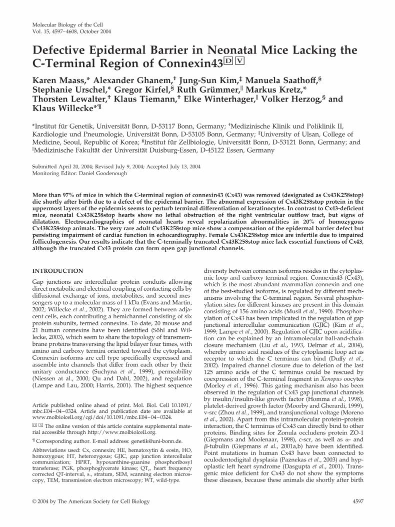
Figure 1. Gene targeting and transcription analysis of thecx43K258stop allele. In a first homologous recombination in hprt-deficient embryonic stem cells, the cx43 coding region was replacedby a PGK-hprt-cassette (Plum et al., 2000). In the second homologousrecombination, the PGK-hprt-cassette was replaced by the truncatedcx43 coding region. Recombined clones were enriched by 6-thiogua-nine selection, validated by PCR analysis and Southern blot byusing a 3� external and a cx43 internal probe. S, SacI; H, HindIII; N,NotI; int, internal; ext., external; CDR, coding region; UTR, untrans-lated region; HR, homologous region; PGK-hprt, phosphoglyceratekinase promoter-hypoxanthine-guanine phosphoribosyl trans-ferase-minigene; PGK-hsvtk, phosphoglycerate kinase promoter-hepes simplex virus thymidine kinase cassette; Ki, knockin. (B)Total RNA blot of heart, brain and liver of WT, HT, and HO adultanimals was probed with a radiolabeled DNA fragment cor-responding to cx43 5�-UTR. The 2.6-kb cx43K258stop transcript wasdetected proportional to gene dosis. No 3.0-kb cx43 wild-type tran-script was detected in homozygous mutant animals. Equal amountsof loaded total RNA were verified by reprobing with a �-actinprobe.
K. Maass et al.
Molecular Biology of the Cell4598
(Immunotec, Marseille, France). Samples were photographed at RT by usingan Axiophot microscope (Carl Zeiss) equipped with a CCD camera (CarlZeiss) and AxioVision software (Carl Zeiss). Controls in the absence ofprimary antibodies were routinely performed and yielded no signals. Figureswere composed with the help of Adobe Photoshop software (version 6.0;image procession restricted to changes in brightness and contrast of wholeimages).
Immunoblot AnalysisEqual protein amounts were determined using the bicinchoninic acid proteindetermination kit (Sigma Chemie) according to the manufacturer’s instruc-tions and separated by SDS-PAGE (Laemmli, 1970) at 25 mA per gel andelectroblotted for 2 h at 100 V at 4°C onto nitrocellulose membranes (Hybond,0.45 �m; Amersham Biosciences UK, Little Chalfont, Buckinghamshire,United Kingdom). Blots were incubated with antibodies overnight at 4°C andimmunoreactive proteins were visualized by species-specific horseradish per-oxidase-conjugated secondary antibodies (Dianova, Hamburg, Germany) andsubsequent enhanced chemiluminescence (Amersham Biosciences UK) asrecommended by the manufacturer. ECL blots were developed on x-ray film(SuperRX; Fujifilm, Tokyo, Japan). Blot could be reused after incubation withstripping buffer (RestoreTM; Pierce Chemical, Rockford, IL).
DNA Isolation and AnalysisGenomic DNA was isolated as described by Laird et al. (1991b). For Southernblot analysis, DNA was digested with HindIII, fractionated on 0.7% agarosegels and transferred onto nylon membranes (Hybond�, 0.4 �m; AmershamBiosciences UK), and filters were probed with 32P-radiolabeled cx43 probes(first 772 base pairs of Cx43 coding region as internal probe, 550 base pairsoutside the 3� homologous region as external probe), washed, sealed intoplastic wrap, and exposed to x-ray film (X-OMAT; Eastman Kodak, Rochester,NY). For genotyping of mice, DNA was subjected to polymerase chain reac-tion (PCR) analysis using the primers delCT-HO (5�-gcatcctcttcaagtctgtcttcg)and RO-delCT (5�-caaaacaccccccaaggaacctag), resulting in an 851-base pairamplicon for the cx43 allele and a 452-base pair amplicon for the cx43K258stopallele.
RNA Isolation and Northern Blot AnalysisTotal RNA was isolated using TRIzol (Invitrogen, Renfew, United Kingdom)according to manufacturer’s instructions. Fifty micrograms of RNA was frac-tionated on 1% agarose/0.9% formaldehyde gels, transferred to nylon mem-branes (Hybond N, 0.45 �m; Amersham Biosciences UK), UV-cross-linked(UV-Stratalinker 2400; Stratagene, La Jolla, CA), and hybridized to 32P-labeledprobes (corresponding to the 3�-untranslated region of cx43 exon 2 or to�-actin). Filters were washed and exposed to x-ray film (X-OMAT; EastmanKodak).
HeLa Cell Culture and TransfectionHeLa connexin transfectants were grown in DMEM (Life Technologies, Egg-enstein, Germany), supplemented with 10% calf serum (Life Technologies),100 �g/ml streptomycin, 100 �g/ml penicillin, and 1 �g/ml puromycin(Sigma Chemie). Cells were passaged three times per week and maintained ina 37°C incubator in a moist atmosphere with 10% CO2. For generation ofCx43K258stop-expressing cells, the truncated version of cx43 was generatedby PCR mutagenesis and inserted into the pBEHpac18 expression vector(Horst et al., 1991) under control of a SV40 promoter. HeLa wild-type cellswere transfected by Lipofection (Tfx-50 reagent; Promega, Madison, WI)according to manufacturer’s instructions. Puromycin-positive clones wereisolated and grown under selection conditions.
Pulse-Chase Analysis and Determination of Half-LifeHeLa cells were metabolically labeled with [35S]methionine for 1 h, as de-scribed by Hertlein et al. (1998). Medium was replaced by nonradioactivemedium, supplemented with additional 15 mg/l methionine (final concen-tration 45 mg/l). Cells were harvested at different time points of chase.Connexin proteins were immunoprecipitated and subjected to SDS-PAGE.After fixation, gels were treated with Amplify (Amersham Biosciences,Freiburg, Germany), dried, and used for autoradiography. Densitometricsignal values of scanned autoradiographies were calculated using Imagemas-ter 2.0 software (Amersham Biosciences). Signal intensity (intensity of opticaldensity; IOD) was correlated to the amount of protein in the lysates. Thedensiometric value at 0-h chase was defined as 100%. Logarithms of thecorrelated IOD values were calculated and the parameters of the mathemat-ical function lnIOD � a � bx were determined by the regression methodusing GraphPad Prism for the IBM-PC (GraphPad Software, San Diego, CA).The half-life was calculated on the slope of the regression function as mean ofat least three pulse-chase experiments.
ECG Analysis of Neonatal MiceNeonates (14 wild type, 29 heterozygous, and 18 homozygous) were subjectedto surface ECG recordings according to Hagendorff et al. (1999). Surface
six-lead ECG was acquired on a multichannel amplifier and converted to adigital signal for analysis (PowerLab system; ADInstruments, Milford, MA).ECG channels were amplified, filtered between 10 and 100 Hz, and sampledwith a rate of 1 kHz. Serial ECG recordings were obtained from day 1 to day6; spontaneous cycle length, heart rate, P-wave duration, PQ-interval, QRS-duration, and QT-interval were determined off-line, with QRS-duration start-ing at the Q-wave onset and lasting to the return of the S-wave to theisoelectric line. QT-interval was measured from the onset of the Q-wave to theend of the T-wave, which was defined as the final return of the T-wave tobaseline level. The QT-interval was rate corrected (QTc) according to Mitchellet al. (1998). ECG parameters were compared between the three genotypes bymeans of one-way analysis of variance (ANOVA) and post hoc Tukey-Kramermultiple comparisons test. Unpaired Student’s t test was performed fordifferentiation within a genotype. P values � 0.05 were considered significant.
RESULTS
Targeted Replacement of cx43 Coding DNA bycx43K258stop Mutant DNA in Transgenic MiceTargeted replacement of cx43 by cx43K258stop cDNA wasachieved by the double replacement strategy (Figure 1A).Previously, the cx43 open reading frame had been replacedby a hypoxanthine-guanine phosphoribosyl transferase (hprt)-minigene under control of a phosphoglycerate kinase (PGK)promoter in hprt-deficient HM1 embryonic stem cells (Plumet al., 2000) in a first homologous recombination. To intro-duce the cx43K258stop mutation, a truncated version of cx43was generated by PCR mutagenesis by using isolated cx43DNA from a mouse 129/Sv genomic library (Stratagene) astemplate. Codon 258 was mutated into a stop codon, intro-ducing an A-to-T transversion at nucleotide 772 of the cod-ing region; the remaining downstream coding region wasdeleted. The targeting construct included the genomic cx43intron-exon 2 boundary, which had been partially deleted inthe first homologous recombination. In the second homolo-gous recombination, the PGK-hprt minigene was subse-quently replaced, leading to the cx43K258stop allele (Figure1A). One of the obtained embryonic stem cell clones, posi-tive in PCR, Southern blot, and Northern blot analyses, wasused for blastocyst injection and gave rise to viable heterozy-gous Cx43K258stop mice. Northern blot analysis of adultmouse tissues (Figure 1B) showed that the wild-type cx43transcript was decreased to 50% in heterozygous animalsand absent in homozygous animals. The abundance ofcx43K258stop transcript increased in a gene dosage-depen-dent manner from heterozygous to homozygous animals.
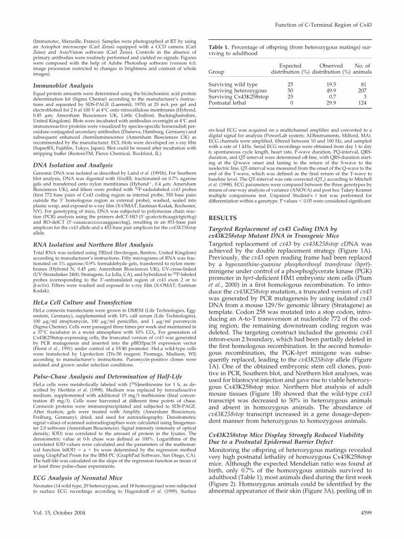
Cx43K258stop Mice Display Strongly Reduced ViabilityDue to a Postnatal Epidermal Barrier DefectMonitoring the offspring of heterozygous matings revealedvery high postnatal lethality of homozygous Cx43K258stopmice. Although the expected Mendelian ratio was found atbirth, only 0.7% of the homozygous animals survived toadulthood (Table 1); most animals died during the first week(Figure 2). Homozygous animals could be identified by theabnormal appearance of their skin (Figure 3A), peeling off in
Table 1. Percentage of offspring (from heterozygous matings) sur-viving to adulthood
GroupExpected
distribution (%)Observed
distribution (%)No. of
animals
Surviving wild type 25 19.5 81Surviving heterozygous 50 49.9 207Surviving Cx43K258stop 25 0.7 3Postnatal lethal 0 29.9 124
Function of C-Terminal Region of Cx43
Vol. 15, October 2004 4599
big squames, predominantly on forehead, posterior back(Figure 3B) and extremities. Constriction bands were oftenfound around the tails of neonatal animals (Figure 3C).Because of the observed epidermal abnormalities, animalswere subjected to the dye penetration assay with toluidineblue to analyze the functionality of the epidermal perme-ability barrier. An obvious deficiency of the epidermal per-meability barrier that increased over the first days after birthwas detected in homozygous Cx43K258stop mice but not inwild-type mice (Figure 3, D and E). Dye penetration was
always found in the posterior back and in several casesalong the spine and on the forehead.
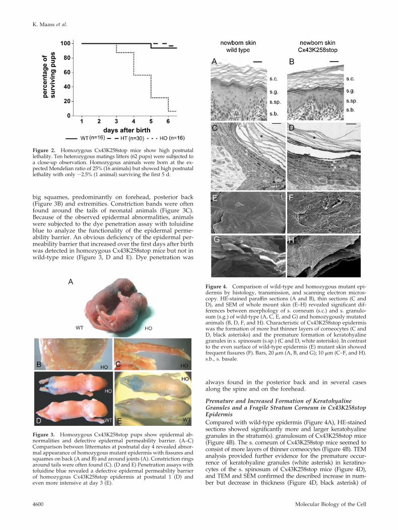
Premature and Increased Formation of KeratohyalineGranules and a Fragile Stratum Corneum in Cx43K258stopEpidermisCompared with wild-type epidermis (Figure 4A), HE-stainedsections showed significantly more and larger keratohyalinegranules in the stratum(s). granulosum of Cx43K258stop mice(Figure 4B). The s. corneum of Cx43K258stop mice seemed toconsist of more layers of thinner corneocytes (Figure 4B). TEManalysis provided further evidence for the premature occur-rence of keratohyaline granules (white asterisk) in keratino-cytes of the s. spinosum of Cx43K258stop mice (Figure 4D),and TEM and SEM confirmed the described increase in num-ber but decrease in thickness (Figure 4D, black asterisk) of
Figure 2. Homozygous Cx43K258stop mice show high postnatallethality. Ten heterozygous matings litters (62 pups) were subjected toa close-up observation. Homozygous animals were born at the ex-pected Mendelian ratio of 25% (16 animals) but showed high postnatallethality with only �2.5% (1 animal) surviving the first 5 d.
Figure 3. Homozygous Cx43K258stop pups show epidermal ab-normalities and defective epidermal permeability barrier. (A–C)Comparison between littermates at postnatal day 4 revealed abnor-mal appearance of homozygous mutant epidermis with fissures andsquames on back (A and B) and around joints (A). Constriction ringsaround tails were often found (C). (D and E) Penetration assays withtoluidine blue revealed a defective epidermal permeability barrierof homozygous Cx43K258stop epidermis at postnatal 1 (D) andeven more intensive at day 3 (E).
Figure 4. Comparison of wild-type and homozygous mutant epi-dermis by histology, transmission, and scanning electron micros-copy. HE-stained paraffin sections (A and B), thin sections (C andD), and SEM of whole mount skin (E–H) revealed significant dif-ferences between morphology of s. corneum (s.c.) and s. granulo-sum (s.g.) of wild-type (A, C, E, and G) and homozygously mutatedanimals (B, D, F, and H). Characteristic of Cx43K258stop epidermiswas the formation of more but thinner layers of corneocytes (C andD, black asterisks) and the premature formation of keratohyalinegranules in s. spinosum (s.sp.) (C and D, white asterisks). In contrastto the even surface of wild-type epidermis (E) mutant skin showedfrequent fissures (F). Bars, 20 �m (A, B, and G); 10 �m (C–F, and H).s.b., s. basale.
K. Maass et al.
Molecular Biology of the Cell4600
corneocyte layers (Figure 4, D and H). SEM of whole mountepidermis (Figure 4, E–H) displayed pronounced differences ofthe upper surface of the s. corneum between wild-type (Figure4, E and G) and homozygous epidermis (Figure 4, F and H). Incontrast to the continuous surface of wild-type epidermis withsingle desquamating corneocytes, the homozygous mutantepidermis seemed brittle and fragile with numerous fissuresspanning several layers of corneocytes (Figure 4F).
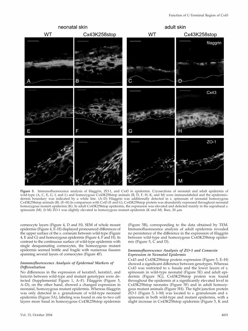
Immunofluorescence Analysis of Epidermal Markers ofDifferentiationNo differences in the expression of keratin5, keratin1, andloricrin between wild-type and mutant genotypes were de-tected (Supplemental Figure 1, A–F). Filaggrin (Figure 5,A–D), on the other hand, showed a changed expression inneonatal, homozygous mutant epidermis. Whereas filaggrinwas only detected in s. granulosum of wild-type neonatalepidermis (Figure 5A), labeling was found in one to two celllayers more basal in homozygous Cx43K258stop epidermis
(Figure 5B), corresponding to the data obtained by TEM.Immunofluorescence analysis of adult epidermis revealedno persistence of the difference in the expression of filaggrinbetween wild-type and homozygous Cx43K258stop epider-mis (Figure 5, C and D).
Immunofluorescence Analysis of ZO-1 and ConnexinExpression in Neonatal EpidermisCx43 and Cx43K258stop protein expression (Figure 5, E–H)showed a significant difference between genotypes. WhereasCx43 was restricted to s. basale and the lower layers of s.spinosum in wild-type neonatal (Figure 5E) and adult epi-dermis (Figure 5G), Cx43K258stop protein was foundthroughout the epidermis at a significantly elevated level inCx43K258stop neonates (Figure 5F) and in adult homozy-gous mutant animals (Figure 5H). The tight junction proteinZO-1 (Figure 5, I–M) was located to s. granulosum and s.spinosum in both wild-type and mutant epidermis, with aslight increase in Cx43K258stop epidermis (Figure 5, K and
Figure 5. Immunofluorescence analysis of filaggrin, ZO-1, and Cx43 in epidermis. Cryosections of neonatal and adult epidermis ofwild-type (A, C, E, G, I, and L) and homozygous Cx43K258stop animals (B, D, F, H, K, and M) were immunolabeled and the epidermis–dermis boundary was indicated by a white line. (A–D) Filaggrin was additionally detected in s. spinosum of neonatal homozygousCx43K258stop animals (B). (E–H) In comparison with Cx43 (E and G), Cx43K258stop protein was abundantly expressed throughout neonatalhomozygous mutant epidermis (K). In adult Cx43K258stop epidermis, the expression was elevated and detected mainly in the suprabasal s.spinosum (M). (I–M) ZO-1 was slightly elevated in homozygous mutant epidermis (K and M). Bars, 20 �m.
Function of C-Terminal Region of Cx43
Vol. 15, October 2004 4601
M). Analysis of other connexins expressed in epidermisrevealed an up-regulation of Cx26 (Supplemental Figure 2,A and B), but no change in Cx30 and Cx31 expression(Supplemental Figure 2, C–F) in homozygous mutant epider-mis. Analysis of proliferation and transition time of keratino-cytes by 5-bromo-2-deoxyuridine (BrdU) labeling showed nodifferences between wild-type and Cx43K258stop epidermis(Supplemental Figure 1, I–M).
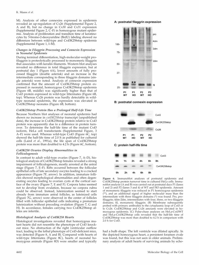
Changes in Filaggrin Processing and Connexin Expressionin Neonatal EpidermisDuring terminal differentiation, high-molecular-weight pro-filaggrin is proteolytically processed to monomeric filaggrinthat associates with keratin filaments. Western blot analysesrevealed no difference in total filaggrin expression, but atpostnatal day 1 (Figure 6A), lower amounts of fully pro-cessed filaggrin (double asterisk) and an increase in theintermediate corresponding to three filaggrin domains (sin-gle asterisk) were noted. Analysis of connexin expressionconfirmed that the amount of Cx43K258stop protein ex-pressed in neonatal, homozygous Cx43K258stop epidermis(Figure 6B, middle) was significantly higher than that ofCx43 protein expressed in wild-type littermates (Figure 6B,top). Whereas Cx26 protein was hardly detectable in wild-type neonatal epidermis, the expression was elevated inCx43K258stop neonates (Figure 6B, bottom).
Cx43K258stop Protein Has a Prolonged Half-Life TimeBecause Northern blot analyses of neonatal epidermis hadshown no increase in cx43K258stop transcript (unpublisheddata), the increase in Cx43K258stop protein relative to Cx43protein was apparently due to a difference in protein turn-over. To determine the half-life time of the mutant Cx43isoform, HeLa cell transfectants (Supplemental Figure, 3A–F) were used. Whereas wild-type Cx43 (Figure 6C, top)showed the half-life time of 2.0 h as published for culturedcells (Laird et al., 1991a), the life span of Cx43K258stopprotein was more than doubled to 4.2 h (Figure 6C, bottom).
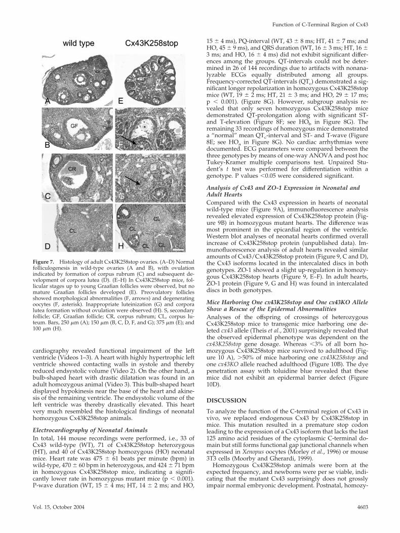
Cx43K258 Ovaries Display Abnormalities inFolliculogenesisIn contrast to adult wild-type ovaries (Figure 7, A–D), his-tological analysis of Cx43K258stop females revealed a strongimpairment of folliculogenesis, mostly arrested at the antralstage (Figure 7, E–F). Rifts occurred between the follicularepithelial cells of late secondary oocytes leading to a crackedappearance (Figure 7F, arrow). In addition, immature folli-cles showed morphological abnormalities and often degen-erating oocytes leading to ovarian cysts at the cortical sur-face of the ovary (Figure 7, E and F). Corpora lutea seemednot to develop from ovulation, because no corpora rubracould be observed. Instead, luteinization seemed to startalready from immature antral follicles, as luteinized cells(Figure 7G, arrow) were observed in the center of folliclesfilled with follicular epithelial cells indicating a prematureluteinization without preceding ovulation (Figure 7, G andH). In accordance, females carrying two Cx43K258stop al-leles are infertile.
Histological Analysis of Cx43K258 HeartsHistological investigations revealed that homozygous mu-tant hearts did not resemble the phenotype of Cx43 knock-out mice. No obstruction of the right ventricular outflowtract, leading to the lethal phenotype of Cx43-deficient mice,was detected (Figure 8, A and B). Compared with hearts ofwild-type littermates (Figure 8C), hearts of neonatal ho-mozygous animals (Figure 8D) were smaller and typically
had a bulb shape. The left ventricle was dilated apically. Inthe depicted homozygous heart, a persistent foramen ovalewas found, but this was not seen in older animals. Prelimi-nary analysis of adult hearts of surviving animals by echo-
Figure 6. Immunoblot analyses of postnatal epidermis andCx43K258stop protein turnover time in cultured HeLa cells. Immu-noblot analysis (A and B) was carried out at postnatal days P1 (lanes1 and 2) and P2 (lanes 3 and 4) of WT and HO epidermis. Amountof monumeric filaggrin was reduced in P1 homozygous epidermis(**), and an additional signal of higher molecular mass than theintermediate with three filaggrin domains (*) was found. Pro, pro-filaggrin; 4dm-2dm, intermediates with four, three, or two filaggrindomains; fil, monumeric filaggrin. (B) Membrane subsequentlyprobed with primary antibodies to the cytoplasmic loop of Cx43 orto Cx26. Cx43K258stop and Cx26 amounts were elevated in ho-mozygous epidermis. (C) Pulse-chase analyses on HeLa-Cx43WTand HeLa-Cx43K258stop cells revealed that the half-life time ofCx43K258stop was more than doubled to 4.2 h in comparison with2.0 h for Cx43.
K. Maass et al.
Molecular Biology of the Cell4602
cardiography revealed functional impairment of the leftventricle (Videos 1–3). A heart with highly hypertrophic leftventricle showed contacting walls in systole and therebyreduced endsystolic volume (Video 2). On the other hand, abulb-shaped heart with drastic dilatation was found in anadult homozygous animal (Video 3). This bulb-shaped heartdisplayed hypokinesis near the base of the heart and akine-sis of the remaining ventricle. The endsystolic volume of theleft ventricle was thereby drastically elevated. This heartvery much resembled the histological findings of neonatalhomozygous Cx43K258stop animals.
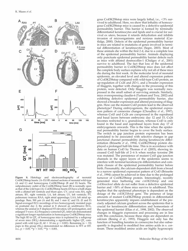
Electrocardiography of Neonatal AnimalsIn total, 144 mouse recordings were performed, i.e., 33 ofCx43 wild-type (WT), 71 of Cx43K258stop heterozygous(HT), and 40 of Cx43K258stop homozygous (HO) neonatalmice. Heart rate was 475 � 61 beats per minute (bpm) inwild-type, 470 � 60 bpm in heterozygous, and 424 � 71 bpmin homozygous Cx43K258stop mice, indicating a signifi-cantly lower rate in homozygous mutant mice (p � 0.001).P-wave duration (WT, 15 � 4 ms; HT, 14 � 2 ms; and HO,
15 � 4 ms), PQ-interval (WT, 43 � 8 ms; HT, 41 � 7 ms; andHO, 45 � 9 ms), and QRS duration (WT, 16 � 3 ms; HT, 16 �3 ms; and HO, 16 � 4 ms) did not exhibit significant differ-ences among the groups. QT-intervals could not be deter-mined in 26 of 144 recordings due to artifacts with nonana-lyzable ECGs equally distributed among all groups.Frequency-corrected QT-intervals (QTc) demonstrated a sig-nificant longer repolarization in homozygous Cx43K258stopmice (WT, 19 � 2 ms; HT, 21 � 3 ms; and HO, 29 � 17 ms;p � 0.001). (Figure 8G). However, subgroup analysis re-vealed that only seven homozygous Cx43K258stop micedemonstrated QT-prolongation along with significant ST-and T-elevation (Figure 8F; see HOb in Figure 8G). Theremaining 33 recordings of homozygous mice demonstrateda “normal” mean QTc-interval and ST- and T-wave (Figure8E; see HOa in Figure 8G). No cardiac arrhythmias weredocumented. ECG parameters were compared between thethree genotypes by means of one-way ANOVA and post hocTukey-Kramer multiple comparisons test. Unpaired Stu-dent’s t test was performed for differentiation within agenotype. P values �0.05 were considered significant.
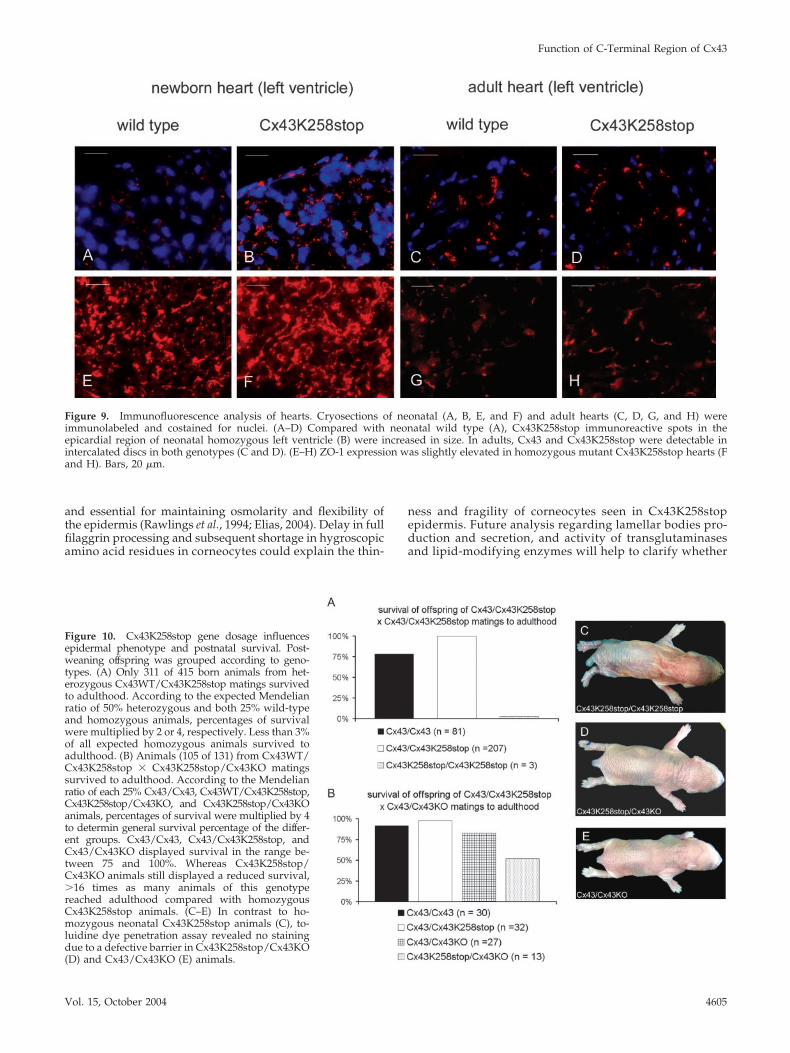
Analysis of Cx43 and ZO-1 Expression in Neonatal andAdult HeartsCompared with the Cx43 expression in hearts of neonatalwild-type mice (Figure 9A), immunofluorescence analysisrevealed elevated expression of Cx43K258stop protein (Fig-ure 9B) in homozygous mutant hearts. The difference wasmost prominent in the epicardial region of the ventricle.Western blot analyses of neonatal hearts confirmed overallincrease of Cx43K258stop protein (unpublished data). Im-munofluorescence analysis of adult hearts revealed similaramounts of Cx43/Cx43K258stop protein (Figure 9, C and D),the Cx43 isoforms located in the intercalated discs in bothgenotypes. ZO-1 showed a slight up-regulation in homozy-gous Cx43K258stop hearts (Figure 9, E–F). In adult hearts,ZO-1 protein (Figure 9, G and H) was found in intercalateddiscs in both genotypes.
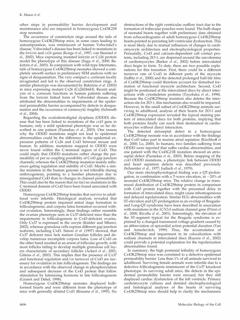
Mice Harboring One cx43K258stop and One cx43KO AlleleShow a Rescue of the Epidermal AbnormalitiesAnalyses of the offspring of crossings of heterozygousCx43K258stop mice to transgenic mice harboring one de-leted cx43 allele (Theis et al., 2001) surprisingly revealed thatthe observed epidermal phenotype was dependent on thecx43K258stop gene dosage. Whereas �3% of all born ho-mozygous Cx43K258stop mice survived to adulthood (Fig-ure 10 A), �50% of mice harboring one cx43K258stop andone cx43KO allele reached adulthood (Figure 10B). The dyepenetration assay with toluidine blue revealed that thesemice did not exhibit an epidermal barrier defect (Figure10D).
DISCUSSION
To analyze the function of the C-terminal region of Cx43 invivo, we replaced endogenous Cx43 by Cx43K258stop inmice. This mutation resulted in a premature stop codonleading to the expression of a Cx43 isoform that lacks the last125 amino acid residues of the cytoplasmic C-terminal do-main but still forms functional gap junctional channels whenexpressed in Xenopus oocytes (Morley et al., 1996) or mouse3T3 cells (Moorby and Gherardi, 1999).
Homozygous Cx43K258stop animals were born at theexpected frequency, and newborns were per se viable, indi-cating that the mutant Cx43 surprisingly does not grosslyimpair normal embryonic development. Postnatal, homozy-
Figure 7. Histology of adult Cx43K258stop ovaries. (A–D) Normalfolliculogenesis in wild-type ovaries (A and B), with ovulationindicated by formation of corpus rubrum (C) and subsequent de-velopment of corpora lutea (D). (E–H) In Cx43K258stop mice, fol-licular stages up to young Graafian follicles were observed, but nomature Graafian follicles developed (E). Preovulatory folliclesshowed morphological abnormalities (F, arrows) and degeneratingoocytes (F, asterisk). Inappropriate luteinization (G) and corporalutea formation without ovulation were observed (H). S, secondaryfollicle; GF, Graafian follicle; CR, corpus rubrum; CL, corpus lu-teum. Bars, 250 �m (A); 150 �m (B, C, D, F, and G); 375 �m (E); and100 �m (H).
Function of C-Terminal Region of Cx43
Vol. 15, October 2004 4603
gous Cx43K258stop mice were largely lethal, i.e., �3% sur-vived to adulthood. Here, we show that lethality of homozy-gous Cx43K258stop mice is caused by a defective epidermalpermeability barrier. This barrier is formed by terminallydifferentiated keratinocytes and lipids and is crucial for sur-vival ex utero, because it retards dehydration and inhibitsinvasion of microorganisms and noxious material (Car-tlidge, 2000). Defects of the epidermal permeability barrierin mice are related to mutations of genes involved in termi-nal differentiation of keratinocytes (Segre, 2003). Most ofthese animals die within the first 2 d, due to a complete lossof the epidermal permeability barrier. Animals displayingonly punctuate epidermal permeability barrier defects suchas mice with ablated desmocollin-1 (Chidgey et al., 2001)survive to adulthood. The fact that loss of the epidermalpermeability barrier in Cx43K258stop mice does not affectthe complete body surface explains why not all of these micedie during the first week. At the molecular level of neonatalepidermis, an elevated level and altered expression patternof Cx43K258stop compared with wild-type Cx43 protein, anup-regulation of Cx26 and ZO-1, and a broader expressionof filaggrin, together with changes in the processing of thisprotein, were detected. Only filaggrin was normally reex-pressed in the small subset of surviving animals. Similarly,mice overexpressing claudin-6 (Turksen and Troy, 2002) andexhibiting defective epidermal permeability barrier, alsoshowed a broader expression and altered processing of filag-grin. How can the mutant Cx43 protein lead to the observedphenotype? During embryogenesis, the epidermal expres-sion pattern of connexins changes (Choudhry et al., 1997),whereas Cx43 and Cx26 are coexpressed in the suprabasaland basal layers between embryonic day 12 and 15, Cx26becomes restricted to s. granulosum, whereas Cx43 is onlyfound in the basal and suprabasal layers from day 17 ofembryogenesis onwards. This is the time when the epider-mal permeability barrier begins to cover the body surface.The switch in gap junction protein expression has beenpostulated to be associated with selective changes in gapjunctional channel permeability during keratinocyte differ-entiation (Brissette et al., 1994). Cx43K258stop protein dis-played a prolonged half-life time. This is in accordance withdata on human Cx43 by Thomas et al. (2003), showing in-creased Cx43 half-life of 2–6 h when residue tyrosine 286was mutated. The persistence of mutant Cx43 gap junctionchannels in the upper layers of the epidermis seems tointerfere with terminal kerationcyte differentiation and com-plete closure of the epidermal permeability barrier beforebirth. Presumably, the calcium-dependent shift from a broadto a narrow epidermal expression pattern of Cx43 (Brissetteet al., 1994) cannot be achieved in time due to the prolongedturnover of Cx43K258stop protein. Interestingly, recentlygenerated mice harboring one cx43K258stop and onecx43knockout allele do not suffer from a defective epidermalbarrier and �50% of these mice survive to adulthood. Thisimplies that the epidermal phenotype is dependent on thedosage of the cx43K258stop gene. The presence of largeamounts of mutant Cx43K258stop protein in differentiatedkeratinocytes apparently impairs establishment of the pre-cisely adjusted calcium gradient across the epidermis that iscrucial for keratinocyte differentiation and homeostasis ofthe epidermal barrier (Elias et al., 1998, 2002). The observedchanges in filaggrin expression and processing are in linewith this conclusion, because these steps are dependent oncalcium (Resing et al., 1993). Filaggrin not only has thefunction of bundling keratins in corneocytes but also subse-quently is degraded to modified free amino acids in s. cor-neum. These modified amino acids are highly hygroscopic
Figure 8. Histology and electrocardiography of neonatalCx43K258stop hearts. (A–D) HE-stained sections of neonatal wild-type(A and C) and homozygous Cx43K258stop (B and D) hearts. Thesubpulmonary outlet of the Cx43K258stop heart (B) is normally openas that of the wild type (A). Cx43K258stop hearts (D) have a bulb shapewith a dilated left ventricle and blunt apex. CV, caval vein; RA, rightatrium; RV, right ventricle; LA, left atrium; LV, left ventricle; PT,pulmonary truncus; RAA, right atrial appendage; LAA, left atrial ap-pendage. Bars, 500 �m (A and B); and 1 mm (C and D). (E and F)Signal-averaged ECG recordings of two homozygously mutated pupson postnatal day 2; the animal in E showed an unobtrusive ECG,whereas the animal in F displayed ST-elevation, QT-prolongation, andenlarged T-wave. (G) Frequency-corrected QTc-intervals demonstrateda significant longer repolarization in homozygous Cx43K258stop mice.The high SD in QTc of homozygous mice is explained by a subgroupof seven mice (HOb) demonstrating a significantly higher QTc com-pared with wild type (E, 60 � 10 ms; p � 0.001). Remaining (n � 33)pups in this group (Hoa) demonstrated no differences to WT or HT.n.s., p � 0.05; **p � 0.01; ***p � 0.001.
K. Maass et al.
Molecular Biology of the Cell4604
and essential for maintaining osmolarity and flexibility ofthe epidermis (Rawlings et al., 1994; Elias, 2004). Delay in fullfilaggrin processing and subsequent shortage in hygroscopicamino acid residues in corneocytes could explain the thin-
ness and fragility of corneocytes seen in Cx43K258stopepidermis. Future analysis regarding lamellar bodies pro-duction and secretion, and activity of transglutaminasesand lipid-modifying enzymes will help to clarify whether
Figure 9. Immunofluorescence analysis of hearts. Cryosections of neonatal (A, B, E, and F) and adult hearts (C, D, G, and H) wereimmunolabeled and costained for nuclei. (A–D) Compared with neonatal wild type (A), Cx43K258stop immunoreactive spots in theepicardial region of neonatal homozygous left ventricle (B) were increased in size. In adults, Cx43 and Cx43K258stop were detectable inintercalated discs in both genotypes (C and D). (E–H) ZO-1 expression was slightly elevated in homozygous mutant Cx43K258stop hearts (Fand H). Bars, 20 �m.
Figure 10. Cx43K258stop gene dosage influencesepidermal phenotype and postnatal survival. Post-weaning offspring was grouped according to geno-types. (A) Only 311 of 415 born animals from het-erozygous Cx43WT/Cx43K258stop matings survivedto adulthood. According to the expected Mendelianratio of 50% heterozygous and both 25% wild-typeand homozygous animals, percentages of survivalwere multiplied by 2 or 4, respectively. Less than 3%of all expected homozygous animals survived toadulthood. (B) Animals (105 of 131) from Cx43WT/Cx43K258stop � Cx43K258stop/Cx43KO matingssurvived to adulthood. According to the Mendelianratio of each 25% Cx43/Cx43, Cx43WT/Cx43K258stop,Cx43K258stop/Cx43KO, and Cx43K258stop/Cx43KOanimals, percentages of survival were multiplied by 4to determin general survival percentage of the differ-ent groups. Cx43/Cx43, Cx43/Cx43K258stop, andCx43/Cx43KO displayed survival in the range be-tween 75 and 100%. Whereas Cx43K258stop/Cx43KO animals still displayed a reduced survival,�16 times as many animals of this genotypereached adulthood compared with homozygousCx43K258stop animals. (C–E) In contrast to ho-mozygous neonatal Cx43K258stop animals (C), to-luidine dye penetration assay revealed no stainingdue to a defective barrier in Cx43K258stop/Cx43KO(D) and Cx43/Cx43KO (E) animals.
Function of C-Terminal Region of Cx43
Vol. 15, October 2004 4605
other steps in permeability barrier development andmaintenance also are impaired in homozygous Cx43K258stop neonates.
The occurrence of constriction rings around the tails ofhomozygous Cx43K258stop mice, in some cases leading toautoamputation, was reminiscent of human Vohwinkel’sdisease. Vohwinkel’s disease has been linked to mutations inthe loricrin and cx26 genes (Korge et al., 1997; van Steensel etal., 2002). Mice expressing mutated loricrin or Cx26 proteinmodel the phenotype of this disease (Suga et al., 2000; Ba-kirtzis et al., 2003). In comparison with wild-type littermates,tails of homozygous Cx43K258stop neonates showed a com-pletely smooth surface in preliminary SEM analysis with nosigns of desquamation. The very compact s. corneum locallyinvaginated and led to the observed constriction rings. Asimilar phenotype was documented by Bakirtzis et al. (2003)in mice expressing mutant Cx26 (Cx26D66H). Recent anal-ysis of s. corneum functions in human patients sufferingfrom the loricrin linked form of Vohwinkel disease alsoattributed the abnormalities to impairments of the epider-mal permeability barrier accompanied by defects in desqua-mation and the occurrence of corneocytic fragility (Schmuthet al., 2004).
Regarding the oculodentodigital dysplasia (ODDD) dis-ease that has been linked to mutations of the cx43 gene inhumans, only a mild epidermal phenotype was so far de-scribed in one patient (Paznekas et al., 2003). One reasonwhy the ODDD mutations might not lead to epidermalabnormalities could be the difference in the epidermal ex-pression patterns of Cx26 and Cx43 between mouse andhuman. In addition, mutations mapped to ODDD werenever found within the C-terminal region of Cx43. Thiscould imply that ODDD mutations rather change the per-meability or per se coupling possibility of Cx43 gap junctionchannels, whereas the Cx43K258stop mutation mainly influ-ences gating regulation and stability of Cx43. Maybe a sim-ilar mutation in the human genome is not tolerable duringembryogenesis, pointing to a harsher phenotype due tomisregulated Cx43 than to changes in channel permeability.This could also be the reason that (so far) no mutations in theC-terminal domain of Cx43 have been found associated withODDD.
Homozygous Cx43K258stop females that survive to adult-hood were infertile. Histological analysis revealed thatCx43K258stop protein impaired antral stage formation infolliculogenesis, and corpora lutea formation occurred with-out ovulation. Interestingly, these findings rather resemblethe ovarian phenotype seen in Cx37-deficient mice than theimpairments in folliculogenesis in Cx43-deficient ovaries.Only Cx37 is expressed in the oocyte (Kidder and Mhawi,2002), whereas granulosa cells express different gap junctionisoforms, including Cx43. Simon et al. (1997) showed, thatCx37 deficient mice lack mature Graafian follicles and de-velop numerous incomplete corpora lutea. Loss of Cx43 onthe other hand resulted in an arrest of follicular growth, withmost follicles failing to develop multiple granulosa cell lay-ers characteristic of secondary follicles (Ackert et al., 2001;Gittens et al., 2003). This implies that the presence of Cx37and functional regulation and/or turnover of Cx43 are nec-essary for ovulation in late folliculogenesis. The findings arein accordance with the observed change in phosphorylationand subsequent decrease of the Cx43 protein that followstimulation by luteinizing hormone in late folliculogenesis(Granot and Dekel, 1998).
Homozygous Cx43K258stop neonates displayed bulb-formed hearts and were different from the phenotype ofmice with generally ablated Cx43 (Reaume et al., 1995). No
obstructions of the right ventricular outflow tract due to theformation of trabecular pouches were found. The bulb shapeof neonatal hearts together with preliminary data obtainedfrom echocardiograms of adult homozygous Cx43K258stophearts pointed to persisting left ventricular dysfunction. Thisis most likely due to mutual influences of changes in cardi-omyocyte architecture and electrophysiological properties.Perinatally, Cx43 and calcium-dependent cell contact pro-teins, including ZO-1, are dispersed around the sarcolemmaof cardiomyocytes (Barker et al., 2002) before intercalateddiscs begin to form. To date, there are two possible expla-nations for this transition. First, there could be a differentturnover rate of Cx43 in different parts of the myocyte(Saffitz et al., 2000) and the detected prolonged half-life timeof Cx43K258stop could therefore account for disturbed for-mation of functional myocyte architecture. Second, Cx43might be positioned at the intercalated discs by direct inter-actions with cytoskeleton proteins (Toyofuku et al., 1998).Because the Cx43K258stop mutation also deletes the inter-action site for ZO-1, this mechanism also would be impaired.However, in the small subset of Cx43K258stop animals sur-viving to adulthood, analysis of their hearts for ZO-1 andCx43K258stop expression revealed the typical staining pat-tern of intercalated discs for both proteins, implying thatboth proteins finally can reach their destination in cardio-myocytes without direct interaction.
The detected atrioseptal defect in a homozygousCx43K258stop neonate was in accordance with the findingsthat Cx43 takes part in murine atrial septation (Kirchhoff etal., 2000; Lo, 2000). In humans, two families suffering fromODDD were reported that suffer cardiac abnormalities, andone patient with the Cx43G21R mutation showed an atrio-septal defect (Paznekas et al., 2003). Before mapping of thecx43 ODDD mutations, a phenotypic link between ODDDand heart septation defects was described previously(Schneider et al., 1977; Judisch et al., 1979).
Our main electrophysiological finding was a QT-prolon-gation, in combination with a T-wave elevation, in �20% ofneonatal Cx43K258stop mice. The relative change in trans-mural distribution of Cx43K258stop protein in comparisonwith Cx43 protein together with the presumed delay information of intercalated discs, might cause inhomogeneousand delayed repolarization. Similar ECG abnormalities withST-elevation and QT-prolongation as an overlap of Brugada-and Long-QT-syndrome have been described in associationwith mutations in the SCN5A sodium channel gene (Priori etal., 2000; Rivolta et al., 2001). Interestingly, the elevation ofthe ST-segment typical for the Brugada syndrome is ex-plained by a changed transmural voltage gradient caused byan abbreviation of epicardial action potential duration (Yanand Antzelevitch, 1999). Thus, the accumulation ofCx43K258stop and impairment in its colocalization withsodium channels in intercalated discs (Kucera et al., 2002)could provide a potential explanation for the repolarizationabnormalities found.
In summary, the high postnatal lethality of homozygousCx43K258stop mice was correlated to a defective epidermalpermeability barrier. Less than 1% of all animals survived toadulthood. Surviving female animals were infertile due to adefective folliculogenesis reminiscent of the Cx37 knockoutphenotype. In surviving adult mice, the defects in the epi-dermal permeability barrier were rescued, but they stilldisplayed cardiac dysfunction of the left ventricle. Primarycardiomyocyte cultures and detailed electrophysiologicaland histological analyses of the hearts of survivingCx43K258stop mice should help to clarify the mechanisms
K. Maass et al.
Molecular Biology of the Cell4606
leading to the observed cardiac changes due to the trunca-tion of Cx43.
ACKNOWLEDGMENTS
We thank Drs. Peter Nielsen and Nalin Kumar (Scripps Research Institute,La Jolla, CA) for a sample of antibodies directed to the cytoplasmic loop ofCx43 (Yeager and Gilula, 1992). We gratefully acknowledge the technical helpof Ina Fiedler and Gaby Schwarz. This work was supported by grants of theGerman Research Association (SFB 284, C1, Wi 270/25-1,2) and the researchgroup on “keratinocytes” to K.W.
REFERENCES
Ackert, C.L., Gittens, J.E., O’Brien, M.J., Eppig, J.J., and Kidder, G.M. (2001).Intercellular communication via connexin43 gap junctions is required forovarian folliculogenesis in the mouse. Dev. Biol. 233, 258–270.
Bakirtzis, G., et al. (2003). Targeted epidermal expression of mutant Connexin26(D66H) mimics true Vohwinkel syndrome and provides a model for thepathogenesis of dominant connexin disorders. Hum. Mol. Genet. 12, 1737–1744.
Barker, R.J., Price, R.L., and Gourdie, R.G. (2002). Increased association ofZO-1 with connexin43 during remodeling of cardiac gap junctions. Circ. Res.90, 317–324.
Brissette, J.L., Kumar, N.M., Gilula, N.B., Hall, J.E., and Dotto, G.P. (1994).Switch in gap junction protein expression is associated with selective changesin junctional permeability during keratinocyte differentiation. Proc. Natl.Acad. Sci. USA 91, 6453–6457.
Cartlidge, P. (2000). The epidermal barrier. Semin. Neonatol. 5, 273–280.
Chidgey, M., et al. (2001). Mice lacking desmocollin 1 show epidermal fragilityaccompanied by barrier defects and abnormal differentiation. J. Cell Biol. 155,821–832.
Choudhry, R., Pitts, J.D., and Hodgins, M.B. (1997). Changing patterns of gapjunctional intercellular communication and connexin distribution in mouseepidermis and hair follicles during embryonic development. Dev. Dyn. 210,417–430.
Dasgupta, C., A.M. Martinez, C.W. Zuppan, M.M. Shah, L.L. Bailey, and W.H.Fletcher. 2001. Identification of connexin43 (alpha1) gap junction gene muta-tions in patients with hypoplastic left heart syndrome by denaturing gradientgel electrophoresis (DGGE). Mutat. Res. 479, 173–186.
Delmar, M., Coombs, W., Sorgen, P., Duffy, H.S., and Taffet, S.M. (2004).Structural bases for the chemical regulation of Connexin43 channels. Cardio-vasc. Res. 62, 268–275.
Duffy, H.S., Sorgen, P.L., Girvin, M.E., O’Donnell, P., Coombs, W., Taffet,S.M., Delmar, M., and Spray, D.C. (2002). pH-dependent intramolecular bind-ing and structure involving Cx43 cytoplasmic domains. J. Biol. Chem. 277,36706–36714.
Elias, P.M. (2004). The epidermal permeability barrier: from the early days atHarvard to emerging concepts. J. Investig. Dermatol. 122, 36–39.
Elias, P.M., Ahn, S.K., Denda, M., Brown, B.E., Crumrine, D., Kimutai, L.K.,Komuves, L., Lee, S.H., and Feingold, K.R. (2002). Modulations in epidermalcalcium regulate the expression of differentiation-specific markers. J. Investig.Dermatol. 119, 1128–1136.
Elias, P.M., Nau, P., Hanley, K., Cullander, C., Crumrine, D., Bench, G.,Sideras-Haddad, E., Mauro, T., Williams, M.L., and Feingold, K.R. (1998).Formation of the epidermal calcium gradient coincides with key milestones ofbarrier ontogenesis in the rodent. J. Investig. Dermatol. 110, 399–404.
Evans, W.H., and Martin, P.E. (2002). Lighting up gap junction channels in aflash. Bioessays 24, 876–880.
Giepmans, B.N., Hengeveld, T., Postma, F.R., and Moolenaar, W.H. (2001a).Interaction of c-Src with gap junction protein connexin-43. Role in the regu-lation of cell-cell communication. J. Biol. Chem. 276, 8544–8549.
Giepmans, B.N., and Moolenaar, W.H. (1998). The gap junction protein con-nexin43 interacts with the second PDZ domain of the zona occludens-1protein. Curr. Biol. 8, 931–934.
Giepmans, B.N., Verlaan, I., Hengeveld, T., Janssen, H., Calafat, J., Falk, M.M.,and Moolenaar, W.H. (2001b). Gap junction protein connexin-43 interactsdirectly with microtubules. Curr. Biol. 11, 1364–1368.
Gittens, J.E., Mhawi, A.A., Lidington, D., Ouellette, Y., and Kidder, G.M.(2003). Functional analysis of gap junctions in ovarian granulosa cells: distinctrole for connexin43 in early stages of folliculogenesis. Am. J. Physiol. 284,C880–C887.
Goldberg, G.S., Bechberger, J.F., and Naus, C.C. (1995). A pre-loading methodof evaluating gap junctional communication by fluorescent dye transfer.Biotechniques 18, 490–497.
Granot, I., and Dekel, N. (1998). Cell-to-cell communication in the ovarianfollicle: developmental and hormonal regulation of the expression of con-nexin43. Hum. Reprod. 13 (suppl 4), 85–97.
Hagendorff, A., Schumacher, B., Kirchhoff, S., Luderitz, B., and Willecke, K.(1999). Conduction disturbances and increased atrial vulnerability in Con-nexin40-deficient mice analyzed by transesophageal stimulation. Circulation99, 1508–1515.
Hardman, M.J., Sisi, P., Banbury, D.N., and Byrne, C. (1998). Patterned acqui-sition of skin barrier function during development. Development 125, 1541–1552.
Harris, A.L. (2001). Emerging issues of connexin channels: biophysics fills thegap. Q. Rev. Biophys. 34, 325–472.
Hertlein, B., Butterweck, A., Haubrich, S., Willecke, K., and Traub, O. (1998).Phosphorylated carboxy terminal serine residues stabilize the mouse gapjunction protein connexin45 against degradation. J. Membr. Biol. 162, 247–257.
Hogan B., Beddington R., Costantini F., and Lacy E. 1994. Manipulating theMouse Embryo: A Laboratory Manual, Cold Spring Harbor, NY: Cold SpringHarbor Laboratory Press.
Homma, N., Alvarado, J.L., Coombs, W., Stergiopoulos, K., Taffet, S.M., Lau,A.F., and Delmar, M. (1998). A particle-receptor model for the insulin-inducedclosure of connexin43 channels. Circ. Res. 83, 27–32.
Horst, E., Wijngaard, P.L., Metzelaar, M., Bast, E.J., and Clevers, H.C. (1991).A method for cDNA cloning in COS cells irrespective of subcellular site ofexpression. Nucleic Acids Res. 19, 4556.
Judisch, G.F., Martin-Casals, A., Hanson, J.W., and Olin, W.H. (1979). Oculo-dentodigital dysplasia. Four new reports and a literature review. Arch. Oph-thalmol. 97, 878–884.
Kidder, G.M., and Mhawi, A.A. (2002). Gap junctions and ovarian folliculo-genesis. Reproduction 123, 613–620.
Kim, D.Y., Kam, Y., Koo, S.K., and Joe, C.O. (1999). Gating connexin 43channels reconstituted in lipid vesicles by mitogen-activated protein kinasephosphorylation. J. Biol. Chem. 274, 5581–5587.
Kirchhoff, S., Kim, J.S., Hagendorff, A., Thonnissen, E., Kruger, O., Lamers,W.H., and Willecke, K. (2000). Abnormal cardiac conduction and morpho-genesis in connexin40 and connexin43 double-deficient mice. Circ. Res. 87,399–405.
Korge, B.P., Ishida-Yamamoto, A., Punter, C., Dopping-Hepenstal, P.J.,Iizuka, H., Stephenson, A., Eady, R.A., and Munro, C.S. (1997). Loricrinmutation in Vohwinkel’s keratoderma is unique to the variant with ichthyo-sis. J. Investig. Dermatol. 109, 604–610.
Kucera, J.P., Rohr, S., and Rudy, Y. (2002). Localization of sodium channels inintercalated disks modulates cardiac conduction. Circ. Res. 91, 1176–1182.
Laemmli, U.K. (1970). Cleavage of structural proteins during the assembly ofthe head of bacteriophage T4. Nature 227, 680–685.
Laird, D.W., Puranam, K.L., and Revel, J.P. (1991a). Turnover and phosphor-ylation dynamics of connexin43 gap junction protein in cultured cardiacmyocytes. Biochem. J. 273, 67–72.
Laird, P.W., Zijderveld, A., Linders, K., Rudnicki, M.A., Jaenisch, R., andBerns, A. (1991b). Simplified mammalian DNA isolation procedure. NucleicAcids Res. 19, 4293.
Lampe, P.D., and Lau, A.F. (2000). Regulation of gap junctions by phosphor-ylation of connexins. Arch. Biochem. Biophys. 384, 205–215.
Lampe, P.D., TenBroek, E.M., Burt, J.M., Kurata, W.E., Johnson, R.G., and Lau,A.F. (2000). Phosphorylation of connexin43 on serine368 by protein kinase Cregulates gap junctional communication. J. Cell Biol. 149, 1503–1512.
Liu, S., Taffet, S., Stoner, L., Delmar, M., Vallano, M.L., and Jalife, J. (1993). Astructural basis for the unequal sensitivity of the major cardiac and liver gapjunctions to intracellular acidification: the carboxyl tail length. Biophys. J. 64,1422–1433.
Lo, C.W. (2000). Role of gap junctions in cardiac conduction and develop-ment: insights from the connexin knockout mice. Circ. Res. 87, 346–348.
Mitchell, G.F., Jeron, A., and Koren, G. (1998). Measurement of heart rate andQ-T interval in the conscious mouse. Am. J. Physiol. 274, H747–H751.
Moorby, C.D., and Gherardi, E. (1999). Expression of a Cx43 deletion mutantin 3T3 A31 fibroblasts prevents PDGF-induced inhibition of cell communica-tion and suppresses cell growth. Exp. Cell Res. 249, 367–376.
Function of C-Terminal Region of Cx43
Vol. 15, October 2004 4607
Moreno, A.P., Chanson, M., Elenes, S., Anumonwo, J., Scerri, I., Gu, H., Taffet,S.M., and Delmar, M. (2002). Role of the carboxyl terminal of connexin43 intransjunctional fast voltage gating. Circ. Res. 90, 450–457.
Morley, G.E., Taffet, S.M., and Delmar, M. (1996). Intramolecular interactionsmediate pH regulation of connexin43 channels. Biophys. J. 70, 1294–1302.
Musil, L.S., Cunningham, B.A., Edelman, G.M., and Goodenough, D.A.(1990). Differential phosphorylation of the gap junction protein connexin43 injunctional communication-competent and -deficient cell lines. J. Cell Biol. 111,2077–2088.
Niessen, H., Harz, H., Bedner, P., Kramer, K., and Willecke, K. (2000). Selec-tive permeability of different connexin channels to the second messengerinositol 1,4,5-trisphosphate. J. Cell Sci. 113, 1365–1372.
Paznekas, W.A., et al. (2003). Connexin 43 (GJA1) mutations cause the pleio-tropic phenotype of oculodentodigital dysplasia. Am. J. Hum. Genet. 72,408–418.
Plum, A., et al. (2000). Unique and shared functions of different connexins inmice. Curr. Biol. 10, 1083–1091.
Priori, S.G., et al. (2000). Clinical and genetic heterogeneity of right bundlebranch block and S.T-segment elevation syndrome: a prospective evaluationof 52 families. Circulation 102, 2509–2515.
Qu, Y., and Dahl, G. (2002). Function of the voltage gate of gap junctionchannels: selective exclusion of molecules. Proc. Natl. Acad. Sci. USA 99,697–702.
Rawlings, A.V., Scott, I.R., Harding, C.R., and Bowser, P.A. (1994). Stratumcorneum moisturization at the molecular level. J. Investig. Dermatol. 103,731–741.
Reaume, A.G., de Sousa, P.A., Kulkarni, S., Langille, B.L., Zhu, D., Davies,T.C., Juneja, S.C., Kidder, G.M., and Rossant, J. (1995). Cardiac malformationin neonatal mice lacking connexin43. Science 267, 1831–1834.
Resing, K.A., al Alawi, N., Blomquist, C., Fleckman, P., and Dale, B.A. (1993).Independent regulation of two cytoplasmic processing stages of the interme-diate filament-associated protein filaggrin and role of Ca2� in the secondstage. J. Biol. Chem. 268, 25139–25145.
Rivolta, I., Abriel, H., Tateyama, M., Liu, H., Memmi, M., Vardas, P., Napoli-tano, C., Priori, S.G., and Kass, R.S. (2001). Inherited Brugada and long QT-3syndrome mutations of a single residue of the cardiac sodium channel conferdistinct channel and clinical phenotypes. J. Biol. Chem. 276, 30623–30630.
Saffitz, J.E., Laing, J.G., and Yamada, K.A. (2000). Connexin expression andturnover: implications for cardiac excitability. Circ. Res. 86, 723–728.
Schmuth, M., et al. (2004). Structural and functional consequences of loricrinmutations in human loricrin keratoderma (Vohwinkel syndrome with ichthy-osis). J. Investig. Dermatol. 122, 909–922.
Schneider, J.A., Shaw, G.G., and Van Reken, D.E. (1977). Congenital heartdisease in oculodentodigital dysplasia. Va Med. 104, 262–263.
Segre, J. (2003). Complex redundancy to build a simple epidermal permeabil-ity barrier. Curr. Opin. Cell Biol. 15, 776–782.
Simon, A.M., Goodenough, D.A., Li, E., and Paul, D.L. (1997). Female infer-tility in mice lacking connexin 37. Nature 385, 525–529.
Sohl, G., and Willecke, K. (2003). An update on connexin genes and theirnomenclature in mouse and man. Cell Commun. Adhes. 10, 173–180.
Stacey, A., Schnieke, A., McWhir, J., Cooper, J., Colman, A., and Melton, D.W.(1994). Use of double-replacement gene targeting to replace the murine alpha-lactalbumin gene with its human counterpart in embryonic stem cells andmice. Mol. Cell. Biol. 14, 1009–1016.
Suchyna, T.M., Nitsche, J.M., Chilton, M., Harris, A.L., Veenstra, R.D., andNicholson, B.J. (1999). Different ionic selectivities for connexins 26 and 32produce rectifying gap junction channels. Biophys. J. 77, 2968–2987.
Suga, Y., Jarnik, M., Attar, P.S., Longley, M.A., Bundman, D., Steven, A.C., Koch,P.J., and Roop, D.R. (2000). Transgenic mice expressing a mutant form of loricrinreveal the molecular basis of the skin diseases, Vohwinkel syndrome and pro-gressive symmetric erythrokeratoderma. J. Cell Biol. 151, 401–412.
Svitkina, T.M., Shevelev, A.A., Bershadsky, A.D., and Gelfand, V.I. (1984).Cytoskeleton of mouse embryo fibroblasts. Electron microscopy of platinumreplicas. Eur. J. Cell Biol. 34, 64–74.
Theis, M., Mas, C., Doring, B., Kruger, O., Herrera, P., Meda, P., and Willecke,K. (2001). General and conditional replacement of connexin43-coding DNA bya lacZ reporter gene for cell-autonomous analysis of expression. Cell Com-mun. Adhes. 8, 383–386.
Thomas, M.A., Zosso, N., Scerri, I., Demaurex, N., Chanson, M., and Staub, O.(2003). A tyrosine-based sorting signal is involved in connexin43 stability andgap junction turnover. J. Cell Sci. 116, 2213–2222.
Toyofuku, T., Yabuki, M., Otsu, K., Kuzuya, T., Hori, M., and Tada, M. (1998).Direct association of the gap junction protein connexin-43 with ZO-1 incardiac myocytes. J. Biol. Chem. 273, 12725–12731.
Turksen, K., and Troy, T.C. (2002). Permeability barrier dysfunction in trans-genic mice overexpressing claudin 6. Development 129, 1775–1784.
van Steensel, M.A., van Geel, M., Nahuys, M., Smitt, J.H., and Steijlen, P.M.(2002). A novel connexin 26 mutation in a patient diagnosed with keratitis-ichthyosis-deafness syndrome. J. Investig. Dermatol. 118, 724–727.
Willecke, K., Eiberger, J., Degen, J., Eckardt, D., Romualdi, A., Guldenagel, M.,Deutsch, U., and Sohl, G. (2002). Structural and functional diversity of con-nexin genes in the mouse and human genome. Biol. Chem. 383, 725–737.
Yan, G.X., and Antzelevitch, C. (1999). Cellular basis for the Brugada syn-drome and other mechanisms of arrhythmogenesis associated with ST-seg-ment elevation. Circulation 100, 1660–1666.
Yeager, M., and Gilula, N.B. (1992). Membrane topology and quaternarystructure of cardiac gap junction ion channels. J. Mol. Biol. 223, 929–948.
Zhou, L., Kasperek, E.M., and Nicholson, B.J. (1999). Dissection of the molec-ular basis of pp60(v-src) induced gating of connexin 43 gap junction channels.J. Cell Biol. 144, 1033–1045.
K. Maass et al.
Molecular Biology of the Cell4608