Seem - focalpointlights.com · luminance data ) ® ® luminance data )

Deep-brain photoreception links luminance detectionto motor output in Xenopus frog tadpolesStephen P. Curriea,1, Gayle H. Dohertya, and Keith T. Sillara,2
aSchool of Psychology and Neuroscience, University of St Andrews, St Andrews KY19 9TS, United Kingdom
Edited by Christopher M. Ciarleglio, The Gunnery, Washington, CT, and accepted by the Editorial Board April 5, 2016 (received for review August 5, 2015)
Nonvisual photoreceptors are widely distributed in the retina andbrain, but their roles in animal behavior remain poorly understood.Here we document a previously unidentified form of deep-brainphotoreception in Xenopus laevis frog tadpoles. The isolated ner-vous system retains sensitivity to light even when devoid of inputfrom classical eye and pineal photoreceptors. These preparationsproduce regular bouts of rhythmic swimming activity in ambientlight but fall silent in the dark. This sensitivity is tuned to short-wavelength UV light; illumination at 400 nm initiates motor activ-ity over a broad range of intensities, whereas longer wavelengthsdo not cause a response. The photosensitive tissue is located in asmall region of caudal diencephalon—this region is necessary to re-tain responses to illumination, whereas its focal illumination is suffi-cient to drive them. We present evidence for photoreception via thelight-sensitive proteins opsin (OPN)5 and/or cryptochrome 1, becausepopulations of OPN5-positive and cryptochrome-positive cells residewithin the caudal diencephalon. This discovery represents a hith-erto undescribed vertebrate pathway that links luminance detec-tion to motor output. The pathway provides a simple mechanism forlight avoidance and/or may reinforce classical circadian systems.
photoreception | locomotion | CPG | opsin 5 | cryptochrome
Animals use spatiotemporally patterned light information toform images using their eyes, whereas slower changes in
illumination can be detected by additional photosensitive regionsincluding the pineal organ. Both visual processing and luminancedetection depend on specialized opsin proteins, which are widelyexpressed in the animal kingdom and located in multiple tissues(1, 2). The idea that regions of the brain other than the pinealcomplex or retina are sensitive to light was proposed over a centuryago when von Frisch demonstrated that blinded and pinealectomizedEuropean minnows (Phoxinus phoxinus) retained an ability to changecolor in response to light (3). In addition, it was demonstrated thatlesions to the diencephalon removed this response, implying that theperiventricular tissue of the brain was directly light-sensitive. Sincethen, deep-brain photoreception, specifically in the hypothalamus,has been studied extensively in relation to its role in gonadalinduction in birds (4–9).Movement in response to light is potentially as ancient as
photosensitivity itself. It is reasonable to assume that cyanobacteria,which have existed for around 2.8 billion years, were some of thefirst organisms to sense light (10). In vertebrates, the first evidencefor extraretinal, extrapineal “photomotor” behavior came from ex-periments on blinded and pinealectomized lampreys (11, 12). Asimilar study of blinded, pinealectomized eels (Anguilla anguilla)showed they too responded to illumination of the head with achange in motor behavior (13). In zebrafish, both positive andnegative phototaxis occurs (14, 15). The fish will swim away froma bright light and generally prefer dark conditions, but in a darkenvironment will swim toward a localized region of light. Althoughthe eyes are required for proper orientation toward a light stim-ulus, a general increase in motor activity upon loss of illumination,termed dark photokinesis, persists in enucleated fish (16). Usinggenetic manipulations, Fernandes et al. (16) were able to narrowthe photosensitive region to a population of melanopsin-positiveneurons of the anterior preoptic area. Another light-driven but
nonvisual, nonpineal motor behavior displayed by larval zebrafishis the photomotor response [PMR (17)]. The PMR is characterizedby low-frequency, high-amplitude coiling and higher-frequency,lower-amplitude swimming behaviors, which are both increasedin response to flashes of bright light. The response occurs tran-siently during development and is mediated by cells within thecaudal hindbrain, which are both necessary and sufficient for thebehavior (18).We have studied the effects of ambient light on spontaneously
generated fictive locomotion produced by the isolated nervoussystem of prometamorphic Xenopus laevis larvae (19). This prepa-ration, devoid of visual and pineal afferent inputs, retains photo-sensitivity; episodes of locomotor activity occur spontaneously in thelight, but preparations fall relatively quiescent or completely silentin the dark. The response is found to be tuned to short-wavelength(390–410 nm) UV illumination, and focal illumination experimentsreveal that a confined region of caudal diencephalon is required togenerate the response. Moreover, immunostaining for OPN5, aknown UV-sensitive opsin (8, 9), and cryptochrome 1, a blue-lightsensor found inDrosophila (20, 21), reveals cells in this region of thetadpole diencephalon that express proteins with an appropriatespectral sensitivity. Together, these results suggest that Xenopuslarvae are equipped with short wavelength-sensitive neurons deepwithin the brain that directly link environmental luminance to motoroutput and may underlie a simple light avoidance response and/orpotentially overlay classical circadian systems.
ResultsThe isolated nervous system of prometamorphic (stage 53–62)X. laevis tadpoles (Fig. 1 A, ii) generates periodic episodes ofrhythmic locomotor-like activity (Fig. 1 B, i; n = 23). As previouslyshown at embryonic and early larval stages of development (22),
Significance
Detecting and responding to light are basic requirements of nearlyall life forms. Species from bacteria to man use light to regulatediverse behaviors from acute phototropism and visual processingto seasonal breeding cycles. Here, we describe a previously un-identified form of photoreception in the brain of frog tadpoles. Thephotoreceptors are preferentially activated by UV light and linkambient light levels to swimming activity. The pathway may be asimple method to optimize lighting conditions for feeding andavoiding predation, or may overlay and reinforce classical circadiansystems. Deep-brain photoreception is of broad significance be-cause the proteins involved are phylogenetically conserved.
Author contributions: S.P.C. and K.T.S. designed research; S.P.C. and G.H.D. performedresearch; S.P.C. and G.H.D. analyzed data; and S.P.C. and K.T.S. wrote the paper.
The authors declare no conflict of interest.
This article is a PNAS Direct Submission. C.M.C. is a guest editor invited by the EditorialBoard.1Present address: Centre for Integrative Physiology, University of Edinburgh, Edinburgh EH89XD, United Kingdom.
2To whom correspondence should be addressed. Email: [email protected].
This article contains supporting information online at www.pnas.org/lookup/suppl/doi:10.1073/pnas.1515516113/-/DCSupplemental.
www.pnas.org/cgi/doi/10.1073/pnas.1515516113 PNAS | May 24, 2016 | vol. 113 | no. 21 | 6053–6058
NEU
ROSC
IENCE
Dow
nloa
ded
by g
uest
on
Mar
ch 4
, 202
0

motor bursts recorded from spinal ventral roots display left–rightalternation between opposing sides of the spinal cord and a briefrostro–caudal delay as activity propagates from head to tail (Fig. 1B, ii). However, instead of requiring sensory stimulation to triggerlocomotor activity, episodes at these later larval stages occurspontaneously (19).Despite being devoid of input from all known photoreceptive
tissues including the lateral eyes and the pineal complex, thepreparations are sensitive to changes in ambient light. When illu-minated with a broad-spectrum halogen light source, preparationsproduced periodic episodes of coordinated locomotor activity (Fig.1B). However, when placed in the dark (Fig. 1 B, i, gray box),preparations generally fell silent. Data from 23 preparations wherethere were at least two 15-minute periods alternating between lightand dark reveal a significant increase in time spent active, from1.39 ± 0.40% in the dark to 9.44 ± 2.29% in the light (Fig. 1 B,iii; P < 0.01). This effect relates specifically to the probability of
fictive locomotion occurring; other parameters of swimmingwere unaffected by the changing light conditions. Relative to thevalue in the dark, the burst duration was 100.72 ± 3.37% (n = 18);the cycle period was 100.12 ± 2.60% (n = 16); and the episodeduration was 112.75 ± 11.75% (n = 23). Following a periodof darkness (Fig. 1 B, v, Upper, gray box), spontaneous, rhythmiclocomotor-like activity was initiated with a short delay. The delay toactivation was variable between preparations but was consistentwithin the same preparation (Fig. 1 B, v). The shortest delay beforeactivation of swimming was 3.94 ± 0.47 s, whereas the longest was122.43 ± 37.51 s (n = 9). Given the link between light and heat, andknowing that swimming in Xenopus is temperature-sensitive (22), itwas important to rule out a thermal contribution to the light sen-sitivity of these preparations. The experiments were thereforedesigned to minimize the effect of temperature in two ways: (i) Allexperiments were carried out in a bath controlled by a Peltiercooler, which maintained the saline at 16.5 ± 0.5 °C; and (ii) thecold light source used generated negligible amounts of heat from thedistal end of the fiber optic light pipe, which was positioned ∼10 cmfrom the recording bath.Because classical light sensitivity in the nervous system depends
on opsin proteins, which have stereotypical spectral fingerprints(1), a first step in exploring the phototransduction mechanism wasto test responsiveness to different wavelengths of light. The halo-gen light source used in the initial experiments emitted a broadspectrum of white light, so a series of relatively narrow wavelengthLEDs were used instead to generate a basic action spectrum ofthe light sensitivity. Illumination of the nervous system (Fig. 2 A, i)with short-wavelength UV light (390–410 nm; 39 lx) produced arobust locomotor response: The time spent active increased to16.56 ± 6.76%, compared with 1.24 ± 0.63% before illuminationand 1.68 ± 1.26% immediately after the lights-on period (n = 7;P < 0.05; Fig. 2 A, ii and iii, purple). Illumination of the same areawith blue (468 nm; 461 lx), green (523 nm; 136 lx), or red (635 nm;36 lx) light did not increase activity above the value recorded in thedark (Fig. 2 A, ii and iii; color corresponds to wavelength).The intensity of light applied depended upon the specific LED
used. Compared with the white light source (∼13,000 lx), UV lightelicited a ventral root motor response even at 39 lx (total timespent active increased to 11.09 ± 1.72%, compared with 0.05 ±0.05% before illumination and 0.30 ± 0.18% immediately after thelights-on period; n = 4; P < 0.01) and 23 lx (total time spent activeincreased to 3.89 ± 1.56%, compared with 0.19 ± 0.19% beforeillumination and 0.28 ± 0.28% immediately after the lights-onperiod; n = 4; P < 0.05) (Fig. 2 B, i and ii). In addition, 2/4preparations tested showed activity in response to UV light at 10and 5 lx (Fig. 2 B, i). In comparison, blue, green, and red lightfailed to cause a response to light at their maximum intensity valuesof 461, 136, and 36 lx, respectively (Fig. 2 A, ii and iii). This tightspectral tuning is particularly clear comparing the robust UV lightresponses to the next-shortest wavelength, blue light, which did notelicit a response, even at 10 times the light intensity.As well as the total time spent active, the intensity of UV light
also dictated the latency to onset of the first swimming episodewhen the illumination is turned on (Fig. 2 B, i and iii). The meanlatency to activity was significantly shorter at 39 lx (32.63 ± 11.27 s)than at 5 lx (121.50 ± 4.5 s; n = 4; P < 0.01). This graded responseto the illumination intensity could be important behaviorally,allowing the animal to respond appropriately to the relative amountof light in the environment.Having established that UV wavelengths produce a maximal
response to illumination, the next step was to localize the sen-sitivity within the isolated nervous system. When light was shoneon the spinal cord alone, no response could be elicited at anyintensity or wavelength, including broad-spectrum white light.The standard dissection in these experiments involved making acut level with the caudal extent of the third ventricle (Fig. 3 A, i).Shining UV light on these preparations produced a reliable,
Fig. 1. Fictive locomotion in prometamorphic X. laevis larvae is sensitive tolight. (A, i) Cartoon of stage 56 larva showing the approximate location ofthe central nervous system. (A, ii) Schematic depicting preparation with glasssuction electrodes on ventral motor roots. (B, i) Extracellular record fromthree ventral motor roots showing spontaneous episodes of fictive loco-motion. (B, ii) Expanded time base to show coordination of spontaneousrhythm and various parameters, including burst duration (BD), cycle period(CP), and episode duration (ED). Spontaneous activity is sensitive to ambientlight levels. In the light, episodes of activity occur regularly every fewminutes, whereas in the dark (gray box) the preparation falls silent. (B, iii)Graph of time spent active in light and dark, expressed as a percentage oftotal recording period, for 23 larval preparations (light-gray lines). Thepopulation mean is shown in black. (B, iv) Other parameters of fictive motoractivity are unaffected; BD (n = 18), CP (n = 16), and ED (n = 23) areexpressed as mean percentage in light relative to dark. (B, v) Graph of meanlatency to motor activity from nine different preparations where at leastthree transitions between dark and light were recorded. In each example,the latency to activity was measured following 10 min in the dark. (Upper)Example response from a stage 54 larva following 10 min in the dark (graybox). Error bars represent ±SEM; ***P < 0.01.
6054 | www.pnas.org/cgi/doi/10.1073/pnas.1515516113 Currie et al.
Dow
nloa
ded
by g
uest
on
Mar
ch 4
, 202
0
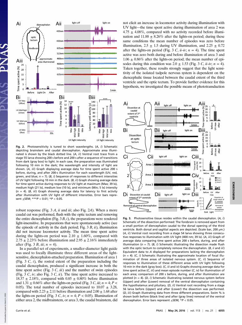
robust response (Fig. 3 A, ii and iii; also Fig. 2A). When a morecaudal cut was performed, flush with the optic tectum and removingthe entire diencephalon (Fig. 3 B, i), the preparations were renderedlight-insensitive. In preparations that were spontaneously active (seethe episode of activity in the dark period; Fig. 3 B, ii), illuminationdid not increase locomotor activity. The mean time spent activeduring the lights-on period was 2.10 ± 1.60%, compared with2.75 ± 2.23% before illumination and 2.95 ± 2.16% immediatelyafter (Fig. 3 B, iii; n = 4).In a parallel set of experiments, a smaller-diameter light guide
was used to focally illuminate three different areas of the light-sensitive, diencephalon-attached preparation. Illumination of area 1(Fig. 3 C, i), the rostral extent of the preparation including thecaudal diencephalon, produced a significant increase in both thetime spent active (Fig. 3 C, iii) and the number of swim episodes(Fig. 3 C, iv; also Fig. 3 C, ii). The time spent active increased to18.37 ± 2.18%, compared with 0.85 ± 0.80% before illuminationand 1.31 ± 0.66% after the lights-on period (Fig. 3 C, iii; n = 4; P <0.05). The total number of episodes increased to 10.07 ± 3.28,compared with 2.23 ± 2.11 before illumination and 2.00 ± 1.68 afterthe lights-on period (Fig. 3 C, iv; n = 4; P < 0.05). Illumination ofeither area 2, the midbrainstem, or area 3, the caudal brainstem, did
not elicit an increase in locomotor activity during illumination withUV light—the time spent active during illumination of area 2 was4.75 ± 4.08%, compared with no activity recorded before illumi-nation and 11.88 ± 8.26% after the lights-on period; during thesesame conditions the mean number of episodes was zero beforeillumination, 2.5 ± 1.5 during UV illumination, and 2.25 ± 0.72after the lights-on period (Fig. 3 C, ii–iv; n = 4). The time spentactive was zero both during and before illumination of area 3 and1.06 ± 0.86% after the lights-on period; the mean number of epi-sodes during this condition was 2.0 ± 1.53 (Fig. 3 C, ii–iv; n = 4).Taken together, these results strongly suggest that the light sensi-tivity of the isolated tadpole nervous system is dependent on thediencephalic tissue located between the caudal extent of the thirdventricle and the optic tectum. To provide further evidence for thishypothesis, we investigated the possible means of phototransduction
Fig. 2. Photosensitivity is tuned to short wavelengths. (A, i) Schematicdepicting brainstem and caudal diencephalon. Approximate area illumi-nated is shown by the black dotted line. (A, ii) Ventral root trace from astage 55 larva showing 200 s before and 200 s after a sequence of transitionsfrom dark (gray box) to light. In each case, the preparation was illuminatedfollowing 10 min in the dark; the wavelength and intensity of light areshown. (A, iii) Graph displaying average data for time spent active 200 sbefore, during, and after 200-s illumination for each wavelength (UV, red,green, and blue; n = 7). (B, i) Sequence of responses to different intensitiesof UV light following 10 min in the dark. (B, ii) Graph showing average datafor time spent active during responses to UV light at maximum (Max; 39 lx),medium high (21 lx), medium low (10 lx), and minimum (Min; 5 lx) intensity(n = 4). (B, iii) Graph showing average data for latency to first activityafter illumination with UV light of different intensities. Error bars repre-sent ±SEM; ***P < 0.01; *P < 0.05.
Fig. 3. Photosensitive tissue resides within the caudal diencephalon. (A, i)Schematic of the dissection performed: The forebrain is removed apart froma small portion of diencephalon caudal to the dorsal opening of the thirdventricle. Both dorsal and sagittal aspects are depicted. (Scale bar, 200 μm.)(A, ii) Ventral root recording from a stage 54 larva showing three consecu-tive responses to illumination with UV light (400 nm; 39 lx). (A, iii) Graph ofaverage data comparing time spent active 200 s before, during, and afterillumination (n = 7). (B, i) Schematic illustrating the dissection made flushwith the optic tectum to completely remove the diencephalon. (B, ii and iii)Equivalent data to A displayed for preparations lacking the diencephalon(n = 4). (C, i) Schematic illustrating the approximate location of focal illu-mination of three areas of isolated nervous system. (C, ii) Sequence ofresponses to illumination of these different areas with UV light following10 min in the dark (gray box). (C, iii and iv) Graphs showing average data fortime spent active (C, iii) and mean episode number (C, iv) for illumination ofeach area; comparison of 200 s before, during, and after illumination areplotted (n = 4). (D, i) Schematic illustrating isolated nervous system before(Upper) and after (Lower) removal of the ventral diencephalon containingthe hypothalamus and pituitary. (D, ii) Ventral root recording from a stage56 larva before (Upper) and after (Lower) the dissection was performed.(D, iii) Graph illustrating data from three different preparations. Swim % isshown both before (black line) and after (gray lines) removal of the ventraldiencephalon. Error bars represent ±SEM; *P < 0.05.
Currie et al. PNAS | May 24, 2016 | vol. 113 | no. 21 | 6055
NEU
ROSC
IENCE
Dow
nloa
ded
by g
uest
on
Mar
ch 4
, 202
0
in the tadpole diencephalon, paying particular attention to the re-gion where the light sensitivity resides.Because most known phototransduction in the vertebrate
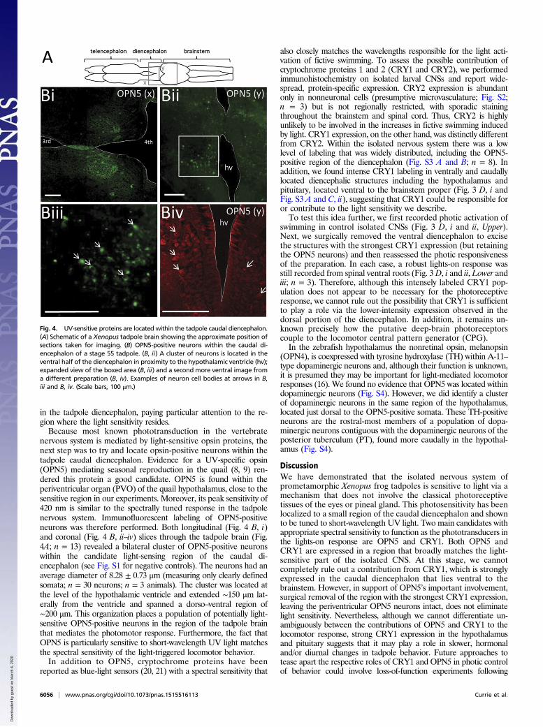
nervous system is mediated by light-sensitive opsin proteins, thenext step was to try and locate opsin-positive neurons within thetadpole caudal diencephalon. Evidence for a UV-specific opsin(OPN5) mediating seasonal reproduction in the quail (8, 9) ren-dered this protein a good candidate. OPN5 is found within theperiventricular organ (PVO) of the quail hypothalamus, close to thesensitive region in our experiments. Moreover, its peak sensitivity of420 nm is similar to the spectrally tuned response in the tadpolenervous system. Immunofluorescent labeling of OPN5-positiveneurons was therefore performed. Both longitudinal (Fig. 4 B, i)and coronal (Fig. 4 B, ii–iv) slices through the tadpole brain (Fig.4A; n = 13) revealed a bilateral cluster of OPN5-positive neuronswithin the candidate light-sensing region of the caudal di-encephalon (see Fig. S1 for negative controls). The neurons had anaverage diameter of 8.28 ± 0.73 μm (measuring only clearly definedsomata; n = 30 neurons; n = 3 animals). The cluster was located atthe level of the hypothalamic ventricle and extended ∼150 μm lat-erally from the ventricle and spanned a dorso–ventral region of∼200 μm. This organization places a population of potentially light-sensitive OPN5-positive neurons in the region of the tadpole brainthat mediates the photomotor response. Furthermore, the fact thatOPN5 is particularly sensitive to short-wavelength UV light matchesthe spectral sensitivity of the light-triggered locomotor behavior.In addition to OPN5, cryptochrome proteins have been
reported as blue-light sensors (20, 21) with a spectral sensitivity that
also closely matches the wavelengths responsible for the light acti-vation of fictive swimming. To assess the possible contribution ofcryptochrome proteins 1 and 2 (CRY1 and CRY2), we performedimmunohistochemistry on isolated larval CNSs and report wide-spread, protein-specific expression. CRY2 expression is abundantonly in nonneuronal cells (presumptive microvasculature; Fig. S2;n = 3) but is not regionally restricted, with sporadic stainingthroughout the brainstem and spinal cord. Thus, CRY2 is highlyunlikely to be involved in the increases in fictive swimming inducedby light. CRY1 expression, on the other hand, was distinctly differentfrom CRY2. Within the isolated nervous system there was a lowlevel of labeling that was widely distributed, including the OPN5-positive region of the diencephalon (Fig. S3 A and B; n = 8). Inaddition, we found intense CRY1 labeling in ventrally and caudallylocated diencephalic structures including the hypothalamus andpituitary, located ventral to the brainstem proper (Fig. 3 D, i andFig. S3 A and C, ii), suggesting that CRY1 could be responsible foror contribute to the light sensitivity we describe.To test this idea further, we first recorded photic activation of
swimming in control isolated CNSs (Fig. 3 D, i and ii, Upper).Next, we surgically removed the ventral diencephalon to excisethe structures with the strongest CRY1 expression (but retainingthe OPN5 neurons) and then reassessed the photic responsivenessof the preparation. In each case, a robust lights-on response wasstill recorded from spinal ventral roots (Fig. 3 D, i and ii, Lower andiii; n = 3). Therefore, although this intensely labeled CRY1 pop-ulation does not appear to be necessary for the photoreceptiveresponse, we cannot rule out the possibility that CRY1 is sufficientto play a role via the lower-intensity expression observed in thedorsal portion of the diencephalon. In addition, it remains un-known precisely how the putative deep-brain photoreceptorscouple to the locomotor central pattern generator (CPG).In the zebrafish hypothalamus the nonretinal opsin, melanopsin
(OPN4), is coexpressed with tyrosine hydroxylase (TH) within A-11–type dopaminergic neurons and, although their function is unknown,it is presumed they may be important for light-mediated locomotorresponses (16). We found no evidence that OPN5 was located withindopaminergic neurons (Fig. S4). However, we did identify a clusterof dopaminergic neurons in the same region of the hypothalamus,located just dorsal to the OPN5-positive somata. These TH-positiveneurons are the rostral-most members of a population of dopa-minergic neurons contiguous with the dopaminergic neurons of theposterior tuberculum (PT), found more caudally in the hypothal-amus (Fig. S4).
DiscussionWe have demonstrated that the isolated nervous system ofprometamorphic Xenopus frog tadpoles is sensitive to light via amechanism that does not involve the classical photoreceptivetissues of the eyes or pineal gland. This photosensitivity has beenlocalized to a small region of the caudal diencephalon and shownto be tuned to short-wavelength UV light. Two main candidates withappropriate spectral sensitivity to function as the phototransducers inthe lights-on response are OPN5 and CRY1. Both OPN5 andCRY1 are expressed in a region that broadly matches the light-sensitive part of the isolated CNS. At this stage, we cannotcompletely rule out a contribution from CRY1, which is stronglyexpressed in the caudal diencephalon that lies ventral to thebrainstem. However, in support of OPN5’s important involvement,surgical removal of the region with the strongest CRY1 expression,leaving the periventricular OPN5 neurons intact, does not eliminatelight sensitivity. Nevertheless, although we cannot differentiate un-ambiguously between the contributions of OPN5 and CRY1 to thelocomotor response, strong CRY1 expression in the hypothalamusand pituitary suggests that it may play a role in slower, hormonaland/or diurnal changes in tadpole behavior. Future approaches totease apart the respective roles of CRY1 and OPN5 in photic controlof behavior could involve loss-of-function experiments following
Fig. 4. UV-sensitive proteins are located within the tadpole caudal diencephalon.(A) Schematic of a Xenopus tadpole brain showing the approximate position ofsections taken for imaging. (B) OPN5-positive neurons within the caudal di-encephalon of a stage 55 tadpole. (B, ii) A cluster of neurons is located in theventral half of the diencephalon in proximity to the hypothalamic ventricle (hv);expanded view of the boxed area (B, iii) and a second more ventral image froma different preparation (B, iv). Examples of neuron cell bodies at arrows in B,iii and B, iv. (Scale bars, 100 μm.)
6056 | www.pnas.org/cgi/doi/10.1073/pnas.1515516113 Currie et al.
Dow
nloa
ded
by g
uest
on
Mar
ch 4
, 202
0

knockdown of the genes for these proteins, for example using theclustered regularly interspaced short palindromic repeats (CRISPR)/CRISPR-associated protein 9 (CAS9) system. However, this ap-proach is beyond the scope of the present study and would best betackled in a genetically more tractable model animal such asXenopus tropicalis.The discovery of neurons within this light-sensitive region of
the tadpole brain expressing the UV-specific opsin OPN5 stronglyimplies this protein is an important mediator of phototransduction.Because photosensitivity in vertebrates is thought to originate fromperiventricular neurons of the diencephalon, it seems plausible thatthis mechanism is phylogenetically conserved and may represent alight-detecting component present in the brain of a primitiveaquatic protovertebrate (23). An important facet of these experi-ments is that light sensitivity only links directly to the probability ofoccurrence of spontaneous locomotor activity. Upon illumination,the isolated nervous system produced regular episodes of fictivelocomotion, whereas in the dark the preparations were generallysilent. There were no differences between the coordination or basicparameters of the locomotor rhythm in the different light condi-tions, suggesting that the photic system of the brain controls merelyhow likely the animal is to swim.This deep-brain light sensitivity could function as a simple
mechanism to maintain the tadpole in an optimal photic environ-ment. It could, for example, help avoid exposure to UV radiationfrom the sun, which can cause DNA damage and is a remarkablywell conserved trait found even in bacteria (24). In addition, it mayhelp to avoid the brightest-lit areas of the environment, wheredetection by predators is likely to occur. This form of light-avoid-ance strategy is found in many fish species, where it is thought toconfer a specific advantage in the face of aerial predation (25). Inembryonic Xenopus tadpoles, light avoidance is achieved by apineal-driven motor response that causes upward swimming inresponse to shadows cast in the water (26, 27). Although thisbehavior is sufficient to maintain the relatively dormant embryosin an optimum environment for survival, the addition or pre-dominance of other light-sensitive systems during developmentmay aid survival in highly active, free-feeding larvae. Another,nonmutually exclusive, possibility is that the deep-brain lightsensitivity could overlay classical circadian control mechanisms,which regulate behavior in response to predictable diurnal fluc-tuations in the environment. Given the tuning of this response toshort wavelengths, it may be appropriate to detect subtle changesin the lighting conditions in an aquatic environment, where theinfluence of longer wavelengths is filtered out by the water.An important next step will be to determine which neuronal
pathway links the photoreceptive neurons to the activation of themotor system. The expression of the UV/blue light-sensitiveproteins OPN5 and CRY1 was located in relatively close proximityto a set of dopaminergic neurons potentially related to the A-11–type population, known to project to the spinal cord and controlmotor output in other species (28). However, it is also plausiblethat OPN5- and/or CRY-positive neurons activate other supra-spinal centers involved in vertebrate locomotion, such as themesencephalic locomotor region (MLR) in the midbrain and/orreticulospinal nuclei in the hind brain (29), for example. Both ofthese possibilities could be involved simultaneously, as dopami-nergic neurons within the PT of the lamprey project to and excitethe MLR directly (30).In zebrafish, the photoreceptors underlying dark photokinesis
have been localized to the anterior preoptic area, and they trans-duce light via the photopigment melanopsin (16). The photosen-sitivity we report in Xenopus is not mediated by the equivalentregion of the brain because the preoptic area has been removed inthese light-sensitive preparations. However, melanopsin was alsofound more caudally in zebrafish, in neurons of the PT (16), an areathat is present in the light-sensitive Xenopus preparations. Thisfinding is particularly relevant because the cells in question were
A-11–type dopaminergic neurons that comprise a diencephalospinalpopulation implicated in motor control (28). However, they areunlikely to mediate the phototransduction we document here. First,the original work that identified melanopsin as a photopigmentwas carried out in Xenopus and, although it was found in both thepreoptic and suprachiasmatic nuclei, there is no evidence for it beingpresent in the caudal hypothalamus (31). Second, because thephotomotor behavior in Xenopus is tuned to short-wavelength UVlight, it does not correspond to the profile of a melanopsin-mediatedresponse, which peaks around 480 nm (1, 32, 33).Alternatively, OPN5 is a UV-specific opsin that has recently
been shown to be a component of the photoperiodic response inquail (8, 9). OPN5 was located within the quail PVO, a caudalhypothalamic structure also present in the photosensitive tadpolepreparation. Moreover, the PVO cells of other species have beenshown to contain dopamine, noradrenaline, and/or serotonin (34),all known modulators of locomotion in Xenopus (35–37). An in-teresting example is the three-spined stickleback, which has largedopaminergic neurons in the PVO forming a contiguous group withthe dopaminergic neurons of the PT (38). This more caudal group isthought to be homologous to the dopaminergic neurons of themammalian zona incerta, which makes up the subthalamic di-encephalic locomotor region, an area important in the supraspinalcontrol of locomotion (39, 40).What is the behavioral significance of this novel photomotor
response in Xenopus tadpoles? The lighting conditions were atphysiological levels for a species native to ponds in South Africa:The broad-spectrum, white light was around 13,000 lx, within therange of intensities experienced during the day although not indirect sunlight [10,000–25,000 lx (41)]; the brightest LED (blue;468 nm) was ∼460 lx and therefore similar to the light intensityexperienced at sunrise or sunset; and the UV LED (390–410 nm)that elicited the maximal response to light only emitted 39 lx, andoccasionally elicited a response as low as 5 lx.Deep-brain photoreception may promote light-avoidance behav-
ior by increasing locomotor activity relative to light intensity, andthus increasing the likelihood of navigating to and settling in dimly litareas. A role for deep-brain photoreception in negative phototaxishas been shown in eels (13); however, the generalized increase inlocomotor activity seen in the isolated Xenopus nervous system ismore similar to the dark photokinesis behavior displayed by larvalzebrafish (16). In the eel, deep-brain photoreception was also shownto mediate photoentrainment to a circadian cycle of increasednocturnal activity (13). Although there is no evidence for circadianvariation in activity during larval life, adult Xenopus are nocturnal(42). Tadpoles of the American toad (Bufo americanus) display in-creased activity and feeding during the day and are generally inactiveovernight (43). In tadpoles of X. laevis, we propose that deep-brainphotoreception serves the dual purpose of reducing exposure to thedamaging influences of both predation and UV on the one hand andautomatically adjusting energetically expensive bouts of locomotoractivity to diurnal changes in light intensity on the other.
Materials and MethodsAnimals and Husbandry. Experiments were performed on a range ofprometamorphic stages of the SouthAfrican clawed frogX. laevis. Animalswereobtained as described previously (19) from an in-house breeding colony. Allprocedures conformed to UK Animals (Scientific Procedures) Act 1986 and theEuropean Community Council directive of November 24, 1986 (86/609/EEC) andhave been approved by the University of St Andrews Animal Welfare EthicsCommittee (AWEC).
Extracellular Electrophysiology Apparatus. After removal of the forebrain apartfrom the caudal-most portion of the diencephalon under Ethyl 3-aminobenzoatemethanesulfonate (MS-222; Sigma-Aldrich) anesthesia (19), the remaining ner-vous systemwas dissected free of the carcass, apart from the caudal-most portionof the tail, which was left attached to verify the preparation was capable ofnormal motor output. Recording conditions were as described previously (19).
Currie et al. PNAS | May 24, 2016 | vol. 113 | no. 21 | 6057
NEU
ROSC
IENCE
Dow
nloa
ded
by g
uest
on
Mar
ch 4
, 202
0

Light Sources. For experiments where the lighting conditions were manip-ulated, the recording apparatus was housed in a modified Faraday cagecovered with aluminum foil and blackout cloth. The light level in the cageduring lights-off was negligible (0 lx). Experiments with white light wereperformed with a standard halogen cold-light source (Olympus; Highlight2000), which emitted broad-spectrum light at ∼13,000 lx (low-voltage hal-ogen projection lamp; 14.5 V, 90 W; Phillips). When investigating the spectralsensitivity of the preparations, a series of LEDs were used (RS Components; allcatalog numbers are provided). The specifications were as follows: blue LED(466-3532), peak λ was 468 nm, brightness was 15,000 millicandela (mcd) or461 lx; green (671-6852), 523 nm, 21,000 mcd (136 lx); red (496-6178), 635 nm,16,000 mcd (36 lx); UV (713-5043), 400 nm (39 lx).
Immunohistochemistry. Tadpole brains were harvested from animals at stage55. The tissue was fixed overnight at 4 °C in FAA fixative [50% (vol/vol)ethanol; 10% (vol/vol) 37–40% formaldehyde; 5% (vol/vol) acetic acid indH2O], dehydrated through a graded alcohol series, and cleared in chloroformbefore wax embedding. Sections were cut at 8 μm on a rotary microtome andthen mounted on electrically charged slides. Sections were deparaffinized inxylene, rehydrated through a graded alcohol series, and washed in phosphate-buffered saline with Triton X-100 (PBS-T). High-temperature antigen retrievalwas performed in 0.1 M citrate buffer. Ten percent horse serum in PBS-T wasused to block nonspecific antibody binding, and then the primary antibody(1:1,000 rabbit anti-OPN5; 1:1,000 rabbit anti-CRY1; or 1:500 anti-CRY2; allAviva Systems Biology) was introduced and left overnight at 4 °C before de-tection with the secondary antibody (1:200 FITC–anti-rabbit; Vector Labs).Verification of the OPN5 antibody in Xenopus was carried out (Figs. S5 and S6);
cryptochrome antibodies were commercially verified as able to cross-react withXenopus proteins (Fig. S7). For double labeling, the previous two steps wererepeated with a second set of antibodies (1:1,000 mouse anti-TH, Sigma-Aldrichand 1:200 TRITC–anti-mouse, Vector Labs; or 1:1,000 rabbit anti-OPN5 and 1:200Texas red–anti-rabbit, Vector Labs). Following a final wash in PBS-T (5 ×5min), thesections were mounted in Citifluor and the coverslip was sealed with ethyl ace-tate. Following immunohistochemistry, images were obtained on a Zeiss AxioImager Ax10 at 40×magnification. Neuronal measurements, as well as tiling andstitching of individual images to create larger composites, were performed usingZEN Imaging Pro (v10; Zeiss) software.
Data Acquisition and Statistical Analysis. Extracellular signals were amplifiedusing differential AC amplifiers (A-M Systems; model 1700; low cutoff, 300 Hz;high cutoff, 500 Hz), digitized using a 1401 analog-to-digital acquisitionsystem (CED; Cambridge Electronic Design), and stored and processed on a PCusing Spike2 (CED) software (sampling rate 8–10 kHz). Electrophysiologicaldata were analyzed using DataView software (v8.62; courtesy of W.J. Heitler,University of St Andrews), and all raw data were imported into Excel (Micro-soft). Statistical analysis was performed in SPSS (IBM, v21). For comparison ofaverage data, either a paired t test or a repeated-measures ANOVA withBonferroni post hoc corrections was used. Error bars represent ±SEM. Due tolarge interpreparation variation, data were sometimes normalized to the valuein control (100%) for a more thorough comparison.
ACKNOWLEDGMENTS. We thank the University of St Andrews for support,and Laurence Picton for help with the figures. S.P.C. was supported by aBiotechnology and Biological Sciences Research Council (BBSRC) studentship.
1. Peirson SN, Halford S, Foster RG (2009) The evolution of irradiance detection: Mela-nopsin and the non-visual opsins. Philos Trans R Soc Lond B Biol Sci 364(1531):2849–2865.
2. Fernald RD (2000) Evolution of eyes. Curr Opin Neurobiol 10(4):444–450.3. von Frisch K (1911) Beiträge zur Physiologie der Pigmentzellen in der Fischhaut.
Gesamte Physiol Menschen Tiere 138(7):319–387.4. Benoit J (1935) Stimulation par la lumiere artificielle du developpement testiculaire
chez des canards aveugles par section du nerf optique. C R Seances Soc Biol Fil 120:133–136.
5. Menaker M, Keatts H (1968) Extraretinal light perception in the sparrow. II. Photo-periodic stimulation of testis growth. Proc Natl Acad Sci USA 60(1):146–151.
6. Siopes TD, Wilson WO (1974) Extraocular modification of photoreception in intactand pinealectomized coturnix. Poult Sci 53(6):2035–2041.
7. Foster RG, Follett BK, Lythgoe JN (1985) Rhodopsin-like sensitivity of extra-retinalphotoreceptors mediating the photoperiodic response in quail. Nature 313(5997):50–52.
8. Nakane Y, et al. (2010) A mammalian neural tissue opsin (Opsin 5) is a deep brainphotoreceptor in birds. Proc Natl Acad Sci USA 107(34):15264–15268.
9. Nakane Y, Shimmura T, Abe H, Yoshimura T (2014) Intrinsic photosensitivity of a deepbrain photoreceptor. Curr Biol 24(13):R596–R597.
10. Olson JM (2006) Photosynthesis in the Archean era. Photosynth Res 88(2):109–117.11. Young JZ (1935) The photoreceptors of lampreys I. Light-sensitive fibres in the lateral
line nerves. J Exp Biol 12:229–238.12. Young JZ (1935) The photoreceptors of lampreys II. The functions of the pineal
complex. J Exp Biol 12:254–270.13. van Veen T, Hartwig HG, Müller K (1976) Light-dependent motor activity and pho-
tonegative behavior in the eel (Anguilla anguilla L.). J Comp Physiol 111(2):209–219.14. Serra EL, Medalha CC, Mattioli R (1999) Natural preference of zebrafish (Danio rerio)
for a dark environment. Braz J Med Biol Res 32(12):1551–1553.15. Burgess HA, Schoch H, Granato M (2010) Distinct retinal pathways drive spatial ori-
entation behaviors in zebrafish navigation. Curr Biol 20(4):381–386.16. Fernandes AM, et al. (2012) Deep brain photoreceptors control light-seeking behavior
in zebrafish larvae. Curr Biol 22(21):2042–2047.17. Kokel D, et al. (2010) Rapid behavior-based identification of neuroactive small mol-
ecules in the zebrafish. Nat Chem Biol 6(3):231–237.18. Kokel D, et al. (2013) Identification of nonvisual photomotor response cells in the
vertebrate hindbrain. J Neurosci 33(9):3834–3843.19. Combes D, Merrywest SD, Simmers J, Sillar KT (2004) Developmental segregation of
spinal networks driving axial- and hindlimb-based locomotion in metamorphosingXenopus laevis. J Physiol 559(Pt 1):17–24.
20. Fogle K, Parson K, Dahm N, Holmes T (2011) CRYPTOCHROME is a blue-light sensorthat regulates neuronal firing rate. Science 331(6023):1409–1413.
21. VanVickle-Chavez SJ, Van Gelder RN (2007) Action spectrum of Drosophila crypto-chrome. J Biol Chem 282(14):10561–10566.
22. Sillar KT, Robertson RM (2009) Thermal activation of escape swimming in post-hatching Xenopus laevis frog larvae. J Exp Biol 212(Pt 15):2356–2364.
23. Vigh B, et al. (2002) Nonvisual photoreceptors of the deep brain, pineal organs andretina. Histol Histopathol 17(2):555–590.
24. Ng WO, Grossman AR, Bhaya D (2003) Multiple light inputs control phototaxis inSynechocystis sp. strain PCC6803. J Bacteriol 185(5):1599–1607.
25. Clark CW, Levy DA (1988) Diel vertical migrations by juvenile sockeye salmon and theantipredation window. Am Nat 131(2):271–290.
26. Foster RG, Roberts A (1982) The pineal eye in Xenopus laevis embryos and larvae:A photoreceptor with a direct excitatory effect on behaviour. J Comp Physiol 145(3):413–419.
27. Jamieson D, Roberts A (2000) Responses of young Xenopus laevis tadpoles to lightdimming: Possible roles for the pineal eye. J Exp Biol 203(Pt 12):1857–1867.
28. Clemens S, Rye D, Hochman S (2006) Restless legs syndrome: Revisiting the dopaminehypothesis from the spinal cord perspective. Neurology 67(1):125–130.
29. Jordan LM (1998) Initiation of locomotion in mammals. Ann N Y Acad Sci 860:83–93.30. Ryczko D, et al. (2013) Forebrain dopamine neurons project down to a brainstem
region controlling locomotion. Proc Natl Acad Sci USA 110(34):E3235–E3242.31. Provencio I, Jiang G, De Grip WJ, Hayes WP, Rollag MD (1998) Melanopsin: An opsin in
melanophores, brain, and eye. Proc Natl Acad Sci USA 95(1):340–345.32. Berson DM, Dunn FA, Takao M (2002) Phototransduction by retinal ganglion cells that
set the circadian clock. Science 295(5557):1070–1073.33. Hattar S, et al. (2003) Melanopsin and rod-cone photoreceptive systems account for
all major accessory visual functions in mice. Nature 424(6944):76–81.34. Vigh B, Vigh-Teichmann I (1998) Actual problems of the cerebrospinal fluid-contact-
ing neurons. Microsc Res Tech 41(1):57–83.35. Sillar KT, Wedderburn JF, Simmers AJ (1992) Modulation of swimming rhythmicity by
5-hydroxytryptamine during post-embryonic development in Xenopus laevis. ProcBiol Sci 250(1328):107–114.
36. McDearmid JR, Scrymgeour-Wedderburn JF, Sillar KT (1997) Aminergic modulation ofglycine release in a spinal network controlling swimming in Xenopus laevis. J Physiol503(1):111–117.
37. Clemens S, Belin-Rauscent A, Simmers J, Combes D (2012) Opposing modulatory ef-fects of D1- and D2-like receptor activation on a spinal central pattern generator.J Neurophysiol 107(8):2250–2259.
38. Ekström P, Honkanen T, Borg B (1992) Development of tyrosine hydroxylase-, dopa-mine- and dopamine beta-hydroxylase-immunoreactive neurons in a teleost, thethree-spined stickleback. J Chem Neuroanat 5(6):481–501.
39. Parker SM, Sinnamon HM (1983) Forward locomotion elicited by electrical stimulationin the diencephalon and mesencephalon of the awake rat. Physiol Behav 31(5):581–587.
40. Milner KL, Mogenson GJ (1988) Electrical and chemical activation of the mesence-phalic and subthalamic locomotor regions in freely moving rats. Brain Res 452(1–2):273–285.
41. Clark RN (1990) Visual Astronomy of the Deep Sky (Sky, Cambridge, MA).42. Casterlin M, Reynolds W (1980) Diel activity and thermoregulatory behavior of a fully
aquatic frog: Xenopus laevis. Hydrobiologia 75(2):247–254.43. Beiswenger RE (1977) Diel patterns of aggregative behaviour in tadpoles of Bufo
americanus, in relation to light and temperature. Ecology 58(1):98–108.
6058 | www.pnas.org/cgi/doi/10.1073/pnas.1515516113 Currie et al.
Dow
nloa
ded
by g
uest
on
Mar
ch 4
, 202
0