DEDICATED EXTREMITY 1.5T M.R. IMAGING: THE FUTURE OF ANKLE IMAGING
42
DEDICATED EXTREMITY 1.5T M.R. IMAGING: THE FUTURE OF ANKLE IMAGING Wilf d CG P h Wilfred CG Peh Khoo Teck Puat Hospital, Singapore & Khoo Teck Puat Hospital, Singapore & National University of Singapore
Transcript of DEDICATED EXTREMITY 1.5T M.R. IMAGING: THE FUTURE OF ANKLE IMAGING

DEDICATED EXTREMITY 1.5T M.R. IMAGING: THE FUTURE
OF ANKLE IMAGINGWilf d CG P hWilfred CG Peh
Khoo Teck Puat Hospital, Singapore &Khoo Teck Puat Hospital, Singapore &National University of Singapore
LECTURE OUTLINELECTURE OUTLINE• Introduction• Why dedicated extremity MRI?• Applications in the ankle• Summary• Summary

ACKNOWLEDGEMENTACKNOWLEDGEMENTI thank my colleagues Mr Michael Chin for his input and Dr Mohammed Isham Othman for hisMohammed Isham Othman for his help in doing some of the background research for thisbackground research for this presentation
INTRODUCTIONINTRODUCTIONDedicated extremity MR systems • Were developed as an
y y
alternative to high-field whole b dbody systems• cost savingscost savings• patient comfort
INTRODUCTIONINTRODUCTIONHistorical perspectives• Introduced in 1993 by Esaote
p p
Biomedica [Genoa, Italy]A B i (1992)• Artoscan Basic (1992)
• 0 2 T• 0.2 T• No fat suppression pp

INTRODUCTIONINTRODUCTIONHistorical perspectives• MagneVu MV1000 [Carlsbad,
p p
CA] (0.18T) Bankrupt 2007
Used MRI advert. Price USD9,000!
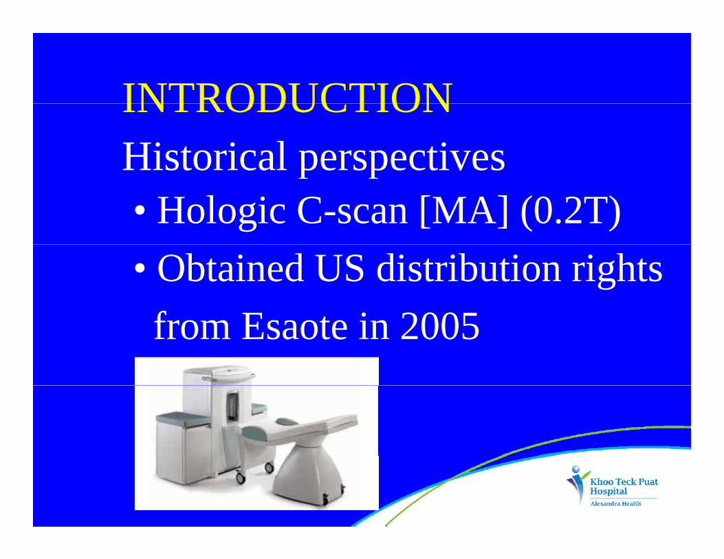
INTRODUCTIONINTRODUCTIONHistorical perspectives• Hologic C-scan [MA] (0.2T)
p p
• Obtained US distribution rights f E i 2005from Esaote in 2005
INTRODUCTIONINTRODUCTIONHistorical perspectives• 0.2T disadvantages
p p
• lower image resolution• fewer image contrast mechanisms• fewer image contrast mechanisms• but met need of rheumatologistsg(occult erosions, synovitis)

INTRODUCTIONINTRODUCTIONHistorical perspectives• 1st high-field extremity MRI
p p
• superconducting magnet• developed by ONI [MA] (1997)• developed by ONI [MA] (1997)• Ortho One (1.0T) in 2000( )

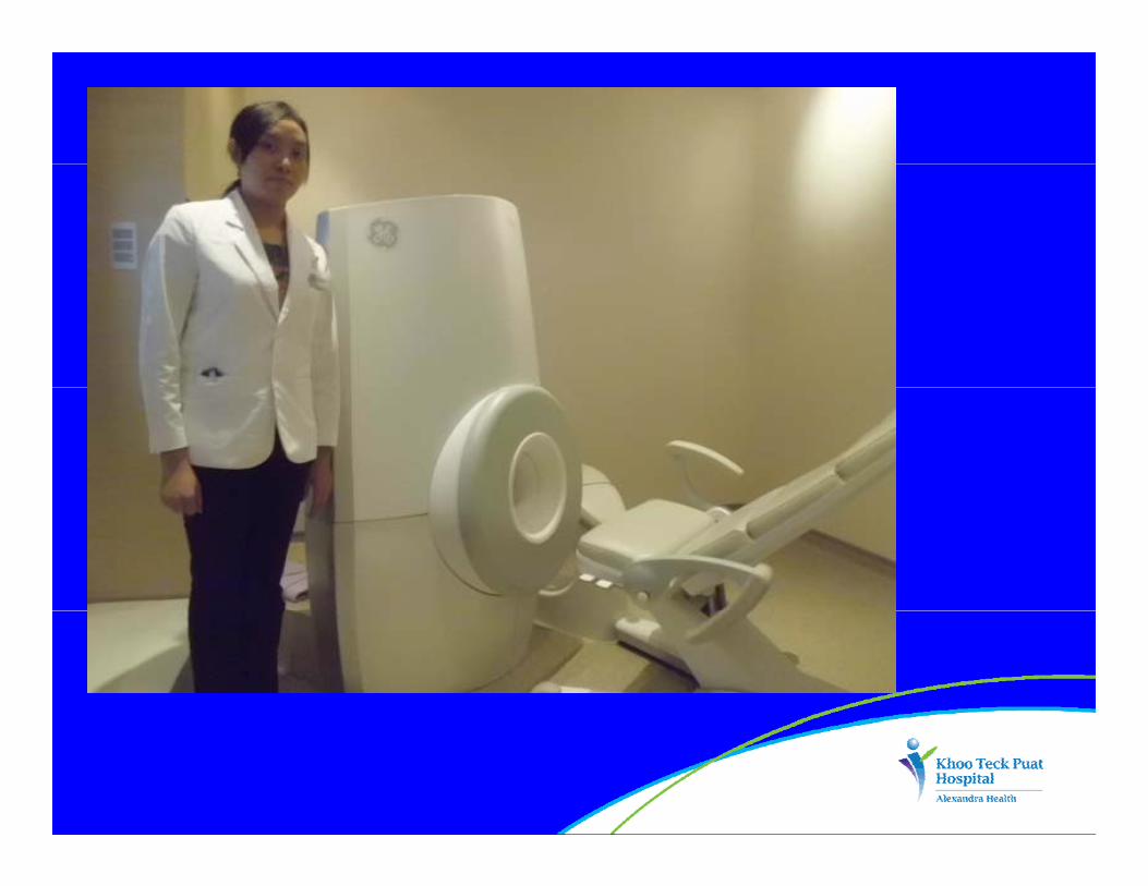
INTRODUCTIONINTRODUCTIONHistorical perspectives• 1st high-field extremity MRI
p p
• acquired by GE in 2009• Optima MR430s (1 5T)• Optima MR430s (1.5T) • installed at KTPH in Jan 2012

Other MRI units at DDR, KTPH• Siemens MagnetomAvanto (1.5T whole body) June 2010
• GE 750W wide bore (3.0T whole body) January 2012

WHY EXTREMITY MRI?WHY EXTREMITY MRI?High image quality• Slew rate 300mT/m/ms
g g q y
• Max gradient power 70mT/m• RF power 2000 peak rms 75W ave• RF power 2000 peak rms, 75W ave• Sequences: spin echo (SE), FSE, q p ( )FSE fat/water, 2D GE, 3D GE, among others

WHY EXTREMITY MRI?WHY EXTREMITY MRI?Cost effectiveness• Relieves whole-body MRI of imaging extremities
• Knees ankles wrists hands arms• Knees, ankles, wrists, hands, arms comprise 20-25% of all scansp
• Price: approx 50% of whole body scanner

WHY EXTREMITY MRI?WHY EXTREMITY MRI?Cost effectiveness• Small footprint: site 15.5 sq m• Light weight: 408 kg• Small scale magnetic shielding• Small scale magnetic shielding• Easy MRI set-up & site preparationy p p p• Fits through standard doorways and elevators
WHY EXTREMITY MRI?WHY EXTREMITY MRI?Cost effectiveness• Uses standard commercial electric power
• Less helium to cool magnet• Less helium to cool magnet

WHY EXTREMITY MRI?WHY EXTREMITY MRI?Operator friendly• Windows-based user interfacep y
• Quick and easy patient positioning• Target anatomy placed in isocentre• Target anatomy placed in isocentreof magnet bore - choice of different gsized volume coils to choose from
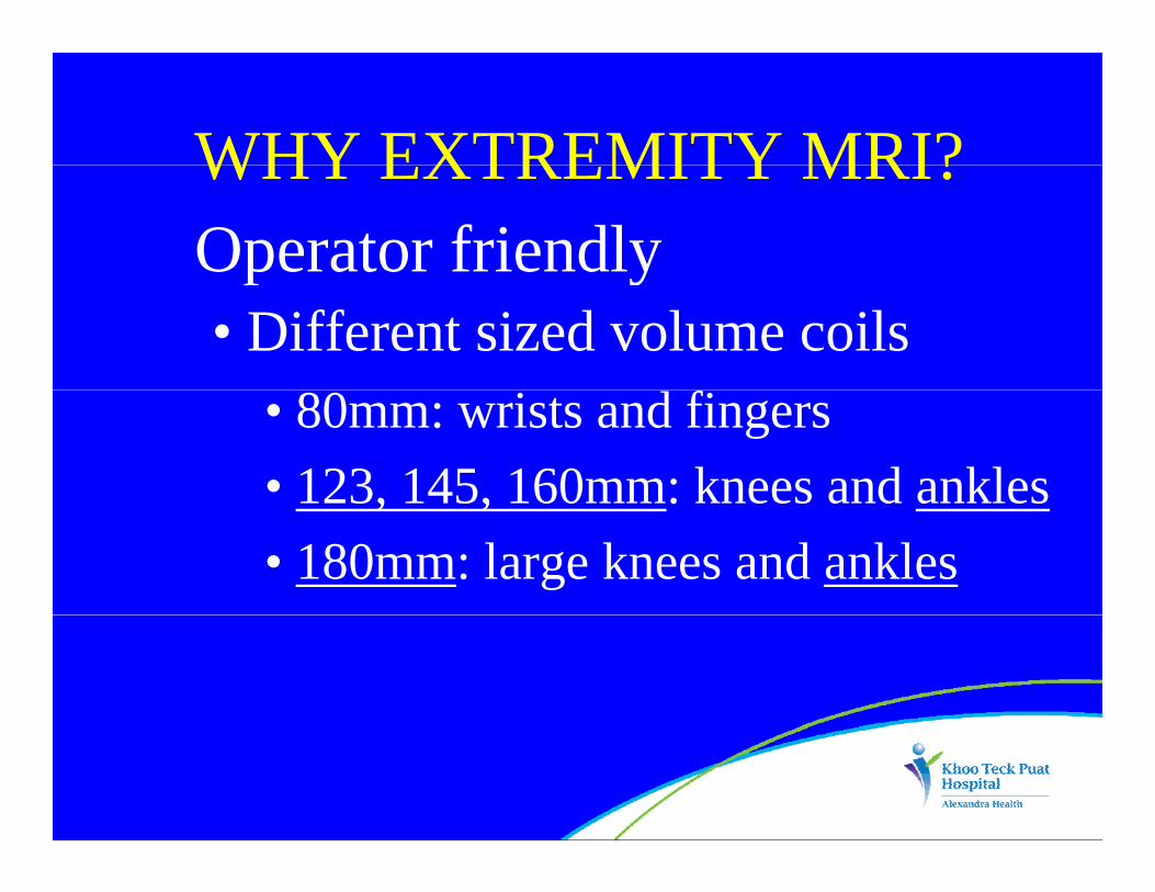
WHY EXTREMITY MRI?WHY EXTREMITY MRI?Operator friendly• Different sized volume coilsp y
• 80mm: wrists and fingers• 123, 145, 160mm: knees and ankles123, 145, 160mm: knees and ankles• 180mm: large knees and ankles


WHY EXTREMITY MRI?WHY EXTREMITY MRI?Patient comfort• Extremity is placed in magnet bore• Rest of patient is outside: sits or lies on reclinable padded chairon reclinable padded chair
• Quiet operation: no loud noisep• Good for claustrophobic patients
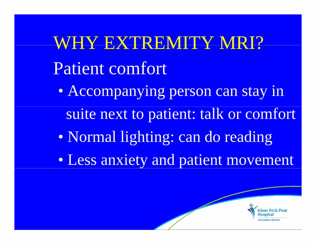
WHY EXTREMITY MRI?WHY EXTREMITY MRI?Patient comfort• Accompanying person can stay in suite next to patient: talk or comfort
• Normal lighting: can do reading• Normal lighting: can do reading• Less anxiety and patient movement y p


ANKLE APPLICATIONSANKLE APPLICATIONSOne year of clinical experience• Number of ankles/feet scanned
y p
to date: Jan 2012-Jan 2013 = 165• Main indication• Main indication
• sports injuriesp j• ligament and tendon injuries, ?OCDs

ANKLE APPLICATIONSANKLE APPLICATIONSStructures • Ligaments• Tendons• Bone marrow• Bone marrow• Subchondral and osteochondrallesions

24-year-old man: sagittal images24 year old man: sagittal images

24-year-old man: yaxial images

ANKLE APPLICATIONSANKLE APPLICATIONSScan performance • Fast scan sequences
p
• Homogeneous fat saturation (using the IDEAL sequence)(using the IDEAL sequence)
• Some ‘free’ sequencesq

41-year-old man: Achilles tendinopathy

28 ld fl h ll i l28-year-old man: flexor hallucis longustenosynovitis and talus bone bruising

26-year-old man: ATFL complete tear

61-year-old man: talar dome early osteochondral lesions

20 ld20-year-old man: accessory navicularsyndrome with normal tibialis posterior tendon

ANKLE APPLICATIONSANKLE APPLICATIONSPotential pitfalls • Patient too big: >150 kg
p
• Foot is plantar-flexed• buckling of Achilles tendonbuckling of Achilles tendon• mildly oblique axial images of ankle
• Incomplete fat saturation for 3D GE images3D GE images
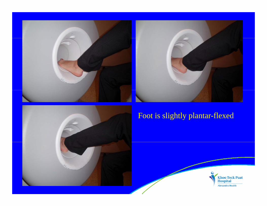
Foot is slightly plantar flexedFoot is slightly plantar-flexed

Plantar-flexed foot• buckling of Achilles tendon• oblique axial imageq g

3D GE images: incomplete fat saturation

ANKLE APPLICATIONSANKLE APPLICATIONSPotential pitfalls • Experience and training need to
p
place target anatomy in isocentre• Positioning of ankle to ensure that• Positioning of ankle to ensure that there is no movement
• Software does not allow double echo sequences

FUTUREFUTURE• Niche market for extremity MRIy
• cost-effective• One-stop service
li i t di i / h b• clinics: sports medicine/rehab, rheumatology, orthopaedicseu a o ogy, o opaed cs
• sports institutes and teams

FUTUREFUTURE• Clinical trials on small animals• Veterinary applications• Neonatal imaging

SUMMARYSUMMARYDedicated extremity 1.5T MRIy• Supplements a standard high-field whole-body MRI scannerC ff i• Cost-effective
• Produces high quality images• Produces high quality images