Decision-making processes following damage ... - Facundo...
16
Decision-making processes following damage to the prefrontal cortex Facundo Manes, 1, * Barbara Sahakian, 1 Luke Clark, 3 Robert Rogers, 3,4 Nagui Antoun, 2 Mike Aitken 3 and Trevor Robbins 3 1 University of Cambridge Psychiatry Department and 2 Department of Radiology, Addenbrooke’s Hospital, Cambridge, 3 Department of Experimental Psychology, University of Cambridge and 4 Department of Psychiatry, University of Oxford, Warneford Hospital, Oxford, UK Correspondence to: Dr Barbara Sahakian, University of Cambridge Psychiatry Department, Addenbrooke’s Hospital, Box 189, Cambridge CB2 2QQ, UK E-mail: [email protected] *Present address: Cognitive Neurology Division, Department of Neurology, Raul Carrea Institute of Neurological Research, Montan ˜eses 2325 (1428), Buenos Aires, Argentina Summary Recent work has suggested an association between the orbitofrontal cortex in humans and practical decision making. The aim of this study was to investigate the profile of cognitive deficits, with particular emphasis on decision-making processes, following damage to differ- ent sectors of the human prefrontal cortex. Patients with discrete orbitofrontal (OBF) lesions, dorsolateral (DL) lesions, dorsomedial (DM) lesions and large fron- tal lesions (Large) were compared with matched con- trols on three different decision-making tasks: the Iowa Gambling Task and two recently developed tasks that attempt to fractionate some of the cognitive components of the Iowa task. A comprehensive battery including the assessment of recognition memory, working memory, planning ability and attentional set-shifting was also administered. Whilst combined frontal patients were impaired on several of the tasks employed, distinct pro- files emerged for each patient group. In contrast to pre- vious data, patients with focal OBF lesions performed at control levels on the three decision-making tasks (and the executive tasks), but showed some evidence of prolonged deliberation. DL patients showed pronounced impairment on working memory, planning, attentional shifting and the Iowa Gambling Task. DM patients were impaired at the Iowa Gambling Task and also at planning. The Large group displayed diffuse impair- ment, but were the only group to exhibit risky decision making. Methodological differences from previous stud- ies of OBF patient groups are discussed, with particular attention to lesion laterality, lesion size and psychiatric presentation. Ventral and dorsal aspects of prefrontal cortex must interact in the maintenance of rational and ‘non-risky’ decision making. Keywords: prefrontal cortex; decision making; risk taking; orbitofrontal; executive function Abbreviations: AcoA = anterior communicating artery; BA = Brodmann area; CANTAB = Cambridge Neuropsycho- logical Test Automated Battery; DL = dorsolateral; DM = dorsomedial; EDS = extra-dimensional shift; ID-ED = intra-dimensional/extra-dimensional shift; IDS = intra-dimensional shift; OBF = orbitofrontal; PFC = prefrontal cortex Introduction Patients with damage involving orbitofrontal (OBF) cortex have been reported to display severe impairments in real-life decision making, despite remaining unimpaired intellectually and on traditional neuropsychological measures (Eslinger and Damasio, 1985; Shallice and Burgess, 1991). This syndrome has been labelled ‘acquired sociopathy’, and is characterized by repeated engagement in high-risk behaviours that are rewarding in the short term but have likely negative consequences for the patient’s well-being. The engagement in such behaviours has been proposed to arise from impaired decision making between various response options on the basis of faulty ‘somatic marking’ (Damasio, 1994; Bechara et al., 2000). These behaviours may be quantifiable using neuropsychological measures derived from everyday deci- sion making. Bechara et al. (1994) developed a task (the Iowa Gambling Task) where subjects must make a series of card ª Guarantors of Brain 2002 Brain (2002), 125, 624–639
Transcript of Decision-making processes following damage ... - Facundo...

Decision-making processes following damage tothe prefrontal cortex
Facundo Manes,1,* Barbara Sahakian,1 Luke Clark,3 Robert Rogers,3,4 Nagui Antoun,2 Mike Aitken3
and Trevor Robbins3
1University of Cambridge Psychiatry Department and2Department of Radiology, Addenbrooke's Hospital,
Cambridge, 3Department of Experimental Psychology,
University of Cambridge and 4Department of Psychiatry,
University of Oxford, Warneford Hospital, Oxford, UK
Correspondence to: Dr Barbara Sahakian, University of
Cambridge Psychiatry Department, Addenbrooke's
Hospital, Box 189, Cambridge CB2 2QQ, UK
E-mail: [email protected]
*Present address: Cognitive Neurology Division,
Department of Neurology, Raul Carrea Institute of
Neurological Research, MontanÄeses 2325 (1428), Buenos
Aires, Argentina
SummaryRecent work has suggested an association between theorbitofrontal cortex in humans and practical decisionmaking. The aim of this study was to investigate thepro®le of cognitive de®cits, with particular emphasis ondecision-making processes, following damage to differ-ent sectors of the human prefrontal cortex. Patientswith discrete orbitofrontal (OBF) lesions, dorsolateral(DL) lesions, dorsomedial (DM) lesions and large fron-tal lesions (Large) were compared with matched con-trols on three different decision-making tasks: the IowaGambling Task and two recently developed tasks thatattempt to fractionate some of the cognitive componentsof the Iowa task. A comprehensive battery including theassessment of recognition memory, working memory,planning ability and attentional set-shifting was alsoadministered. Whilst combined frontal patients wereimpaired on several of the tasks employed, distinct pro-
®les emerged for each patient group. In contrast to pre-vious data, patients with focal OBF lesions performedat control levels on the three decision-making tasks(and the executive tasks), but showed some evidence ofprolonged deliberation. DL patients showed pronouncedimpairment on working memory, planning, attentionalshifting and the Iowa Gambling Task. DM patientswere impaired at the Iowa Gambling Task and also atplanning. The Large group displayed diffuse impair-ment, but were the only group to exhibit risky decisionmaking. Methodological differences from previous stud-ies of OBF patient groups are discussed, with particularattention to lesion laterality, lesion size and psychiatricpresentation. Ventral and dorsal aspects of prefrontalcortex must interact in the maintenance of rational and`non-risky' decision making.
Keywords: prefrontal cortex; decision making; risk taking; orbitofrontal; executive function
Abbreviations: AcoA = anterior communicating artery; BA = Brodmann area; CANTAB = Cambridge Neuropsycho-
logical Test Automated Battery; DL = dorsolateral; DM = dorsomedial; EDS = extra-dimensional shift; ID-ED =
intra-dimensional/extra-dimensional shift; IDS = intra-dimensional shift; OBF = orbitofrontal; PFC = prefrontal cortex
IntroductionPatients with damage involving orbitofrontal (OBF) cortex
have been reported to display severe impairments in real-life
decision making, despite remaining unimpaired intellectually
and on traditional neuropsychological measures (Eslinger and
Damasio, 1985; Shallice and Burgess, 1991). This syndrome
has been labelled `acquired sociopathy', and is characterized
by repeated engagement in high-risk behaviours that are
rewarding in the short term but have likely negative
consequences for the patient's well-being. The engagement
in such behaviours has been proposed to arise from impaired
decision making between various response options on the
basis of faulty `somatic marking' (Damasio, 1994; Bechara
et al., 2000). These behaviours may be quanti®able using
neuropsychological measures derived from everyday deci-
sion making. Bechara et al. (1994) developed a task (the Iowa
Gambling Task) where subjects must make a series of card
ã Guarantors of Brain 2002
Brain (2002), 125, 624±639
selections resulting in winning and losing money. The four
card decks are characterized by different reward±punishment
pro®les, such that decks A and B offer high rewards but
higher penalties, resulting in overall loss, whereas decks C
and D offer smaller rewards but minimal penalties, resulting
in overall pro®t. Healthy controls developed a preference for
the `safe' decks by about trial 40 (of 100 choices). However, a
small group of frontal patients, with damage including the
medial OBF cortex, typically preferred the riskier decks for
the duration of the task, and also failed to develop anticipa-
tory skin responses prior to risky decisions (Bechara et al.,
1994, 1996). The de®cit cannot be readily explained in terms
of working memory impairment, as these patients were
capable of performing a delay task, sensitive to more dorsal
prefrontal damage (Bechara et al., 1998).
Although the working memory component of the Iowa
Gambling Task may have been controlled for, `risky'
decision making on the task could still be associated with a
number of potentially dissociable mechanisms, including
reduced deliberation, poor learning of outcome probabilities,
genuine preference for risky outcomes, and de®cits in
strategy acquisition and maintenance. We have developed a
task with the aim of dissecting these components further
(Rogers et al., 1999a). In the `Gamble' task, subjects must
initially make a fairly simple probabilistic decision, and must
then gamble points on their con®dence in this decision. A
strength of the task is that all the information needed to make
the decision and place the bet is visually presented on the
screen, and each trial is relatively independent of the last;
hence, working memory and learning processes are mini-
mized. The task also aims to dissociate motor impulsivity
from genuine risk-taking behaviour: the bets are presented in
both ascending and descending series, and therefore in the
ascending condition, subjects must suppress their response to
place a higher bet. Using the `Gamble' task we have
demonstrated that patients with OBF cortex damage show
impaired quality of decision making (choosing the likely
outcome on fewer trials), deliberate for longer about their
decisions and bet reduced amounts (Rogers et al., 1999a).
Patients with dorsolateral (DL) and dorsomedial (DM)
prefrontal lesions behaved similarly to controls on the task
(Rogers et al., 1999a).
The Bechara et al. and Rogers et al. investigations of
decision-making cognition in frontal patients have both been
limited in several ways. In particular, patient groups with
damage including OBF cortex rather than restricted to OBF
cortex have been studied. In the case series examined by
Bechara et al. (1994, 1996, 1998), the lesion overlap between
patients was greatest in the medial orbitofrontal region, but
lesions extended in several patients to anterior dorsolateral
cortex, cingulate gyrus and temporal pole. In the Rogers et al.
(1999a) study, 10 patients with frontal damage including
OBF cortex were compared with 10 patients with damage
restricted to the DL and DM prefrontal cortex (PFC). To
con®rm the involvement of OBF cortex in decision making it
is necessary to study patients with more focal lesions. The
effect of lesion laterality has also been inadequately assessed:
the Bechara group case series had mostly bilateral lesions,
whilst the Rogers group had a mixture of unilateral and
bilateral damage. A recent abstract from Tranel et al. (2000)
has indicated that right-lateralized damage may preferentially
affect decision-making processes. Finally, previous studies
have overlooked the potentially confounding effects of
psychiatric symptoms. Secondary mood disturbance (both
depression and mania) is well-documented following frontal
cortex damage (e.g. see Robinson et al., 1988) and may
modulate decision-making processes independently of the
organic damage, e.g. see Murphy et al., 2001).
The `Gamble' task was subsequently adapted for use in
functional neuroimaging. In the modi®ed `Risk' task (Rogers
et al., 1999b), the subject must again choose between two
mutually exclusive options, but the decision making and
betting responses are now combined, and the level of reward
associated with each bet is systematically pitted against the
likelihood of reward. The larger reward is always associated
with the least likely outcome to ensure an element of con¯ict,
which was not an original feature of the Gamble task. A PET
investigation in healthy subjects revealed signi®cant activa-
tions associated with resolution of reward con¯ict in three
foci in inferior frontal cortex: the anterior part of the middle
frontal gyrus [Brodmann area (BA) 10], in the orbital gyrus
(BA 11) and in the anterior portion of the inferior frontal
gyrus (BA 47) (Rogers et al., 1999b).
The impetus for the present investigation was to compare
decision-making cognition on the three tasks described
above, in patients with unilateral lesions restricted to speci®c
subregions of the prefrontal cortex. The modi®ed Risk task
(Rogers et al., 1999b) has not previously been used in brain-
damaged patients. In order to re®ne the measurement of
decision-making cognition, it is critical to compare directly
the sensitivity and speci®city of the tasks currently in use. The
present study examined the effects of restricted OBF, DL and
DM lesions on decision-making performance. A fourth group
of patients with large frontal lesions involving both dorsal and
ventral aspects of PFC was also examined. Patients with
bilateral lesions or lesions extending outside of the frontal
lobe were speci®cally excluded.
The second objective of the present study was to examine
the effects of discrete damage to prefrontal subregions on
other aspects of executive function. This would enable
comparison of the OBF de®cit pro®le with the impairments
traditionally seen following frontal lobe damage, and also
permit investigation of the relationship between decision
making and other forms of executive function. Human lesion
studies have robustly implicated PFC in the mediation of
working memory (Owen et al., 1990, 1999; D'Esposito and
Postle, 1999), planning (Owen et al., 1990; Morris et al.,
1997), attentional set-shifting (Milner, 1963; Owen et al.,
1991; Stuss et al., 2000) and verbal ¯uency (Borkowski et al.,
1967; Stuss et al., 1998), and whilst neuroimaging research
has suggested focal (and dissociable) activations within the
PFC (e.g. Baker et al., 1996), there is a paucity of data on the
Prefrontal cortex and decision making 625

effects of lesions to PFC subregions on these tasks (for recent
exceptions, see Stuss et al., 1998, 2000). Neuroimaging
research is unable to demonstrate the necessary involvement
of an activated brain region for task performance, and whilst
repetitive transcranial magnetic stimulation may have the
capacity to induce highly localized `transient lesions' in
healthy subjects during cognitive task performance, repetitive
transcranial magnetic stimulation is at present only effective
at regions lying near the scalp and therefore cannot be used to
stimulate inferior PFC. Human lesion research therefore
remains particularly valuable as a means of assessing the role
of PFC in cognition, and moreover, provides direct informa-
tion useful for diagnosis and rehabilitation after brain
damage.
MethodsSubjectsPatients were recruited from the CCNRP (Cambridge
Cognitive Neuroscience Research Panel) at the MRC
Cognition and Brain Sciences Unit (n = 18) and from the
King's College Hospital in London (n = 1). The CCNRP is an
accumulating database of volunteers with focal brain lesions.
The study was approved by the Cambridge Local Research
Ethics Committee and patients gave informed consent to
participate. All patients had a single focal lesion, veri®ed by
MRI, con®ned to frontal structures. Lesion aetiology was
mostly cerebrovascular haemorrhage or tumour resection
(Table 1). Exclusion criteria were current/previous psychiat-
ric diagnoses, colour blindness, neurological disease other
than that determining inclusion in the study and history of
diffuse brain damage.
Healthy control volunteers from the Cambridge area were
obtained through an advertisement in the local newspaper and
were paid. Controls were closely matched with patients for
age and National Adult Reading Test (NART) (Nelson, 1982)
estimate of premorbid verbal IQ (Table 2). There were no
differences between frontal subgroups and controls in terms
of age [F(4,27) = 1.48, P = 0.235] or NART score [F(4,27) =
0.966, P = 0.442].
Neuroradiological assessmentEighteen patients received MRI scans of the brain, with 3D
set acquisition in the coronal plane using a SPGR (spin
gradient echo) T1-weighted sequence and a T2-weighted axial
sequence. One subject had his lesion location determined
from a previous MRI scan. MRI scans were interpreted by a
neurologist with experience in structural neuroimaging
(F.M.) and a senior neuroradiologist (N.A.) who were both
blind to the experimental results. Lesions were traced using
Table 1 Characteristics of individual patients
Subjects Age (years) Sex Aetiology Side Years post-onset Medication
OrbitofrontalC.E. 61 M Excs. oligodendroglioma L 12 CarbamazepineM.B. 47 M Haemorrhage L 5 Phenytoin, folic acidE.H. 44 F Excs. meningioma L 6 CarbamazepineE.T. 49 F Haemorrhage (AcoA) R 3 PhenytoinS.D. 39 F Haemorrhage (AcoA) L 2 Phenytoin
DorsolateralA.D. 63 F Infarct L 5 AspirinS.H. 44 F AVM R 8 Phenytoin, lamotrigineC.G. 52 F Excs. oligodendroglioma R 40 Aspirin, atenolol, oestrogens, carbamazepineK.G. 64 F Haemorrhage (AcoA) R 2 None
DorsomedialP.P. 59 F Excs. meningioma L 3 NoneG.D. 46 F Excs. oligodendroglioma L 14 PhenytoinS.J. 73 M Infarction L 4 AspirinD.T. 69 M Infarction R 4 Warfarin, simvastatimJ.T. 58 M Excs. meningioma L 5 None
LargeM.M. 46 M Haemorrhage (AcoA) L 2 NoneJ.K. 53 F Haemorrhage (AcoA) R 3 CarbamazepineD.R. 55 M Excs. meningioma R 2 NoneM.K. 59 F Excs. oligodendroglioma R 2 NoneC.P. 37 F Haemorrhage (AcoA) R 8 Sodium valproate
Table 2 Subject characteristics [mean (SEM)]
Group n Gender (M : F) Age (years) NART IQ
Orbitofrontal 5 2 : 3 47.8 (8.5) 119 (4.6)Dorsolateral 4 0 : 4 55.7 (9.5) 120 (4.6)Dorsomedial 5 3 : 2 61.0 (10.6) 111 (9.9)Large lesion 5 2 : 3 50.0 (8.7) 115 (9.0)Controls 13 6 : 7 52.7 (10.9) 116 (9.6)
626 F. Manes et al.

MRIcro v. 1.25 (available at http://www.psychology.nottin-
gham.ac.uk/staff/cr1/mricro.html) and normalized to a stand-
ard template using SPM96 (statistical parametric mapping)
(Friston et al., 1995) with cost function masking (Brett et al.,
2002) to mask the lesion from the calculation of the
normalization parameters. Lesions were then localized with
standard atlases (Talairach and Tournoux, 1988; Damasio,
1995) and four groups were identi®ed: ®ve patients with
discrete OBF lesions, four patients with discrete DL lesions,
®ve patients with discrete DM lesions and ®ve patients with
lesions involving the ventral and dorsal areas of the prefrontal
cortex (Large lesions group) (see Figs 1±4).
Neuropsychological testingWith the exception of verbal ¯uency, neuropsychological
testing was computerized and run on a Datalux 486 PC with a
26.7 cm touch-sensitive monitor. Several tasks were taken
from the Cambridge Neuropsychological Test Automated
Battery (CANTAB) (CeNeS PLC, Cambridge, UK; Robbins
et al., 1994, 1998). Testing lasted 3±4 h and was consequently
split over two sessions at least 1 week apart. Patients were
tested at home. Testing was administered in a ®xed order,
with the exception that the order of the Gamble and Risk tasks
was counterbalanced across subjects.
Verbal ¯uency (Benton and Hamsher, 1976)This is a traditional neuropsychological assessment in which
subjects are required to generate as many words as possible
beginning with the letters F, A and S, each in 1 min. Scores
are summed across the 3 min. In the second part of this test
subjects are asked to produce as many exemplars as they can
from the semantic category `Animals' in a period of 90 s.
Spatial span (CANTAB; Owen et al., 1990)This is a visuospatial short-term memory test based upon the
Corsi Block Tapping Task (Milner, 1971). Subjects are
shown a series of white boxes in a spatial array that change
Fig. 1 Location of lesions in patients included in the OBF group. Patient 1: limited damage involving the left medial orbital surface,including the gyrus rectus. Patient 2: damage to the left inferior orbital PFC, including the orbital gyrus. Patient 3: cortical loss involvingmost of inferior right PFC including the gyrus rectus and orbital gyrus, but sparing the middle frontal gyrus in the middle surface and thecingulate gyrus on the medial surface. Patient 4: damage involving the left lateral orbital surface and extending laterally. Patient 5:damage involving the left orbital surface and extending along the centrum semiovale.
Prefrontal cortex and decision making 627

colour one by one. They must reproduce the sequence by
touching the boxes in the same order that they changed
colour. Sequence length increases from two to nine boxes; the
task terminates if subjects make three errors at any one level.
Dependent variables are the ®nal level at which the subject
correctly reproduces a sequence (i.e. the spatial span), and
numbers of errors.
Pattern/spatial recognition memory (CANTAB)(Sahakian et al., 1988)In the pattern recognition task, two sets of 12 geometric
patterns are displayed in a box in the centre of the screen for
3 s each. Subjects must look carefully at the patterns and try to
remember them. In the test phase each pattern is presented
alongside a novel pattern, and the subject must touch the
recognized pattern. In the spatial recognition task, four sets of
®ve white boxes are displayed at various positions on the
screen, one box at a time. Subjects must remember the place
on the screen where each box appeared. In the test phase, the
boxes are presented again together with other boxes in novel
locations; subjects must touch the box in the recognized
location. Percentage correct responses and response latencies
constitute the performance indices on both tasks.
Spatial working memory (CANTAB) (Owen et al.,1990)In this self-ordered working memory task, subjects must
search through a series of boxes to ®nd coloured tokens.
Boxes yielding tokens must be marked mentally and avoided
on subsequent trials. The task becomes more dif®cult as the
number of boxes increases from three to four, six and eight.
Dependent variables are between search errors (returning to
boxes which have yielded tokens on previous trials) and a
strategy score derived from the number of search patterns
started from each box (where a low score indicates good use
of strategy).
Fig. 2 Location of lesions in patients included in the DL group. Patient 6: area of cortical loss involving the right superior DL PFC.Medial areas are spared. Patient 7: large area of cortical loss involving the right superior DL PFC. Medial and polar areas are spared.Patient 8: limited damage to the right DL PFC. Patient 9: limited damage to the left DL PFC.
628 F. Manes et al.
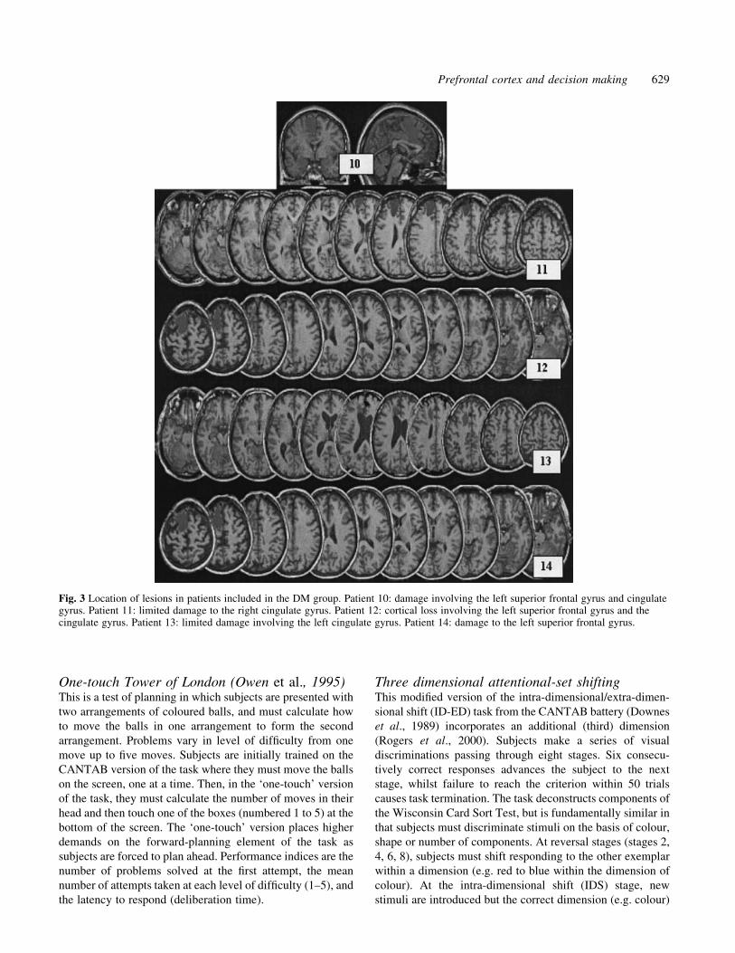
One-touch Tower of London (Owen et al., 1995)This is a test of planning in which subjects are presented with
two arrangements of coloured balls, and must calculate how
to move the balls in one arrangement to form the second
arrangement. Problems vary in level of dif®culty from one
move up to ®ve moves. Subjects are initially trained on the
CANTAB version of the task where they must move the balls
on the screen, one at a time. Then, in the `one-touch' version
of the task, they must calculate the number of moves in their
head and then touch one of the boxes (numbered 1 to 5) at the
bottom of the screen. The `one-touch' version places higher
demands on the forward-planning element of the task as
subjects are forced to plan ahead. Performance indices are the
number of problems solved at the ®rst attempt, the mean
number of attempts taken at each level of dif®culty (1±5), and
the latency to respond (deliberation time).
Three dimensional attentional-set shiftingThis modi®ed version of the intra-dimensional/extra-dimen-
sional shift (ID-ED) task from the CANTAB battery (Downes
et al., 1989) incorporates an additional (third) dimension
(Rogers et al., 2000). Subjects make a series of visual
discriminations passing through eight stages. Six consecu-
tively correct responses advances the subject to the next
stage, whilst failure to reach the criterion within 50 trials
causes task termination. The task deconstructs components of
the Wisconsin Card Sort Test, but is fundamentally similar in
that subjects must discriminate stimuli on the basis of colour,
shape or number of components. At reversal stages (stages 2,
4, 6, 8), subjects must shift responding to the other exemplar
within a dimension (e.g. red to blue within the dimension of
colour). At the intra-dimensional shift (IDS) stage, new
stimuli are introduced but the correct dimension (e.g. colour)
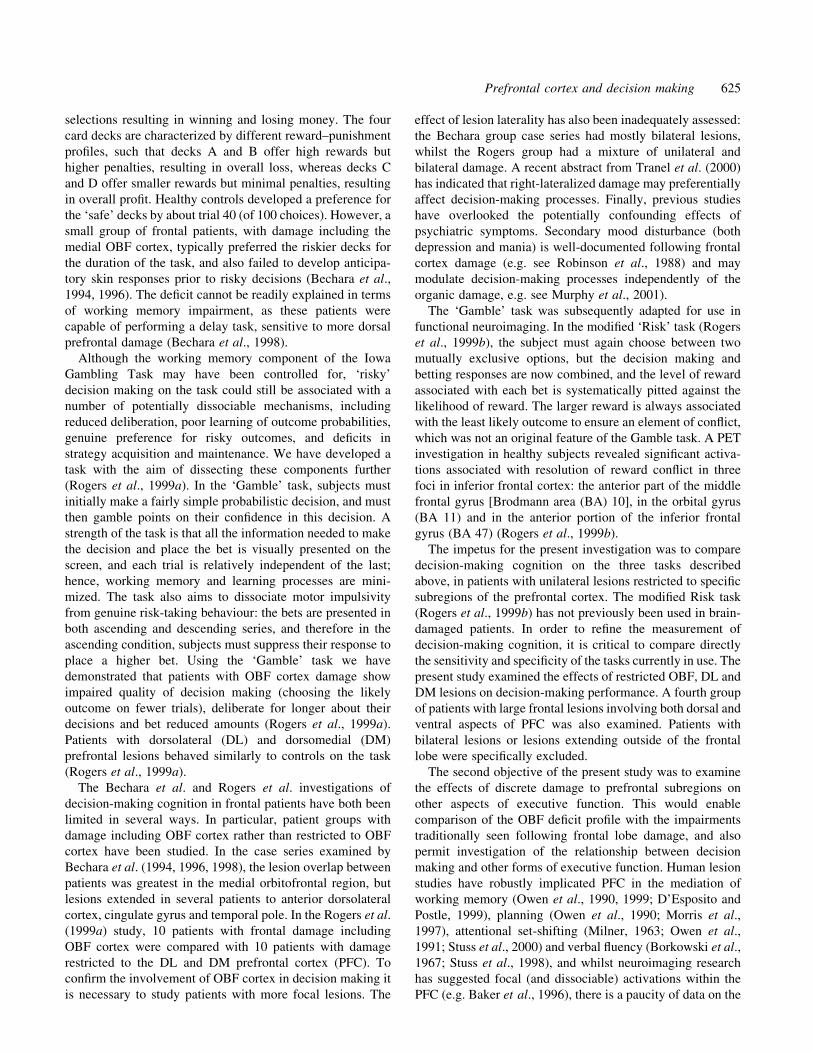
Fig. 3 Location of lesions in patients included in the DM group. Patient 10: damage involving the left superior frontal gyrus and cingulategyrus. Patient 11: limited damage to the right cingulate gyrus. Patient 12: cortical loss involving the left superior frontal gyrus and thecingulate gyrus. Patient 13: limited damage involving the left cingulate gyrus. Patient 14: damage to the left superior frontal gyrus.
Prefrontal cortex and decision making 629

remains the same. At the extra-dimensional shift (EDS) stage,
new stimuli are introduced and subjects must shift responding
to a new dimension (e.g. colour to shape). Dependent
variables are the number of stages passed, and the number
of errors at reversal stages (combined), IDS and EDS.
Decision making tasksIowa Gambling Task (Bechara et al., 1994)Here the subject is required to sample repeatedly from four
decks of cards (A±D). Each card selection results in the
subject winning an amount of money, while after some
selections subjects also lose an amount of money. Decks A
and B are characterized by large wins ($100 on each trial) but
occasional large punishments (e.g. $1250 on deck B), such
that subjects lose money over repeated choices. Decks C and
D are associated with smaller wins (only $50 per trial) but
smaller losses, so that subjects make a pro®t over repeated
choices. The main dependent variable on the task is the
number of choices from decks A and B, the risky decks.
Gamble task (Rogers et al., 1999a)Subjects are presented with a display of a mixture of 10 red
and blue boxes, and must decide whether they think a yellow
token is hidden under a red box or a blue box. This is a
relatively simple probabilistic decision, and the ratio of red to
blue boxes (9 : 1, 8 : 2, 7 : 3, 6 : 4) varies from trial to trial in a
randomized manner. Token location is pre-speci®ed and
pseudo-randomized; hence the probability of the subject
choosing correctly is independent on each trial. The subject
indicates his decision by touching a response panel marked
either `red' or `blue'. After making this initial choice the
subject attempts to increase a points score by placing a bet on
Fig. 4 Location of lesions in patients included in the large frontal lesions group. All patients have lesions involving ventral and moreposterior areas of the prefrontal cortex.
630 F. Manes et al.

their con®dence in their decision being correct. Possible bets
are presented by the computer in a sequence (either ascending
or descending) of 5, 25, 50, 75 and 95% of the points
available at the time of the decision. Each bet is displayed for
a period of 5 s before being replaced by its successor, and
subjects must touch the box when they feel the displayed
amount is an appropriate bet. Following bet selection, the
token location is revealed, accompanied by either a `You
win!' message and a short rising musical scale, or a `You
lose!' message and a low tone. Correct choices increase the
points total by the amount bet whilst incorrect choices
decrease the points total by the amount bet.
The task enables the probabilistic element of decision
making to be dissected from the risk-taking element, as the
choice and bet responses are separate. Deliberation times (to
make the decision) are also assessed as a third dependent
variable. The three variables may each interact with the ratio
of red to blue boxes: at the 9 : 1 ratio, subjects should pick the
likely outcome more consistently, be more con®dent in their
decisions and hence bet more, and may deliberate less, in
comparison with trials at the 6 : 4 ratio. Furthermore,
comparison of ascending and descending conditions enables
impulsive behaviour to be separated from genuine risk-taking
behaviour (genuine risk takers must inhibit motor responding
for many seconds in the ascending condition).
Risk task (Rogers et al., 1999b)This task is similar to the Gamble task described above, with
the core difference being that there is a ®xed bet available
with each choice of box colour, and these bets vary with the
box ratio across trials. The subject is told that the computer
has hidden a token inside one of six red or blue boxes and the
subject must decide whether the token is hidden inside a red
box or a blue box. The gamble is now intrinsic to the decision
made; for example, there may be a 4 : 2 ratio of red to blue
boxes, with a gamble of 10 points for choosing red and 90
points for choosing blue (the gamble is displayed in each
response panel). If the subject correctly chose red he would
win only 10 points but a wrong decision would only lose 10
points. Correct choice of blue would win the subject 90 points
whereas a wrong decision would lose 90 points. Whilst the
token is more likely to be hidden under a red box, will the
subject be tempted into choosing blue by the higher reward at
stake? It is emphasized to subjects that choices might involve
either conservative or risk-taking behaviour, and that they
should try to maximize pro®ts from an initial loan of 100
points. The ratio of coloured boxes (5 : 1, 4 : 2 and 3 : 3) and
the balance between the associated rewards (10 vs 90, 20 vs
80, 30 vs 70, 40 vs 60 and 50 vs 50) varies independently
from trial to trial according to a ®xed pseudo-random
sequence. This sequence ensures that each balance of reward
and ratio of coloured boxes co-occurs an equal number of
times, with the restriction that on all trials with an unequal
ratio of red and blue boxes (i.e. 5 : 1 or 4 : 2), the larger
reward was always associated with the least likely outcome
(i.e. the colour with the fewest number of boxes), thus
capturing the con¯ict inherent in risk-taking situations. Speed
of decision making and percentage choice of most likely
outcome were assessed at each reward value, for the 4 : 2 and
5 : 1 ratios only (the 3 : 3 ratio is not analysed as neither
choice is more or less likely).
Statistical analysisTest scores in Tables 3 and 4 are presented as means with
standard errors of the mean. Effects were considered signi®-
cant at P < 0.05, using two-tailed tests. Raw data were tested
for conformity to the normal distribution using the
Kolmogorov±Smirnov test. Scores were transformed to
reduce skewness where normality was violated {latency
scores were subjected to the logarithmic transformation [log
10 (latency)] and proportion scores to the arcsine transform-
ation [2arcsine (proportion score)]; see Howell, 1997}. Core
dependent variables for each task were subjected to two sets
of analyses. First, an independent samples t-test was used to
compare the combined frontal patient group with controls, to
examine the comparability of the present study group with the
traditional literature on frontal lesions. Secondly, a one-way
ANOVA (analysis of variance) (unweighted means) com-
pared the ®ve groups (four frontal subgroups and controls).
Assessment of performance on Gamble and Risk tasks was
based on published work (Rogers et al., 1999a, b), thus all
comparisons were regarded as `planned' and investigated
using Helmert orthogonal linear contrasts, in addition to post
hoc tests. Simple effects were examined on the other tasks
Fig. 5 Deliberation times on the One-touch Tower of London taskin frontal subgroups, by level of dif®culty (two to ®ve moveproblems), in controls (®lled diamonds), OBF (®lled squares), DL(®lled trianges), DM (crosses) and Large lesion groups (opencircles). SED = standard error of the difference between means.
Prefrontal cortex and decision making 631
using post hoc comparisons (Tukey's HSD), where the main
effect of group was signi®cant. In the ®gures, the index of
variation used is not the standard error of the mean, but the
standard error of the difference of the means, which is
appropriate when one is interested in the (within-subjects)
relationship between variables rather than the variables
themselves. This is calculated by the formula from Cochran
and Cox (1957):
(MSe/n1) + (MSe/n2) + (MSe/n3). . .,
where MSe is the mean square error of the interaction term,
and n is the number of subjects per group.
ResultsNeuropsychological testsVerbal ¯uencyThe combined frontal group was signi®cantly impaired at
letter ¯uency (F, A, S) [t(26) = 2.13, P = 0.046] but not
category ¯uency (Animals) [t(26) = 1.46, P = 0.156]. There
was no statistically signi®cant difference among the four
frontal subgroups and controls on letter ¯uency [F(4,23) =
1.63, P = 0.201] or category ¯uency [F(4,23) = 1.36, P =
0.277].
Pattern and spatial recognitionPattern recognition performance (percentage correct) was not
signi®cantly impaired in either the combined frontal group
[t(25) = 0.600, P = 0.558] or the ®ve subgroups [F(4,22) =
1.61, P = 0.208]. There was similarly no effect on pattern
recognition response latency (both P > 0.10). On spatial
recognition (percentage correct), the combined frontal group
were unimpaired relative to controls [t(25) = 1.26, P = 0.219]
but subgroup effects were apparent [F(4,22) = 4.76, P = 0.06]
due to poor performance of the Large group compared with
the controls (P = 0.007), OBF group (P = 0.011) and DM
group (P = 0.021). Both the OBF and DM subgroups
performed very well, thus masking differences between
controls and all frontals combined. Spatial recognition
response latency was similar between frontals and controls
(P > 0.10) and across subgroups [F(4,22) = 2.36, P = 0.09].
Spatial spanThere were no signi®cant differences between controls and
combined frontals in their spatial span [frontal group mean
4.82 (SD 1.07), control mean 5.22 (SD 1.09); t(24) = 0.895, P
= 0.380] or total number of errors [frontal mean 11.6 (SD
4.0), control mean 12.8 (SD 5.36); t(24) = 0.38, P = 0.546].
There were similarly no apparent differences across sub-
groups, [F(4,21) = 1.08, P = 0.392] and [F(4,21) = 0.917, P =
0.472], respectively (see Table 3).
Spatial working memoryThe combined frontal group made more between search
errors than the controls, but this was not signi®cant because of
the large variability in the data [frontals mean 41.6 (SD 21.7),
control mean 28.1 (SD 15.4); t(26) = 1.67, P = 0.108]. When
the ®ve groups were compared (see Table 3) there was a
signi®cant difference between groups [F(4,23) = 5.67, P =
0.003]. Post hoc comparisons showed that the Large lesion
group made more errors overall than controls (P = 0.018) and
the OBF group (P = 0.006). The DL group also made more
errors than the OBF group (P = 0.026).
There was no signi®cant group difference between controls
and the combined frontals for the strategy score [t(26) =
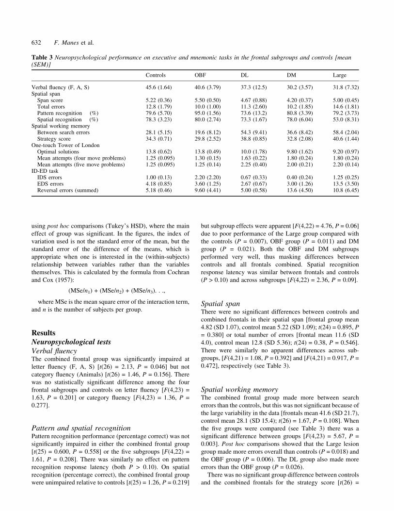
Table 3 Neuropsychological performance on executive and mnemonic tasks in the frontal subgroups and controls [mean(SEM)]
Controls OBF DL DM Large
Verbal ¯uency (F, A, S) 45.6 (1.64) 40.6 (3.79) 37.3 (12.5) 30.2 (3.57) 31.8 (7.32)Spatial span
Span score 5.22 (0.36) 5.50 (0.50) 4.67 (0.88) 4.20 (0.37) 5.00 (0.45)Total errors 12.8 (1.79) 10.0 (1.00) 11.3 (2.60) 10.2 (1.85) 14.6 (1.81)Pattern recognition (%) 79.6 (5.70) 95.0 (1.56) 73.6 (13.2) 80.8 (3.39) 79.2 (3.73)Spatial recognition (%) 78.3 (3.23) 80.0 (2.74) 73.3 (1.67) 78.0 (6.04) 53.0 (8.31)
Spatial working memoryBetween search errors 28.1 (5.15) 19.6 (8.12) 54.3 (9.41) 36.6 (8.42) 58.4 (2.04)Strategy score 34.3 (0.71) 29.8 (2.52) 38.8 (0.85) 32.8 (2.08) 40.6 (1.44)
One-touch Tower of LondonOptimal solutions 13.8 (0.62) 13.8 (0.49) 10.0 (1.78) 9.80 (1.62) 9.20 (0.97)Mean attempts (four move problems) 1.25 (0.095) 1.30 (0.15) 1.63 (0.22) 1.80 (0.24) 1.80 (0.24)Mean attempts (®ve move problems) 1.25 (0.095) 1.25 (0.14) 2.25 (0.40) 2.00 (0.21) 2.20 (0.14)
ID-ED taskIDS errors 1.00 (0.13) 2.20 (2.20) 0.67 (0.33) 0.40 (0.24) 1.25 (0.25)EDS errors 4.18 (0.85) 3.60 (1.25) 2.67 (0.67) 3.00 (1.26) 13.5 (3.50)Reversal errors (summed) 5.18 (0.46) 9.60 (4.41) 5.00 (0.58) 13.6 (4.50) 10.8 (6.45)
632 F. Manes et al.

0.478, P = 0.636], but there was a signi®cant difference
between the ®ve subgroups [F(4,23) = 7.23, P = 0.001),
obscured in the combined frontal group because of the
superior performance of the OBF and DM groups. The DL
group were impaired relative to OBF group (P = 0.009), while
the Large group were impaired relative to controls (P =
0.036), the OBF group (P = 0.001) and the DM group (P =
0.018).
One-touch Tower of LondonThe combined frontal group solved signi®cantly fewer
problems on the ®rst attempt relative to controls [frontal
mean 10.7 (SD 3.16), controls 13.8 (SD 1.75); t(25) = 2.52, P
= 0.018], and this effect was also signi®cant for the
comparison across frontal subgroups and controls [F(4,22)
= 4.71, P = 0.007] (see Table 3). The Large lesion group were
impaired relative to controls (P = 0.030). Group by level of
dif®culty interaction terms were signi®cant for both sets of
analyses, [F(4,100) = 3.72, P = 0.007] for combined frontals,
[F(16,88) = 2.29, P = 0.008] for subgroups, due to particular
impairment in three frontal groups on the four and ®ve move
problems, which place greatest demands on forward plan-
ning. The mean number of attempts taken on ®ve move
problems was greater in the DL group relative to controls (P =
0.008), DM group relative to controls (P = 0.041) and Large
group relative to controls (P = 0.006). The OBF group
performed accurately on the task, and were signi®cantly
better than DL (P = 0.017) and Large (P = 0.016) subgroups
on the most dif®cult problems. However, the OBF group
speci®cally showed longer deliberation times on the task
relative to the other groups (see Fig. 5). They took nearly
twice as long as controls to solve the ®ve move problems (50 s
compared with 29 s), whilst the DM and Large groups
performed similarly to controls (24 s and 26 s, respectively).
The subgroup ANOVA was signi®cant [F(4,22) = 3.31, P =
0.029] and the OBF group were impaired relative to controls
(P = 0.078) and DM patients (P = 0.039).
Three-dimensional ID-ED set shiftingFour frontal patients failed to complete the task: one DL
patient and three Large patients. The DL patient failed at
compound discrimination, and the Large lesion patients failed
at compound reversal, EDS and extra-dimensional reversal.
The two patients failing the task prior to the EDS stage were
excluded from analysis of errors at each stage (see Table 3).
The combined frontal group were not signi®cantly impaired
at IDS [t(26) = 0.221, P = 0.827], EDS [t(26) = 0.743, P =
0.464] or summed reversal trials [t(26) = 1.74, P = 0.093].
However, analysis by location did reveal a signi®cant
difference across the frontal groups at the EDS stage
[F(4,23) = 6.73, P = 0.001]: the Large group were impaired
relative to all other subgroups (P < 0.005 for each compari-
son). There was no difference among the ®ve groups at the
IDS stage [F(4,23) = 0.525, P = 0.718] or at combined
reversal stages [F(4,23) = 1.36, P = 0.278].
Decision making tasksIowa Gambling TaskThe combined frontal group made signi®cantly more selec-
tions from the risky decks than controls [t(28) = 3.88, P <
Fig. 6 Iowa Gambling Task performance in controls (®lleddiamonds), OBF (®lled squares), DL (®lled trianges), DM(crosses) and Large lesion groups (open circles). Fig. 7 Risk adjustment (percentage of current points total bet on
each decision) on the Gamble task in controls (®lled diamonds),OBF (®lled squares), DL (®lled trianges), DM (crosses) and Largelesion groups (open circles). Across all groups, subjects placehigher bets with increasing box ratio. Patients with large frontallesions place higher bets at all box ratios than the other groups.SED = standard error of the difference between means.
Prefrontal cortex and decision making 633

0.001] and there was a signi®cant effect across the ®ve
subgroups [F(4,25) = 5.42, P = 0.003]. Three subgroups were
impaired relative to controls: the DL group (P = 0.037), the
DM group (P = 0.030) and the Large group (P = 0.012). These
three groups made, on average, more choices from the risky
decks than from the safe decks. The OBF group, in contrast,
performed similarly to controls (see Fig. 6).
Gamble task(i) Quality of decision making. The combined frontal group
did not show poorer decision making than controls [F(1,27) =
0.894, P = 0.353], and there were no signi®cant differences
among the ®ve subgroups [F(4,24) = 0.636, P = 0.642],
although the DL and DM groups showed a tendency to make
optimal choices less often than the OBF and Large groups
(see Table 3). The main effect of ratio on the quality of
decision making approached signi®cance [F(3,72) = 2.57, P =
0.07], such that subjects were more likely to pick the likely
outcome at higher ratios (i.e. 9 : 1 over 6 : 4), but there was no
interaction of group by ratio.
(ii) Risk adjustment. There was a highly signi®cant effect
of ratio on the amount bet [F(3,72) = 21.9, P < 0.001], with
larger ratios generally producing higher bets. There were no
signi®cant differences for either the combined frontals
compared with controls, or among the ®ve separate subgroups
(both P > 0.10). There was also no signi®cant interaction of
group with ratio. However, the Large lesion group placed
signi®cantly higher bets than the other patient subgroups
[Helmert contrast: t(24) = 2.07, P = 0.049] (see Fig. 7). All
subjects placed larger bets in the descending compared with
the ascending condition [F(1,84) = 40.7, P < 0.001], but
group by condition (ascend versus descend) interaction terms
were not signi®cant for either analysis (both P > 0.10), thus
impulsivity of responding did not differ between groups.
(iii) Speed of decision making. Deliberation time was
marginally slower in the combined frontal group than
controls [F(1,27) = 3.42, P = 0.076], but there were no
differences among frontal subgroups [F(4,24) = 0.810, P =
0.531]. Deliberation time was only weakly affected by ratio
(P = 0.07) such that subjects took longer to decide at lower
ratios, but there was no interaction of group by ratio.
Risk taskA three-factor ANOVA model with box ratio as a two-level
(5 : 1 vs 4 : 2, within-subjects) factor, reward proportion as a
®ve-level (50 : 50, 60 : 40, 70 : 30, 80 : 20, 90 : 10, within-
subjects) factor, and group as between subjects factor,
revealed no differences between subgroups [F(4,26) = 2.08,
P = 0.112] but a number of signi®cant interaction terms: box
ratio 3 group [F(4,26) = 3.12; P = 0.032]; box ratio 3 reward
proportion [F(4,104) = 2.81; P = 0.029]; and group 3 reward
proportion [F(16,104) = 1.73, P = 0.05]. Because subjects
performed differently at the two box ratios [F(1,26) = 3.32, P
= 0.08], separate ANOVAs for each ratio condition were
conducted to elucidate the nature of the interaction effects.
For the 5 : 1 box ratio, the combined frontal group was
impaired relative to controls [F(1,29) = 5.73, P = 0.023], and
there was an effect of subgroup [F(4,26) = 2.79, P = 0.047]
due to the Large lesion group choosing the likely outcome
less than controls (P = 0.029). There were no signi®cant main
effects of reward proportion or group by reward proportion
interactions. For the 4 : 2 ratio, the combined frontal group
chose the likely outcome less often [F(1,29) = 4.78, P =
0.037], but there were no differences among frontal
subgroups [F(4,26) = 1.51, P = 0.229]. Although the choice
Fig. 8 Performance on the Risk task in controls (®lled diamonds), OBF (®lled squares), DL (®lled trianges), DM (crosses) and Largelesion groups (open circles), at the 5 : 1 box ratios. Error bars represent standard error of the difference between means.
634 F. Manes et al.

of the most likely outcome was reduced in the Large and DL
compared with OBF and DM groups (see Fig. 8), these
differences were not signi®cant. There were no signi®cant
main effects of reward or group by reward interactions. The
combined frontal group also deliberated longer before
making decisions on the 5 : 1 box trials [frontals mean
3198 ms (SD 1455 ms), controls mean 2220 ms (SD 896 ms);
F(1,29) = 4.81, P = 0.037], but this effect was not signi®cant
for the 4 : 2 box trials (P = 0.199), and there were no
differences between the ®ve subgroups on deliberation (both
P > 0.10), although the OBF group deliberated longest at both
box ratios (see Table 4).
Summary of ®ndings on decision making tasksPatients in the dorsomedial, dorsolateral, and large lesion
groups selected more cards from risky decks than controls on
the Iowa Gambling Task. This effect was also seen in the
combined group of frontal patients. On the Gamble task, the
group with large frontal lesions placed higher bets than
the other groups, and the combined frontal group deliberated
for longer. Quality of decision making did not signi®cantly
differ among the groups. On the Risk task, the Large lesion
group again showed risk taking behaviour, choosing the less
likely, but higher rewarding, outcome more often than
controls. This effect was also seen in the combined frontal
group, who also deliberated for longer over decisions.
DiscussionThe present investigation is the ®rst to compare directly three
decision-making tasks in patients with focal damage to
prefrontal subregions. One, the Iowa Gambling Task, has
well-established sensitivity to medial orbitofrontal cortex
damage. Two further tasks developed in this laboratory
attempt to fractionate the component processes of the Iowa
Gambling Task, and control for the working memory and
learning processes inherent in that task by employing a visual
format where all the information required to make the
decisions is explicitly presented to subjects. A group of
patients with large lesions to frontal cortex was impaired on
all three decision-making tasks: they selected more cards
from risky decks on the Iowa Gambling Task, they placed
higher bets on simple probabilistic decisions on the Gamble
task and they chose the less likely, but more rewarding,
option more frequently on the Risk task. However, a
surprising and important ®nding was that a group of patients
with unilateral lesions restricted to orbitofrontal cortex
performed similarly to controls on all three tasks.
Neuroradiological assessment of lesion location in the 19
patients tested identi®ed four groups: an OBF group, a DM
PFC group, a DL PFC group and a group with large frontal
pathology involving both ventral and dorsal cortex.
Performance on a neuropsychological battery including
executive and mnemonic measures as well as the three
decision-making tasks was examined across the frontal
subgroups, but a second analysis of the combined frontal
patients was also performed to assess comparability with
previous data on frontal damage. Our combined frontal group
was signi®cantly impaired on verbal ¯uency and One-touch
Tower of London, consistent with previous ®ndings
(Borkowski et al., 1967; Shallice, 1982). The combined
group also showed impaired decision making on the Iowa
Gambling Task and the Risk task, and deliberated for longer
on both the Gamble and Risk tasks. The OBF group was
remarkably unimpaired on this wide-ranging test battery.
Indeed, the superior performance of the OBF group appears to
have masked effects in the combined frontal group on spatial
recognition memory and spatial working memory. The only
cognitive abnormality seen in the OBF group was lengthened
deliberation times on several tasks. While this effect was
relatively speci®c to the OBF group on the One-touch Tower
of London task (where the other groups, if anything,
deliberated less than controls), lengthened deliberation
times on the Gamble and Risk tasks were seen in all frontal
subgroups.
Patients with selective dorsomedial damage chose more
cards from risky decks on the Iowa Gambling Task than
controls, but no de®cits were revealed on the Gamble or Risk
tasks. The DM group was also impaired at forward planning
on the One-touch Tower of London task, but performed well
on spatial recognition, spatial span and spatial working
Table 4 Cognitive performance on the decision-making tasks in frontal subgroups and controls [mean (SEM)]
Controls OBF DL DM Large
Iowa Gambling TaskChoices from risky decks (A and B) 33.6 (3.51) 40.6 (5.30) 55.3 (6.96) 52.0 (2.72) 54.4 (4.32)
Gamble task% likely choice 90.6 (14.2) 92.2 (8.17) 81.5 (24.6) 81.5 (11.4) 89.1 (9.55)% bet (of total) 50.3 (14.5) 48.2 (11.5) 51.4 (17.6) 49.2 (14.1) 68.2 (6.43)Decision latency (ms) 2517 (1023) 3245 (1222) 3057 (860) 3165 (509) 3384 (1721)
Risk task4 : 2 ratioÐ% likely choice 86.3 (3.04) 77.5 (11.7) 67.5 (11.8) 78.1 (6.81) 67.6 (12.4)4 : 2 ratioÐdecision latency (ms) 2563 (333.9) 4091 (1541) 2432 (715.8) 3570 (546.6) 3379 (1268)5 : 1 ratioÐ% likely choice 90.5 (3.29) 85.9 (7.22) 75.9 (11.0) 78.7 (8.82) 60.0 (12.6)5 : 1 ratioÐdecision latency (ms) 2220 (239.6) 3822 (1063) 2384 (605.2) 3388 (403.9) 3150 (827.9)
Prefrontal cortex and decision making 635

memory tasks. Patients with DL PFC lesions displayed Iowa
Gambling Task and One-touch Tower of London impair-
ments, but additional de®cits were apparent on spatial
working memory and a novel task measuring attentional
set-switching. Finally, the group with large prefrontal lesions
showed impairment across a broad range of the tasks,
including spatial recognition memory, spatial working mem-
ory, One-touch Tower of London and ID-ED attentional set-
shifting, as well as the three decision-making tasks. Effects in
the large lesion group included those not apparent in the
combined frontal analysis, as subjects with discrete lesions
performing the tasks adequately would dilute effects in a
combined group. This clearly highlights a potential confound
in previous studies using mixed groups of frontal patients
with varying lesion foci and lesion aetiologies, and could
explain inconsistencies across reports.
Prefrontal contributions to decision makingThe patients with large lesions were the only group to show
impaired decision making on the Gamble and Risk tasks.
Their pro®le of performance on the Gamble task resembled
that of patients with fvFTD (frontal variant frontotemporal
dementia) (Rahman et al., 1999) and aneurysmal subarach-
noid haemorrhage of the anterior communicating artery
(AcoA) (Mavaddat et al., 2000). Both of these groups also
typically have rather diffuse frontal pathology, albeit prefer-
entially affecting the ventral aspects. The Large lesion,
fvFTD and AcoA patients all placed higher bets under both
ascending and descending conditions, thereby demonstrating
genuine risk-taking behaviour. Our ®nding that patients with
selective damage either to OBF or to DL or DM PFC did not
also behave like this suggests that the size of the lesion may
be critical, and that damage to both dorsal and ventral
prefrontal systems is necessary to disrupt decision making.
The Large lesion group was also impaired at the Iowa
Gambling Task, but so too were the DL and DM patients. The
Iowa Gambling Task requires a number of extraneous
cognitive processes that are not involved in the Gamble
task, for example, working memory for the bad decks, and the
assimilation of reward±punishment information into a suc-
cessful response strategy. Whereas Bechara et al. (1998)
demonstrated gambling task impairments in the presence of
intact working memory, our DL patients were impaired at a
speci®c test of working memory, which may contribute to
their de®cit on the Iowa Gambling Task task. Likewise, the
DM group were de®cient at the One-touch Tower of London
task, which may resemble the Iowa Gambling Task in
requiring prospective consideration of the outcomes of
responses. In conclusion, we do not consider the evidence
for a localized dorsolateral or dorsomedial PFC contribution
to decision making to be compelling.
While an intuitive explanation of the present data might be
in terms of lesion size or `mass action' (e.g. the OBF group
showed mainly intact performance due to having the smallest
lesions), it can be seen from Figs 1±4 that this was clearly not
the case. The DM group, which showed clear cognitive
impairment, had the smallest lesions. The only effect seen in
the OBF group was lengthened deliberation times in planning
and a tendency towards longer response latencies on the
Gamble and Risk tasks. Rogers et al. (1999a) reported that
OBF patients show lengthened deliberation time on the
Gamble task, although in the presence of altered quality of
decision making and betting. Murphy et al. (2001) also
showed that manic patients deliberated longer on dif®cult
decisions. Although it is dif®cult to interpret precisely,
increased deliberation clearly indicates that OBF damage is
not merely associated with motor impulsivity, which would
be expected to reduce deliberation. The role of OBF cortex in
inhibitory control is likely to be multifaceted. One possibility
that we are unable to rule out is that the retarded latencies
re¯ect an incipient de®cit in decision making, which is
compensated by lengthened deliberation.
Our ®nding that patients with selective OBF lesions
displayed intact quality of decision making and appropriate
risk-taking behaviour across three tasks assessing such
performance was unexpected, and apparently contrasts with
previous reports by Bechara et al. (1994) and Rogers et al.
(1999a). While the group sizes in the present study were
admittedly small, patients with discrete OBF damage are
unusual, and our group sizes are comparable with the other
studies in the ®eld (Bechara et al., 1994, 1996; Stuss et al.,
2000). Impairments were detected on the tasks in at least one
of the other frontal groups, and thus lack of test sensitivity or
statistical power does not seem the most obvious explanation.
To reconcile our data with the previous studies, it should ®rst
be noted that on the basis of the stringent inclusion criteria in
the present study, many of the patients used in the studies by
Bechara et al. and Rogers et al. would have been categorized
in our Large lesion group, for which signi®cant decision-
making impairments were evident. For example, Damasio's
patient (E.V.R.) with acquired sociopathy had a large bilateral
lesion involving the entire right OBF and extending to medial
and dorsolateral areas. In the Bechara et al. case series, the
medial OBF cortex represented only the area of lesion overlap
between all the patients. Similarly, of the 10 OBF patients
studied by Rogers et al. (1999a), six also had damage
extending outside OBF.
Secondly, laterality of the lesion may be crucial: whilst the
Bechara et al. patient series had bilateral lesions, four of the
®ve patients in the OBF group in the present study had a left-
sided lesion, whilst four of the ®ve patients in the Large
group, with decision-making de®cits, had right-sided lesions.
A recent abstract by Tranel et al. (2000) also highlights the
importance of laterality in patients with unilateral damage:
Iowa Gambling Task impairment was observed in patients
with right, but not left, ventromedial PFC damage. Whilst our
single patient with a right-sided OBF lesion did not seem
anomalous compared with the rest of the group, a laterality
effect is also consistent with the PET imaging study by
Rogers et al. (1999b), which demonstrated predominantly
right-sided OBF activation associated with resolution of
636 F. Manes et al.

reward con¯ict on the Risk task. In the Rogers et al. (1999a)
lesion study, which also demonstrated OBF-associated deci-
sion-making impairments, one patient had a bilateral lesion,
while four of the unilateral lesions were right sided. Patients
with selective OBF damage are very unusual and a multi-
centre or meta-analytic approach may be required to con®rm
the laterality effect suggested by these data.
There are at least two further factors that may contribute to
differences between the present data and the previous studies.
In the Bechara et al. investigations, patients were only
included if they demonstrated real-life decision-making
de®cits. The extent to which their ®ndings generally apply
to the cognitive sequelae of OBF damage therefore remains
unclear. Finally, in the present study subjects were screened
for psychiatric symptomatology which, if present, led to
exclusion. The emergence of affective syndromes secondary
to lateralized frontal pathology is well documented: left-sided
lesions are associated with depressive symptomatology and
right-sided lesions are (less commonly) associated with
secondary mania (Robinson et al., 1988). Decision-making
de®cits (on the Gamble task) have been demonstrated as state
effects in patients with (`primary') mania and depression
(Murphy et al., 2001). In neurological patients, although one
may argue that the decision-making impairment and mood
disorder may both be the result of the frontal pathology, it is
equally possible that the mood disorder itself has caused the
decision-making impairment. Previous studies have not
reported psychiatric status in the frontal patients and may
consequently be confounded by affective syndromes.
Other executive function de®cits in patients withfocal prefrontal damageInvestigation of the effects of frontal lesions on other
neuropsychological measures enables the speci®city of the
decision-making de®cits to be assessed. The Iowa
Gambling Task in particular has working memory,
contingency learning and possible set-shifting components,
which are tapped separately by the present test battery.
The self-ordered spatial working memory and One-touch
Tower of London tests both require strategic choice
among different candidate response sequences, although
each sequence or `search' is associated with only a single
goal, rather than the need to evaluate the balance between
probabilistic outcomes and variable reward magnitudes as
in the three decision-making tasks. Large frontal lesions
signi®cantly impaired spatial working memory strategy
scores and One-touch Tower of London performance, as
well as performance on the Iowa Gambling Task. The
more focal DL and DM lesions also impaired One-touch
Tower of London performance, but only the DL lesion
affected self-ordered spatial working memory. Thus, only
a limited dissociation from the Iowa Gambling Task was
found. However, these ®ndings do not necessarily
contradict the data of Bechara et al. (1998), which
show a dissociation between their task and spatial
working memory performance, as performance on the
spatial span task was intact in our Large, DL and DM
groups. The greater sensitivity of the self-ordered spatial
working memory task, which has been associated with
ventrolateral and DL PFC foci from a PET study (Owen
et al., 1996), is probable because it contains an additional
strategic element (Owen et al., 1990; Robbins, 1996),
which was not a major component of the delay tasks used
by Bechara et al. (1998). However, the ®ndings do raise
the possibility that (impaired) executive processes may
contribute to performance on tasks with decision-making
cognition. This would be at least partly consistent with
the conclusions of a recent meta-analytical review of
functional imaging data, implicating a network of mid-
dorsolateral PFC, mid-ventrolateral PFC and anterior
cingulate foci that contribute to a broad range of
cognitive functions including working memory, problem
solving, episodic memory, control processes and response
selection (Duncan and Owen, 2000). Finally, only the
patients with large frontal lesions were also impaired at
attentional set-shifting. The de®cit was speci®c to the
extra-dimensional shift stage of the ID-ED task, which is
fully consistent with previous ®ndings that frontal patients
with regionally heterogeneous lesions were signi®cantly
impaired in their ability to shift response set to a
previously irrelevant dimension (EDS), but not in shifting
attention to novel exemplars of the same dimension (IDS)
(Owen et al., 1991). From our data, there is clearly no
reason to link the Iowa Gambing Task de®cit to a
shifting impairment per se, and this is consistent with a
previous report of dissociated performance on the Iowa
Gambling Task and the Wisconsin Card Sort Test in
Patient E.V.R. (Damasio, 1994).
ConclusionsPatients with restricted, predominantly left-sided, OBF cortex
damage performed at control levels on a range of cognitive
tasks assessing decision making, working memory, planning
and attentional shifting. These results seem to contradict
previous ®ndings using decision-making tasks, but may be
explained by possible laterality effects and the focal nature of
these lesions compared with those of previous studies. In
contrast, patients with large frontal lesions and selective DL
lesions were impaired across a range of tasks requiring
working memory, planning, and attentional shifting. Patients
with large frontal lesions placed higher bets and made less
rational decisions on two recently developed decision-making
tasks. Of the three decision-making tasks employed in the
present study, the Iowa Gambling Task appears to be the most
sensitive, but may detect impairment on the basis of its extra
load on working memory and associative learning in addition
to its capacity for measuring decision making that involves
risk taking.
Prefrontal cortex and decision making 637

AcknowledgementsWe wish to thank Drs A. R. Damasio and A. Bechara for
generously allowing us the use of the Iowa Gambling Task,
and Dr Bechara for helpful comments on a draft of the
manuscript. Dr Matthew Brett provided assistance with the
neuroanatomical analysis. This research was funded by a
programme grant from the Wellcome Trust to T.R., B. J.
Everitt, B.S. and A. C. Roberts, and was completed within the
Medical Research Council Co-operative Group in Brain,
Behaviour and Neuropsychiatry. F.M.'s work was supported
by the Raul Carrea Institute of Neurological Research and the
Medical Research Council Cognition and Brain Sciences
Unit.
References
Baker SC, Rogers RD, Owen AM, Frith CD, Dolan RJ, Frackowiak
RS, et al. Neural systems engaged by planning: a PET study of the
Tower of London task. Neuropsychologia 1996; 34: 515±26.
Bechara A, Damasio AR, Damasio H, Anderson SW. Insensitivity
to future consequences following damage to human prefrontal
cortex. Cognition 1994; 50: 7±15.
Bechara A, Tranel D, Damasio H, Damasio AR. Failure to respond
autonomically to anticipated future outcomes following damage to
prefrontal cortex. Cereb Cortex 1996; 6: 215±25.
Bechara A, Damasio H, Tranel D, Anderson SW. Dissociation of
working memory from decision making within the human prefrontal
cortex. J Neurosci 1998; 18: 428±37.
Bechara A, Damasio H, Damasio AR. Emotion, decision making
and the orbitofrontal cortex. [Review]. Cereb Cortex 2000; 10: 295±
307.
Benton AL, Hamsher K. Multilingual aphasia examination. Iowa
City: University of Iowa Press; 1976.
Borkowski JG, Benton AL, Spreen OL. Word ¯uency and brain
damage. Neuropsychologia 1967; 5: 135±40.
Brett M, Leff A, Ashburner J. Spatial normalization of brain images
with focal lesions using cost function masking. Neuroimage. In
press 2002.
Cochran WG, Cox GM. Experimental designs. 2nd ed. New York:
Wiley; 1957.
Damasio AR. Descartes' error: emotion, reason and the human
brain. New York: G.P. Putnam; 1994.
Damasio H. Human brain anatomy in computerized images.
Oxford: Oxford University Press; 1995.
D'Esposito M, Postle BR. The dependence of span and delayed-
response performance on prefrontal cortex. Neuropsychologia 1999;
37: 1303±15.
Downes JJ, Roberts AC, Sahakian BJ, Evenden JL, Morris RG,
Robbins TW. Impaired extra-dimensional shift performance in
medicated and unmedicated Parkinson's disease: evidence for a
speci®c attentional dysfunction. Neuropsychologia 1989; 27: 1329±
43.
Duncan J, Owen AM. Common regions of the human frontal lobe
recruited by diverse cognitive demands. [Review]. Trends Neurosci
2000; 23: 475±83.
Eslinger PJ, Damasio AR. Severe disturbance of higher cognition
after bilateral frontal lobe ablation: patient EVR. Neurology 1985;
35: 1731±41.
Friston KJ, Holmes AP, Worsley KJ, Poline JB, Frith CD,
Frackowiak RJ. Statistical parametric maps in functional imaging:
a general linear approach. Hum Brain Mapp 1995; 2: 189±210.
Howell DC. Statistical methods for psychology. Belmont:
Wadsworth; 1997.
Mavaddat N, Kirkpatrick PJ, Rogers RD, Sahakian BJ. De®cits in
decision-making in patients with aneurysms of the anterior
communicating artery. Brain 2000; 123: 2109±17.
Milner B. Effects of different brain lesions on card sorting. Arch
Neurol 1963; 9: 90±100.
Milner B. Interhemispheric differences in the localization of
psychological processes in man. [Review]. Brit Med Bull 1971;
27: 272±7.
Morris RG, Miotto EC, Feigenbaum JD, Bullock P, Polkey CE.
Planning ability after frontal and temporal lobe lesions in humans:
the effects of selection equivocation and working memory load.
Cogn Neuropsychol 1997; 14: 1007±27.
Murphy FC, Rubinsztein JS, Michael A, Rogers RD, Robbins TW,
Paykel ES, et al. Decision-making cognition in mania and
depression. Psychol Med 2001; 31: 679±93.
Nelson HE. NART: National Adult Reading Test manual. Windsor
(UK): NFER-Nelson; 1982.
Owen AM, Downes JJ, Sahakian BJ, Polkey CE, Robbins TW.
Planning and spatial working memory following frontal lobe lesions
in man. Neuropsychologia 1990; 28: 1021±34.
Owen AM, Roberts AC, Polkey CE, Sahakian BJ, Robbins TW.
Extra-dimensional versus intra-dimensional set shifting
performance following frontal lobe excisions, temporal lobe
excisions or amygdalo-hippocampectomy in man. Neuro-
psychologia 1991; 29: 993±1006.
Owen AM, Sahakian BJ, Semple J, Polkey CE, Robbins TW.
Visuo-spatial short-term recognition memory and learning after
temporal lobe excisions, frontal lobe excisions or amygdalo-
hippocampectomy in man. Neuropsychologia 1995; 33: 1±24.
Owen AM, Evans AC, Petrides M. Evidence for two-stage model of
spatial working memory processing within the lateral frontal cortex:
a positron emission tomography study. Cereb Cortex 1996; 6: 31±8.
Owen AM, Herrod NJ, Menon DK, Clark JC, Downey SP,
Carpenter TA, et al. Rede®ning the functional organization of
working memory processes within human lateral prefrontal cortex.
Eur J Neurosci 1999; 11: 567±74.
Rahman S, Sahakian BJ, Hodges JR, Rogers RD, Robbins TW.
Speci®c cognitive de®cits in mild frontal variant frontotemporal
dementia. Brain 1999; 122: 1469±93.
Robbins TW. Dissociating executive functions of the prefrontal
cortex. [Review]. Philos Trans R Soc Lond B Biol Sci 1996; 351:
1463±71.
638 F. Manes et al.

Robbins TW, James M, Owen AM, Sahakian BJ, McInnes L,
Rabbitt P. Cambridge Neuropsychological Test Automated Battery
(CANTAB): a factor analytic study of a large sample of normal
elderly volunteers. Dementia 1994; 5: 266±81.
Robbins TW, James M, Owen AM, Sahakian BJ, Lawrence AD,
McInnes L, et al. A study of performance on tests from the
CANTAB battery sensitive to frontal lobe dysfunction in a large
sample of normal volunteers: implications for theories of executive
functioning and cognitive aging. J Int Neuropsychol Soc 1998; 4:
474±90.
Robinson RG, Boston JD, Starkstein SE, Price TR. Comparison of
mania and depression after brain injury: causal factors. Am J
Psychiatry 1988; 145: 172±8.
Rogers RD, Everitt BJ, Baldacchino A, Blackshaw AJ, Swainson R,
Wynne, K et al. Dissociable de®cits in the decision-making
cognition of chronic amphetamine abusers, opiate abusers,
patients with focal damage to prefrontal cortex, and tryptophan-
depleted normal volunteers: evidence for monoaminergic
mechanisms. Neuropsychopharmacology 1999a; 20: 322±39.
Rogers RD, Owen AM, Middleton HC, Williams EJ, Pickard JD,
Sahakian BJ, et al. Choosing between small, likely rewards and
large, unlikely rewards activates inferior and orbital prefrontal
cortex. J Neurosci 1999b; 19: 9029±38.
Rogers RD, Andrews TC, Grasby PM, Brooks DJ, Robbins TW.
Contrasting cortical and subcortical activations produced by
attentional-set shifting and reversal learning in humans. J Cogn
Neurosci 2000; 12: 142±62.
Sahakian BJ, Morris RG, Evenden JL, Heald A, Levy R, Philpot M,
et al. A comparative study of visuospatial memory and learning in
Alzheimer-type dementia and Parkinson's disease. Brain 1988; 111:
695±718.
Shallice T. Speci®c impairments of planning. Philos Trans R Soc
Lond B Biol Sci 1982; 298: 199±209.
Shallice T, Burgess PW. De®cits in strategy application following
frontal lobe damge in man. Brain 1991; 114: 727±41.
Stuss DT, Alexander MP, Hamer L, Palumbo C, Dempster R, Binns
M, et al. The effects of focal anterior and posterior brain lesions on
verbal ¯uency. J Int Neuropsychol Soc 1998; 4: 265±78.
Stuss DT, Levine B, Alexander MP, Hong J, Palumbo C, Hamer L,
et al. Wisconsin Card Sorting Test performance in patients with
focal frontal and posterior brain damage: effects of lesion location
and test structure on separable cognitive processes.
Neuropsychologia 2000; 38: 388±402.
Talairach J, Tournoux P. Co-planar stereotaxic atlas of the human
brain. Stuttgart: Thieme; 1988.
Tranel D, Bechara A, Damasio H, Damasio AR. Decision-making
in patients with unilateral ventromedial prefrontal cortex lesions
[abstract]. Soc Neurosci Abstr 2000; 26: S49.
Received April 11, 2001. Revised August 16, 2001.
Second revision October 19, 2001. Accepted October 22, 2001
Prefrontal cortex and decision making 639