De novo actin polymerization is required for model Hirano...
12
RESEARCH ARTICLE De novo actin polymerization is required for model Hirano body formation in Dictyostelium Yun Dong 1 , Sonbol Shahid-Salles 1, *, Dan Sherling 2, ‡ , Nathan Fechheimer 1,§ , Nathan Iyer 1,¶ , Lance Wells 2 , Marcus Fechheimer 1 and Ruth Furukawa 1, ** ABSTRACT Hirano bodies are eosinophilic, actin-rich inclusions found in autopsied brains in numerous neurodegenerative diseases. The mechanism of Hirano body formation is unknown. Mass spectrometry analysis was performed to identify proteins from partially purified model Hirano bodies from Dictyostelium. This analysis identified proteins primarily belonging to ribosomes, proteasomes, mitochondria and cytoskeleton. Profilin, Arp/2/3 and WASH identified by mass spectrometry were found to colocalise with model Hirano bodies. Due to their roles in actin regulation, we selected these proteins for further investigation. Inhibition of the Arp2/3 complex by CK666 prevented formation of model Hirano bodies. Since Arp2/3 activation occurs via the WASH or WAVE complex, we next investigated how these proteins affect Hirano body formation. Whereas model Hirano bodies could form in WASH- deficient cells, they failed to form in cells lacking HSPC300, a member of the WAVE complex. We identified other proteins required for Hirano body formation that include profilin and VASP, an actin nucleation factor. In the case of VASP, both its G- and F-actin binding domains were required for model Hirano body formation. Collectively, our results indicate that de novo actin polymerization is required to form model Hirano bodies. KEY WORDS: Dictyostelium, Hirano body, Actin binding protein, Mass spectrometry, Arp2/3, Profilin, WASH, HSPC300 INTRODUCTION Neurodegenerative diseases affect millions of people worldwide and are caused by loss of structure or function of neurons. Some of the most widespread diseases exhibit hallmark protein aggregates such as Alzheimer’s disease (AD) with β-amyloid plaques (Glenner and Wong, 1984) and neurofibrillary tau tangles (Kosik et al., 1986), Parkinson’s disease with Lewy bodies containing α-synuclein (Spillantini et al., 1998), and amyotrophic lateral sclerosis (ALS) with the aggregation of TDP43 (Neumann et al., 2006). Actin inclusions are also present as pathological structures in the form of Hirano bodies (Galloway et al., 1987a; Goldman, 1983; Hirano et al., 1968) and actin-cofilin rods (reviewed in Bamburg et al., 2010). Hirano bodies are eosinophilic cytoplasmic inclusions that are composed primarily of F-actin, with a characteristic electron dense paracrystalline ultrastructure (Goldman, 1983; Schochet and McCormick, 1972). Hirano bodies colocalize with actin-binding proteins as well as proteins that are significant for neurodegenerative diseases, such as tau in AD (Galloway et al., 1987a,b); however since investigation of Hirano bodies was limited to postmortem tissues, the mechanism of Hirano body formation is unknown. Models to study Hirano bodies in living cells were developed by expressing altered forms of a 34 kDa Dictyostelium actin-binding protein in Dictyostelium and mammalian cell lines (Davis et al., 2008; Maselli et al., 2002, 2003) and mice (Furgerson et al., 2014; Ha et al., 2011b). These altered forms exhibit gain-of-function activated F-actin binding (Griffin et al., 2014; Maselli et al., 2002, 2003). Proteins present in Hirano bodies in postmortem tissues were also found in model Hirano bodies (Davis et al., 2008; Furgerson et al., 2012; Ha et al., 2011a; Maselli et al., 2002, 2003; Spears et al., 2014). These results prompted further investigation of some proteins found in brain specimens utilizing modern reagents in live cells expressing model Hirano bodies and have shed light on the possible physiological role(s) of Hirano bodies in neurodegenerative diseases. The presence of model Hirano bodies protected cells from death induced by AICD (intracellular domain of the amyloid precursor protein) (Furgerson et al., 2012; Ha et al., 2011a). The presence of model Hirano bodies and AICD and/or various forms of tau either protected cells from cell death or enhanced cell death depending whether the form of tau had a propensity to aggregate through enhanced phosphorylation (Spears et al., 2014). Since valuable information about the physiological role of Hirano bodies was obtained by re-examining proteins found to colocalize to Hirano bodies, we have developed a partial purification of model Hirano bodies and utilized mass spectrometry to elucidate the protein composition of model Hirano bodies in the model organism Dictyostelium. From the results of mass spectrometry, we focused on the role of proteins that regulate actin polymerization. The results support the hypothesis that de novo actin polymerization is required for model Hirano body formation. RESULTS Model Hirano bodies in Dictyostelium are large (1-3 μm) F-actin- rich inclusions, readily detected by labeling with TRITC-labeled phalloidin. This property was utilized to follow their purification and enrichment by sedimentation following cell lysis and density gradient fractionation. Purification of model Hirano bodies to homogeneity was not possible due to the temporal instability of the model Hirano bodies after cell lysis. Observation of model Hirano bodies labeled with TRITC-phalloidin with time yielded a total time of approximately 1 h from cell lysis until the model Hirano bodies Received 28 September 2015; Accepted 3 May 2016 1 Department of Cellular Biology, University of Georgia, Athens, GA, USA 30602. 2 Complex Carbohydrate Research Center, Department of Biochemistry and Molecular Biology, University of Georgia, Athens, GA USA 30602. *Present address: St. Mary’s of Michigan, Grand Blanc, MI 48439. ‡ Present address: West Jefferson Medical Center, LA 70072. § Present address: Wofford College, Spartanburg, SC 29303. ¶ Present address: Vanderbilt University, Nashville, TN 37235. **Author for correspondence ([email protected]) R.F., 0000-0002-9233-7641 This is an Open Access article distributed under the terms of the Creative Commons Attribution License (http://creativecommons.org/licenses/by/3.0), which permits unrestricted use, distribution and reproduction in any medium provided that the original work is properly attributed. 807 © 2016. Published by The Company of Biologists Ltd | Biology Open (2016) 5, 807-818 doi:10.1242/bio.014944 Biology Open by guest on May 8, 2018 http://bio.biologists.org/ Downloaded from
Transcript of De novo actin polymerization is required for model Hirano...

RESEARCH ARTICLE
De novo actin polymerization is required for model Hirano bodyformation in DictyosteliumYun Dong1, Sonbol Shahid-Salles1,*, Dan Sherling2,‡, Nathan Fechheimer1,§, Nathan Iyer1,¶, Lance Wells2,Marcus Fechheimer1 and Ruth Furukawa1,**
ABSTRACTHirano bodies are eosinophilic, actin-rich inclusions found inautopsied brains in numerous neurodegenerative diseases. Themechanism of Hirano body formation is unknown. Mass spectrometryanalysis was performed to identify proteins from partially purifiedmodel Hirano bodies from Dictyostelium. This analysis identifiedproteins primarily belonging to ribosomes, proteasomes,mitochondria and cytoskeleton. Profilin, Arp/2/3 and WASHidentified by mass spectrometry were found to colocalise withmodel Hirano bodies. Due to their roles in actin regulation, weselected these proteins for further investigation. Inhibition of theArp2/3 complex by CK666 prevented formation of model Hiranobodies. Since Arp2/3 activation occurs via the WASH or WAVEcomplex, we next investigated how these proteins affect Hirano bodyformation. Whereas model Hirano bodies could form in WASH-deficient cells, they failed to form in cells lackingHSPC300, amemberof theWAVE complex.We identified other proteins required for Hiranobody formation that include profilin and VASP, an actin nucleationfactor. In the case of VASP, both its G- and F-actin binding domainswere required for model Hirano body formation. Collectively, ourresults indicate that de novo actin polymerization is required to formmodel Hirano bodies.
KEY WORDS: Dictyostelium, Hirano body, Actin binding protein,Mass spectrometry, Arp2/3, Profilin, WASH, HSPC300
INTRODUCTIONNeurodegenerative diseases affect millions of people worldwideand are caused by loss of structure or function of neurons. Some ofthe most widespread diseases exhibit hallmark protein aggregatessuch as Alzheimer’s disease (AD) with β-amyloid plaques (Glennerand Wong, 1984) and neurofibrillary tau tangles (Kosik et al.,1986), Parkinson’s disease with Lewy bodies containingα-synuclein (Spillantini et al., 1998), and amyotrophic lateralsclerosis (ALS) with the aggregation of TDP43 (Neumann et al.,2006). Actin inclusions are also present as pathological structures in
the form of Hirano bodies (Galloway et al., 1987a; Goldman, 1983;Hirano et al., 1968) and actin-cofilin rods (reviewed in Bamburget al., 2010). Hirano bodies are eosinophilic cytoplasmic inclusionsthat are composed primarily of F-actin, with a characteristic electrondense paracrystalline ultrastructure (Goldman, 1983; Schochet andMcCormick, 1972). Hirano bodies colocalize with actin-bindingproteins as well as proteins that are significant for neurodegenerativediseases, such as tau in AD (Galloway et al., 1987a,b); howeversince investigation of Hirano bodies was limited to postmortemtissues, the mechanism of Hirano body formation is unknown.
Models to study Hirano bodies in living cells were developed byexpressing altered forms of a 34 kDa Dictyostelium actin-bindingprotein in Dictyostelium and mammalian cell lines (Davis et al.,2008; Maselli et al., 2002, 2003) and mice (Furgerson et al., 2014;Ha et al., 2011b). These altered forms exhibit gain-of-functionactivated F-actin binding (Griffin et al., 2014; Maselli et al., 2002,2003). Proteins present in Hirano bodies in postmortem tissues werealso found in model Hirano bodies (Davis et al., 2008; Furgersonet al., 2012; Ha et al., 2011a;Maselli et al., 2002, 2003; Spears et al.,2014). These results prompted further investigation of someproteins found in brain specimens utilizing modern reagentsin live cells expressing model Hirano bodies and have shed lighton the possible physiological role(s) of Hirano bodies inneurodegenerative diseases. The presence of model Hirano bodiesprotected cells from death induced by AICD (intracellular domainof the amyloid precursor protein) (Furgerson et al., 2012; Ha et al.,2011a). The presence of model Hirano bodies and AICD and/orvarious forms of tau either protected cells from cell death orenhanced cell death depending whether the form of tau had apropensity to aggregate through enhanced phosphorylation (Spearset al., 2014). Since valuable information about the physiologicalrole of Hirano bodies was obtained by re-examining proteins foundto colocalize to Hirano bodies, we have developed a partialpurification of model Hirano bodies and utilized mass spectrometryto elucidate the protein composition of model Hirano bodies inthe model organism Dictyostelium. From the results of massspectrometry, we focused on the role of proteins that regulate actinpolymerization. The results support the hypothesis that de novoactin polymerization is required for model Hirano body formation.
RESULTSModel Hirano bodies in Dictyostelium are large (1-3 µm) F-actin-rich inclusions, readily detected by labeling with TRITC-labeledphalloidin. This property was utilized to follow their purificationand enrichment by sedimentation following cell lysis and densitygradient fractionation. Purification of model Hirano bodies tohomogeneity was not possible due to the temporal instability of themodel Hirano bodies after cell lysis. Observation of model Hiranobodies labeled with TRITC-phalloidin with time yielded a total timeof approximately 1 h from cell lysis until the model Hirano bodiesReceived 28 September 2015; Accepted 3 May 2016
1Department of Cellular Biology, University of Georgia, Athens, GA, USA 30602.2Complex Carbohydrate Research Center, Department of Biochemistry andMolecular Biology, University of Georgia, Athens, GA USA 30602.*Present address: St. Mary’s of Michigan, Grand Blanc, MI 48439. ‡Presentaddress: West Jefferson Medical Center, LA 70072. §Present address: WoffordCollege, Spartanburg, SC 29303. ¶Present address: Vanderbilt University,Nashville, TN 37235.
**Author for correspondence ([email protected])
R.F., 0000-0002-9233-7641
This is an Open Access article distributed under the terms of the Creative Commons AttributionLicense (http://creativecommons.org/licenses/by/3.0), which permits unrestricted use,distribution and reproduction in any medium provided that the original work is properly attributed.
807
© 2016. Published by The Company of Biologists Ltd | Biology Open (2016) 5, 807-818 doi:10.1242/bio.014944
BiologyOpen
by guest on May 8, 2018http://bio.biologists.org/Downloaded from

completely disassembled. Fractions from the Opti-prep gradientwith model Hirano bodies also contained particles that were stainedwith DAPI, a general DNA fluorescent marker. Thus, it wasexpected to identify contaminant proteins/particles from massspectrometry that localize to the nucleus and/or mitochondria thatcontain DNA.Identification of approximately 135 proteins with two or more
fragmented peptides was achieved; of these proteins, 37 hadpredicted sequences identifying them as ribosomal, 13 wereproteasome components, 34 were mitochondrial proteins, 33 wereproteins found in the cytoplasm and 18 were identified as linked toother pathways (Appendix Table A2). These proteins were acompilation of five runs. Approximately 270 proteins wereidentified with a single fragmented peptide. Several of theseproteins (66) had been previously identified with two or morefragmented peptides. Several proteins comprise components ofpreviously identified contaminants such as the mitochondria (28proteins), ribosomes (34), endosomes (13), and proteasome (10).There were several putative and hypothetical proteins (27) and avariety of cytoplasmic proteins (52) identified. There were also 21cytoskeletal proteins identified. Some of these proteins had beenpreviously identified with multiple fragments, or in multiple massspectrometry analysis, or that were subunits of proteins withmultiple polypeptides such as myosin II. We investigated several ofthe cytoskeleton-associated proteins due to their role in actinpolymerization (see below).
Mitochondria do not colocalize with model Hirano bodiesTo verify whether mitochondria are in model Hirano bodies orwhether they were contaminants in the fraction containing them, weinduced the expression of E60K-GFP (E60K-34 kDa protein fused toGFP, see Table S1) using the discoidin promoter for 24 h. The cellswere stainedwithMitoTracker®RedCMXRos (Invitrogen, Carlsbad,CA), a live cell dye, and fixed. The mitochondria did not colocalizewithmodel Hirano bodies in fixed cells (Fig. S1). Thus, mitochondriaand its associated proteins identified by mass spectrometry appear tobe contaminants in the model Hirano body purification. Allmitochondrial proteins were eliminated from the list of possibleproteins in model Hirano bodies identified by mass spectrometry.
The role of profilin I in model Hirano body formationUsing inducible promoters, it has been observed that model Hiranobodies begin as small actin foci that coalesce to form a largeinclusion (Griffin et al., 2014; Reyes et al., 2009). It is not knownwhether the foci disassemble and reassemble to form the largeinclusion or whether the foci coalesce into one inclusion. Uponexamination of the mass spectrometry analysis, we observedthat profilin and Arp2/3 were putatively present in model Hiranobodies. We further examined these proteins since they are importantin the regulation of actin filament formation. We examinedthe colocalization of profilin I with model Hirano bodies inDictyostelium expressing CT (c-terminal portion of 34 kDa protein,amino acids 129-295, seeTable S1) by immunofluorescence. ProfilinI was enriched and colocalized well with the model Hirano bodies(Fig. 1A-D). This result verified the presence of profilin I in modelHirano bodies. To determine if profilin I affects model Hirano bodyformation, profilin I knockdown strain ([AS]proA) (Haugwitz et al.,1994) was transformed with plasmid encoding CT under aconstitutive promoter (Maselli et al., 2002). Transformed cells([AS]proA-CT), [AS]proA and wild-type cells expressing CT wereimmunoblotted to confirm that expression of profilin I was reduced(data not shown). Model Hirano bodies still formed in transformed
cells (Fig. 1E-H); however, compared with wild-type cells thatexpressed CT, model Hirano bodies that were formed when profilinI was knocked down were significantly smaller (I, P<0.001). Thisresult suggests that profilin I promotes model Hirano bodyformation.
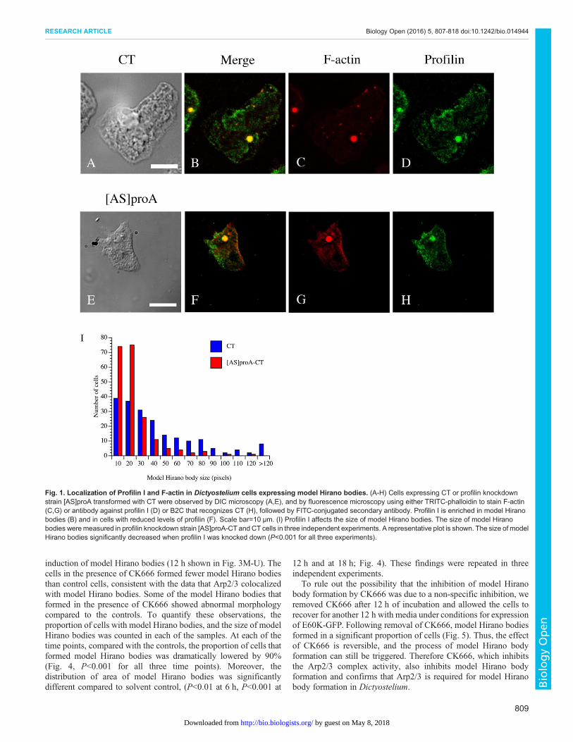
Colocalization of the Arp2/3 complex with model HiranobodiesThe subunits of the Arp2/3 complex Arp3 and p21 (also namedARPC3) were identified by one peptide fragment each in the massspectrometry analysis. Since Arp2/3 complex has an importantregulatory role in actin polymerization, the presence of Arp2 andArp3 in model Hirano bodies was tested to verify these results.Dictyostelium with CT constitutively expressed were transformedwith plasmid encoding GFP-Arp2 (Insall et al., 2001) and stainedwith TRITC-labeled phalloidin. To also determine the localization ofArp3 compared to the model Hirano bodies, Dictyosteliumconstitutively expressing GFP-Arp3 (Insall et al., 2001) weretransformed with plasmid encoding CT and stained with TRITC-labeled phalloidin. GFP-Arp2 was enriched in model Hirano bodies(Fig. 2A-D). GFP-Arp3 localized at similar regions of cells asGFP-Arp2, and was enriched in model Hirano bodies (Fig. 2E-H).These results support that the Arp2/3 complex colocalizes withmodelHirano bodies, providing confirmation of themass spectrometry data.
Inhibition of the Arp2/3 complex activity inhibits modelHirano body formationTo determine the role of the Arp2/3 complex in model Hirano bodyformation, we utilized an Arp2/3 complex inhibitor, CK666, sincethe knockout of either Arp2 or Arp3 is lethal. It has been shown thatCK666 specifically inhibits the actin nucleation activity of theArp2/3 complex (Nolen et al., 2009). A mechanism was proposedthat CK666 inhibits Arp2/3 complex activity by preventing aconformational reorganization crucial for activation (Hetrick et al.,2013); however it has never been used inDictyostelium. In addition,the previous studies utilizing CK666 (Nolen et al., 2009; Yang et al.,2012) incubated it with the cells for approximately 30 min; howeverit takes at least 3 h to see the small F-actin foci forming, and morethan 6 h to see a significant number of model Hirano bodies withlarger sizes (Griffin et al., 2014; Reyes et al., 2009). Therefore, itwas necessary to test whether CK666 inhibits the DictyosteliumArp2/3 complex for a length of time comparable to model Hiranobody formation before utilizing it to investigate the role of Arp2/3complex in model Hirano body formation.
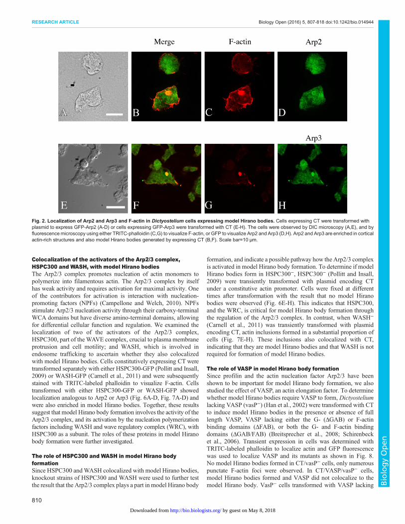
To determine if CK666 inhibits Dictyostelium Arp2/3 complexactivity and the time span of efficacy, Dictyostelium cellsconstitutively expressing GFP-Arp3 were incubated with 100 µMCK666, with 0.2% DMSO/media as the solvent control, or mediaonly, for different lengths of time, followed by fixation and stainingwith TRITC-labeled phalloidin to localize F-actin. In agreementwith published data, GFP-Arp3 colocalized with F-actin at thecortex and the lamellipodia in the controls (Fig. 3G,K) (Insall et al.,2001). In contrast, in the cells incubated in the presence of CK666,the colocalization of F-actin and GFP-Arp3 was reduced andGFP-Arp3 was not localized in the cortex or lamellipodia (Fig. 3C).This shows that CK666 inhibited the activity of the Arp2/3 complexin Dictyostelium, an effect lasting for at least 18 h. Therefore,CK666 could be used as a tool to examine the role of the Arp2/3complex in model Hirano body formation.
Subsequently, E60K-GFP cells were induced in the presence orabsence of 100 µM CK666. Cells were fixed and stained withTRITC-phalloidin to visualize the F-actin at 6 h, 12 h and 18 h after
808
RESEARCH ARTICLE Biology Open (2016) 5, 807-818 doi:10.1242/bio.014944
BiologyOpen
by guest on May 8, 2018http://bio.biologists.org/Downloaded from
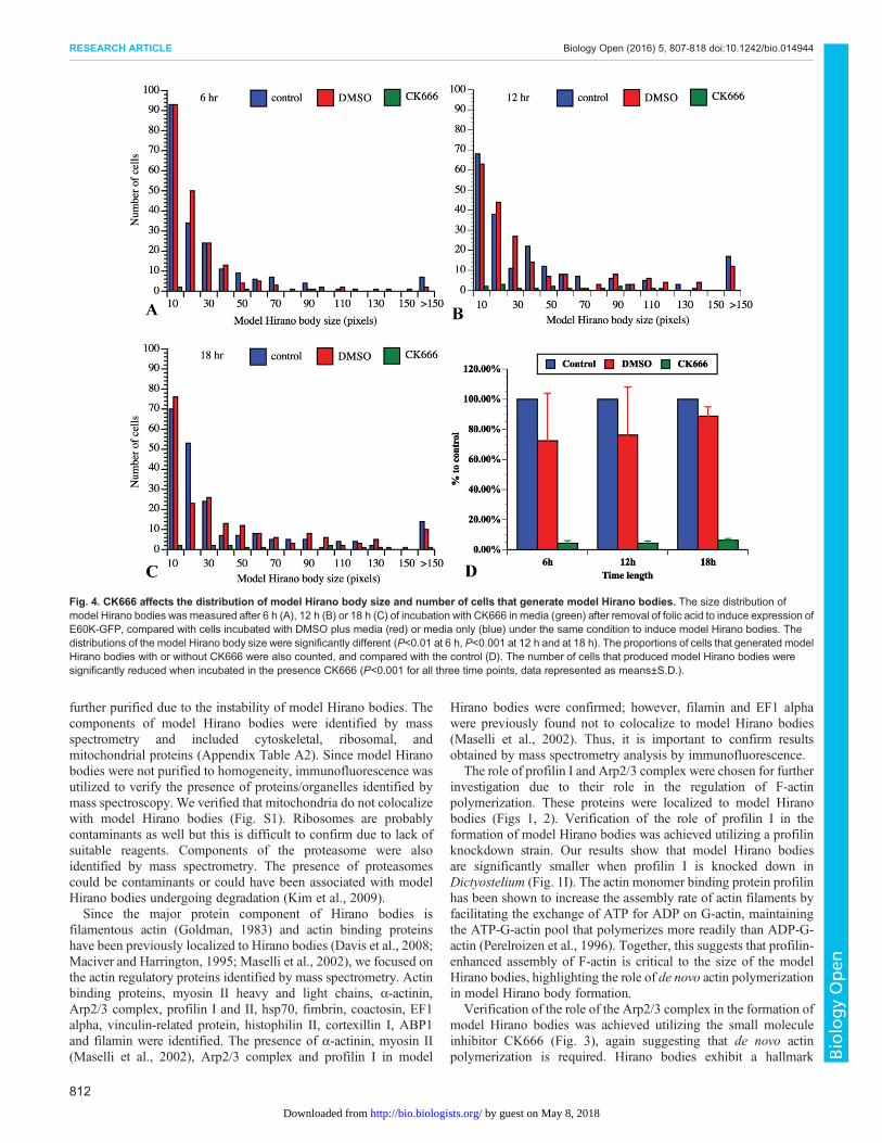
induction of model Hirano bodies (12 h shown in Fig. 3M-U). Thecells in the presence of CK666 formed fewer model Hirano bodiesthan control cells, consistent with the data that Arp2/3 colocalizedwith model Hirano bodies. Some of the model Hirano bodies thatformed in the presence of CK666 showed abnormal morphologycompared to the controls. To quantify these observations, theproportion of cells with model Hirano bodies, and the size of modelHirano bodies was counted in each of the samples. At each of thetime points, compared with the controls, the proportion of cells thatformed model Hirano bodies was dramatically lowered by 90%(Fig. 4, P<0.001 for all three time points). Moreover, thedistribution of area of model Hirano bodies was significantlydifferent compared to solvent control, (P<0.01 at 6 h, P<0.001 at
12 h and at 18 h; Fig. 4). These findings were repeated in threeindependent experiments.
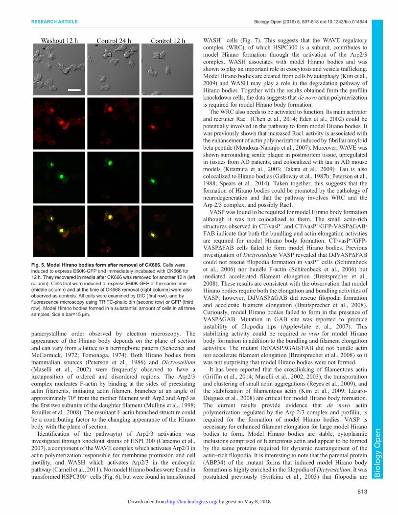
To rule out the possibility that the inhibition of model Hiranobody formation by CK666 was due to a non-specific inhibition, weremoved CK666 after 12 h of incubation and allowed the cells torecover for another 12 h with media under conditions for expressionof E60K-GFP. Following removal of CK666, model Hirano bodiesformed in a significant proportion of cells (Fig. 5). Thus, the effectof CK666 is reversible, and the process of model Hirano bodyformation can still be triggered. Therefore CK666, which inhibitsthe Arp2/3 complex activity, also inhibits model Hirano bodyformation and confirms that Arp2/3 is required for model Hiranobody formation in Dictyostelium.
Fig. 1. Localization of Profilin I and F-actin in Dictyostelium cells expressing model Hirano bodies. (A-H) Cells expressing CT or profilin knockdownstrain [AS]proA transformed with CT were observed by DIC microscopy (A,E), and by fluorescence microscopy using either TRITC-phalloidin to stain F-actin(C,G) or antibody against profilin I (D) or B2C that recognizes CT (H), followed by FITC-conjugated secondary antibody. Profilin I is enriched in model Hiranobodies (B) and in cells with reduced levels of profilin (F). Scale bar=10 µm. (I) Profilin I affects the size of model Hirano bodies. The size of model Hiranobodies weremeasured in profilin knockdown strain [AS]proA-CT and CT cells in three independent experiments. A representative plot is shown. The size of modelHirano bodies significantly decreased when profilin I was knocked down (P<0.001 for all three experiments).
809
RESEARCH ARTICLE Biology Open (2016) 5, 807-818 doi:10.1242/bio.014944
BiologyOpen
by guest on May 8, 2018http://bio.biologists.org/Downloaded from
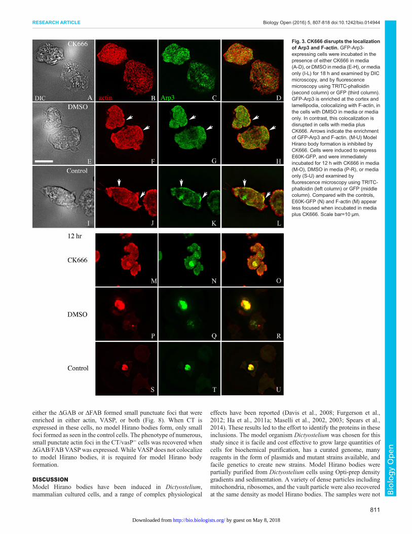
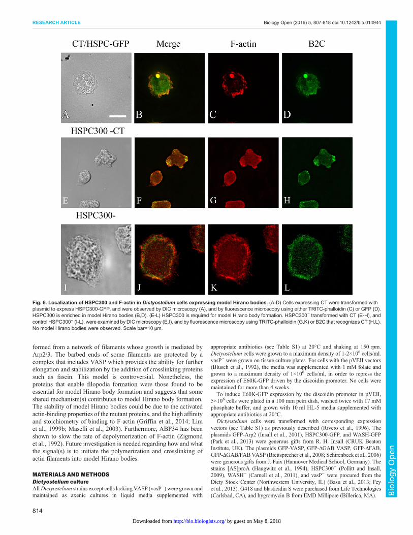
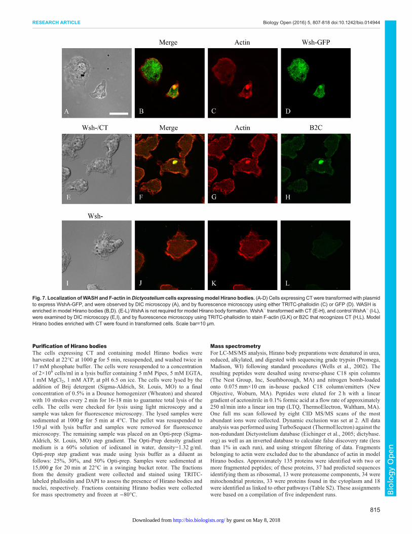
Colocalization of the activators of the Arp2/3 complex,HSPC300 and WASH, with model Hirano bodiesThe Arp2/3 complex promotes nucleation of actin monomers topolymerize into filamentous actin. The Arp2/3 complex by itselfhas weak activity and requires activation for maximal activity. Oneof the contributors for activation is interaction with nucleation-promoting factors (NPFs) (Campellone and Welch, 2010). NPFsstimulate Arp2/3 nucleation activity through their carboxy-terminalWCA domains but have diverse amino-terminal domains, allowingfor differential cellular function and regulation. We examined thelocalization of two of the activators of the Arp2/3 complex,HSPC300, part of the WAVE complex, crucial to plasma membraneprotrusion and cell motility; and WASH, which is involved inendosome trafficking to ascertain whether they also colocalizedwith model Hirano bodies. Cells constitutively expressing CT weretransformed separately with either HSPC300-GFP (Pollitt and Insall,2009) or WASH-GFP (Carnell et al., 2011) and were subsequentlystained with TRITC-labeled phalloidin to visualize F-actin. Cellstransformed with either HSPC300-GFP or WASH-GFP showedlocalization analogous to Arp2 or Arp3 (Fig. 6A-D, Fig. 7A-D) andwere also enriched in model Hirano bodies. Together, these resultssuggest that model Hirano body formation involves the activity of theArp2/3 complex, and its activation by the nucleation polymerizationfactors including WASH and wave regulatory complex (WRC), withHSPC300 as a subunit. The roles of these proteins in model Hiranobody formation were further investigated.
The role of HSPC300 and WASH in model Hirano bodyformationSince HSPC300 and WASH colocalized with model Hirano bodies,knockout strains of HSPC300 and WASH were used to further testthe result that the Arp2/3 complex plays a part in model Hirano body
formation, and indicate a possible pathway how the Arp2/3 complexis activated in model Hirano body formation. To determine if modelHirano bodies form in HSPC300−, HSPC300− (Pollitt and Insall,2009) were transiently transformed with plasmid encoding CTunder a constitutive actin promoter. Cells were fixed at differenttimes after transformation with the result that no model Hiranobodies were observed (Fig. 6E-H). This indicates that HSPC300,and the WRC, is critical for model Hirano body formation throughthe regulation of the Arp2/3 complex. In contrast, when WASH−
(Carnell et al., 2011) was transiently transformed with plasmidencoding CT, actin inclusions formed in a substantial proportion ofcells (Fig. 7E-H). These inclusions also colocalized with CT,indicating that they are model Hirano bodies and that WASH is notrequired for formation of model Hirano bodies.
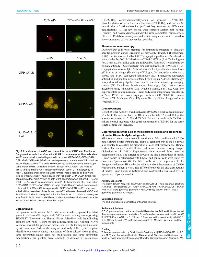
The role of VASP in model Hirano body formationSince profilin and the actin nucleation factor Arp2/3 have beenshown to be important for model Hirano body formation, we alsostudied the effect of VASP, an actin elongation factor. To determinewhether model Hirano bodies require VASP to form, Dictyosteliumlacking VASP (vasP−) (Han et al., 2002) were transformed with CTto induce model Hirano bodies in the presence or absence of fulllength VASP, VASP lacking either the G- (ΔGAB) or F-actinbinding domains (ΔFAB), or both the G- and F-actin bindingdomains (ΔGAB/FAB) (Breitsprecher et al., 2008; Schirenbecket al., 2006). Transient expression in cells was determined withTRITC-labeled phalloidin to localize actin and GFP fluorescencewas used to localize VASP and its mutants as shown in Fig. 8.No model Hirano bodies formed in CT/vasP− cells, only numerouspunctate F-actin foci were observed. In CT/VASP/vasP− cells,model Hirano bodies formed and VASP did not colocalize to themodel Hirano body. VasP− cells transformed with VASP lacking
Fig. 2. Localization of Arp2 and Arp3 and F-actin in Dictyostelium cells expressing model Hirano bodies. Cells expressing CT were transformed withplasmid to express GFP-Arp2 (A-D) or cells expressing GFP-Arp3 were transformed with CT (E-H). The cells were observed by DIC microscopy (A,E), and byfluorescencemicroscopy using either TRITC-phalloidin (C,G) to visualize F-actin, or GFP to visualize Arp2 and Arp3 (D,H). Arp2 and Arp3 are enriched in corticalactin-rich structures and also model Hirano bodies generated by expressing CT (B,F). Scale bar=10 µm.
810
RESEARCH ARTICLE Biology Open (2016) 5, 807-818 doi:10.1242/bio.014944
BiologyOpen
by guest on May 8, 2018http://bio.biologists.org/Downloaded from
either the ΔGAB or ΔFAB formed small punctuate foci that wereenriched in either actin, VASP, or both (Fig. 8). When CT isexpressed in these cells, no model Hirano bodies form, only smallfoci formed as seen in the control cells. The phenotype of numerous,small punctate actin foci in the CT/vasP− cells was recovered whenΔGAB/FABVASPwas expressed. While VASP does not colocalizeto model Hirano bodies, it is required for model Hirano bodyformation.
DISCUSSIONModel Hirano bodies have been induced in Dictyostelium,mammalian cultured cells, and a range of complex physiological
effects have been reported (Davis et al., 2008; Furgerson et al.,2012; Ha et al., 2011a; Maselli et al., 2002, 2003; Spears et al.,2014). These results led to the effort to identify the proteins in theseinclusions. The model organism Dictyostelium was chosen for thisstudy since it is facile and cost effective to grow large quantities ofcells for biochemical purification, has a curated genome, manyreagents in the form of plasmids and mutant strains available, andfacile genetics to create new strains. Model Hirano bodies werepartially purified from Dictyostelium cells using Opti-prep densitygradients and sedimentation. A variety of dense particles includingmitochondria, ribosomes, and the vault particle were also recoveredat the same density as model Hirano bodies. The samples were not
Fig. 3. CK666 disrupts the localizationof Arp3 and F-actin. GFP-Arp3-expressing cells were incubated in thepresence of either CK666 in media(A-D), or DMSO inmedia (E-H), or mediaonly (I-L) for 18 h and examined by DICmicroscopy, and by fluorescencemicroscopy using TRITC-phalloidin(second column) or GFP (third column).GFP-Arp3 is enriched at the cortex andlamellipodia, colocalizing with F-actin, inthe cells with DMSO in media or mediaonly. In contrast, this colocalization isdisrupted in cells with media plusCK666. Arrows indicate the enrichmentof GFP-Arp3 and F-actin. (M-U) ModelHirano body formation is inhibited byCK666. Cells were induced to expressE60K-GFP, and were immediatelyincubated for 12 h with CK666 in media(M-O), DMSO in media (P-R), or mediaonly (S-U) and examined byfluorescence microscopy using TRITC-phalloidin (left column) or GFP (middlecolumn). Compared with the controls,E60K-GFP (N) and F-actin (M) appearless focused when incubated in mediaplus CK666. Scale bar=10 µm.
811
RESEARCH ARTICLE Biology Open (2016) 5, 807-818 doi:10.1242/bio.014944
BiologyOpen
by guest on May 8, 2018http://bio.biologists.org/Downloaded from
further purified due to the instability of model Hirano bodies. Thecomponents of model Hirano bodies were identified by massspectrometry and included cytoskeletal, ribosomal, andmitochondrial proteins (Appendix Table A2). Since model Hiranobodies were not purified to homogeneity, immunofluorescence wasutilized to verify the presence of proteins/organelles identified bymass spectroscopy. We verified that mitochondria do not colocalizewith model Hirano bodies (Fig. S1). Ribosomes are probablycontaminants as well but this is difficult to confirm due to lack ofsuitable reagents. Components of the proteasome were alsoidentified by mass spectrometry. The presence of proteasomescould be contaminants or could have been associated with modelHirano bodies undergoing degradation (Kim et al., 2009).Since the major protein component of Hirano bodies is
filamentous actin (Goldman, 1983) and actin binding proteinshave been previously localized to Hirano bodies (Davis et al., 2008;Maciver and Harrington, 1995; Maselli et al., 2002), we focused onthe actin regulatory proteins identified by mass spectrometry. Actinbinding proteins, myosin II heavy and light chains, α-actinin,Arp2/3 complex, profilin I and II, hsp70, fimbrin, coactosin, EF1alpha, vinculin-related protein, histophilin II, cortexillin I, ABP1and filamin were identified. The presence of α-actinin, myosin II(Maselli et al., 2002), Arp2/3 complex and profilin I in model
Hirano bodies were confirmed; however, filamin and EF1 alphawere previously found not to colocalize to model Hirano bodies(Maselli et al., 2002). Thus, it is important to confirm resultsobtained by mass spectrometry analysis by immunofluorescence.
The role of profilin I and Arp2/3 complex were chosen for furtherinvestigation due to their role in the regulation of F-actinpolymerization. These proteins were localized to model Hiranobodies (Figs 1, 2). Verification of the role of profilin I in theformation of model Hirano bodies was achieved utilizing a profilinknockdown strain. Our results show that model Hirano bodiesare significantly smaller when profilin I is knocked down inDictyostelium (Fig. 1I). The actin monomer binding protein profilinhas been shown to increase the assembly rate of actin filaments byfacilitating the exchange of ATP for ADP on G-actin, maintainingthe ATP-G-actin pool that polymerizes more readily than ADP-G-actin (Perelroizen et al., 1996). Together, this suggests that profilin-enhanced assembly of F-actin is critical to the size of the modelHirano bodies, highlighting the role of de novo actin polymerizationin model Hirano body formation.
Verification of the role of the Arp2/3 complex in the formation ofmodel Hirano bodies was achieved utilizing the small moleculeinhibitor CK666 (Fig. 3), again suggesting that de novo actinpolymerization is required. Hirano bodies exhibit a hallmark
Fig. 4. CK666 affects the distribution of model Hirano body size and number of cells that generate model Hirano bodies. The size distribution ofmodel Hirano bodies was measured after 6 h (A), 12 h (B) or 18 h (C) of incubation with CK666 in media (green) after removal of folic acid to induce expression ofE60K-GFP, compared with cells incubated with DMSO plus media (red) or media only (blue) under the same condition to induce model Hirano bodies. Thedistributions of the model Hirano body size were significantly different (P<0.01 at 6 h, P<0.001 at 12 h and at 18 h). The proportions of cells that generated modelHirano bodies with or without CK666 were also counted, and compared with the control (D). The number of cells that produced model Hirano bodies weresignificantly reduced when incubated in the presence CK666 (P<0.001 for all three time points, data represented as means±S.D.).
812
RESEARCH ARTICLE Biology Open (2016) 5, 807-818 doi:10.1242/bio.014944
BiologyOpen
by guest on May 8, 2018http://bio.biologists.org/Downloaded from
paracrystalline order observed by electron microscopy. Theappearance of the Hirano body depends on the plane of sectionand can vary from a lattice to a herringbone pattern (Schochet andMcCormick, 1972; Tomonaga, 1974). Both Hirano bodies frommammalian sources (Peterson et al., 1986) and Dictyostelium(Maselli et al., 2002) were frequently observed to have ajuxtaposition of ordered and disordered regions. The Arp2/3complex nucleates F-actin by binding at the sides of preexistingactin filaments, initiating actin filament branches at an angle ofapproximately 70° from the mother filament with Arp2 and Arp3 asthe first two subunits of the daughter filament (Mullins et al., 1998;Rouiller et al., 2008). The resultant F-actin branched structure couldbe a contributing factor to the changing appearance of the Hiranobody with the plane of section.Identification of the pathway(s) of Arp2/3 activation was
investigated through knockout strains of HSPC300 (Caracino et al.,2007), a component of theWAVE complex which activates Arp2/3 inactin polymerization responsible for membrane protrusion and cellmotility, and WASH which activates Arp2/3 in the endocyticpathway (Carnell et al., 2011). Nomodel Hirano bodies were found intransformed HSPC300− cells (Fig. 6), but were found in transformed
WASH− cells (Fig. 7). This suggests that the WAVE regulatorycomplex (WRC), of which HSPC300 is a subunit, contributes tomodel Hirano formation through the activation of the Arp2/3complex. WASH associates with model Hirano bodies and wasshown to play an important role in exocytosis and vesicle trafficking.Model Hirano bodies are cleared from cells by autophagy (Kim et al.,2009) and WASH may play a role in the degradation pathway ofHirano bodies. Together with the results obtained from the profilinknockdown cells, the data suggests that de novo actin polymerizationis required for model Hirano body formation.
The WRC also needs to be activated to function. Its main activatorand recruiter Rac1 (Chen et al., 2014; Eden et al., 2002) could bepotentially involved in the pathway to form model Hirano bodies. Itwas previously shown that increased Rac1 activity is associated withthe enhancement of actin polymerization induced by fibrillar amyloidbeta peptide (Mendoza-Naranjo et al., 2007). Moreover, WAVE wasshown surrounding senile plaque in postmortem tissue, upregulatedin tissues from AD patients, and colocalized with tau in AD mousemodels (Kitamura et al., 2003; Takata et al., 2009). Tau is alsocolocalized to Hirano bodies (Galloway et al., 1987b; Peterson et al.,1988; Spears et al., 2014). Taken together, this suggests that theformation of Hirano bodies could be promoted by the pathology ofneurodegeneration and that the pathway involves WRC and theArp 2/3 complex, and possibly Rac1.
VASP was found to be required for model Hirano body formationalthough it was not colocalized to them. The small actin-richstructures observed in CT/vasP– and CT/vasP–/GFP-VASPΔGAB/FAB indicate that both the bundling and actin elongation activitiesare required for model Hirano body formation. CT/vasP–/GFP-VASPΔFAB cells failed to form model Hirano bodies. Previousinvestigation of Dictyostelium VASP revealed that DdVASPΔFABcould not rescue filopodia formation in vasP− cells (Schirenbecket al., 2006) nor bundle F-actin (Schirenbeck et al., 2006) butmediated accelerated filament elongation (Breitsprecher et al.,2008). These results are consistent with the observation that modelHirano bodies require both the elongation and bundling activities ofVASP; however, DdVASPΔGAB did rescue filopodia formationand accelerate filament elongation (Breitsprecher et al., 2008).Curiously, model Hirano bodies failed to form in the presence ofVASPΔGAB. Mutation in GAB site was reported to produceinstability of filopodia tips (Applewhite et al., 2007). Thisstabilizing activity could be required in vivo for model Hiranobody formation in addition to the bundling and filament elongationactivities. The mutant DdVASPΔGAB/FAB did not bundle actinnor accelerate filament elongation (Breitsprecher et al., 2008) so itwas not surprising that model Hirano bodies were not formed.
It has been reported that the crosslinking of filamentous actin(Griffin et al., 2014; Maselli et al., 2002, 2003), the transportationand clustering of small actin aggregations (Reyes et al., 2009), andthe stabilization of filamentous actin (Kim et al., 2009; Lázaro-Diéguez et al., 2008) are critical for model Hirano body formation.The current results provide evidence that de novo actinpolymerization regulated by the Arp 2/3 complex and profilin, isrequired for the formation of model Hirano bodies. VASP isnecessary for enhanced filament elongation for large model Hiranobodies to form. Model Hirano bodies are stable, cytoplasmicinclusions comprised of filamentous actin and appear to be formedby the same proteins required for dynamic rearrangement of theactin–rich filopodia. It is interesting to note that the parental protein(ABP34) of the mutant forms that induced model Hirano bodyformation is highly enriched in the filopodia ofDictyostelium. It waspostulated previously (Svitkina et al., 2003) that filopodia are
Fig. 5. Model Hirano bodies form after removal of CK666. Cells wereinduced to express E60K-GFP and immediately incubated with CK666 for12 h. They recovered in media after CK666 was removed for another 12 h (leftcolumn). Cells that were induced to express E60K-GFP at the same time(middle column) and at the time of CK666 removal (right column) were alsoobserved as controls. All cells were examined by DIC (first row), and byfluorescence microscopy using TRITC-phalloidin (second row) or GFP (thirdrow). Model Hirano bodies formed in a substantial amount of cells in all threesamples. Scale bar=15 µm.
813
RESEARCH ARTICLE Biology Open (2016) 5, 807-818 doi:10.1242/bio.014944
BiologyOpen
by guest on May 8, 2018http://bio.biologists.org/Downloaded from
formed from a network of filaments whose growth is mediated byArp2/3. The barbed ends of some filaments are protected by acomplex that includes VASP which provides the ability for furtherelongation and stabilization by the addition of crosslinking proteinssuch as fascin. This model is controversial. Nonetheless, theproteins that enable filopodia formation were those found to beessential for model Hirano body formation and suggests that someshared mechanism(s) contributes to model Hirano body formation.The stability of model Hirano bodies could be due to the activatedactin-binding properties of the mutant proteins, and the high affinityand stoichiometry of binding to F-actin (Griffin et al., 2014; Limet al., 1999b; Maselli et al., 2003). Furthermore, ABP34 has beenshown to slow the rate of depolymerization of F-actin (Zigmondet al., 1992). Future investigation is needed regarding how and whatthe signal(s) is to initiate the polymerization and crosslinking ofactin filaments into model Hirano bodies.
MATERIALS AND METHODSDictyostelium cultureAllDictyostelium strains except cells lacking VASP (vasP−) were grown andmaintained as axenic cultures in liquid media supplemented with
appropriate antibiotics (see Table S1) at 20°C and shaking at 150 rpm.Dictyostelium cells were grown to a maximum density of 1-2×10⁶ cells/ml.vasP− were grown on tissue culture plates. For cells with the pVEII vectors(Blusch et al., 1992), the media was supplemented with 1 mM folate andgrown to a maximum density of 1×10⁶ cells/ml, in order to repress theexpression of E60K-GFP driven by the discoidin promoter. No cells weremaintained for more than 4 weeks.
To induce E60K-GFP expression by the discoidin promoter in pVEII,5×106 cells were plated in a 100 mm petri dish, washed twice with 17 mMphosphate buffer, and grown with 10 ml HL-5 media supplemented withappropriate antibiotics at 20°C.
Dictyostelium cells were transformed with corresponding expressionvectors (see Table S1) as previously described (Rivero et al., 1996). Theplasmids GFP-Arp2 (Insall et al., 2001), HSPC300-GFP, and WASH-GFP(Park et al., 2013) were generous gifts from R. H. Insall (CRUK BeatonInstitute, UK). The plasmids GFP-VASP, GFP-ΔGAB VASP, GFP-ΔFAB,GFP-ΔGAB/FABVASP (Breitsprecher et al., 2008; Schirenbeck et al., 2006)were generous gifts from J. Faix (Hannover Medical School, Germany). Thestrains [AS]proA (Haugwitz et al., 1994), HSPC300− (Pollitt and Insall,2009), WASH− (Carnell et al., 2011), and vasP− were procured from theDicty Stock Center (Northwestern University, IL) (Basu et al., 2013; Feyet al., 2013). G418 and blasticidin S were purchased from Life Technologies(Carlsbad, CA), and hygromycin B from EMD Millipore (Billerica, MA).
Fig. 6. Localization of HSPC300 and F-actin in Dictyostelium cells expressing model Hirano bodies. (A-D) Cells expressing CT were transformed withplasmid to express HSPC300-GFP, and were observed by DIC microscopy (A), and by fluorescence microscopy using either TRITC-phalloidin (C) or GFP (D).HSPC300 is enriched in model Hirano bodies (B,D). (E-L) HSPC300 is required for model Hirano body formation. HSPC300− transformed with CT (E-H), andcontrol HSPC300− (I-L), were examined by DICmicroscopy (E,I), and by fluorescencemicroscopy using TRITC-phalloidin (G,K) or B2C that recognizes CT (H,L).No model Hirano bodies were observed. Scale bar=10 µm.
814
RESEARCH ARTICLE Biology Open (2016) 5, 807-818 doi:10.1242/bio.014944
BiologyOpen
by guest on May 8, 2018http://bio.biologists.org/Downloaded from
Purification of Hirano bodiesThe cells expressing CT and containing model Hirano bodies wereharvested at 22°C at 1000 g for 5 min, resuspended, and washed twice in17 mM phosphate buffer. The cells were resuspended to a concentrationof 2×10⁶ cells/ml in a lysis buffer containing 5 mM Pipes, 5 mM EGTA,1 mM MgCl2, 1 mM ATP, at pH 6.5 on ice. The cells were lysed by theaddition of Brij detergent (Sigma-Aldrich, St. Louis, MO) to a finalconcentration of 0.5% in a Dounce homogenizer (Wheaton) and shearedwith 10 strokes every 2 min for 16-18 min to guarantee total lysis of thecells. The cells were checked for lysis using light microscopy and asample was taken for fluorescence microscopy. The lysed samples weresedimented at 1000 g for 5 min at 4°C. The pellet was resuspended to150 µl with lysis buffer and samples were removed for fluorescencemicroscopy. The remaining sample was placed on an Opti-prep (Sigma-Aldrich, St. Louis, MO) step gradient. The Opti-Prep density gradientmedium is a 60% solution of iodixanol in water, density=1.32 g/ml.Opti-prep step gradient was made using lysis buffer as a diluent asfollows: 25%, 30%, and 50% Opti-prep. Samples were sedimented at15,000 g for 20 min at 22°C in a swinging bucket rotor. The fractionsfrom the density gradient were collected and stained using TRITC-labeled phalloidin and DAPI to assess the presence of Hirano bodies andnuclei, respectively. Fractions containing Hirano bodies were collectedfor mass spectrometry and frozen at −80°C.
Mass spectrometryFor LC-MS/MS analysis, Hirano body preparations were denatured in urea,reduced, alkylated, and digested with sequencing grade trypsin (Promega,Madison, WI) following standard procedures (Wells et al., 2002). Theresulting peptides were desalted using reverse-phase C18 spin columns(The Nest Group, Inc, Southborough, MA) and nitrogen bomb-loadedonto 0.075 mm×10 cm in-house packed C18 column/emitters (NewObjective, Woburn, MA). Peptides were eluted for 2 h with a lineargradient of acetonitrile in 0.1% formic acid at a flow rate of approximately250 nl/min into a linear ion trap (LTQ, ThermoElectron, Waltham, MA).One full ms scan followed by eight CID MS/MS scans of the mostabundant ions were collected. Dynamic exclusion was set at 2. All dataanalysis was performed using TurboSequest (ThermoElectron) against thenon-redundant Dictyostelium database (Eichinger et al., 2005; dictybase.org) as well as an inverted database to calculate false discovery rate (lessthan 1% in each run), and using stringent filtering of data. Fragmentsbelonging to actin were excluded due to the abundance of actin in modelHirano bodies. Approximately 135 proteins were identified with two ormore fragmented peptides; of these proteins, 37 had predicted sequencesidentifying them as ribosomal, 13 were proteasome components, 34 weremitochondrial proteins, 33 were proteins found in the cytoplasm and 18were identified as linked to other pathways (Table S2). These assignmentswere based on a compilation of five independent runs.
Fig. 7. Localization ofWASH and F-actin inDictyostelium cells expressingmodel Hirano bodies. (A-D) Cells expressing CTwere transformed with plasmidto express WshA-GFP, and were observed by DIC microscopy (A), and by fluorescence microscopy using either TRITC-phalloidin (C) or GFP (D). WASH isenriched in model Hirano bodies (B,D). (E-L) WshA is not required for model Hirano body formation. WshA− transformed with CT (E-H), and control WshA− (I-L),were examined by DIC microscopy (E,I), and by fluorescence microscopy using TRITC-phalloidin to stain F-actin (G,K) or B2C that recognizes CT (H,L). ModelHirano bodies enriched with CT were found in transformed cells. Scale bar=10 µm.
815
RESEARCH ARTICLE Biology Open (2016) 5, 807-818 doi:10.1242/bio.014944
BiologyOpen
by guest on May 8, 2018http://bio.biologists.org/Downloaded from
Data analysisFor protein identification, MSn data were searched against translatedgenomic database (Eichinger et al., 2005; curated at dictybase.org) usingSEQUEST (Bioworks 3.3, Thermo Fisher Scientific) with the followingsettings: 1000 ppm (10 ppm for data acquired using LTQ Orbitrap XL™)tolerance was set for precursor masses and 0.5 Da for fragment masses;trypsin was specified as the enzyme and only fully tryptic peptideidentifications were retained; a maximum of three missed cleavage sites,three differential amino acids per modification, and three differentialmodifications per peptide were allowed; oxidization of methionine
(+15.99 Da), carboxyamidomethylation of cysteine (+57.02 Da),phosphorylation of serine/threonine/tyrosine (+79.97 Da), and O-GlcNAcmodification of serine/threonine (+203.08 Da) were set as differentialmodifications. All the raw spectra were searched against both normal(forward) and reverse databases under the same parameters. Peptides werefiltered to 1% false-discovery rate and protein assignments were required tohave a minimum of two independent peptides.
Fluorescence microscopyDictyostelium cells were prepared for immunofluorescence to visualizespecific proteins and/or structures as previously described (Fechheimer,1987). F-actin was labeled by TRITC-conjugated phalloidin. Mitochondriawere labeled by 200 nM MitoTracker® Red CMXRos (Life Technologies)for 30 min at 20°C in live cells and followed by fixation. CT was labeled byprimary antibodyB2Cgenerated inmouse (Furukawa et al., 1992) andFITC-conjugated anti-mouse IgG. Profilin I was labeled by antibody obtained as agift from A. A. Noegel (University of Cologne, Germany) (Haugwitz et al.,1994), and FITC conjugated anti-mouse IgG. Fluorescent-conjugatedantibodies and phalloidin were obtained from Sigma-Aldrich. Microscopywas performed using Applied Precision DeltaVision I microscope-imagingsystem (GE Healthcare Bio-Sciences, Pittsburgh, PA). Images wereassembled using Photoshop CS6 (Adobe Systems, San Jose, CA). Forexperiments to determinemodel Hirano body sizes, imageswere recorded ona Zeiss IM35 microscope equipped with a CCD 300-T-RC camera(Dage MTI, Michigan City, IN) controlled by Scion Image software(Fredrick, MD).
Drug treatmentCK666 (Sigma-Aldrich) was dissolved in DMSO to a stock concentration of50 mM. Cells were incubated in HL-5 media for 6 h, 12 h and 18 h in theabsence or presence of 100 µM CK666. For each sample with CK666, asolvent control incubated with equal concentration of DMSO for the samelength of time was included.
Determination of the size of model Hirano bodies and proportionof model Hirano body-forming cellsMicroscopy images were taken in contiguous fields until a total of 200model Hirano bodies were imaged. The total cell number in all the fields wasalso counted to calculate the proportion of cells that formed model Hiranobodies. The area of model Hirano bodies was measured using ImageJ(Schneider et al., 2012). Experiments were repeated three times inindependent trials. The difference between the size distributions of modelHirano bodies in cells treated with CK666 and control cells were tested byexact test of goodness-of-fit. The difference between the proportions of cellsthat generated model Hirano bodies with or without the presence of CK666was tested by Student’s t-test. The difference between the size distributionsof model Hirano bodies in [AS]proA and control cells was tested by Chisquare test of goodness-of-fit.
AcknowledgementsThe plasmidsGFP-Arp2, HSPC300-GFP, andWASH-GFPwere generous gifts fromR. H. Insall. The plasmids GFP-VASP, GFP-ΔGAB VASP, GFP-ΔFAB, GFP-ΔGAB/FAB VASP were generous gifts from J. Faix. Antibody against profilin I was agenerous gift from A. A. Noegel.
Competing interestsThe authors declare no competing or financial interests.
Author contributionsS.S.-S. performed the purification of model Hirano bodies; D.S. and L.W. performedthe mass spectrometry and analysis; Y.D. performed experiments with profilin, Arp2/3, HSPC300 andWASH. N.F., N.I., and R.F. performed the experiments with VASP;Y.D., R.F., M.F., and L.W. wrote the manuscript. RF, MF, and LW supervised theexperiments.
FundingThis work was supported by Public Health Service grant [1RO1-NS0465101 to R.F.and M.F.] from the National Institute of Neurological Disorders and Stroke and byfunds for mass spectrometry equipment from the Georgia Research Alliance (L.W.).
Fig. 8. Localization of VASP and mutant forms of VASP and F-actin inDictyostelium cells transformed with CT to induce model Hirano bodies.vasP− were transformed with plasmid to express GFP-VASP, GFP-ΔGAB,GFP-ΔFAB, GFP-ΔGAB/FAB and in the presence or absence of CT to inducemodel Hirano bodies. The cells were observed by fluorescence microscopyusing either TRITC-phalloidin or GFP. Except for CT/vasP–, the mergedTRITC-phalloidin and GFP images are shown. When CT is expressed invasP−, punctate small actin foci were formed. Model Hirano bodies wereformed when CT/vasP− was rescued with full length GFP-VASP. Small focicontaining either actin, VASP, or both were observed when either GFP-ΔGABor GFP-ΔFAB VASP was expressed in vasP−. In the presence of CT and eitherGFP-ΔGAB or GFP-ΔFAB VASP, no large model Hirano bodies were formed,only small foci. When CT is expressed in GFPΔGAB/FAB vasP−, punctateactin foci that resembled those formed in vasP– cells were observed. VASPandits ability to bind actin is required either for F-actin foci to coalesce or elongatefrom F-actin foci to form model Hirano bodies. Arrowheads indicate either actinfoci or model Hirano bodies. Scale bar=5 µm.
816
RESEARCH ARTICLE Biology Open (2016) 5, 807-818 doi:10.1242/bio.014944
BiologyOpen
by guest on May 8, 2018http://bio.biologists.org/Downloaded from
Supplementary informationSupplementary information available online athttp://bio.biologists.org/lookup/doi/10.1242/bio.014944.supplemental
ReferencesApplewhite, D. A., Barzik, M., Kojima, S.-I., Svitkina, T. M., Gertler, F. B. andBorisy, G. G. (2007). Ena/VASP proteins have an anti-capping independentfunction in filopodia formation. Mol. Biol. Cell 18, 2579-2591.
Bamburg, J. R., Bernstein, B. W., Davis, R. C., Flynn, K. C., Goldsbury, C.,Jensen, J. R., Maloney, M. T., Marsden, I. T., Minamide, L. S., Pak, C. W. et al.(2010). ADF/Cofilin-actin rods in neurodegenerative diseases. Curr. AlzheimerRes. 7, 241-250.
Basu, S., Fey, P., Pandit, Y., Dodson, R. J., Kibbe, W. A. and Chisholm, R. L.(2013). DictyBase 2013: integrating multiple Dictyostelid species. Nucleic AcidsRes. 41, D676-D683.
Blusch, J., Morandini, P. and Nellen, W. (1992). Transcriptional regulation byfolate: inducible gene expression in Dictyostelium transformants during growthand early development. Nucleic Acids Res. 20, 6235-6238.
Breitsprecher, D., Kiesewetter, A. K., Linkner, J., Urbanke, C., Resch, G. P.,Small, J. V. and Faix, J. (2008). clustering of VASP actively drives processive,WH2 domain-mediated actin filament elongation. EMBO J. 27, 2943-2954.
Campellone, K. G. andWelch, M. D. (2010). A nucleator arms race: cellular controlof actin assembly. Nat. Rev. Mol. Cell Biol. 11, 237-251.
Caracino, D., Jones, C., Compton, M. and Saxe, C. L. (2007). The N-terminus ofDictyostelium Scar interacts with Abi and HSPC300 and is essential for properregulation and function. Mol. Biol. Cell 18, 1609-1620.
Carnell, M., Zech, T., Calaminus, S. D., Ura, S., Hagedorn, M., Johnston, S. A.,May, R. C., Soldati, T., Machesky, L. M. and Insall, R. H. (2011). Actinpolymerization driven by WASH causes V-ATPase retrieval and vesicleneutralization before exocytosis. J. Cell Biol. 193, 831-839.
Chen, B., Brinkmann, K., Chen, Z., Pak, C. W., Liao, Y., Shi, S., Henry, L.,Grishin, N. V., Bogdan, S. and Rosen, M. K. (2014). The WAVE regulatorycomplex links diverse receptors to the actin cytoskeleton. Cell 156, 195-207.
Davis, R. C., Furukawa, R. and Fechheimer, M. (2008). A cell culture model forinvestigation of Hirano bodies. Acta Neuropathol. 115, 205-217.
Eden, S., Rohatgi, R., Podtelejnikov, A. V., Mann, M. and Kirschner, M. W.(2002). Mechanism of regulation of WAVE1-induced actin nucleation by Rac1 andNck. Nature 418, 790-793.
Eichinger, L., Pachebat, J. A., Glockner, G., Rajandream, M.-A., Sucgang, R.,Berriman, M., Song, J., Olsen, R., Szafranski, K., Xu, Q. et al. (2005). Thegenome of the social amoeba Dictyostelium discoideum. Nature 435, 43-57.
Fechheimer, M. (1987). The Dictyostelium discoideum 30,000-dalton protein is anactin filament-bundling protein that is selectively present in filopodia. J. Cell Biol.104, 1539-1551.
Fey, P., Dodson, R. J., Basu, S. and Chisholm, R. L. (2013). One stop shop foreverything Dictyostelium: dictyBase and the Dicty Stock Center in 2012.MethodsMol. Biol. 983, 59-92.
Furgerson, M., Fechheimer, M. and Furukawa, R. (2012). Model Hirano bodiesprotect against tau-independent and tau-dependent cell death initiated by theamyloid precursor protein intracellular domain. PLoS ONE 7, e44996.
Furgerson, M., Clark, J. K., Crystal, J. D., Wagner, J. J., Fechheimer, M. andFurukawa, R. (2014). Hirano body expression impairs spatial working memory ina novel mouse model. Acta Neuropathol. Commun. 2, 131.
Furukawa, R., Butz, S., Fleischmann, E. and Fechheimer, M. (1992). TheDictyostelium discoideum 30,000 dalton protein contributes to phagocytosis.Protoplasma 169, 18-27.
Galloway, P. G., Perry, G. andGambetti, P. (1987a). Hirano body filaments containactin and actin-associated proteins. J. Neuropathol. Exp. Neurol. 46, 185-199.
Galloway, P. G., Perry, G., Kosik, K. S. and Gambetti, P. (1987b). Hirano bodiescontain tau protein. Brain Res. 403, 337-340.
Glenner, G. G. and Wong, C. W. (1984). Alzheimer’s disease: initial report of thepurification and characterization of a novel cerebrovascular amyloid protein.Biochem. Biophys. Res. Commun. 120, 885-890.
Goldman, J. E. (1983). The association of actin with Hirano bodies. J. Neuropathol.Exp. Neurol. 42, 146-152.
Griffin, P., Furukawa, R., Piggott, C., Maselli, A. and Fechheimer, M. (2014).Requirements for Hirano body formation. Eukaryot. Cell 13, 625-634.
Ha, S., Furukawa, R. and Fechheimer, M. (2011a). Association of AICD and Fe65with Hirano bodies reduces transcriptional activation and initiation of apoptosis.Neurobiol. Aging 32, 2287-2298.
Ha, S., Furukawa, R., Stramiello, M., Wagner, J. J. and Fechheimer, M. (2011b).Transgenic mouse model for the formation of Hirano bodies. BMC Neurosci. 12,97.
Han, Y.-H., Chung, C. Y., Wessels, D., Stephens, S., Titus, M. A., Soll, D. R. andFirtel, R. A. (2002). Requirement of a vasodilator-stimulated phosphoproteinfamily member for cell adhesion, the formation of filopodia, and chemotaxis inDictyostelium. J. Biol. Chem. 277, 49877-49887.
Haugwitz, M., Noegel, A. A., Karakesisoglou, J. and Schleicher, M. (1994).Dictyostelium amoebae that lack G-actin sequestering profilins show defects in F-Actin content, cytokinesis, and development. Cell 79, 303-314.
Hetrick, B., Han, M. S., Helgeson, L. A. and Nolen, B. J. (2013). Small moleculesCK-666 and CK-689 inhibit actin-related protein ⅔ complex by blocking anactivating conformational change. Chem. Biol. 20, 701-712.
Hirano, A., Dembitzer, H. M., Kurland, L. T. and Zimmerman, H. M. (1968). Thefine structure of some intraganglionic alterations. J. Neuropathol. Expt. Neurol. 27,167-182.
Insall, R., Mueller-Taubenberger, A., Machesky, L., Kohler, J., Simmeth, E.,Atkinson, S. J., Weber, I. andGerisch, G. (2001). Dynamics of the DictyosteliumArp2/3 complex in endocytosis, cytokinesis, and chemotaxis.Cell Motil. Cytoskel.50, 115-128.
Kim, D.-H., Davis, R. C., Furukawa, R. and Fechheimer, M. (2009). Autophagycontributes to degradation of Hirano bodies. Autophagy 5, 44-51.
Kitamura, Y., Tsuchiya, D., Takata, K., Shibagaki, K., Taniguichi, T., Smith,M. A., Perry, G., Miki, H., Takenawa, T. and Shimohama, S. (2003). Possibleinvolvement of Wiskott-Aldrich syndrome protein family in aberrant neuronalsprouting in Alzheimer’s disease. Neurosci. Lett. 346, 149-152.
Kosik, K. S., Joachim, C. L. and Selkoe, D. J. (1986). Microtubule-associatedprotein tau (tau) is a major antigenic component of paired helical filaments inAlzheimer disease. Proc. Natl. Acad. Sci. USA 83, 4044-4048.
Lazaro-Dieguez, F., Aguado, C., Mato, E., Sanchez-Ruiz, Y., Esteban, I.,Alberch, J., Knecht, E. and Egea, G. (2008). Dynamics of an F-actin aggresomegenerated by the actin-stabilizing toxin jasplakinolide. J. Cell Sci. 121, 1415-1425.
Lim, R. W. L., Furukawa, R. and Fechheimer, M. (1999b). Evidence ofintramolecular regulation of the Dictyostelium discoideum 34,000 Da F-actin-bundling protein. Biochemistry 38, 16323-16332.
Maciver, S. K. and Harrington, C. R. (1995). Two actin binding proteins, actindepolymerizing factor and cofilin, are associated with Hirano bodies. Neuroreport6, 1985-1988.
Maselli, A. G., Davis, R., Furukawa, R. and Fechheimer, M. (2002). Formation ofHirano bodies in Dictyostelium and mammalian cells induced by expression of amodified form of an actin cross-linking protein. J. Cell Sci. 115, 1939-1952.
Maselli, A. G., Furukawa, R., Thomson, S. A. M., Davis, R. C. and Fechheimer,M. (2003). Formation of Hirano bodies induced by expression of an actin cross-linking protein with a gain-of-function mutation. Eucaryot. Cell 2, 778-787.
Mendoza-Naranjo, A., Gonzalez-Billault, C. and Maccioni, R. B. (2007). Abeta1-42 stimulates actin polymerization in hippocampal neurons through Rac1 andCdc42 Rho GTPases. J. Cell Sci. 120, 279-288.
Mullins, R. D., Heuser, J. A. and Pollard, T. D. (1998). The interaction of Arp2/3complex with actin: nucleation, high affinity pointed end capping, and formation ofbranching networks of filaments. Proc. Natl. Acad. Sci. USA 95, 6181-6186.
Neumann, M., Sampathu, D. M., Kwong, L. K., Truax, A. C., Micsenyi, M. C.,Chou, T. T., Bruce, J., Schuck, T., Grossman, M., Clark, C. M. et al. (2006).Ubiquitinated TDP-43 in frontotemporal lobar degeneration and amyotrophiclateral sclerosis. Science 314, 130-133.
Nolen, B. J., Tomasevic, N., Russell, A., Pierce, D. W., Jia, Z., McCormick, C. D.,Hartman, J., Sakowicz, R. and Pollard, T. D. (2009). Characterization of twoclasses of small molecule inhibitors of Arp2/3 complex. Nature 460, 1031-1034.
Park, L., Thomason, P. A., Zech, T., King, J. S., Veltman, D. M., Carnell, M., Ura,S., Machesky, L. M. and Insall, R. H. (2013). Cyclical action of the WASHcomplex: FAM21 and capping protein drive WASH recycling, not initialrecruitment. Dev. Cell 24, 169-181.
Perelroizen, I., Didry, D., Christensen, H., Chua, N.-H. and Carlier, M.-F. (1996).Role of nucleotide exchange and hydrolysis in the function of profilin in actinassembly. J. Biol. Chem. 271, 12302-12309.
Peterson, C., Suzuki, K., Kress, Y. and Goldman, J. E. (1986). Abnormalities ofdendritic actin organization in the brindled mouse. Brain Res. 382, 205-212.
Peterson, C., Kress, Y., Vallee, R. and Goldman, J. E. (1988). High molecularweight microtubule-associated proteins bind to actin lattices (Hirano bodies). ActaNeuropathol. 77, 168-174.
Pollitt, A. Y. and Insall, R. H. (2009). Loss of Dictyostelium HSPC300 causes ascar-like phenotype and loss of SCAR protein. BMC Cell Biol. 10, 13.
Reyes, J. F., Stone, K., Ramos, J. and Maselli, A. (2009). Formation of Hiranobodies after inducible expression of a modified form of an actin-cross-linkingprotein. Eukaryot. Cell 8, 852-857.
Rivero, F., Furukawa, R., Noegel, A. A. and Fechheimer, M. (1996). Dictyosteliumdiscoideum cells lacking the 34,000-dalton actin-binding protein can grow,locomote, and develop, but exhibit defects in regulation of cell structure andmovement: a case of partial redundancy. J. Cell Biol. 135, 965-980.
Rouiller, I., Xu, X.-P., Amann, K. J., Egile, C., Nickell, S., Nicastro, D., Li, R.,Pollard, T. D., Volkmann, N. and Hanein, D. (2008). The structural basis of actinfilament branching by the Arp2/3 complex. J. Cell Biol. 180, 887-895.
Schirenbeck, A., Arasada, R., Bretschneider, T., Stradai, T. E. B., Schleicher, M.and Faix, J. (2006). The bundling activity of vasodilator-stimulatedphosphoprotein is required for filopodium formation. Proc. Natl. Acad. Sci. USA103, 7694-7699.
Schneider, C. A., Rasband,W. S. and Eliceiri, K. W. (2012). NIH Image to ImageJ:25 years of image analysis. Nat. Methods 9, 671-675.
817
RESEARCH ARTICLE Biology Open (2016) 5, 807-818 doi:10.1242/bio.014944
BiologyOpen
by guest on May 8, 2018http://bio.biologists.org/Downloaded from

Schochet, S. S., Jr and McCormick, W. F. (1972). Ultrastructure of Hirano bodies.Acta Neuropathol. 21, 50-60.
Spears, W., Furgerson, M., Sweetnam, J. M., Evans, P., Gearing, M.,Fechheimer, M. and Furukawa, R. (2014). Hirano bodies differentiallymodulate cell death induced by tau and the amyloid precursor proteinintracellular domain. BMC Neurosci. 15, 74.
Spillantini, M. G., Crowther, R. A., Jakes, R., Hasegawa, M. and Goedert, M.(1998). alpha-Synuclein in filamentous inclusions of Lewy bodies fromParkinson’s disease and dementia with lewy bodies. Proc. Natl. Acad. Sci. USA95, 6469-6473.
Svitkina, T. M., Bulanova, E. A., Chaga, O. Y., Vignjevic, D. M., Kojima, S.-I.,Vasiliev, J. M. and Borisy, G. G. (2003). Mechanism of filopodia initiation byreorganization of a dendritic network. J. Cell Biol. 160, 409-421.
Takata, K., Kitamura, Y., Nakata, Y., Matsuoka, Y., Tomimoto, H., Taniguichi, T.and Shimohama, S. (2009). Involvement of WAVE accumulation in Ab/APP
pathology-dependent tangle modification in Alzheimer’s disease. Am. J. Pathol.175, 17-24.
Tomonaga, M. (1974). Ultrastructure of Hirano bodies. Acta Neuropathol. 28,365-366.
Wells, L., Vosseller, K., Cole, R. N., Cronshaw, J. M., Matunis, M. J. and Hart,G. W. (2002). Mapping sites of O-Glc-Nac modification using affinity tags forserine and threonine post-translational modifications. Mol Cell. Proteomics 1,791-804.
Yang, Q., Zhang, X.-F., Pollard, T. D. and Forscher, P. (2012). Arp2/3 complex-dependent actin networks constrain myosin II function in driving retrograde actinflow. J. Cell Biol. 197, 939-956.
Zigmond, S. H., Furukawa, R. and Fechheimer, M. (1992). Inhibition of actinfilament depolymerization by the Dictyostelium 30,000 dalton actin-bundlingprotein. J. Cell Biol. 119, 559-567.
818
RESEARCH ARTICLE Biology Open (2016) 5, 807-818 doi:10.1242/bio.014944
BiologyOpen
by guest on May 8, 2018http://bio.biologists.org/Downloaded from