Date: 7 September, 2021 at 13:20-16:30 JST Venue: On the Web
20
Transcript of Date: 7 September, 2021 at 13:20-16:30 JST Venue: On the Web


Opening speech
13:20-13:23 Speech by the Representative of International Session of JSP 3
13:23-14:20 [ Session 1 "Free Paper" ] Chairman: Tomohiro Ogawa (Department of Vascular Surgery, Fukushima Daiichi Hospital)
Time Table Title Presenter Commentator min
IS-1-1 13:23-13:30Elevated platelet-derived sGPVI is a biomarker of venous in-stent stenosis in patients with post-
thrombotic syndromeAdam M. Gwozdz Javier Serralde Gallegos 5+2
IS-1-2 13:30-13:37Radical resection of tumor thrombus from de inferior vena cava and kidney carcinoma under real
time monitoring by intraabdominal vascular sonographyJorge Flores Sergio Gianesini 5+2
IS-1-3 13:37-13:44 Treatment of the Venous Insufficiency CEAP C6 with Inelastic Compression System Javier Serralde Gallegos Daisuke Akagi 5+2
IS-1-4 13:44-13:51New disease entity of severe choric vein insufficiency of the left leg: Manifestation of severe
choric vein insufficiency by arteriovenous fistula after the left iliac vein thrombosisShouma Tanaka 5+2
IS-1-5 13:51-13:58Short-term result and trend of length from saphenofemoral junction to the end of plugged great
saphenous vein after cyanoacrylate closure (CC)Shinji Tomita Takashi Yamaki 5+2
IS-1-6 13:58-14:05Strategy to prevent nerve injury and deep vein thrombosis in radiofrequency segmental thermal
ablation of the saphenous veins using a new objective pain scaleKenji Yamamoto Daisuke Akagi 5+2
IS-1-7 14:05-14:12 Cyanoacrylate glue ablation for venous leak erectile dysfunction Sriram Narayanann Takahiro Imai 5+2
IS-1-8 14:12-14:17 Genetic Determinants of Venous Thromboembolisms and Varicose Veins Eri Fukaya 5
14:25-14:40 [ Session 2 "EVF Prize Winner Presentation" ] Chairman: Takashi Yamaki (Department of Plastic and Reconstructive Surgery, Tokyo Women's Medical University Center East)
Time Table Title Presenter min
IS-2-1 14:25-14:40 Quantitative and structural characteristics of mitochondrial DNA in varicose veins Mariya A. Smetanina Takashi Yamaki 10+5
14:40-14:55 [ Session 3 "Lecture request from Medtoronic" ] Chairman: Masayuki Hirokawa (Ochanomizu Vascular & Vein Clinic)
Time Table Title Presenter min
IS-3-1 14:40-14:55A Comparison of Cyanoacrylate Glue and Radiofrequency Ablation Endovenous Saphenous Vein
Closure Techniques in CEAP 6 PatientsMisaki M Kiguchi 10+5
14:55-15:37 [ Session 4 "Free Paper" ] Chairman: Takashi Yamaki (Department of Plastic and Reconstructive Surgery, Tokyo Women's Medical University Center East)
Time Table Title Presenter Commentator min
IS-4-1 14:55-15:02Noninvasive near-infrared spectroscopic evaluation of calf muscle oxygenation in patients with
advanced chronic venous insufficiency associated with tricuspid regurgitationTakashi Yamaki Malay Patel 5+2
IS-4-2 15:02-15:09 Resection of venous malformations in bulbar conjunctiva Mine Ozaki Takashi Yamaki 5+2
IS-4-3 15:09-15:16 The treatment of Venous Malformation in the lower limbs Iwashina Yuki Luis René Arias Villarroel 5+2
IS-4-4 15:16-15:23 Outcome Analyses of 2381 Supermicrosurgical Lymphovenous Anastomoses Reiko Tsukuura Daisuke Akagi 5+2
IS-4-5 15:23-15:30 Effectiveness of Lymphatic Drainage and KINESIOTAPE Luis René Arias Villarroel Sergio Gianesini 5+2
IS-4-6 15:30-15:37Technical Considerations for Quality Scans for CVT Patients
– An Australian Vascular Sonographer Experience –Gary Liu Yoshiko Watanabe 5+2
15:40-16:19 [ Session 5 "COVID-19 and Thrombosis" ] Chairman: Makoto Mo (Department of Cardiovascular Surgery, Yokohama Minami Kyosai Hospital), Sergio Gianesini (University of Ferrara, ITALY)
Time Table Title Presenter Commentator min
IS-5-1 15:40-15:47 Thrombosis Risk Assessment During COVID-19: American Perspective Joseph A. Caprini 5+2
IS-5-2 15:47-15:54 Covid-19 and thrombosis - evidence from most recent anticoagulation trials Eduardo Ramacciotti Sergio Gianesini 5+2
IS-5-3 15:54-16:01 Covid-19 and thrombosis Armando Mansilha 5+2
IS-5-4 16:01-16:08Incidence and Clinical Features of Venous Thromboembolism in Hospitalized Patients with
COVID-19 in JapanYugo Yamashita Elna Masuda (HAWAII PACIFIC HEALTH, Honolulu, HI) 5+2
IS-5-5 16:08-16:15 COVID-19 and Thrombosis in Turkey Suat Doğancı Tomohiro Ogawa 5+2
IS-5-6 16:15-16:22 Aggressive Treatment for Proximal Deep Vein Thrombosis in COVID-19 Patient Nyityasmono Tri Nugroho Takahiro Imai 5+2
16:22-16:27 Discussion 5
Closing speech
16:27-16:30 Speech by the Vice President of JSP 3
All speakers
Makoto Mo(Department of Cardiovascular Surgery, Yokohama Minami Kyosai Hospital)
Date: 7 September, 2021 at 13:20-16:30 JST Venue: On the Web
The 41st Annual Meeting of the Japanese Society of Phlebology
International Session 2021
Takashi Yamaki(Department of Plastic and Reconstructive Surgery, Tokyo Women's Medical University Center East)

13:20~16:30 International Session
Chairs:Sergio Gianesini(University of Ferrara, ITALY)
Chairs:Elna Masuda(Hawaii Pacific Health, Honolulu, HI)
Chairs:Masayuki Hirokawa(Ochanomizu Vascular & Vein Clinic)
Chairs:Makoto Mo(Department of Cardiovascular Surgery, Yokohama Minami Kyosai Hospital/
Chairs:Makoto Mo(Department of Surgery, Yokohama City University)
Chairs:Tomohiro Ogawa(Department of Vascular Surgery, Fukushima Daiichi Hospital)
Chairs:Takashi Yamaki(Department of Plastic and Reconstructive Surgery,
Chairs:Takashi Yamaki(Tokyo Women's Medical University Center East)
Opening Speech Department of Plastic and Reconstructive Surgery, Tokyo Women’ Medical University Center East Takashi Yamaki
Session 1:Free Paper IS-1-1 Elevated platelet-derived sGPVI is a biomarker of venous in-stent stenosis in patients with post- thrombotic syndrome Academic Department of Vascular Surgery, Section of Vascular Risk and Surgery, 00000000000, School of Cardiovascular Medicine and Science, St Thomas’ospital, King’ College London, UK Adam M. Gwozdz
Discussant; Javier Serralde Gallegos (Hospital MAC Periferico Sur, Mexico City/ Colegio de Medicos Cirujanos J. Raymond Tourna)
IS-1-2 Radical resection of tumor thrombus from de inferior vena cava and kidney carcinoma under real time monitoring by intraabdominal vascular sonography Unit of Vascular Surgery of the Baptist Medical Center. Asunción, Paraguay Jorge Flores
Discussant; Sergio Gianesini (University of Ferrara, ITALY)
IS-1-3 Treatment of the Venous Insufficiency CEAP C6 with Inelastic Compression System Hospital MAC Periferico Sur, Mexico City/ Colegio de Medicos Cirujanos J. Raymond Tourna
Javier Serralde Gallegos
Discussant; Daisuke Akagi (Department of Cardiovascular Surgery, Kawasaki Medical School,
Kurashiki, Okayama, Japan)
IS-1-4 New disease entity of severe choric vein insufficiency of the left leg: Manifestation of severe choric vein insufficiency by arteriovenous fistula after the left iliac vein thrombosis Department of Cardiovascular Surgery, Yokohama Minami Kyosai Hospital/ Department of Surgery Yokohama City University,,,,,,,,,,,,,,,,,,,,,,,,,,,,,,,,,,,,,,,,,,,, Shouma Tanaka IS-1-5 Short-term result and trend of length from saphenofemoral junction to the end of plugged great saphenous vein after cyanoacrylate closure (CC) Department of Cardiovascular Surgery, Gifu Heart Center, Gifu, Japan Shinji Tomita
Discussant; Takashi Yamaki (Department of Plastic and Reconstructive Surgery,
Tokyo Women's Medical University Center East, Japan) IS-1-6 Strategy to prevent nerve injury and deep vein thrombosis in radiofrequency segmental thermal ablation of the saphenous veins using a new objective pain scale Okamura Memorial Hospital, Department of Cardiovascular Surgery, Center of Varicose Veins Kenji Yamamoto
Discussant; Daisuke Akagi (Department of Cardiovascular Surgery, Kawasaki Medical School,
Kurashiki, Okayama, Japan)
IS-1-7 Cyanoacrylate glue ablation for venous leak erectile dysfunction Senior Vascular and Endovascular Surgeon, The Harley Street Heart and Vascular Centre ,,,,,,,,,,,,,,,,,,,,,,,,,,,,,,,,,,,,,,,,, Sriram Narayanan
Discussant; Takahiro Imai (Department of Vascular Surgery, Nishinokyo Hospital, Nara, Japan)
IS-1-8 Genetic Determinants of Venous Thromboembolisms and Varicose Veins Department of Surgery, Division of Vascular Surgery, Stanford University School of Medicine, CA (R.A.B., E.F.)/ Department of Epidemiology, University of Washington, Seattle (N.L.S.)/,,,,,,,,,,,,,,,,,,,,,,,,,,,,,,,,,,,,,,,,,,,,,,,,,,,,,,,,,,,,,,,,,,,,,/ Program in Medical and Population Genetics, Broad Institute of MIT and Harvard, Cambridge, MA (D.K.).,,,,,,,,,,,,,,, Eri Fukaya

Session 2:EVF Prize Winner Presentation
IS-2(EVF) Quantitative and structural characteristics of mitochondrial DNA in varicose veins Institute of Chemical Biology and Fundamental Medicine, Novosibirsk, Russia/ Mariya A. Smetanina Novosibirsk State University, Novosibirsk, Russia
Session 3:Lecture request from Medtoronic
IS-3 A Comparison of Cyanoacrylate Glue and Radiofrequency Ablation Endovenous
Saphenous Vein Closure Techniques in CEAP 6 Patients
MedStar Health Vein Centers Misaki M. Kiguchi
Session 4:Free Paper
IS-4-1 Noninvasive near-infrared spectroscopic evaluation of calf muscle oxygenation in patients with advanced chronic venous insufficiency associated with tricuspid regurgitation Department of Plastic and Reconstructive Surgery, Tokyo Women’ Medical University Center East Takashi Yamaki
Discussant; Malay Patel (Gujarat university, India)
IS-4-2 Resection of venous malformations in bulbar conjunctiva Department of Plastic Surgery, Kyorin University School of Medicine Mine Ozaki
Discussant; Takashi Yamaki (Department of Plastic and Reconstructive Surgery,
Tokyo Women's Medical University Center East, Japan)
IS-4-3 The treatment of Venous Malformation in the lower limbs Department of Plastic, Reconstructive and Aesthetic Surgery, Kyorin University School of Medicine Yuki Iwashina
Discussant; Luis René Arias Villarroel (Director of Arias Medical Clinic)
IS-4-4 Outcome Analyses of 2381 Supermicrosurgical Lymphovenous Anastomoses Department of Plastic and Reconstructive Surgery, National Center for Global Health and Medicine Reiko Tsukuura
Discussant; Daisuke Akagi (Department of Cardiovascular Surgery, Kawasaki Medical School,
Kurashiki, Okayama, Japan)
IS-4-5 Effectiveness of Lymphatic Drainage and KINESIOTAPE Director of Arias Medical Clinic Luis René Arias Villarroel
Discussant; Sergio Gianesini (University of Ferrara, ITALY)
IS-4-6 Technical Considerations for Quality Scans for CVT Patients ‒ An Australian Vascular Sonographer Experience ‒ The University of Sydney, Australia Gary Liu
Discussant; Yoshiko Watanabe (First Department of Physiology, Kawasaki Medical School,
Kurashiki, Okayama, Japan)

Session 5:COVID-19 and Thrombosis
IS-5-1 Thrombosis Risk Assessment During COVID-19: American Perspective Emeritus, NorthShore University Health System, Evanston, IL/,,,,,,,, Senior Clinician Educator, Pritzker School of Medicine, Chicago, IL Joseph A. Caprini
IS-5-2 Covid-19 and thrombosis - evidence from most recent anticoagulation trials Loyola University Medical Center Eduardo Ramacciotti
Discussant; Sergio Gianesini (University of Ferrara, ITALY)
IS-5-3 COVID-19 and Thrombosis Department of Angiology and Vascular Surgery, University of Porto Armando Mansilha
IS-5-4 Incidence and Clinical Features of Venous Thromboembolism in Hospitalized Patients with COVID-19 in Japan Department of Cardiovascular Medicine, Graduate School of Medicine, Kyoto University, Kyoto, Japan Yugo Yamashita
Discussant; Elna Masuda (HAWAII PACIFIC HEALTH, Honolulu, HI) IS-5-5 COVID-19 and Thrombosis in Turkey Flight Surgeon Professor of Venous Surgery Health Sciences University, Gulhane School of Medicine Department of Cardiovascular Surgery Director, Department of Aero-Space Medicine Director, Department of Diving and Hyperbaric Medicine Suat Doğancı
Discussant; Tomohiro Ogawa (Department of Vascular Surgery, Fukushima Daiichi Hospital, Japan) IS-5-6 Aggressive Treatment for Proximal Deep Vein Thrombosis in COVID-19 Patient Vascular and Endovascular Division, Department of Surgery, Cipto Mangunkusumo National Hospital, Jakarta, Indonesia/ Department of Surgery, University Hospital of Universitas Indonesia, Depok, Indonesia Nyityasmono Tri Nugroho
Discussant; Takahiro Imai (Department of Vascular Surgery, Nishinokyo Hospital, Nara, Japan)
Closing Speech Department of Cardiovascular Surgery, Yokohama Minami Kyosai Hospital/ Department of Surgery, Yokohama City University
Makoto Mo

205 2021 Vol. 32 No.2
71
IS-1-1 Elevated platelet-derived sGPVI is a biomarker of venous in-stent stenosis in patients with post-thrombotic syndrome
1Academic Department of Vascular Surgery, Section of Vascular Risk and Surgery, School of Cardiovascular Medicine and Science, St Thomas’Hospital, King’s College London, UK 2Institute for Cardiovascular and Metabolic Research, School of Biological Sciences, University of Reading, UK 3Division of Hematology/Oncology, The Hospital for Sick Children, Toronto, Canada
Adam M. Gwozdz1, Stephen A. Black1, Rachael Morris1, Sophie Messiha1, Mohammed Ikram1, Alexander P. Bye2, Jonathan M. Gibbins2, Margaret L. Rand3, Ashish S. Patel1, Bijan Modarai1, Alberto Smith1, Prakash Saha1
BACKGROUND In-stent stenosis following intervention for post-thrombotic syndrome (PTS) occurs in ~ 30% of cases, despite therapeutic anticoagulation. The aim of this study was to investigate whether platelets have a role in this process. METHODS Case-matched patients undergoing venous stenting were prospectively recruited. Venous in-stent thrombus specimens were excised and immunohistochemical analysis was performed to detect collagen I, collagen III, CD68 and CD41. Blood samples were taken before and after venous stent placement, and platelet activation markers (P-selectin and phosphatidylserine) and reactivity were determined by flow cytometry and plate-based aggregation, respectively. Soluble glycoprotein VI (sGPVI), shed during activation, was measured in plasma. Patients with in-stent stenosis requiring reintervention (>50% diameter reduction) were compared with those who did not during follow-up. RESULTS Forty-five patients were recruited (median age: 43yrs, range: 33-55yrs; 65% female). Re-intervention was required in 19 patients (42%; median time: 3wks, range: 1day-3mths). Immunohistochemical analysis of in-stent thrombosis demonstrated a rich network of platelets, both early (collagen III) and late (collagen I) forms of collagen, and inflammatory cell infiltrates. There was no significant difference in platelet activation or reactivity after stenting, but P-selectin exposure pre-stent was significantly higher in patients who developed in-stent stenosis (2.7%±0.4 vs 1.7%±0.2; P<0.05). sGPVI levels before stent insertion were increased in patients who developed in-stent stenosis (18.9±3.6ng/mL vs. 7.4±0.9ng/ mL; P<0.01). Platelet reactivity to collagen-related
peptide, a GPVI-specific platelet agonist, was reduced in patients who developed in-stent stenosis (logEC50 = -6.5M±0.3 vs -7.2M±0.2; P<0.01; n=33). CONCLUSION Venous stenting does not activate platelets or alter platelet function, but patients who developed in-stent stenosis exhibited greater levels of pre-stent platelet activation, greater loss of platelet surface GPVI in the form of sGPVI and consequent reduction in reactivity to GPVI activation. sGPVI may have potential to risk-stratify patients undergoing deep venous reconstruction and predict who requires closer surveillance.

IS-1-2 Radical resection of tumor thrombus from de inferior vena cava and kidney carcinoma under real time monitoring by intraabdominal vascular sonography
Unit of Vascular Surgery of the Baptist Medical Center. Asunción, Paraguay
Flores J, Samaniego C, Gamarra J, González N, Kurtz L, Centurión F.
Background: Renal cell carcinoma (RCC) represents the most common form of kidney cancer, with a peak incidence in the seventh decade of life and intravascular tumor growth along the renal vein into the inferior vena cava (IVC) occurs in up to 10% of all patients with RCC. Methods: The case of a 69 years-old male patient is reported, who presented with RCC extending to the retrohepatic IVC, diagnosed by computed tomography (CT) (Figure 1). During the operation, the upper and lower level of the tumor thrombus inside de IVC was checked by real time intraabdominal Doppler ultrasonography to assure the appropriate level of vascular clamping (Figure 2). Radical nephrectomy was performed and then, the infradiaphragmatic and infrarenal IVC was clamped, as well as, Pringle maneuver was accomplished to decrease bleeding. A longitudinal incision of the IVC was performed and the tumor thrombus removed. As the IVC wall was infiltrated by the tumor at the level of the ostium of the right renal vein, a segment of 10 cm of the IVC was resected and replaced by a Dacron graft (Figure 3). After declamping, vascular sonography of the IVC was performed again to confirm total resection of the tumor thrombus. Results: Postoperative CT demonstrated total resection of the RCC and IVC tumor thrombus (Figure 4). Discussion: Despite latest research on therapeutic approaches against IVC thrombosis due to RCC, recent outcomes published on medical literature still support the fact that aggressive surgical resection continues to be the only potentially curative treatment for this disease and our strategy of real time monitoring of the operative field by Doppler ultrasonography seems to be a good alternative to ensure total resection of the thrombus from inside the vein lumen.
Fig. 1 Fig. 2 Fig. 3 Fig. 4
72
206 静 脈 学

IS-1-3 Treatment of the Venous Insufficiency CEAP C6 with Inelastic Compression System
1Hospital MAC Periferico Sur, Mexico City, 2Accademia Mexicana de Flebologia y Linfologia, 3Colegio de Medicos Cirujanos J. Raymond Tournay
Serralde Gallegos J.A.1,2,3
BACKGROUND: The compressive therapy has been for several years the main therapeutic strategy for the treatment of venous ulcer that has proven to be able to attack the venous hypertension, improve microcirculation, decrease proinflammatory factors in the endothelium, allowing healing of the ulcer reason for which it has been cataloged with the degree of recommendation I. Currently we have inelastic compression systems made with an adjustable velcro material which have been shown to increase the venous work pressure favoring the activity of the muscular pumps increasing the return to the deep venous system in a primordial way. OBJECTIVES: Demonstrate the effectiveness of inelastic compression in the treatment of venous ulcers using an adjustable velcro system which allows ulcers to heal, attacking the pathophysiology of chronic venous disease generating a cost benefit. METHODS: An Inelastic System was placed in the lower extremities that presented venous ulcers with a variable evolution from 2 months to 4 years, presenting different diameters associated with moderate to severe edema, pain, variable lymphorea and clinically without local infection. In all patients the Ankle-Brachial Index was performed. RESULTS: The healing of the venous ulcers was achieved using an average pressure of 30-40 mmHg at a time similar to elastic and multilayer systems, appreciating a significant decrease in pain after its application with reduction of edema in the first 24 hours of the 15 to 20% and at 7 days thereafter from 20 to 25%, referring good tolerance. After healing, patients continued with Elastic Compressive Therapy type II and III. DISCUSSION: Adjustable inelastic systems made with velcro, prove to be effective and safe to the treatment of the venous insufficiency on stage CEAP C6, allowing tissue healing, significantly reducing pain and edema with better results compared to multilayer therapy with a true cost benefit for the patient long-term.
IS-1-4 New disease entity of severe choric vein insufficiency of the left leg: Manifestation of severe choric vein insufficiency by arteriovenous fistula after the left iliac vein thrombosis
1Department of Cardiovascular Surgery, Yokohama Minami Kyosai Hospital, 2Department of Surgery, Yokohama City University
Shouma Tanaka1,2, Makoto Mo1,2, Yoshiyuki Kobayash1,2, Shinnichi Suzuki1, Kennichirou Aga1,2, Kenichi Fushim1,2, Eri Kikuch1, Norihisa Karube1,2, Naoki Hashiyama1,2
BACKGROUND: We experienced elderly cases of severe chronic venous insufficiency with asymptomatic lower limb arteriovenous fistula (AVF) and deep vein thrombosis (DVT) in the left iliac vein triggered by bed rest. RESULTS: The number of cases was 7 (two males and 5 females), age 70-88 years, 79.7 ± 7.0 Average ± SD), which was more common among females. Activity in daily living (ADL) is decreased in all cases at the onset of symptoms. The cause of decreased ADL was surgery (4 cases), cerebrovascular accident (two cases), psychiatric disorder (one case), and bedridden due to old age (one case). All cases were accompanied by severely tense edema of the entire left lower limbs. CEAP C class were C4b in 3 cases and C6 in 4 cases. CT angiography and Duplex ultrasonography were useful for diagnosis in all cases. In all cases, trans-arterial embolization (TAE) of AVF with coil or n-Butyl-2-cyanoacrylate (NBCA) was performed (1-4 times, 2.4 ± 1.3 Average ± SD). Because TAE alone was not effective enough, femoro-femoral crossover bypass using ePTFE prosthesis was performed in two cases (one case with early occlusion, one case with long-term patency and occlusion 8 years later) previously. More recently iliac vein stenting was performed in two cases with success. In all cases, the pain and edema improved markedly. Three ulcer in other three C6 cases were cured by the combined use of compression therapy. The one patient whose ulcer did not heal died of MRSA sepsis with ePTFE prosthesis infection. DISCUSSION: These cases series have in common that asymptomatic AVF becomes manifest as severe post-thrombotic syndrome due to the left iliac vein DVT at the time of bed rest due to surgery or paralysis in old age patients. Most cases was reported from Asian country with literature review.
207 2021 Vol. 32 No.2
73
IS-1-5 Short-term result and trend of length from saphenofemoral junction to the end of plugged great saphenous vein after cyanoacrylate closure (CC)
Department of Cardiovascular Surgery, Gifu Heart Center, Gifu, Japan
Shinji Tomita, Yutaka Koyama, Masahiro Inagaki, Koushi Sawada, Yasuhide Okawa
[BACKGROUND] It is unclear about the fate of the end of plugged GSV after CC. In this study we investigated short-term result and followed trend of length from SFJ to the end of plugged GSV after CC. [METHODS] We consecutively operated 45 patients (age 64±14, male:female,19:26) for GSV insufficiency by CC from September to December in 2020. We performed echography to measure length from SFJ to the end of plugged GSV (A) just after CC, POD 1, and POD 7. [RESULTS] Fifty two legs were treated with CC. Operation time was 37±17 minutes. Number of glue-shots were 6.7±1.8 and the length of treatment was 15.1±5.3cm. Occlusion rate was 100% at POD 7. Side effects were as follows; redness (7 cases, 13%), pain (3), tenderness (2), foreign body sensation (2), urticaria (0), infection (0), and DVT (0). There were 3 cases (5.8%) with EGIT III at POD 7, who showed EGIT I after 1 month with anticoagulation, while no case with EGIT IV. The patients with redness at thigh were treated with ointment and oral anti-allergic drugs. The A (6.4±2.2) just after operation shortened to 4.5±1.9 at POD1(P<0.001), and 2.7±2.4 at POD7(P<0.001). At POD1 The shape of edge of thrombus toward SFJ was tongue-like, however, that attached to the wall of GSV and shape of edge changed to gulf-like, usually distal to superficial abdominal vein at POD7. [DISCUSSION] Occlusion site gradually proceeded to proximally in GSV and tended to stop just distal to superficial abdominal vein, and ended up with shorter A. The mechanism of shorter A may be mainly due to thrombosis, not glue, which coagulates just after operation. The edge of occlusion site should be monitored in the long term.
IS-1-6 Strategy to prevent nerve injury and deep vein thrombosis in radiofrequency segmental thermal ablation of the saphenous veins using a new objective pain scale
1Okamura Memorial Hospital, Department of Cardiovascular Surgery, Center of Varicose Veins, 2Shiga General Hospital, Department of Cardiovascular Surgery, 3Shizuoka General Hospital, Department of Cardiovascular Surgery
Kenji Yamamoto1, Senri Miwa1, Tomoyuki Yamada2, Shuji Setozaki3 Mamoru Hamuro1, Shunji Kurokawa1, Sakae Enomoto1
74
208 静 脈 学
Backgrounds: Endovenous ablation (EA) is currently used as a treatment method for saphenous vein insufficiency. Most endothermal procedures have been developed to be performed under tumescent local anesthesia (TLA). However, general anesthesia and peripheral nerve blocks with sedation pose risks such as post-procedural delay in discharge, prolonged immobilization, and deep vein thrombosis (DVT). Appropriate treatment strategies are therefore needed to prevent nerve injury and DVT associated with EA. We evaluated the benefit of local anesthesia and active walking soon after surgery in preventing nerve injury and deep vein thrombosis caused during endovenous ablation. Methods: EA was performed in 1,334 consecutive patients. Varicectomy was performed using the stab avulsion technique, including for perforator vein insufficiency without suture and ligation. After surgery, patients were encouraged to walk 100‒200 m inside the ward for 3‒5 times/h. The pain was evaluated objectively using the Okamura pain scale (OPS) and subjectively using the numerical rating scale (NRS). Results: Stab avulsion was performed at 11.8 ± 8.0 sites and the mean operative time was 33.9 ± 15.2 min. The mean OPS and NRS scores were 1.6 ± 1.3 and 3.0 ± 2.0, respectively. DVT and pulmonary embolism were absent. The incidence of nerve injury was 0.3%. However, several patients visited the outpatient department ahead of schedule because of bleeding. In approximately one in 10‒20 patients, there may be oozing to the outer sides of the elastic stockings after surgery. Discussions: In our retrospective analysis involving a total of 1334 patients who underwent EA under local anesthesia using the OPS, we did not observe any occurrences of DVT and PE. The incidence of nerve injury was 0.3%. These incident rates were lower than those reported previously. EA should be performed with the patients under local anesthesia to prevent nerve injury and DVT. References: Yamamoto K, Miwa S, Yamada T, et al. Strategy to prevent nerve injury and deep vein thrombosis in radiofrequency segmental thermal ablation of the saphenous veins using a new objective pain scale [published online ahead of print, 2021 Apr 28]. Phlebology. 2021;2683555211010513. doi:10.1177/026835 55211010513

IS-1-7 Cyanoacrylate glue ablation for venous leak erectile dysfunction
Senior Vascular and Endovascular Surgeon, The Harley Street Heart and Vascular Centre, Singapore General Secretary, Asian Venous Forum
Sriram Narayanan
Background Erectile dysfunction is a common disorder affecting over 300 million men worldwide. Vascular disease is the underlying cause in over half these patients, with about two-thirds due to poor arterial inflow and/ or impaired nitric oxide activity. However, a third are secondary to rapid venous outflow from the penile corpora, resulting in an inability to sustain an erection or in late cases to even initiate an erection. This condition of corpora-veno-occlusive erectile dysfunction (CVOED) or venous leak ED affects younger men in their thirties and forties and is a source of great mental and social distress to them. We describe the results of a minimally invasive procedure of Glue Ablation for Venogenic Impotence (GAVI) to slow down the venous outflow using cyanoacrylate glue ablation of the deep dorsal vein bifurcation with over 1 year follow-up. A short video of the procedure will also be presented. Methods The results of 19 consecutive patients with CVOED who underwent the GAVI procedure are described. Venous leak was diagnosed using hemodynamic testing following a test dose of 10 mcg of Alprostadil to induce a medical erection. Patients with normal penile brachial indices and penile arterial pulse volume recordings, but with end diastolic velocities of greater than 5 cm/second were considered to have CVOED. The Sexual Health Inventory for Men (SHIM) score was used to grade the severity of ED and measured before the GAVI procedure and at 6 weeks, 3 months, 6 months and 1 year after the procedure. Hemodynamic testing was repeated 6 months after the procedure. Results All 19 patients were followed up for 6 months, but only 12 presented for follow up at 1 year. The median SHM, scores increased from 8.1 (moderate to severe ED) to 22.1. 23.1, 23.4 and 23.1 at 6 weeks, 3 months, 6 months and 1 year respectively (a SHIM score of 22-25 is normal i.e no ED). The median end diastolic velocities also returned to normal from 6.4 cm/sec to 0.4 cm/sec on the right side and 5.7 cm/sec to 0.3 cm/sec on the left side at 6 months. No major complications occurred, though minimal bruising at the access site occurred in 7 patients that was managed without intervention. Discussion Cyanoacrylate glue ablation of the deep dorsal vein bifurcation for venous leak ED is a safe and effective procedure with minimal complications. The hemodynamic as well as functional success of the procedure is seen to be maintained at 6 months and 1 year respectively.
IS-1-8 Genetic Determinants of Venous Thromboembolisms and Varicose Veins
Department of Surgery, Division of Vascular Surgery, Stanford University School of Medicine, CA (R.A.B., E.F.). Department of Epidemiology, University of Washington, Seattle (N.L.S.). Program in Medical and Population Genetics, Broad Institute of MIT and Harvard, Cambridge, MA (D.K.).
Richard A. Baylis, Nicholas L. Smith, Derek Klarin, Eri Fukaya
Background: Venous thromboembolism and chronic venous disease are a common and disabling problem and it has been known for decades that family history is an important risk factor, implicating a genetic element to a patient’s risk. Our understanding of the pathogenesis of these diseases has greatly benefited from an expansion of population genetic studies from pioneering familial studies to large genome-wide association studies; we now have multiple risk loci for each venous disease. Methods: We performed a review to highlight the current state of knowledge on the genetics of venous thromboembolism and chronic venous disease by reviewing recent finding highlighted in genome-wide association study (GWAS) in the recent literature. Results: For venous thromboembolism, 9 loci with variants reaching genome-wide significance (ABO, F2, F5, F11, FGG, PROCR, ZFPM2, TSPAN15, and SLC44A2) were identified and a diverse trove of other genes have shown novel association with VTE, some of which point to the role of immune function, platelets, and red blood cells in modifying VTE risk. For varicose veins, GWAS identified over 30 new genome-wide significant loci, identifying pathways involved in vascular development and skeletal/limb biology. Conclusions: With the emergence of modern population genetics, we have started to decode the genetic factors mediating the heritable contribution to these diseases. We now have numerous candidate loci for both diseases but still have significant unexplained heritability and often a rudimentary understanding of the functional role of these loci on venous disease. References: Baylis RA, Smith NL, Klarin D, Fukaya E. Epidemiology and Genetics of Venous Thromboembolism and Chronic Venous Disease. Circ Res. 2021 Jun 11;128(12):1988-2002.
2092021 Vol. 32 No.2
75

76
210 静 脈 学
EVF Prize Winner Presentation
IS-2 Quantitative and structural characteristics of mitochondrial (EVF) DNA in varicose veins Smetanina M.A.1,2, Sevost'ianova K.S.1,2, Shirshova A.N.1, Oscorbin I.P.1, Oskina N.A.1, Zolotukhin I.A.3, Filipenko M.L.1,2 1Institute of Chemical Biology and Fundamental Medicine, Novosibirsk, Russia 2Novosibirsk State University, Novosibirsk, Russia 3Pirogov Russian National Research Medical University, Moscow, Russia
Background: Despite the high prevalence of varicose vein disease, our understanding of the molecular mechanisms underlying its pathogenesis is yet to be clarified. Venous wall composition and morphology of its constituents is substantially changed during vein wall remodeling. Smooth muscle cells of the middle layer that are responsible for venous tone change their contractile function to a secretory one, which may correspond to a decrease in healthy mitochondria in the cells since there is no much need to get energy from them. We aimed to investigate quantitative and structural (in terms of common deletions in the MT-ND4 gene region) characteristics of mitochondrial DNA (mtDNA) in varicose veins. Methods: The study was conducted in accordance with the principles written in the Declaration of Helsinki and approved by our institutional committee. Post-operation material of paired samples (varicose and non-varicose vein segments left after surgery, from a corresponding C2‒C4 CEAP class patient) of great saphenous veins of the lower limbs was used for this study. Total DNAs isolated from 120 samples (or 60 pairs ‒ from 60 patients) were taken for the analysis. Determination of mtDNA level (normalized to genomic DNA level) was performed by real-time qPCR and droplet digital PCR (ddPCR) using Taq-Man probes and primers. Statistical analysis was performed in Excel and STATISTICA packages, using the Wilcoxon-signed rank test. Results: We found that total mtDNA level was decreased in varicose veins compare to non-varicose veins (1.186-fold, p=0.004, according to qPCR). Tiny differences (1.024-fold decrease, p=0.0009) in the no-deletion mtDNA proportion in varicose veins were revealed with ddPCR. Discussion: Our results suggest that quantitative and structural changes in mtDNA are plausibly involved in varicose vein pathogenesis since mtDNA copy number is decreased and mtDNA integrity is compromised in varicose veins. The work was supported by the Russian Science Foundation (project No. 17-75-20223).

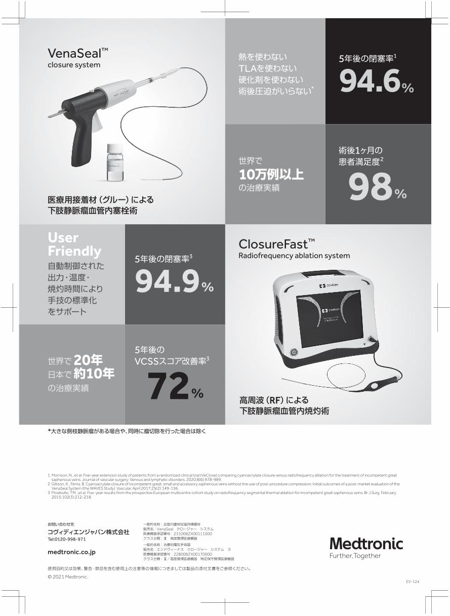
IS-3 Treatment of superficial venous reflux in CEAP 6 patients: a comparison of cyanoacrylate glue and radiofrequency ablation techniques
Leigh Ann O'Banion, Kyle B. Reynolds, Mariya Kochubey, Bianca Cutler, Eshetu A. Tefera, Rachel Dirks, Misaki M. Kiguchi
Objective Venous leg ulcers (CEAP [clinical, etiologic, anatomic, pathophysiologic] class 6) represent the most severe form of chronic venous insufficiency. As closure techniques for superficial venous reflux evolve, direct outcome comparisons of treatments are integral, because many studies have already demonstrated that early endovenous intervention improves wound healing. The present study compared the rates of venous wound healing between two techniques of superficial vein closure: ClosureFast radiofrequency ablation (RFA) and adhesive closure (VenaSeal; both Medtronic, Inc, Minneapolis, Minn). Methods We performed a multi-institutional retrospective review of all patients with CEAP class 6 who had undergone closure of their truncal veins from 2015 to 2020. Patients undergoing ClosureFast RFA were compared with those undergoing VenaSeal adhesive closure. The primary endpoint was the interval to wound healing from initial vein closure. The secondary endpoints included ulcer recurrence and infection rates. Bivariate analysis involved the χ2, Fisher exact, t, and Wilcoxon rank sum tests. Multivariate linear regression analysis was used to examine the factors affecting the time to wound healing in the most predictive model. Statistical significance was defined as P < .05. Results A total of 119 patients with CEAP 6 were included, with a median follow-up of 105 days (interquartile range, 44-208 days). Of the 119 limbs, 68 were treated with RFA and 51 with VenaSeal. Significantly more patients undergoing RFA had had a history of deep vein thrombosis (29% vs 10%; P = .01) and deep venous reflux (82% vs 51%; P = .003). The VenaSeal patients were older (72 years vs 65 years; P = .02) with a greater rate of coronary artery disease (16% vs 37%; P = .01). The median time to wound healing after the procedure was significantly shorter for VenaSeal than for RFA (43 vs 104 days; P = .001). Two RFA patients developed a postprocedure infection. The ulcer recurrence rate was 19.3% (22.1% for RFA vs 13.7% for VenaSeal; P = .25). On multivariate analysis, the treatment modality was the only significant predictor of the time to wound healing. When stratified by ulcer size as small (<3 cm2) vs large (>3 cm2), VenaSeal closure healed the wounds significantly faster for all ulcers.
Conclusion ClosureFast and VenaSeal are both safe and effective treatments to eliminate truncal venous insufficiency. VenaSeal showed a superior time to wound healing compared with ClosureFast in both large and small ulcers.
211 2021 Vol. 32 No.2
77
IS-4-1 Noninvasive near-infrared spectroscopic evaluation of calf muscle oxygenation in patients with advanced chronic venous insufficiency associated with tricuspid regurgitation
Department of Plastic and Reconstructive Surgery, Tokyo Women’s Medical University Medical Center East
Takashi Yamaki, Karin Ikeda, Yurie Wada, Sumire Yagi, Tomoko Uchida, Rie Miyazaki, Yumiko Sasaki
BACKGROUND: Near-infrared spectroscopy (NIRS) allows continuous noninvasive monitoring of changes in the tissue levels of oxygenated (O2Hb) and deoxygenated hemoglobin (HHb). Severe tricuspid regurgitation (TR) has occasionally been known to lead to advanced symptoms of chronic venous disease (CVD). The purpose of this study was to investigate changes in the calf muscle O2Hb and HHb levels during standing and exercise using NIRS and to compare discriminative parameters between patients with TR and those without. METHODS: Forty-eight limbs in 46 patients with CEAP C4a-C6 were studied. Thirteen limbs had CVD associated with TR and the remaining 35 had primary valvular insufficiency with no evidence of TR. On standing, increases in O2Hb and HHb were calculated by subtracting the baseline value from the maximum value (ΔO2Hbst and ΔHHbst). The times taken for the O2Hb and HHb to become maximal (TO2Hbst, and THHbst) were also measured. During 10 tiptoe movements, the change in O2Hb was calculated by subtracting the value measured at the end of exercise from that measured at the beginning of exercise (ΔO2Hbex). On the other hand, 10 tiptoe movements produced venous expulsion (ΔHHbEex) and a subsequent retention (ΔHHbRex). The oxygenation index (HbD; HbD=O2Hb-HHb) was also calculated at the end of standing and at the end of 10 tiptoe movements (ΔHbDst and ΔHbDex). RESULTS: On standing, the △HHbst increased significantly in patients with TR relative to those without (21 ± 24 vs. 8 ± 3, P=0.04). Similarly, △HbDst decreased significantly in patients with TR relative to those without (1 ± 33 vs. 22 ± 19, P=0.04). During ten tiptoe movements, the fall in △HbDex was significantly more pronounced in patients with TR (-38 ± 37 vs. -4 ± 13, P=0.01). There were no significant differences in the remaining parameters between the two groups. CONCLUSIONS: Our previous studies had shown that TO2Hbst was the best parameter for discriminating early from advanced CVD. In the present study, however, TO2Hbst was similar among patients with >CEAP C4 regardless of tricuspid valve insufficiency. A significant increase in △HHbst and a decrease in △HbDst on standing, and a significant decrease in △HbDex were considered to allow discrimination between patients with TR and those without. These results may reflect the impaired wound healing seen in patients who have severe tricuspid valve insufficiency.
IS-4-2 Resection of venous malformations in bulbar conjunctiva
Department of Plastic Surgery, Kyorin University School of Medicine
Mine Ozaki, Yuki Iwashina, Naoya Oshima, Akihiko Takushima
[Background] Venous malformations (VMs) in bulbar conjunctiva may result in impairment of eye movement and disfigurement. Sclerotherapy and/or surgical resection are considered to be main treatment options of Venous malformations (VMs). However, sclerotherapy to periorbital lesions has been regarded as inappropriate due to its risk for major complications including blindness. Therefore, surgical resection is the preferable method to avoid the complications. Herein, we experienced resection of VMs in bulbar conjunctiva in five patients, and the clinical courses and results are reported in this paper. [Patients & Method] Five patients (four females and one male) who suffered from irritable VMs in bulbar conjunctiva were treated in our department between from 2013 to 2018. Average age was 49 (ranged from 28 to 63), and all patients also suffered from eyelid lesion in addition to bulbar conjunctiva. All eyelid lesions were resected and reconstructed by conventional methods and resection of VMs in bulbar conjunctiva were performed under microscope. [Results] VMs in three out of five patients were completely resected owing to the microscopic dissection between bulbar conjunctiva and lesions above sclera. However, total dissection between bulbar conjunctiva and VM was impossible in two patients. Postoperatively, good eye movement without disfigurement were achieved in three patients whose lesions were completely resected. However, partial adhesion between bulbar conjunctiva and palpebral conjunctiva was recognized in the remaining two patients. [Discussion] In 2009, Mehta reported amniotic membrane grafting is a distinctive method for the defect of conjunctiva after resection of venous malformations in bulbar conjunctiva. This method may be useful in cases whose lesion adhered to the bulbar conjunctiva. However, preparation and preservation of the amniotic membrane requires a great deal of tasks to clear the regulations in Japan. Securing the bulbar conjunctiva by dissecting the mucosa from VM can be an alternative and better method.
78
212 静 脈 学
IS-4-3 The treatment of Venous Malformation in the lower limbs
Department of Plastic, Reconstructive and Aesthetic Surgery, Kyorin University School of Medicine
Iwashina Yuki, Ozaki Mine, Oshima Naoya, Takushima Akihiko
BACKGROUND Venous malformations (VMs) are the slow flow lesions that are comprised of a network of serpiginous interconnected veins with ectatic venous channels deficient in vascular smooth muscle1). They can occur throughout the body, and often cause pain and cosmetic concern. Particularly, patients with VMs in the lower limbs suffer from persistent pain, which is difficult to control. Generally, the treatment options of VMs are compression garment, oral aspirin, percutaneous sclerotherapy and surgical resection. In this paper, the treatment of VMs in the lower limbs in our department are described. METHOD Between 2015 and 2019, 69 patients (26 males, 43 females ; mean age 27 years) of VMs in the lower limbs who complain pain are included. 9 of them were diagnosed with Klippel-Trenaunay syndrome. RESULT 60 patients underwent therapeutic intervention. 27 patients were primarily treated with oral aspirin , but 15 of them required surgical intervention subsequently due to uncontrollable pain. 48 patients required surgical intervention(resection 13 cases , sclerotherapy 35 cases , both 9 cases). DISCUSSION Pain with VMs is thought to occur due to thrombophlebitis in the lesions and severely declines QOL of the patients. Oral aspirin was applied to almost half of our cases. The others denied continuous use of aspirin. Oral aspirin acquired only 50% success ratio for pain control, so surgical intervention played an important role for pain control. Sclerotherapy is often the firstline treatment option because it is less invasive method than surgical resection2,3). But resection was effective especially for localized VMs and painful lesions in extended VMs. It is important to adapt surgical intervention appropriately for treatment of VMs in the lower limbs. REFERENCES 1)Wassef M, Blei F, Adams D, et al. Vascular anomalies classification: reccomendations from the International Society for the Study of Vascular Anomalies. Pediatrics. 2015;136:e203-214. 2)Heit JJ, Do HM, Prestigiacomo CJ, et al. Guidelines and parameters: percutaneous sclerotherapy for the treatment of head and neck venous and lymphatic malformation. J Neurointerv Surg. 2016. 3)Steiner F, FitzJohn T, Tan ST. Surgical treatment for venous malformation. J Plast Reconstr Aesthet Surg. 2013;66(12):1741-9.
IS-4-4 Outcome Analyses of 2381 Supermicrosurgical Lymphovenous Anastomoses
Department of Plastic and Reconstructive Surgery, National Center for Global Health and Medicine
Reiko Tsukuura, Takashi Kageyama, Hayahito Sakai, Nana Yamamoto, Takumi Yamamoto
Background: Supermicrosurgical lymphovenous anastomosis (LVA) has become a therapeutic option for compression-refractory lymphedema. Little is known regarding factors associated with better clinical results. This study aimed to investigate efficacy of LVA according to indocyanine green (ICG) lymphography findings. Methods: Secondar lower extremity lymphedema patients who underwent LVA were evaluated. Intraoperative findings and postoperative results were analyzed according to ICG lymphography patterns. Lymphedema index was used to evaluate perioperative lymphedematous volume change. Postoperative outcomes were evaluated according to pathophysiological stages based on ICG lymphography findings. Results: In total, 2381 anastomoses were performed on 454 limbs. All operations were done under local infiltration anesthesia, and there was no postoperative complication. Longer skin incision was required in regions with Stardust/Diffuse pattern (S-region/D-region) than in that with Linear pattern (L-region) on ICG lymphography. Lymph vessels were smaller and more sclerotic in D-region. Postoperatively, 89% of the patients showed volume reduction, and 93% could be free from cellulitis. Patients with lower ICG stage were associated with higher rates of volume reduction and cure. Conclusions: LVA is minimally invasive surgical treatment for compression-refractory lymphedema. ICG lymphography facilitates LVA surgery by guiding lymph vessel location and condition, and allows prediction of postoperative outcomes. Early diagnosis and early treatment is a key to maximize therapeutic effect of LVA.
213 2021 Vol. 32 No.2
79

IS-4-5 Effectiveness of Lymphatic Drainage and KINESIOTAPE
DIRECTOR OF ARIAS MEDICAL CLINIC PRESIDENT OF THE BOLIVIAN COMMUNITY OF PHLEBOLOGY AND LYMPHOLOGY: COBOFLIN
Luis René Arias Villarroel
OBJECTIVE Under normal conditions the transport capacity of the lymphatic system is more than 10 liters, which can be increased to 20-25. When necessary, compensatory mechanisms (collateral lymphatic pathways + lympholymphatic and lymphovenous anastomoses) are triggered. The increase in the diameter of the vessels causes a valvular insufficiency which worsens the transport capacity. The lymphatic system has two functions: an immunological function of transporting antigens from the tissues to the antigens from the tissues to the lymphoid organs to produce immune reactions, and another: extravascular homeostatic, by reabsorbing and returning to the blood circulation the protein bodies and plasma proteins that are continuously leaving the blood capillaries into the interstitium. The treatment of lymphedema is based almost exclusively on rehabilitation measures, monitoring sites to assess progress. Lymphatic drainage pumps, pharmacology of lymphedema, limiting work overload, preventing the outflow of fluids from the blood capillary, help evacuate retained water by reducing oncotic pressure linked to accumulated proteins. PREVENTION: creams, antiseptic agents, fungicides, topical and oral benzopyrones, etc. TREATMENT: diuretics, immunological therapy, benzopyrones. IMMUNOLOGICAL THERAPY: injection of lymphocytes, immune BCG vaccine by scarification. MATERIAL: Kinesiotape bandage RESULTS: Improve muscle function & the injured ligament. Joint alignment, increase spacing improve lymphatic drainage, fibrosis, scars and hematomas.& organic or segmental function. CONCLUCIONS: Increasing temperature: warm colors Decrease temperature: cool colors Provides muscle support. Eliminates pain. Decreases inflammation, edema.
IS-4-6 Technical Considerations for Quality Scans for CVT Patients
‒ An Australian Vascular Sonographer Experience ‒
The University of Sydney, Australia
Gary Liu
Background In Australia, ultrasound examinations are primarily performed by highly trained sonographers. For chronic venous insufficiency (CVI) study, there seemed to be variable levels of expertise and knowledge among the sonographers. The University of Sydney had conducted an online survey to explore how CVI studies are being performed, providing insights into current practices and recommendations for quality improvement. Methods Questionnaires were distributed through the Australasian Sonographers Association to capture demographics, practices of ultrasound, examination techniques, the use of nomenclature, knowledge, and experience of sonographers. Results The study received 97 valid responses and the results showed a heterogeneity in the clinical application of ultrasound. The majority of respondents used standing, sitting and reverse Trendelenburg position. The combination of standing and sitting was used by 48.5%. Four sonographers claimed using the supine position (4.1%). A question specific to reflux provocation methods revealed that manual augmentation was used by 97.9% of respondents. Valsalva manoeuvres and toe-elevation manoeuvre were both used by 47.4% as alternative methods. Sonographers using the elevation-dependency manoeuvre and Parana manoeuvre accounted for 5% and 2% respectively. Automatic pneumatic pump was available to two sonographers from different practices. No respondent used Cremona manoeuvre or recorded any other method as‘other’choice. Discussion Our study is a multi-faceted exploration that identified a need for standardized diagnostic and reporting guidelines. Technical performance and waveforms interpretation for CVI ultrasound will be discussed in the upcoming meeting as they have been identified as the essential elements responsible for the discrepancies in diagnostic findings.
80
214 静 脈 学

IS-5-1 Thrombosis Risk Assessment During COVID-19: American Perspective
Emeritus, NorthShore University Health System, Evanston, IL Senior Clinician Educator, Pritzker School of Medicine, Chicago, IL
Joseph A. Caprini
COVID-19 infection is a serious and life-threatening disorder associated with a complex pathophysiology including arterial and venous thrombosis, organ dysfunction, and in some cases consumptive coagulopathy. The virus attacks endothelial cells systemwide with a predilection for the pulmonary alveolar-endothelial interface. Primary pulmonary thrombosis and alveolar hemorrhages have also been described. COVID-19 infection induces a complex series of biologic responses including activation of platelet, coagulation, and fibrinolytic pathways leading to both thrombosis and bleeding manifestations. Additionally, an inflammatory response including a cytokine storm with triggering of symptomatology includes vasodilatation, hypotension, bradycardia, increased vascular permeability, Histamine release, and angioedema. The coagulation changes in this disease have stimulated an intense series of studies to attempt to minimize the incidence of thrombotic events and mortality. Several large clinical trials have produced conflicting results regarding optimal anticoagulant dosing, however individual detailed thrombosis risk assessment was not part of these studies[1, 2].
Recently a retrospective observational cohort including 184 patients with severe COVID-19 scored with the IMPROVE and Caprini Risk scores was published[3]. Linear increase in both scores according to risk level were associated with both increasing rates of VTE and mortality and were independent predictors of these events as seen below. Hopefully future studies will incorporate risk scoring to refine optimal anticoagulant dosing. References 1.Lawler, P.R., et al., Therapeutic Anticoagulation in Non-Critically Ill Patients with Covid-19. COVID-19 SARS-CoV-2 preprints from medRxiv, 2021. 2.Lopes, R.D., et al., Therapeutic versus prophylactic anticoagulation for patients admitted to hospital with COVID-19 and elevated D-dimer concentration (ACTION): an open-label, multicentre, randomised, controlled trial. The Lancet, 2021. 397(10291): p. 2253-2263. 3.Paz Rios, L.H., et al., Prognostic Value of Venous Thromboembolism Risk Assessment Models in Patients with Severe COVID-19. TH Open, 2021. 5(2): p. e211-e219.
215 2021 Vol. 32 No.2
81

IS-5-2 COVID-19 and Thrombosis Loyola University Medical Center
Ramacciotti Eduardo
IS-5-3 COVID-19 and Thrombosis Department of Angiology and Vascular Surgery, University of Porto
Armando Mansilha
82
216 静 脈 学
IS-5-4 Incidence and Clinical Features of Venous Thromboembolism in Hospitalized Patients with COVID-19 in Japan
1Department of Cardiovascular Medicine, Graduate School of Medicine, Kyoto University, Kyoto, Japan, 2Department of Cardiology, Kuwana City Medical Center, Kuwana, Japan, 3Department of Cardiovascular Surgery, Yokohama Minami Kyosai Hospital, Kanagawa, Japan
Yugo Yamashita1, Norikazu Yamada2, Makoto Mo3
Background: Several studies reported a high prevalence of venous thromboembolism (VTE), including pulmonary embolism (PE) in COVID-19. However, the prevalence of VTE seemed to widely vary according to reports, which might be due to the different study population, ethnic differences and distinct resource availability. Actually, a recent surveillance questionnaire in Japan has reported the small number of patients diagnosed as VTE in COVID-191,2. Further studies have been warranted to clarify the current status of VTE among patients with COVID-19 who were evaluated with imaging examination, where under-diagnosis of VTE could be avoided. Methods: The VTE and COVID-19 in Japan Study is a retrospective, multicenter cohort study enrolling hospitalized patients with COVID-19 who was evaluated with contrast-enhanced computed tomography (CT) examination among 22 centers in Japan between March 2020 and October 2020. Results: Among 1236 patients with COVID-19, 45 patients (3.6%) patients were evaluated with contrast-enhanced CT examination. VTE events occurred in 10 patients (22.2%), and the incidences of VTE in mild, moderate, and severe COVID-19 were 0%, 11.8%, and 40.0%, respectively. COVID-19 patients with VTE showed a higher body weight (81.6 kg versus 64.0 kg, P=0.005) and body mass index (26.9 kg/m2 versus 23.2 kg/m2, P=0.04), and a higher proportion of severe status for COVID-19 compared with those without. There was no significant difference in the proportion of patients alive at discharge between patients with and without VTE (80.0% versus 88.6%, P=0.48). Among 8 PE patients, all of them were low-risk PE. Discussion: Among a relatively small number of patients with contrast-enhanced CT examination in Japanese real-world clinical practice, there were no VTE patients among mild COVID-19 patients, whereas the incidence of VTE seemed to be relatively high among severe COVID-19 patients, although all of PE events were low-risk PE events without significant impact on mortality risk. Reference 1. Yamashita Y, Yamada N, Mo M. Prevalence of venous thromboembolism in patients with severe novel coronavirus pneumonia. The Primary Prevention of Venous Thromboembolism in Patients with COVID-19 in Japan: Current Status and Future Perspective. Ann Vasc Dis. 2021;14:1-4. 2. Yamashita Y, Hara N, Obana M, et al. Clinical Features of Venous Thromboembolism in Patients With Coronavirus Disease 2019 (COVID-19) in Japan - A Case Series Study. Circ J. 2021;85:309-313.
IS-5-5 COVID-19 and Thrombosis in Turkey
Flight Surgeon Professor of Venous Surgery Health Sciences University, Gulhane School of Medicine Department of Cardiovascular Surgery Director, Department of Aero-Space Medicine Director, Department of Diving and Hyperbaric Medicine UIP, Scientific Committee Member Executive Committee Member, Turkish Society of Phlebology
Suat Doğancı
Since the beginning of the pandemic, coronavirus disease 2019 (COVID-19) has posed a multitude of challenges to health care systems globally. COVID-19 caused by the novel severe acute respiratory syndrome coronavirus 2 (SARS-CoV2), is characterized by a dysregulated immune system and hypercoagulability. COVID-associated coagulopathy was recognized based on profound D-dimer elevations and evidence of microthrombi and macrothrombi, both in venous and arterial systems. The underlying mechanisms associated with covid-associated coagulopathy have been suggested, but not clearly defined. The model of immunothrombosis illustrates the elaborate crosstalk between the innate immune system and coagulation. The rendering of a procoagulant state in COVID-19 involves the interplay of many innate immune pathways. COVID-19 virus can directly infect immune and endothelial cells, leading to endothelial injury and dysregulation of the immune system. Activated leukocytes potentiate a procoagulant state via many complicated mechanisms. Immunothrombosis provides a comprehensive perspective of the several synergistic pathways pertinent to the pathogenesis of COVID-associated coagulopathy.
217 2021 Vol. 32 No.2
83

IS-5-6 Aggressive Treatment for Proximal Deep Vein Thrombosis in COVID-19 Patient
1Vascular and Endovascular Division, Department of Surgery, Cipto Mangunkusumo National Hospital, Jakarta, Indonesia, 2Department of Surgery, University Hospital of Universitas Indonesia, Depok, Indonesia
Nyityasmono Tri Nugroho1,2, Dedy Pratama1
COVID-19 has emerged worldwide and along with its sequalae. A systemic injury is stated in critically-ill COVID-19 patients. The endotheliitis or inflammation in the endothelial cell in the capillary system, and in addition a microvascular inflammation could precipitate the thrombosis event in COVID-19. Hypercoagulable state leading in thrombosis event in a non-critically and critically ill COVID-19 patients could increase venous thromboembolism (VTE) events in the worldwide prevalence. The weighted mean prevalence (WMP) for VTE, deep vein thrombosis (DVT), and pulmonary embolism (PE) from a study with 1,988 COVID-19 patients were 31.3% (95% CI: 24.3-39.2%), 19.8% (95% CI: 10.5-34.0%), and 18.9% (95% CI: 14.4-24.3%), respectively. In 88 critically ill patients with Padua score ≥4 and IMPROVE score ≥2 showed DVT prevalence were 53% and 46%, respectively. Proximal DVT could lead a higher mortality rate due to the PE incidence. Proximal DVT including iliofemoral and caval thrombosis is the most dangerous that could lead to PE event. Proximal DVT are difficult to diagnose. The D-Dimer/Fibrinogen (D/F) ratio >1.0 x 10⁻³ could predict the DVT event. Mortality rate due to proximal DVT could be lowered by aggressive therapy. Extremity compression may help in proximal DVT therapy, nevertheless its usage is unsignificant. Protocol for anticoagulant prophylaxis in proximal DVT is not recommended in critically ill COVID-19 patients with high D-Dimer (≥ 10,000 ng/mL), and probable proximal DVT, and Padua score ≥4, despite that, fully anticoagulant is recommended, including low molecular weight heparin (LMWH) or unfractionated heparin (UFH), to decrease the mortality rate. A decreasing of mortality rate until 28% could be achieved with full anticoagulant therapy. Aggressive therapy such as surgical venous thrombectomy, pharmaco-mechanical thrombectomy, or aspiration
thrombectomy may help in selected cases. A proximal DVT event could be concomitant with acute limb ischemia (ALI) episode. Vein and artery assessment in a same time became mandatory in COVID-19 patient, although a routine screening is not necessary. Radiological imaging including Duplex imaging and/or contrast CT veno-angiography might help for early diagnosis in proximal DVT. Proper diagnosis and aggressive treatment are fully recommended in the protocol for proximal DVT in COVID-19 patient. References: Di Minno A, Ambrosino P, Calcaterra I, Di Minno MND. COVID-19 and Venous Thromboembolism: A Meta-analysis of Literature Studies. Semin Thromb Hemost. 2020 Oct;46(7):763-771. doi: 10.1055/s-0040-1715456. Epub 2020 Sep 3. PMID: 32882719; PMCID: PMC7645842. Chen S, Zhang D, Zheng T, Yu Y, Jiang J. DVT incidence and risk factors in critically ill patients with COVID-19. J Thromb Thrombolysis. 2021 Jan;51(1):33-39. doi: 10.1007/s11239-020-02181-w. PMID: 32607652; PMCID: PMC7324310. Suhartono, R. and Nugroho, NT. Dealing with Hypercoagulability Problem in COVID-19 Cases. J. Indonesian Soc. Vasc Endo Surg. 2021,2(1). doi: 10.36864/jinasvs.2021.1.001. Wuillemin WA, Korte W, Waser G, La¨mmle B. Usefulness of the D-dimer/fibrinogen ratio to predict deep venous thrombosis. J Thromb Haemost 2005; 3: 385‒7.
84
218 静 脈 学