Dao danh vinh gist imaging jfim hanoi 2015
54
Journées Francophones d’Imagerie Médical JFIM 2015 Gastro-Intestinal Stroma Tumors - Overview - Dao Danh Vinh, MD Department of Radiology Bach Mai Hospital Ha Noi - Viet Nam
-
Upload
jfim-journees-francophones-dimagerie-medicale -
Category
Health & Medicine
-
view
389 -
download
0
Transcript of Dao danh vinh gist imaging jfim hanoi 2015

Journées Francophones d’Imagerie Médical JFIM 2015
Gastro-Intestinal Stroma Tumors - Overview -
Dao Danh Vinh, MD Department of Radiology
Bach Mai Hospital Ha Noi - Viet Nam

Journées Francophones d’Imagerie Médical JFIM 2015
Introduction • 1983: Mazur and Clark first
introduced the term GISTs • < 1998: – Confused with other tumors in
muscularis propria – Leiomyomas, leiomyosarcomas,
l e i o m y o b l a s t o m a s , schwannomas,…
• 1998: – Hirota et al: dicovered mutation
of KIT – Immunohistochemical staining. – Studies in ultra-stucture of cells – GISTs is difinitely difference
w i t h o t h e r m e s e n c h y m a l neoplasms in GI tract
Slide by Cristina Antonescu, M.D

Journées Francophones d’Imagerie Médical JFIM 2015
Introduction
• United - State – Prevalent: 6,8/1.000.000 – Incidence: 4.500–6.000/y
• No association with:
• Geographic location
• Ethnicity, race, occupation
• Age: 50-60 (rarely < 40). • Sex: M~F • NF type I: increased prevalence,
multiple
Tran T et al. Am J Gastroenterol. 2005 Jan;100(1):162-8

Journées Francophones d’Imagerie Médical JFIM 2015
Introduction Viet Nam: • > 20 papers about GISTs public in domestic journals • Case/seri case report; single centre (no national data study)
Authors Year City N GISTs
Pham Minh Hai et al 2004-‐2008 HCM N/A 41
Ngo Quoc Dat et al 2005-‐2011 HCM N/A 130
Nguyen Van Mao et al 2010-‐2011 Ha Noi 84 73
…
Pham Minh Hai et al. Y Hoc TP. Ho Chi Minh, Vol. 13 Supplement of No 1 -2009:65-68 Ngo Quoc Dat et al. Y Hoc TP. Ho Chi Minh, Vol. 15 Supplement of No 3 -2011:129-135 Nguyen Van Mao et al. Y hoc thuc hanh. 2011; 730: 56-60

Journées Francophones d’Imagerie Médical JFIM 2015
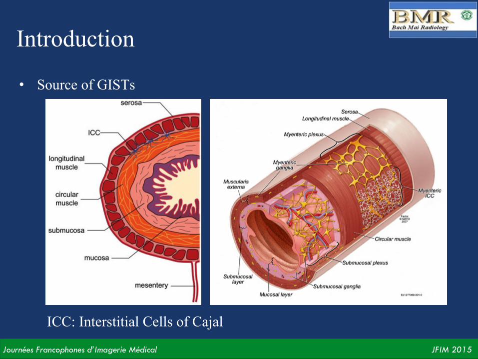
Introduction Source of GISTs: • Interstitial cell of Cajal (ICC) • Pacemaker of smooth muscles
in GI tract Signals to trigger peristalsis: • Stomach: 3 times/min • Jejunum: 11-12 times/min • Ileum: 9-10 times/min • Colon: 3-4 times/min
Journées Francophones d’Imagerie Médical JFIM 2015
Introduction
ICC: Interstitial Cells of Cajal
• Source of GISTs

Journées Francophones d’Imagerie Médical JFIM 2015
Clinical features

Journées Francophones d’Imagerie Médical JFIM 2015
Position • Depend on the normal distribution of ICCs • From oesophagus to annus • Extragastrointestinal stroma tumors: mesentery, peritoneum,
retroperitoneum
Journées Francophones d’Imagerie Médical JFIM 2015
Symptom
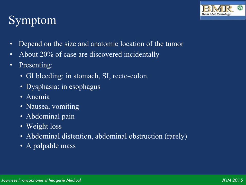
• Depend on the size and anatomic location of the tumor • About 20% of case are discovered incidentally • Presenting:
• GI bleeding: in stomach, SI, recto-colon. • Dysphasia: in esophagus • Anemia • Nausea, vomiting • Abdominal pain • Weight loss • Abdominal distention, abdominal obstruction (rarely) • A palpable mass

Journées Francophones d’Imagerie Médical JFIM 2015
Pathologic features

Journées Francophones d’Imagerie Médical JFIM 2015
Gross features
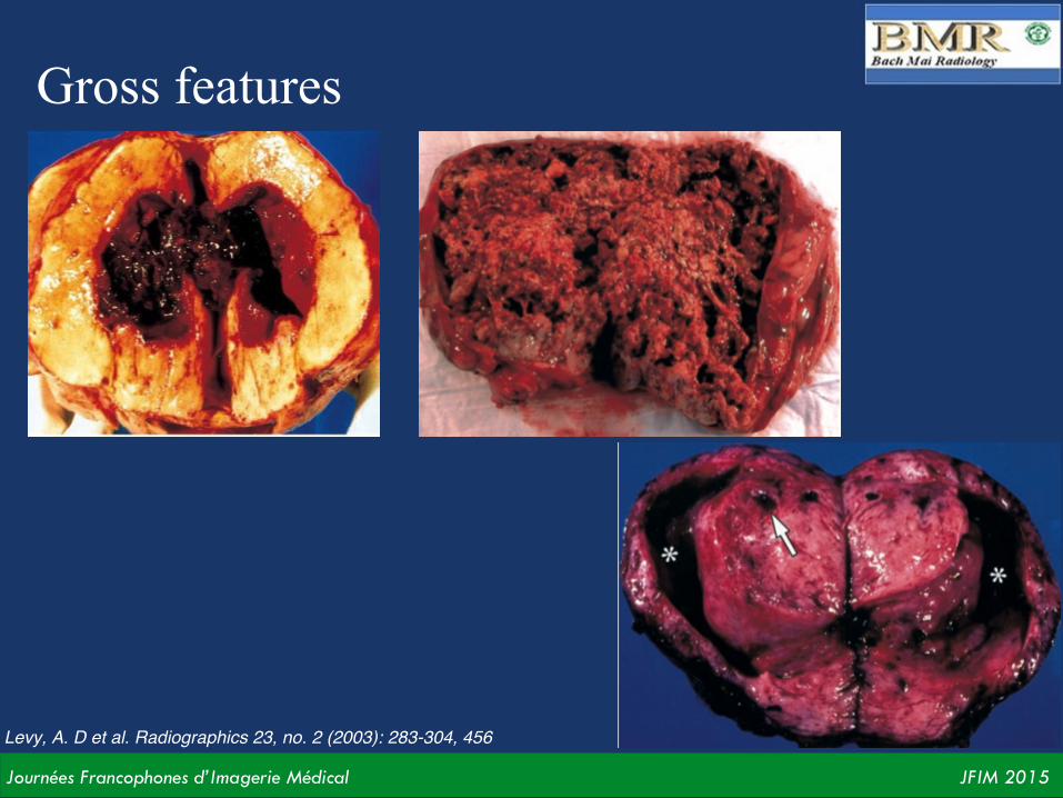
• Exophytic: Sub-serous, sub-mucous • Size: mm - 30 cm • Border: wel-defined, ill-define • Centre: degeneration, necrosis, haemorrhage, cavity…
Levy, A. D et al. Radiographics 23, no. 2 (2003): 283-304, 456
Journées Francophones d’Imagerie Médical JFIM 2015
Gross features
Levy, A. D et al. Radiographics 23, no. 2 (2003): 283-304, 456

Journées Francophones d’Imagerie Médical JFIM 2015
Microscopic features • Three major patterns of histologic features:
– Predominantly spindle cells: 70% – Predominantly epithelioid cells: 20% – Other (mixture, angiomatoid, myxoid): 10%
Hematoxylin - Eosin (x40) Levy, A. D et al. Radiographics 23, no. 2 (2003): 283-304, 456

Journées Francophones d’Imagerie Médical JFIM 2015
Microscopic features • Immunohistochemistry (IHC) • KIT protein (CD117): (+) in 91-95% of cases • PDGFRA - CD34 (70%), DOG1
Type tế bào hình trụ (spindle) Type tế bào dạng biểu mô (epithelioid)
Levy, A. D et al. Radiographics 23, no. 2 (2003): 283-304, 456

Journées Francophones d’Imagerie Médical JFIM 2015
Microscopic features

Journées Francophones d’Imagerie Médical JFIM 2015
Prognosis Mitotic rate (MR): rate of cell division
HE x 400
Levy, A. D et al. Radiographics 23, no. 2 (2003): 283-304, 456

Journées Francophones d’Imagerie Médical JFIM 2015
Prognosis
Levy, A. D et al. Radiographics 23, no. 2 (2003): 283-304, 456
Benign:MR = 0 Low-grade malignant :MR < 5 Malignant :MR > 5

Journées Francophones d’Imagerie Médical JFIM 2015
Prognosis
Factors: size and MR

Journées Francophones d’Imagerie Médical JFIM 2015
GISTs of stomach

Journées Francophones d’Imagerie Médical JFIM 2015
Features
• Stomach tumors: 2-3% • Posistion: 70% in the body,
fundus • Size: 4-25cm • Exophytic (sub-mucous, sub-
serous): 80% • Necrosis, haemorrhage, cavity.
Rarely calcification • Surface ulceration: 60% • Hyper-vascular: peripheral 90%. • Mitoses rate: hight

Journées Francophones d’Imagerie Médical JFIM 2015
Submucous GISTs
M, 67Y F, 67Y
AP view Oblique view
Levy, A. D et al. Radiographics 23, no. 2 (2003): 283-304, 456

Journées Francophones d’Imagerie Médical JFIM 2015
58YO female Chronic epigastric pain Sub-‐mucous mass

Journées Francophones d’Imagerie Médical JFIM 2015

Journées Francophones d’Imagerie Médical JFIM 2015
CLVT
56YO Female Sub-‐mucous mass

Journées Francophones d’Imagerie Médical JFIM 2015
54YO female Chronic epigastric pain Sub-‐serous GISTs

Journées Francophones d’Imagerie Médical JFIM 2015
69YO Female, epigastric pain
Schwanoma
Levy, A. D et al. Radiographics 23, no. 2 (2003): 283-304, 456
Difference diagnosis

Journées Francophones d’Imagerie Médical JFIM 2015
Schwanoma

Journées Francophones d’Imagerie Médical JFIM 2015
Difference diagnosis: AGC • Most common tumor in stomach, especially in the antrum • Large base • Rarely with exophytic (sub-serous, sub-mucous) • Regional adenopathies • Endoscopy with biopsy

Journées Francophones d’Imagerie Médical JFIM 2015
GISTs of small bowel

Journées Francophones d’Imagerie Médical JFIM 2015
Features
• Jejunum, Ilium > Duodenum • Size: 2-20cm • Exophytic: sub-mucous, sub-serous • Hypervascular with arterial feeders • GI obstruction, sub-obstruction • Cavity, necrosis, fistula • Rarely local invasion: malignant • Atypic GIST (infiltration): lymphoma,
adenocarcinoma…

Journées Francophones d’Imagerie Médical JFIM 2015
• Nam, 57 tuổi • Đau bụng âm ỉ, diễn biến 4 tháng

Journées Francophones d’Imagerie Médical JFIM 2015

Journées Francophones d’Imagerie Médical JFIM 2015
D T
GISTs: gastrointescnal stroma tumors (IHC -‐ CD117)

Journées Francophones d’Imagerie Médical JFIM 2015
43YO Female Epigastric distencon Duodenum mass

Journées Francophones d’Imagerie Médical JFIM 2015
52YO Male Epigastric distencon Duodenum mass

Journées Francophones d’Imagerie Médical JFIM 2015

Journées Francophones d’Imagerie Médical JFIM 2015

Journées Francophones d’Imagerie Médical JFIM 2015
Levy, A. D et al. Radiographics 23, no. 2 (2003): 283-304, 456
Ilium GIST: sub-serous feature with cavity and communication with lumen bowel

Journées Francophones d’Imagerie Médical JFIM 2015
Atypic GISTs: adenocarcinoma, lymphoma, metastasis…
Levy, A. D et al. Radiographics 23, no. 2 (2003): 283-304, 456
Difference diagnosis

Journées Francophones d’Imagerie Médical JFIM 2015
Colo-rectum GISTs
Journées Francophones d’Imagerie Médical JFIM 2015
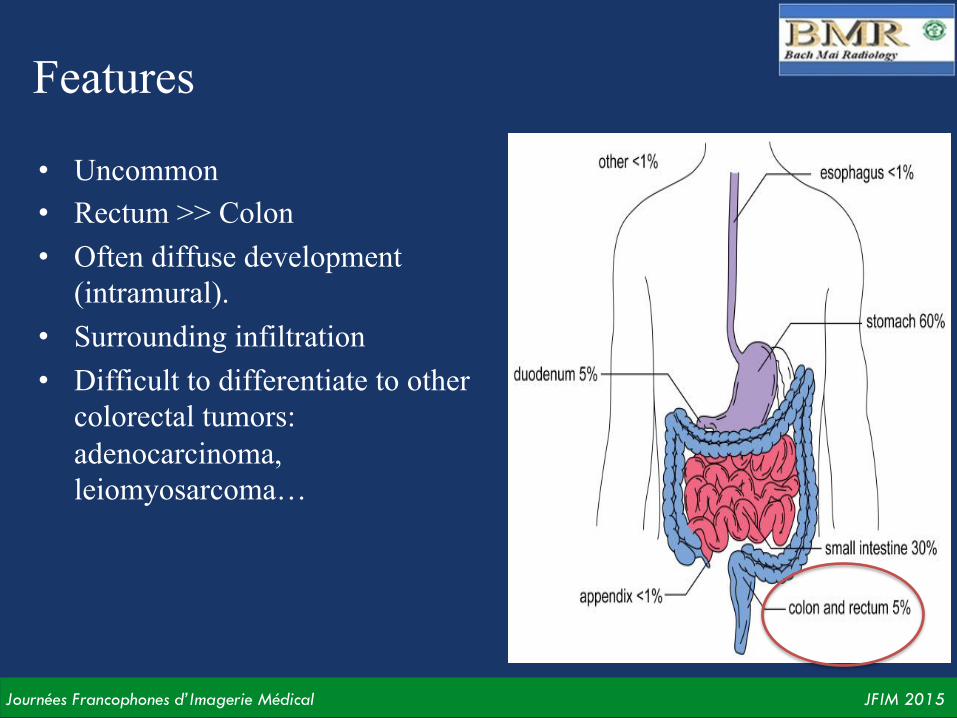
Features
• Uncommon • Rectum >> Colon • Often diffuse development
(intramural). • Surrounding infiltration • Difficult to differentiate to other
colorectal tumors: adenocarcinoma, leiomyosarcoma…

Journées Francophones d’Imagerie Médical JFIM 2015
47YO Female Sub-‐obstruccon Hepacc flexure colon mass
Levy, A. D et al. Radiographics 23, no. 2 (2003): 283-304, 456

Journées Francophones d’Imagerie Médical JFIM 2015
69YO Female Hypogastric pain, hematochezia GIST đoạn cuối trực tràng
nhiều ổ chảy máu
Levy, A. D et al. Radiographics 23, no. 2 (2003): 283-304, 456
Journées Francophones d’Imagerie Médical JFIM 2015
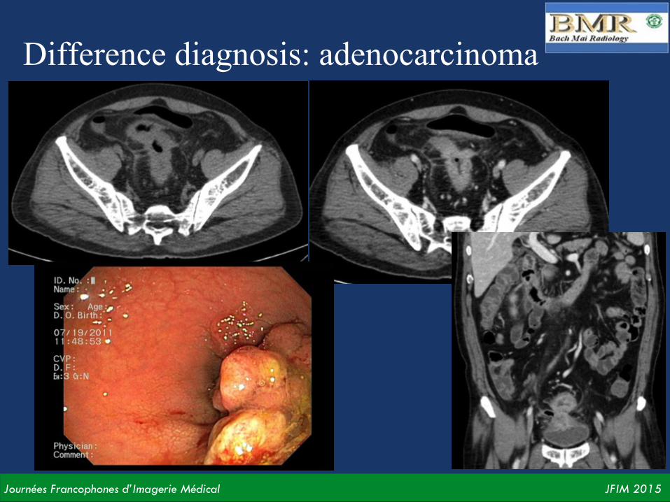
Difference diagnosis: adenocarcinoma

Journées Francophones d’Imagerie Médical JFIM 2015
Metastasis GISTs

Journées Francophones d’Imagerie Médical JFIM 2015
Features
• GISTs < 2cm are often benign. • Some GISTs are malignant, metastasis by many ways • Target organs
– Most often: live, peritoneum (95%). – Uncommon: soft-tissue, lung, pleural membrance – Rare: regional adenopathies, cerebral, bone…
• Characteristic: – Similar to original GISTs – Hyper-attenuation – Hypervascular – Heterogenous: necrosis, hemorrhage, cavity…

Journées Francophones d’Imagerie Médical JFIM 2015
GIST hỗng tràng di căn gan
Nam 55 tuổi Chướng bụng thượng vị

Journées Francophones d’Imagerie Médical JFIM 2015
Nam 68 tuổi, GIST dạ dày di căn gan, phúc mạc
Nam 67 tuổi, GIST dạ dày di căn gan
Hong, X. Radiographics 26, no. 2 (2006): 481-95.

Journées Francophones d’Imagerie Médical JFIM 2015
Nam 56 tuổi, GIST hỗng tràng di căn gan Nữ 64 tuổi, GIST hồi tràng di căn phần mềm, phúc mạc
Hong, X. Radiographics 26, no. 2 (2006): 481-95.

Journées Francophones d’Imagerie Médical JFIM 2015
Nữ, 71 tuổi, đau hạ vị, sờ thấy khối khu trú

Journées Francophones d’Imagerie Médical JFIM 2015
IHC: CD117 (+)
Multiple malignant GISTs
Journées Francophones d’Imagerie Médical JFIM 2015
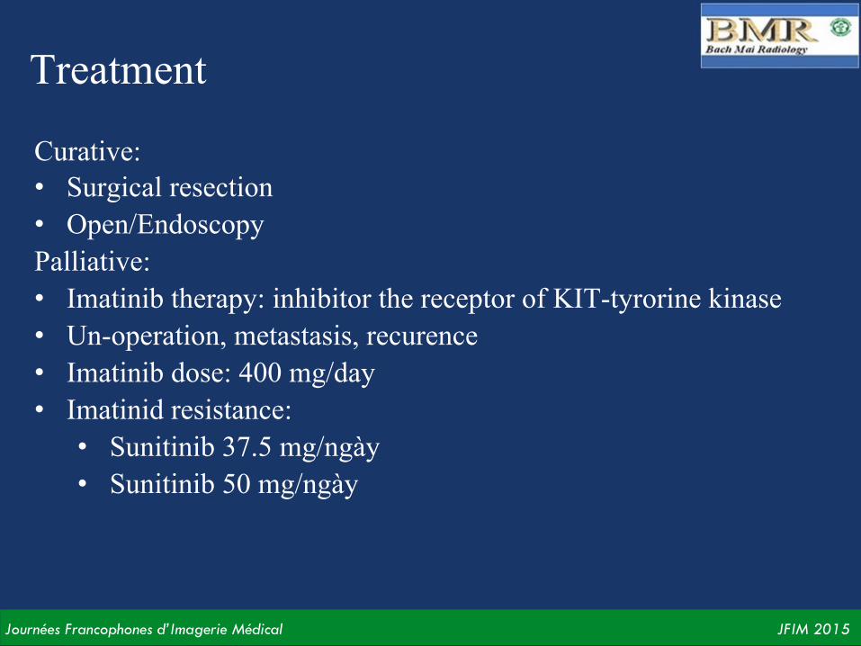
Treatment
Curative: • Surgical resection • Open/Endoscopy Palliative: • Imatinib therapy: inhibitor the receptor of KIT-tyrorine kinase • Un-operation, metastasis, recurence • Imatinib dose: 400 mg/day • Imatinid resistance: • Sunitinib 37.5 mg/ngày • Sunitinib 50 mg/ngày
Journées Francophones d’Imagerie Médical JFIM 2015
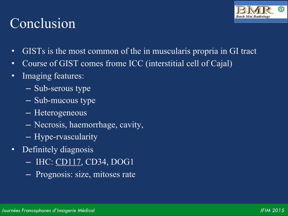
Conclusion
• GISTs is the most common of the in muscularis propria in GI tract • Course of GIST comes frome ICC (interstitial cell of Cajal) • Imaging features:
– Sub-serous type – Sub-mucous type – Heterogeneous – Necrosis, haemorrhage, cavity, – Hype-rvascularity
• Definitely diagnosis – IHC: CD117, CD34, DOG1 – Prognosis: size, mitoses rate

Journées Francophones d’Imagerie Médical JFIM 2015
Thanks so much for your a/en1on!