Intraabdominal Migrating of Intrauterine Contraceptive Devices (IUD)

Linköping University Medical Dissertation No.1085
Experimental studies on
Damage Control Surgery and Intraabdominal Hypertension
Pia Olofsson
Centre for Teaching and Research in Disaster Medicine and Traumatology Division of Surgery
Department of Clinical and Experimental Medicine Faculty of Health Sciences,
SE-581 85, Linköping, Sweden
Linköping 2008
1

SUPERVISORs
Professor Tore Vikström, MD, PhD and Associate Professor Kenth Johansson, MD, PhD
Papers I, II and III are reprinted with permission from the respective journal.
The studies in this thesis were supported by The National Board of Health and Welfare, Sweden and
the Medical Research Council of South-east Sweden.
Printed by LiU-Tryck, Linköping, Sweden 2008
ISBN 978-91-7393-775-7
ISSN 0345-0082
2

ABSTRACT Damage control surgery (DCS) offers an alternative to the traditional surgical management of complex or multiple injuries in critically injured patients. If a patient survives the initial phase of DCS, complications may occur, one of these being intraabdominal hypertension (IAH) and it´s potential development into the abdominal compartment syndrome. The indications for DCS have been widened and DCS principles can be applied in situations where time and resources are essential factors. The DCS principles of rapidly controlling intestinal spillage have not been evaluated. The aim of the studies in Papers I and II was to evaluate the principles of spillage control of intestinal contents according to the DCS concept and more specifically the effects of early rapid control of multiple bowel perforations on cardiovascular and pulmonary function compared with conventional small bowel resections in an animal model with abdominal trauma. In Paper I the animal model using anaesthetised pigs included a gunshot wound to the abdomen which caused multiple small bowel injuries. Haemorrhagic shock was combined with the gunshot wound in Paper II. The results presented in Paper I showed a significant reduction in rise in systemic vascular resistance and pulmonary vascular resistance, and a trend towards higher cardiac output and lower oxygen consumption in the bowel ligation group. In Paper II the results show a longer persistence of lactic acidaemia in the bowel ligation group. The aim of the study in Paper III was to assess visceral (intestinal, gastric and renal) microcirculation parallel with central haemodynamics and respiratory function during stepwise increases in intraabdominal pressure. In Paper IV we studied mucosal barrier function and morphology in the small bowel and colon of the pigs which were subjected to IAH. The IAP in anaesthetised pigs was increased stepwise using CO2 inflation, by 10 mm Hg at 10-minute intervals up to 50 mm Hg, and followed by exsufflation (Paper III). The micro-circulation was selectively studied using a 4-channel laser Doppler flowmeter (Periflex 5000, Perimed, Sweden). The mucosal tissues were mounted in modified Ussing chambers for assessment of barrier function (E.coli K12 uptake and 51Cr-EDTA permeability) (Paper IV). The results showed that the microcirculation of the small bowel mucosa and colon mucosa was significantly less affected compared to the seromuscular layers. The microcirculation of gastric mucosa, renal cortex and the seromuscular layer of small bowel and colon were significantly reduced with each increase. Cardiac output (CO) decreased significantly at IAP levels above 10 mm Hg and the respiratory function data showed an increasing airway pres-sure and a concomitant reduction in thoracic compliance. Transmucosal passage of E. coli
was increased three-fold in the small bowel after ACS with a significant correlation to the degree of mucosal microcirculatory reperfusion after exsufflation. 51Cr-EDTA permeability was unaffected. Bacterial passage in the colon was unchanged, whereas 51Cr-EDTA
3

permeability after ACS increased by up to 181% of baseline and was correlated to significant histopathological changes in the mucosa. In Paper I we have demonstrated that early rapid control of multiple bowel perforations in a model with moderate shock resulted in less impairment of SVR and PVR than conventional resection and anastomosis. The use of DCS principles, however, had no beneficial effect on cardiovascular function when haemorrhagic shock was combined with abdominal missile trauma (Paper II), on the contrary bowel ligation was followed by more prolonged lactic acidosis than conventional repair. The studies in Papers III and IV indicate that the microcirculation of intestinal mucosa and especially small bowel mucosa seem better preserved in response to intraabdominal hypertension caused by CO2 insufflation than other intraabdominal microvascular beds. The short term ACS in this model caused morphological changes in the intestinal mucosa, and mucosal barrier dysfunction. The response pattern concerning barrier function changes after CO2 insufflation differs between small bowel and colonic mucosa. The small bowel mucosa showed increased bacterial passage, and the colonic mucosa an increase in paracellular permeability and secretory response.
4

ABBREVIATIONSANOVA analysis of variance ACS abdominal compartment syndrome APP abdominal perfusion pressure CI cardiac index CO cardiac output CMBC concentration of moving blood cells CVP central venous pressure 51Cr-EDTA 51Chromium ethylene diamine tetra-acetic acid DCS damage control surgery ER emergency room FP filtration gradient GFP glomerular filtration gradient IAH intraabdominal hypertension IAP intraabdominal pressure ICU intensive care unit Isc short circuit current IVP intravesical pressure LDF laser Doppler flowmetry MAP mean arterial pressure MPAP mean pulmonary arterial pressure OR operating room PAOP pulmonary artery occlusion pressure PaO2 partial pressure of oxygen in arterial blood PaCO2 partial pressure of carbon dioxide in arterial blood PCWP pulmonary capillary wedge pressure PD transepithelial potential difference PTP proximal tubular pressure PU perfusion units PVR pulmonary vascular resistance SaO2 arterial oxygen saturation SD standard deviation SEM standard error of the mean SV stroke volume SvO2 mixed venous oxygen saturation SVR systemic vascular resistance TB total backscatter TER transepithelial electrical resistance
5

CONTENTS
LIST OF PAPERS 7
INTRODUCTION 9
BACKGROUND 11
AIMS OF THE THESIS 23
MATERIAL AND METHODS 25
RESULTS 39
DISCUSSION 49
CONCLUSIONS 55
SAMMANFATTNING PÅ SVENSKA 57
ACKNOWLEDGEMENTS 59
REFERENCES 61
6
LIST OF PAPERS
I. The Effects of early rapid control of multiple bowel perforations after high-
energy trauma to the abdomen: implications for damage control surgery.
P Olofsson, F Abu-Zidan, J Wang, N Nagelkerke, S Lennquist, T Vikström
J of Trauma 2006; 61:185-91
II. Muliple small bowel ligation compared to conventional primary repair
after abdominal gunshot wound with hemorrhagic shock.
P Olofsson, T Vikström, N Nagelkerke, J Wang, F Abu-Zidan
Accepted for publication, Scandinavian Journal of Surgery, September 2008
III. Gastrointestinal microcirculation and cardiopulmonary function during
experimentally increased intraabdominal pressure,
P Olofsson, S Berg, H C Ahn, L Brudin, T Vikström, K Johansson
Accepted for publication, Critical Care Medicine, June 2008
IV. Increased transmucosal uptake of E.coli K12 in porcine small bowel
following experimental short-term abdominal compartment syndrome.
P Olofsson, L Mellblom, S Berg, H C Ahn, T Vikström, L Brudin, K Johansson,
J Söderholm
Submitted
7

8

INTRODUCTION Trauma is one of the leading causes of death amongst young people (1). Trauma care has changed and more sophisticated methods have made it possible to survive multiple trauma that previously was not consistent with continuoused life. Surrounding circumstances and complications are better understood and possible to treat (2, 3). Some methods we use are not possible to evaluate by of randomised prospective human studies because of ethical aspects. Furthermore there are no alternatives to best care when a severely injured patient arrives at the trauma bay. Animal studies are therefore required in order to study the principles of trauma care. One concept that has gained interest over recent decades is damage control surgery, DCS or damage control laparotomy as it initially was called. Briefly DCS includes Phase I, rapid control of haemorrhage, control of spilling of intestinal contents or urine, packing and closure of the abdomen; Phase II, is resuscitation in the ICU; and Phase III, definitive repair. The use and development of DCS has successfully changed the outcome of otherwise moribund trauma patients. The method was initially applied to a small number of exsanguinating trauma patients with multiple injuries confined to the abdomen, but it´s use has expanded to include the chest, retroperitoneal space, pelvis and limbs. DCS has also evolved to become a well- known method for general surgical patients whose physiological derangements do not allow completion of an intended operation. DCS can also be considered in situations where requirements exceed the resources available, either in quantity or quality (3-13). DCS principles may be applied in both civilian disaster situations and war surgery however the potential physiological consequences and other side-effects of DCS have not as yet been well- clarified (5, 14). In the beginning of the DCS era mortality was rather high (50-70%) probably due to the “novelty” of the concept and the selection of patients, where only the “worst “trauma cases were chosen. In recent years however there has been a general development of the concept. Patients are now chosen with more experience and methods are more sophisticated, especially with regard to interventional radiography, treatment in the intensive care unit, and management of the open abdomen. The survival rate has increased considerably, but DCS still has a high incidence of morbidity (9). One complication that is recognised after DCS is intraabdominal hypertension (IAH) and its eventual development into the abdominal compartment syndrome (ACS) (3, 9). The Abdominal compartment syndrome is defined as intraabdominal hypertension with associated renal, pulmonary or haemodynamic compromise (15). The widespread use of the DCS concept in abdominal trauma has involuntarily increased the awareness of IAH/ACS and of the “open abdomen”. That there is an increase in ACS frequency is suggested by the rapid increase in literature on the subject, involving patients of all ages, neonates to the elderly, and
9

diverse clinical situations, including non-surgical. Recent interest has helped map the local and systemic effects of increased intraabdominal pressure and the importance of early recognition and treatment. Trauma patients represent a large group who are at risk for the development of both primary and secondary abdominal compartment syndrome, the latter occurring when there is no primary intrabdominal injury. Intraabdominal hypertension has a profound effect on splanchnic organs causing diminished perfusion and mucosal acidosis which may contribute to the development of impaired intestinal barrier function which in its turn could lead to septic complications and organ failure (15). The aim of this thesis was to analyse various aspects of damage control surgery principles as well as the complications of intraabdominal hypertension and short term abdominal compartment syndrome (ACS). In Papers I and II our aim was to develop an experimental animal model to study one of theprinciples of damage control surgery, intestinal spillage control, after penetrating trauma (gunshot wound) with (Paper II) and without (Paper I) controlled haemorrhagic shock. These two studies evaluate the effects of early rapid control of multiple bowel perforations on cardiovascular and pulmonary function in traumatic shock compared to conventional small bowel resection anastomosis. In Paper III our main interest was to further develop the animal model for studies on intraabdominal hypertension and the abdominal compartment syndrome, focusing on the pathophysiology of ACS and especially the microcirculation of the intestinal layers. In Paper IV, specimens from twelve of the initially twenty-one animals in Paper III, were collected before and after the “short term ACS” in order to study the permeability and morphology of the bowel wall.
10

BACKGROUND
Damage Control Surgery, DCS
The damage control surgery (DCS) approach is described by Hirshberg and Walden (16) as an operative sequence in primary trauma surgery where, life- and time-saving techniques are used to arrest haemorrhage and control spillage by deliberately avoiding resection and reconstruction. This is then followed by a period of stabilisation in the intensive care unit and subsequent planned reoperation including reconstruction. The concept is also applied in vascular and orthopaedic surgery, non-traumatic vascular injuries, non-traumatic abdominal, urologic and thoracic surgery. It is discussed in the literature on disaster medicine concerning civilian disaster situations with limited resources, where the saving of both time spent in the operating room (OR) and resources is of great value (5, 8, 11, 13, 17-21). The term Damage Control Surgery (DCS) originates from the Unites States Navy where “Damage control” was used when a ship was damaged and means keeping the ship afloat in order to get to a harbour and once there carry out permanent repairs (22). Pringle (23) was the first to describe the concept of hepatic packing in 1908. Pringle used the technique to control life-threatening liver haemorrhage (sutures over gauze packing to control portal venous haemorrhage caused by trauma). Halsted modified the technique by using rubber sheets between the liver and the packing material (24). During World War II and the Vietnam War the concept was not advocated but in the 1980´s there came reports (25, 26) regarding packing for liver injuries. Stone (27) described the concept of abbreviated laparotomy and intra-abdominal packing in the exsanguinating, hypothermic and coagulopathic trauma patient in 1983. After initial stabilisation the patient subsequently received definitive surgical repair. Rotondo and Schwab (12) named the concept “damage control laparotomy” in 1993 and described the logistics. Phase one (DC I) consists of life-saving surgery i.e. control of haemorrhage and contamina-tion, abdominal packing and abbreviated wound closure. Phase II (DC II) consists of continued resuscitation and rewarming in the ICU, and Phase III (DC III) consists of definitive repair of all injuries. Recently Johnson and Schwab (9) have also described a Phase 0,”Damage Control Zero” (DC 0), which represents the early pre-hospital and the emergency department phase. Damage Control Zero includes injury pattern recognition and the early decision to apply damage control. Strategies undertaken in the early phase include the shortening of pre-hospital time and emergency department resuscitation by the avoidance of time-consuming investigations, and rapid access to the OR. The incidence of ACS is difficult to determine since different methods of measurement and types of patient have been studied, but ranges between 0.1 to more than 30 % of postoperative and trauma patients.
11

Indications. Much has been written about when to apply the strategies of damage control abdominal surgery. (9, 10, 28-32). The main indication is to save life and to improve outcome in selected exsanguinating trauma patients with intraabdominal injuries (25). A patient who is cold (core temperature below 35°C), coagulopathic and acidotic on arrival, whether it be from penetrating or blunt trauma, is a candidate for damage control surgery. However the use of DCS has expanded to include trauma to the chest, retroperitoneal space, pelvis and limbs. DCS is also a well-known strategy in general surgical patients whose physiological derangements do not allow the completion of intended surgery (13), and DCS can be considered in situations where needs exceed the quantity or quality of resources available. DCS principles may be applied in civilian disaster situations as well as in war (5, 14). Hypothermia. The definition of hypothermia is a core temperature of less than 35°C (10). Heat loss begins at the time of injury and continues to increase during the time spent in prehospital care, ER, and OR. The clinical effects of hypothermia include arrhythmias, reduction in cardiac output, increased systemic vascular resistance, and a left shift of the oxygen-haemoglobin saturation curve, indicating a reduction in oxygen availability to the cells. One of the most important physiologic effects of hypothermia is platelet dysfunction and dysfunction of the intrinsic and extrinsic clotting cascades (33, 34). Acidosis. Hypoperfusion of the tissues causes a shift from aerobic to anaerobic metabolism at the cellular level and the production of lactic acid. Metabolic acidosis is a negative prognostic indicator in the traumatised patient (35-37). A lack of clearance of lactate within 48 hours is strongly predictive of mortality (38). A metabolic acidosis also exacerbates the coagulopathic situation. Coagulopathy. Coagulopathy may have its origin in dilution and consumption of coagulation factors and/or hypothermia (see above). Patt et al described (39) three mechanisms by which hypothermia interfered with haemostasis: inhibition of temperature-dependant enzyme-activated coagulation cascades; increased fibrinolytic activity; and reversible platelet dysfunction. Decreased total and ionised calcium concentration, the severity of injury and metabolic acidosis may also contribute to the dysfunction of normal haemostatic mechanisms. The clinical observation of coagulopathy is not always confirmed by laboratory investigation. Coagulation testing is normally performed at 37°C rather than at the patients core temperature and thus underestimates the degree of coagulopathy (3). Platelet dysfunction in hypothermia leads to prolonged bleeding time. Changes in enzyme kinetics may delay the initiation and propagation of platelet aggregation in spite of adequate replacement (3).
12

Damage Control Phase Zero. This phase includes the time spent in the prehospital setting and the trauma admission area. The early decision to implement DCS is believed to be of great importance for the outcome of the patient (9). Damage control zero includes rapid transport and resuscitation, and early decision making. Damage Control Phase I - the initial laparotomy. Initial assessment includes the site and the severity of the injury/bleeding and the organs injured. In general the main principles of damage control surgery that are included in the first phase are:
1. initial four quadrant packing and exploration of the abdomen and control of haemorrhage which may include directed packing, balloon tamponade, stenting, shunting and/or interventional radiographic procedures
2. control of spillage from intestine, biliary tree, and urinary tract 3. definitive packing and rapid temporary closure of the abdomen (rapid skin closure
only, leaving the fascia open) or leaving the abdomen open with a dressing of choice. Packing of the abdomen could be either resuscitative (minutes) or therapeutic. Therapeutic packing is used to tamponade bleeding when it is surgically unmanageable or when the patient has developed coagulopathy. Prompt angiography to assess intraparenchymal or intramuscular bleeding in difficult locations is sometimes required, but is demanding on logistics and the skills of the radiologist (3, 40). Damage Control Phase II. The aim of this phase is simply to restore physiological parameters. This is started at the scene of accident and continues during the abbreviated laparotomy and on the ICU. Rewarming and correction of hypoperfusion, metabolic acidosis and coagulopathy is normally performed within the first 24-48 hours (3, 9). Damage Control Phase III. A patient who is well perfused, normothermic, without coagulopathy and has a normal arterial pH is the ideal patient to transfer to the operating theatre for the last phase of the damage control surgery concept. A systematic and thorough exploration with definitive repair is made and fascial closure when tolerated (3, 9). Complications. Although survival of the severely injured trauma patient has increased with the use of damage control surgery there is still a high risk for morbidity and mortality (28, 32, 41-44). Mortality rates range between 10 and 69 % (3, 4, 6, 12, 45, 46). The complications that are most frequent after damage control surgery are wound infection, intra-abdominal abscess, dehiscence, bile leak, enterocutaneous fistula, sepsis, abdominal compartment syndrome (2-25 %) and multiple organ failure (6, 7, 12, 26, 29, 32, 42-44, 47-55).
13

The abdominal compartment syndrome (ACS) is a well-known and, in some studies, frequent complication after damage control surgery, with an incidence that varies between studies (0-5, 5-14, 5- 33 %) (42, 54, 56-63). Risk factors for the development of ACS include severe abdominal and/or pelvic trauma, high-volume fluid resuscitation (controversial according to which fluids and amounts), preexisting co-morbidities, packing, massive bowel distension, ischaemic reperfusion injury of the gut, hypothermia and primary fascial closure (56, 61). Some trauma centres have abandoned the closure of skin or fascia after the initial surgery (DCS) and use the open abdomen principle for temporary abdominal closure. It is important however to remember the possibility of recurrent ACS despite temporary abdominal closure (9).
14

Intraabdominal Hypertension and Abdominal Compartment
Syndrome
Compartment syndrome is a condition in which increased pressure in a confined anatomical space adversely affects circulation and threatens the perfusion of tissues therein (56). The consequences of elevated intra-abdominal pressure include cardiovascular, respiratory and renal dysfunction, but Intraabdominal hypertension (IAH) will affect all intraabdominal organs as well as the CNS (64). IAH and the abdominal compartment syndrome (ACS) not only cause significant morbidity and mortality in severely injured trauma patients, but also patients with non-traumatic conditions (15). Our understanding of the pathophysiology of IAH and the ACS, and also the use of intraabdominal pressure measurement have increased during recent years with improvement in patient morbidity and mortality as a result (15). The definitions that follow are from the “World Society of the ACS” (WSACS) consensus document (65). Intra- abdominal pressure (IAP). The IAP measured at any point in the abdomen may be assumed to represent the pressure throughout the abdomen. IAP increases with inspiration and decreases with expiration and is also affected by the volume of the solid organs or hollow viscera, and the presence of ascites, blood, or other space-occupying items. The intra-abdominal pressure (IAP) is the steady-state pressure concealed within the abdominal cavity. The abdominal perfusion pressure (APP) is the mean arterial pressure minus intraabdominal pressure, (APP = MAP – IAP) and has been proposed to be a more accurate predictor of visceral perfusion. In a study by Cheatham, an APP > 60 mm Hg represented a potential endpoint for resuscitation (66). IAP should be expressed in mm Hg and measured at end-exspiration in the complete supine position after ensuring that muscle contractions are absent, and with the transducer zeroed at the level of the midaxillary line (65). IAP mesurement may be performed in various ways but is usually done indirectly via the bladder. The recommended volume of fluid in the bladder during measurement is 25 ml in humans (65) and for young pigs 10-15 ml only (67). Measurement via the urinary bladder is considered to be the golden standard but has some disadvantages, such as interference from the urinary system and the fact that it is an indirect measurement. It is believed that the bladder acts as a passive diaphragm when filled with saline (68). Normal IAP is approximately 5-7 mm Hg in critically ill adults. Certain physiological conditions such as morbid obesity or pregnancy may be associated with chronic IAP elevations of 10-15 mm Hg to which the patient has adapted without significant pathophysiology.
15
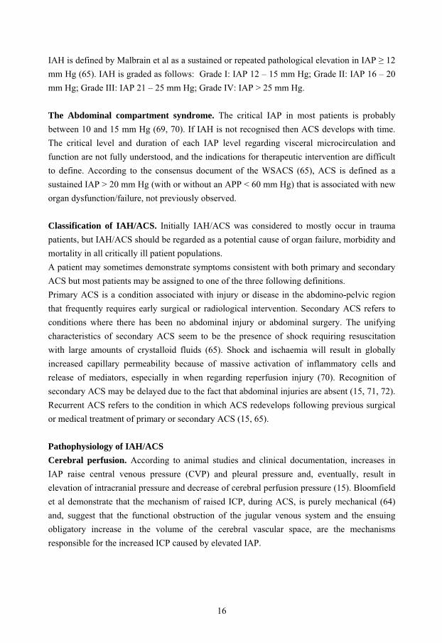
IAH is defined by Malbrain et al as a sustained or repeated pathological elevation in IAP � 12 mm Hg (65). IAH is graded as follows: Grade I: IAP 12 – 15 mm Hg; Grade II: IAP 16 – 20 mm Hg; Grade III: IAP 21 – 25 mm Hg; Grade IV: IAP > 25 mm Hg. The Abdominal compartment syndrome. The critical IAP in most patients is probably between 10 and 15 mm Hg (69, 70). If IAH is not recognised then ACS develops with time. The critical level and duration of each IAP level regarding visceral microcirculation and function are not fully understood, and the indications for therapeutic intervention are difficult to define. According to the consensus document of the WSACS (65), ACS is defined as a sustained IAP > 20 mm Hg (with or without an APP < 60 mm Hg) that is associated with new organ dysfunction/failure, not previously observed. Classification of IAH/ACS. Initially IAH/ACS was considered to mostly occur in trauma patients, but IAH/ACS should be regarded as a potential cause of organ failure, morbidity and mortality in all critically ill patient populations. A patient may sometimes demonstrate symptoms consistent with both primary and secondary ACS but most patients may be assigned to one of the three following definitions. Primary ACS is a condition associated with injury or disease in the abdomino-pelvic region that frequently requires early surgical or radiological intervention. Secondary ACS refers to conditions where there has been no abdominal injury or abdominal surgery. The unifying characteristics of secondary ACS seem to be the presence of shock requiring resuscitation with large amounts of crystalloid fluids (65). Shock and ischaemia will result in globally increased capillary permeability because of massive activation of inflammatory cells and release of mediators, especially in when regarding reperfusion injury (70). Recognition of secondary ACS may be delayed due to the fact that abdominal injuries are absent (15, 71, 72). Recurrent ACS refers to the condition in which ACS redevelops following previous surgical or medical treatment of primary or secondary ACS (15, 65).
Pathophysiology of IAH/ACS
Cerebral perfusion. According to animal studies and clinical documentation, increases in IAP raise central venous pressure (CVP) and pleural pressure and, eventually, result in elevation of intracranial pressure and decrease of cerebral perfusion pressure (15). Bloomfield et al demonstrate that the mechanism of raised ICP, during ACS, is purely mechanical (64) and, suggest that the functional obstruction of the jugular venous system and the ensuing obligatory increase in the volume of the cerebral vascular space, are the mechanisms responsible for the increased ICP caused by elevated IAP.
16

Cardiovascular function. Elevation in IAP impairs venous return, causes pooling of fluid in the lower extremities, and leads to reduction in cardiac output (CO). Reduction in CO results from decreased inferior vena caval flow secondary to direct compression of the inferior vena cava and portal vein as well as from an increased thoracic pressure, which decreases both inferior and superior vena caval flow. Another mechanism to reduce CO involves the direct increase in systemic vascular resistance leading to an increase in afterload and thus a subsequent fall in CO. Systemic vascular resistance may increase through direct compressive effects on aorta and systemic vasculature, but more commonly as compensation for the reduced venous return and falling stroke volume. As a result of this physiologic compen-sation, mean arterial pressure remains stable in the early stages of IAH/ACS despite reductions in venous return and cardiac output. Central venous pressure, pulmonary wedge pressure and left ventricular afterload increase because of increased systemic vascular resistance. However volume status and anaesthesia technique are two important factors to take in consideration when studying CO data from different studies (15, 73-76). Respiratory function. Severely elevated IAP, as in ACS, can cause severe alterations in respiratory system mechanics. Elevated IAP forces the diaphragm upwards into the thoracic cavity, compressing basilar lung segments. This is physiologically manifested as a decrease in functional residual capacity, an increase in alveolar dead space and ventilation perfusion mismatch. The increase in IAP forces the diaphragm upwards into the thoracic cavity and causes a rise in intra-thoracic pressure. As a consequence, respiratory system, chest wall compliance, total lung capacity, and residual volume are reduced. This leads to hypoxia, hypercapnia and the need for mechanical ventilation with increased pressure (15, 75, 77). Renal function. Oliguria/anuria in spite of aggressive fluid resuscitation is the classical sign of ACS. The mechanisms responsible for the impairment of renal function are direct compression of the renal parenchyma, decreased perfusion of the kidneys (since cardiac output is decreased), and increased water and sodium retention due to activation of the renin-angiotensin system (78-81). The filtration gradient (FG) is the mechanical force across the glomerulus and equals the difference beween the glomerular filtration pressure (GFP) and the proximal tubular pressure (PTP). In the presence of IAH, PTP may be replaced by IAP and GFP estimated as MAP minus IAP. FG = GFP – PTP = MAP – 2 x IAP.
Visceral perfusion. Intraabdominal pressures above 10 mm Hg will show adverse effects at a cellular, organ and compartment level. Even at these modest levels of IAP elevation reductions in hepatic and splancnic perfusion are seen (82-85) and this effect appears to be exacerbated by haemorrhage despite fluid resuscitation (86). The exact mechanism of reduced splanchnic perfusion associated with increased IAP is not known (15), but involves a direct
17

effect on mesenteric arterial resistance, myogenic reflexes within the splanchnic vasculature and humoral factors or a combination of the two (57, 59, 63, 68, 84, 85, 87-99). The increased IAP compresses organs within the abdomen. Splanchnic perfusion is reduced secondary to reduced cardiac output, reduced perfusion pressure and increasing splanchnic vascular res-istance. Ivatury et al have suggested that splanchnic hypoperfusion and gut mucosal aci-dosis begin at much lower pressures than other clinical markers of ACS and should therefore be treated early (90). This means that profound splanchnic ischaemia could be present without haemodynamic signs of IAH (15). An IAP greater than 20 mm Hg impairs intestinal perfusion at the mucosal and submucosal levels, leading to a reduction in tissue oxygen tension, anaerobic cell metabolism, acidosis, and free-radical generation (87, 97, 100). Oxygen free radical production and/or bacterial translocation as a consequence of increased IAP have been studied by several authors (83, 100-103). Some studies show a significant level of translocation to mesenterical lymphnodes, liver and spleen (83, 100, 104). It has been suggested that ischaemia at this level could be associated with an increased incidence of multiple organ failure, sepsis and increased mortality (63, 71, 83, 97, 100, 104-109). The ischaemic and reperfused intestine is considered to be a prime initiator of post injury multiple organ failure (110). The potential mechanisms include distant organ injury caused by primed neutrophil leukocytes, and translocation of bacterial debris and toxic products via the mesenteric lymph (111, 112). Lower Extremity perfusion. Increased IAP increases femoral venous pressure and peripheral vascular resistance and diminishes femoral artery blood flow (113).
Intestinal morphology and circulation
Morphology. The intestinal wall of the human gastrointestinal canal is composed of four layers. The mucosa and submucosa are responsible for absorption and excretion. The muscularis layers, with an inner circular and outer longitudinal muscle layer, casue propulsive and non-propulsive motion of the intestine. The mucosa is divided into lamina epithelialis, lamina propria and lamina muscolaris mucosae. In humans the jejunum, the proximal 2/5 of the small intestine, is thicker with more mucosal folds and richer vascularisation than the distal part, the ileum. The total area of the inner surface of the jejunum is increased by 5-8 mm folds, plica circulares, containing mucosa and submucosa, and by finger-like projections, villi. The villi are composed of an epithelial layer with an underlying lamina propria, arterioles and venous plexuses, and centrally placed lymphatic capillaries, lacteals. The intestinal villi and attending microvilli, in the jejunum, augment the mucosal absorption surface. The villi are 0.5-1.2 mm high and 0.1 mm wide and contain lamina epithelialis and lamina propria from the mucosa. Between the villi are the crypts of Lieberkuhn.
18

The small intestine wall consists of epithelial cells, which to 85% consist of enterocytes that is the absorptive epithelial cells. Goblet cells are less frequent (10% of the epithelial cells) and produce mucin. Paneth cells (3,5% of the epithelial cells) are located in the crypts and contain various secretory granules and their function is considered to be prevention of proliferation of bacterial growth. Enteroendocrine cells are located in the epithelium and release gastrointestinal hormones in response to changes in the external environment. The M cells (microfold or membranous cells, found in the follicle-associated epithelium) are considered to have the primary physiological role of rapid uptake and presentation of particular antigens and microorganisms to the immune cells of the lymphoid follicle to induce an effective immune response. The mucosa of the large intestine lacks the villi but has deep crypts. The colonic epithelium is composed of a mixture of columnar absorptive cells (colonocytes), goblet cells and to a minor extent enterochromaffin cells covering the surface and extending into crypts of Lieberkuhn.
Figure 1. Schematic illustration of the small intestinal wall covered by villus epithelium, with kind permission of Åsa Keita.
Near the bottom of the crypt, stem cells give rise to transit cells that differentiate during transfer upwards towards the lumen (114). Porcine intestines are considered to be similar to human but the the jejunum and ileum contain numerous pathches of lymph nodes and the crypts are deeper, contain goblet cells all the way down to the bottom of the crypts and lack paneth cells. The large intestines in pigs are coiled and voluminous and contain a larger amount of lymphocytes at the surface of the epithelium (115).
19

Circulation in general. The vascular supply of the gastrointestinal tract is built up of a system of vascular arcades including mesenteric vessels and intramural vessels (116). The arterial system of the intestine and of the stomach is built like a close-meshed network of small interconnected arteries in the submucosa from which arterioles pass into the various morphological and functional units of the organ wall. From anatomical studies it seems that the intramural vessels consist of parallel-coupled vascular circuits, supplying different wall layers. The degree of vascularisation differs depending on physiological demand. Each parallel-coupled vascular bed consists of several series-coupled vascular sections, precapillary resistance vessels, precapillary sphincters, exchange vessels, postcapillary resistance vessels and capacitance vessels. Colonic blood flow at rest is somewhat lower than that of the small intestine. This has been shown in dog (117), cat (118) and man (119). The flow distribution, however is almost the same as in the stomach and small intestine, with the major portion of blood flow directed to the mucosa. Intestinal mucosal perfusion of the small intestine. Precapillary mucosal arterioles act as resistance vessels and are the site of local metabolic and neurogenic control of mucosal blood flow. Precapillary sphincters control both number of perfused capillaries and distribution of perfusion within the intestinal wall. The tone of the postcapillary resistance vessels, venules, and veins determine the capillary hydrostatic pressure, pooled blood volume and rate of fluid exchange across the capillary wall. The blood flow in the mucosa/submucosa constitutes of approximately 65-90% of total intestinal blood flow during resting conditions (120). The villi and crypts account for 25-35% and 20-30% respectively of the total intestinal blood flow. Each villus is perfused through a central arteriole with surrounding venous plexus. A critical feature of the vasculature of the villus is the close approximation of the main arteriole in the core to the peripherially located capillaries and venules (121,122), which leads to a progressive lowering of PO2 and increasing osmolarity towards the tip of the villus. The most important result of the increased effectiveness of this countercurrent system during arterial hypotension is that more oxygen is shunted. This also means that when more villi are perfused at low arterial pressure the exchange area is increased. The driving force of this extravascular shunting of oxygen is the PO2 difference between the central vessel and the capillary network. This PO2 difference is created by arterial inflow and also by the epitheial consumption of oxygen which takes place along the full length of the villus. The low oxygen tension at the tip of the villi is present at normal resting perfusion pressure and is even lower during hypotension. The extravascular short-circuiting of oxygen in the villous countercurrent exchanger causes tissue hypoxia at the tip of the villus and is considered to lead to mucosal damage (123).
20

Autoregulation of blood flow, the tendency for blood flow to remain adequate despite changes in arterial perfusion pressure is much discussed and studied. The kidney was the first organ that was described to show autoregulation in 1931 and twenty years later autoregulation had been observed in various organs. In a review Johnson thoroughly discusses the various hypotheses for the mechanism of autoregulation (metabolic, myogenic, tuboglomerular feedback and tissue pressure) and also impairment of autoregulation (124). The intestinal vascular bed is also considered to autoregulate blood flow. Under normal conditions, a decrease in arterial pressure induces a redistribution of flow toward the mucosa due to pressure flow autoregulation (125). Autoregulation is defined as the ability to maintain adequate blood flow despite changes in perfusion pressure within a range of 50 and 150 mm Hg and is achieved by contraction and relaxation of the small precapillary resistance vessles, arterioles. The regulation of capillary flow due to autoregulation is considered functionally important since the fenestrated capillary bed of the mucosa would otherwise run a con-siderable risk for rapid oedema formation (120).
Barrier function/permeability.
The intestinal epithelium is an important barrier between the body and the external environment in animals and humans. The intestinal barrier in general performs uptake and absorption of nutrients, water and ions, as well as being a selective barrier against harmful dietary components, bacteria and other antigens. The small intestine is responsible for digestion and absorption of dietary nutrients. The main function of the large intestine is the absorption of water and ions and the storage and transport of faeces and gas. The protective function of the intestinal mucosa also includes intestinal propulsive motility, and the rapid repair and turnover process of epithelial cells (126). The so-called preepithelial defence or barrier consists of the mucous layer secreted by the epithelium (goblet cells), IgA-antibodies that bind to and aggregate bacteria to prevent them from adhering to the epithelial cells, and also localised cell- mediated immune mechanisms. Active chloride transport by epithelial cell promotes intraluminal fluid flux that washes away harmful agents (127). Anaerobic organisms normally cover the lower small intestine and colon walls hence limiting overgrowth and colonisation of other more deleterious bacteria. Intestinal motility is also considered a protective mechanism (128, 129). The intestinal barrier consists of neighboring cells adjoined in series of intercellular junctions commonly called the junctional complex that was first described by Farquar and Palade (130). The junctional complex consists of tight junctions, adherence junctions, desmosomes and gap junctions.
21

The tight junction complex consists of almost 40 distinct proteins or protein families. Tight junctions are located at the most apical part of the lateral cell membrane forming a network of linking strands between the adjacent epithelial cells constituting a paracellular barrier for passage of ions and molecules. Junction tightness is dependant on the number of strands and the number of anastomoses between the strands, as well as by the functional state of the junctions. There is both size-selectivity and a charge-selectively (positively charged ions and molecules pass more easily) within the tight junction permeability barrier. The tightness of the epithelium also depends on the number of cells per unit area and the shape of the cells (114, 131).Possible routes for permeation are: transcellular route (i.e. through the cell membrane); paracellular route (i.e. between the cells via the tight junctions); transcellular via pores; active carrier-mediated absorption (nutrients, e.g. glucose); pinocytosis and transcytosis; and phago-cytosis and/or macropinocytosis (131, 132).
22

AIMS of the thesis • To develop an animal model for studies on the principles of spillage control on intestinal contents according to the DCS concept. • To compare rapid control of multiple bowel perforations, according to DCS, with conven-tional small bowel resections and anastomosis, regarding the effects on cardiovascular and pulmonary function, in an animal model with abdominal gunshot trauma. • To compare rapid control of multiple bowel perforations with conventional small bowel resections and anastomosis, regarding the effects on cardiovascular and pulmonary function, in an animal model with abdominal gunshot trauma and simulated haemorrhagic shock. • To develop an animal model for evaluation of increased intraabdominal pressure and its effect on mucosal blood flow. • To study, in this animal model, visceral microcirculation together with central heamody-namics and respiratory function during stepwise increase of intraabdominal pressure including short term ACS. • To investigate mucosal barrier function and morphology in the porcine small bowel and colon after short term ACS.
23

24

MATERIAL AND METHODS
Ethical issues.
All studies were approved of by the Animal Ethics Committee of the University of Linköping, Sweden.
Animal Model.
Female Swedish Landrace pigs (17.5-30.0 kg), 10-14 weeks old were used. The animals arrived at the laboratory one or two days prior to the experiments and were accommodated in an accredited facility. They had free access to tap water but were fasted 12 hours prior to experiments. After premedication with intramuscular azaperone 4 mg/kg, ketamine 10 mg/kg and atropine 0.02 mg/kg, anesthesia was induced by pentobarbital 12-15 mg/kg via an ear vein. Anaesthesia was maintained with continuous i.v. infusion of pentobarbital 6-10 mg/kg/h and pethidine 2-3 mg/kg/h, administered via an infusion pump. The animals were ventilated with room air, using a time-cycled ventilator (Servo ventilator 900B, Maquet, Stockholm, Sweden). The ventilator was set to a tidal volume of 10 ml/kg, a rate of 20 breaths/minute and an inspiratory:exspiratory ratio of 1:2 to establish normocapnoea or slight hypocapnoea (PaCO2 4.0-5.5 kPa). Thereafter no further changes in ventilator settings were made. Isotonic balanced crystalloid solution was given i.v. at a rate of 5 ml/ kg /h (Paper I) and 8 ml/kg/h (Papers II, III and IV) throughout the experiment. A cystostomy catheter was inserted via a small suprapubic midline incision and the diverted urine was collected via a Foley catheter. Hypothermia was avoided by the use of heating pads and insulation blankets. Preparation included tracheostomy, urinary bladder catheter and catheters for vascular access. A central venous catheter in the left external jugular vein was used for the continuous infusion of drugs, fluids, and for measuring central venous pressure (CVP). Arterial blood samples were drawn from a catheter in the right common carotid artery, which also monitored the mean arterial pressure. An extra catheter in the left common carotid artery was inserted (Paper II) in order to lower the blood volume and cause haemorrhagic shock. A 7.0-7.5 F balloon-tipped Swan-Ganz fiberoptic semicontinuous cardiac output pulmonary artery catheter (Edwards Lifesciences) was placed in the pulmonary artery, and cardiac output and mixed venous oxygen saturation (SvO2) was automatically calculated by a monitoring device (Vigilance Monitor, Edwards Lifesciences). Heart rate (HR), mean arterial blood pressure (MAP), central venous pressure (CVP), mean pulmonary arterial pressure (MPAP), pulmonary capillary wedge pressure (PCWP), (Papers I and II), cardiac output (CO), mixed venous oxygen saturation (Papers I-III), core temperature from the tip of the thermodilution catheter, thoracic compliance and end-inspiratory airway pressure (Papers III and IV) via the ventilator, and intravesical pressure (IVP) (Papers III and IV) were measured continuously. Arterial (Papers I-IV) and mixed venous blood (Papers I and II) gas samples
25

were analysed at 37° C and corrected for blood temperature measured by the pulmonary artery catheter thermistor. Samples of 10 ml venous blood were drawn for measuring lactate, platelets, activated partial prothrombin time, prothrombin time and fibrinogen degradation products (Papers I and II), which were analysed at the laboratory of the University Hospital, Linköping University. The samples were treated according to instructions from the laboratory (lactate tubes at room temperature, platelet tubes in the refrigerator, and the coagulation profile tubes were frozen after centrifugation). Before starting the experiments the animals were left to stabilize for thirty minutes. After the experiments were finished the animals were sacrificed using an overdose of pentobarbital (Papers I-IV).
Calculations
Cardiac output (CO); Heart rate x stroke volume Cardiac index; Cardiac output/ Body surface area Thoracic compliance; Tidal volume divided by plateau pressure minus end
expiratory pressure Stroke volume (SV); CO/HR Systemic vascular resistance (SVR); 80 x (MAP-CVP)/CO Pulmonary vascular resistance (PVR); 80 x (MPAP-PCWP)/CO Oxygen delivery; Oxygen content in arterial blood x CO. (Oxygen
content in blood; 1.34 x haemoglobin x oxygen saturation of blood)
Oxygen consumption; oxygen content in arterial blood - oxygen content in
mixed venous blood x CO Oxygen extraction ratio; oxygen consumption/ oxygen delivery
26

Experimental procedures
Paper I
Fifteen anaesthetised pigs were exposed to a standardised abdominal trauma. The anaesthetised animals were shot in the abdomen with a handgun (9 mm Luger) while lifted up with belts in a prone position so that the abdomen was dependant. The inlet of the bullet was 15 cm above the symphysis pubis (left side) and 10 cm from the midline. Twenty minutes later laparotomy was performed. The animals were randomised into two groups. In the first group, the resection anastomosis group (RA, n=8), small bowel injuries were treated with resection and anastomosis. The abdomen was tightly closed with a running suture (prolene). In the second group, the multiple bowel ligation group (BL, n=7), small-bowel injuries were treated by ligation. The abdomen was rapidly closed with towel clips. The duration of resection anastomosis surgery ranged between 1.5 to 2 hours and for multiple small bowel ligation between 15 and 20 minutes. Systemic haemodynamic parameters were measured, arterial and mixed venous blood gas samples were analysed, and haematological samples for lactate, platelet count and coagulation profile were drawn at baseline (30 minutes after preparation), immediately after the gunshot, immediately after the laparotomy (time 0), 0.5 hours after the laparotomy, and then hourly until 6 hours after laparotomy. Figure 2. Small bowel with multiple ligation (to the left) and small bowel anastomosis (to the right)
27

Paper II
Eighteen anaesthetised pigs were injured with the same standardised abdominal missile trauma as in Paper I. After the missile trauma the animals were gradually bled to a mean arterial pressure of 50 mm Hg for 15 minutes by controlled bleeding. The bleeding was performed directly after the gunshot wound as a second insult to make the model more similar to the clinical situation and also to make the shock state more severe. Total bleeding time was 30 minutes and resuscitation with Ringers lactate solution 50 ml/kg/h within 20-30 minutes was then commenced. After resuscitation the animals were randomised into two groups. The conventional surgery group (resection anastomosis (RA), n=9) had resection and anastomosis of all injured segments of small bowel. Two animals had colonic lesions, one had a colostomy and the other had a small perforation, which was sutured. Skin was closed using continuous suture. The early rapid multiple bowel ligation group (BL, n=9) had quick ligation of the small bowel ends. Three animals in this group had colonic lesions. One animal had a colostomy and the other two animals had minor lesions, which were sutured. The skin was rapidly closed using towel clips. The number of small bowel segments injured and intraabdominal free blood volume were measured. Samples of 10 ml venous blood were drawn after baseline, after gunshot, after bleeding, after resuscitation, and at 0, 3 and 6 hours after laparotomy. Lactate, platelets, activated partial prothrombin time and prothrombin time was analysed. Arterial and mixed venous blood gas samples were analysed. The animals were observed for 6 hours postoperatively as in Paper I. Paper III
Twenty-six (including six control animals) anaesthetised pigs were used. During laparotomy a 5 mm laparoscopic port was placed in the left side of the abdominal wall to inflate CO2 into the peritoneum, using standard laparoscopy equipment to increase the intraabdominal pressure (IAP). Above 30 mm Hg extra CO2 was inflated via a separate tube. The abdomen was closed in two layers to prevent leakage of CO2. After baseline measurements were taken the IAP was increased to 10 mm Hg and then stepwise increased to 20, 30, 40 and 50 mm Hg at 10- minute intervals. IAP was measured directly via the CO2 insufflator (trocar) and indirectly by using the intravesicular pressure (IVP). IVP was measured after installation of 50 ml of water at body temperature into the urinary bladder via the intravesicular catheter which was also connected to a pressure transducer. The microcirculation was measured using a four-channel laser Doppler flowmeter. The probes (fibre diameter 0.125 mm, fibre centre separation 0.250 mm, wavelength 780 nm ) were mounted in a probe holder sutured to the surface of the gastric mucosa (n=15), small bowel mucosa (n=21, (6 controls)), small bowel serosa (seromuscular layer)(n=19, (6 controls)), colon mucosa (n=15, (6 controls)), colon serosa (seromuscular layer)(n=16, (6 controls)) and renal cortex (n=10). In the control group the microcirculation of the small bowel (mucosa and serosa) and colon (mucosa and serosa) only
28

was measured. The enterotomies of the small bowel and colon were closed, the abdominal wall was closed in two layers and the animals were then left to stabilise for thirty minutes before making baseline measurements. The flow probes were connected to a four-channel laser Doppler flowmeter (LDF) (Periflex 5000, Perimed). The flowmeter was connected to a multichannel data acquisition system and analysed using the Perisoft (Perimed) software programme. When analysing the LDF data we compared the mean values of the last two minutes at every pressure level with the whole period at that pressure (ten minutes); there was no difference. After initial movement artifacts, the signal level was stable and we chose to measure the last two minutes of every ten-minute period. The values are expressed in relative units with the biological zero subtracted. At the end of the experiment the zero-line for the microcirculation was measured after which the animals were sacrificed using an intravenous overdose of pentobarbital.
Figure 3. One of the four probes, sutured to the small bowel mucosa and connected to the four channel flowmeter (LDF) (Periflex 5000, Perimed).
The following physiological parameters were registered: end-inspiratory airway pressure and thoracic compliance via the ventilator; heart rate; mean arterial pressure; central venous pressure; cardiac output; body temperature; and IVP. SvO2 was measured by the fibreoptic technique (Vigilance, Edwards Lifesciences). PaO2, PaCO2, pH, SaO2 were measured by arterial blood-gas samples analysed at 37° C. For separate studies (Paper IV), full bowel wall biopsies of 2-5 cm were taken from small bowel and colon. The biopsies were taken at least 10 cm from the circulation recording sites and the bowel closed with continuous suture. The control animals were treated in the same way except that they were not insufflated with CO2 and microcirculation measurements were concentrated to the intestine (small bowel and colon).
29

Laser Doppler flowmetry
The laser Doppler instrument uses near-infrared monochromatic light to illuminate the tissue. It measures perfusion or microcirculatory blood flow. The laser light hits moving blood cells in the microvasculature and this scatters the light and causes the laser light to change frequency due to Doppler shift. The frequency change depends on the number of blood cells in the illuminated area and the speed of those blood cells. Reflected laser light is returned to a photodetector in the instrument. The reading in the photo detector is electronically processed and the signal converted into perfusion value. The theory behind the laser Doppler flowmetry is that an optical fibre leads light generated by a laser to the laser Doppler perfusion monitor probe tip that lies in contact with the tissue. The beam of light enters the tissue and is scattered by blood cells moving within the volume illuminated by the beam causing the light to change frequency; the Doppler shift. Within the tissue there is a mixture of Doppler non-shifted and Doppler shifted light. The proportion of shifted to non-shifted light is related to the number of moving objects in the path of the laser beam. Physiologically the parameters most relevant in the tissue under the study are Blood perfusion, Concentration of moving blood cells (CMBC) and Velocity. Blood perfusion = CMBC x Mean Velocity of the cells. Blood perfusion is a relative value, it is measured in Perfusion units (PU). The CMBC is proportional to the number of blood cells moving through the tissue. Velocity represents the average velocity of the blood cells moving in the field measured. Total Backscatter (TB) is the amount of light, both Doppler shifted and non-shifted, measured by the system. TB values depend, for example on tissue properties such as colour, and the number of blood cells in the tissue. TB values also depend on technical parameters such as laser power, separation of the fibres in the probe, and the total receiving area of the fibres in the probe. The probes in our study (Periflex 5000, Perimed) had a fibre diameter of 0.125 mm and fibre centre separation of 0.250 mm providing a superficial measuring depth (133).
Paper IV
Eighteen anaesthetised pigs from Paper III (among them 6 control pigs) were included in this study. During the initial preparation, a laparotomy was performed and, segments (full-thickness wall biopses) of 2-5 cm were obtained from small bowel and colon for investigation of barrier function and histopathology. After baseline measurements the abdomen was inflated with CO2 and IAP was increased stepwise by 10 mm Hg at 10-minute intervals up to 50 mm Hg. After 10 minutes of recovery after release of IAP (i.e. 60 minutes after baseline), the abdomen was reopened and tissue segments from small bowel and colon were obtained once again. The segments were stripped of muscle layers and mounted in modified Ussing chambers (see below). Viability was assessed according to electrophysiological variables. Barrier function was studied by measuring paracellullar permeability to 51Cr-EDTA and uptake of chemically killed fluoroscent E.coli K-12.
30

Histopathological evaluation was included. Tissue samples were taken adjacent to the area studied in the Ussing chamber, fixed in 4% formaldehyde solution, embedded in paraffin, sectioned, and stained with haematoxylin-eosin. The samples were coded prior to staining. A blinded histopathologist with special experience in gastrointestinal pathology evaluated the mucosal specimens using an 8-grade scale to describe epithelial changes (maximum value 3.5). The scale used was a modification of the eight-grade Park-Haglund scale (134). The scale was modified by further discriminating changes at low levels of the scale, subdividing Grades 1, 2 and 3 mucosal changes into two levels each (i.e. 1.0; 1.5; 2.0; 2.5; 3.0; 3.5) (Table I).
Ussing chamber experiments (IV)
The Ussing chamber was described in 1951 by the Danish physiologists Ussing and Zerhan (135). The original method was modified in 1988 by Grass and Sweetana (136). The technique has been important for the understanding of ion transport in the intestine and it is also used in pharmaceutical research for studies of drug absorption. In recent years there has been an increasing interest in using the method for studies on various pathophysiological processes in both animal and human bowel mucosa. The principle is shown in Figure 4. The modified Ussing chamber consists of two half chambers with the mucosal tissue sample mounted as a semipermeable membrane between them. The two reservoirs on each side of the tissue sample are filled with buffer and oxygenated continuously. This gas flow mixes the buffer solution reducing the thickness of the unstirred water layer (114). Using a heater block system, the tissue samples and solutions are kept at 37°C. The permeability marker is added to the mucosal buffer, and samples are removed from the submucosal (serosal) buffer at defined time intervals for assessment of permeation.
IPt
PDAg/AgCl
Mucosa
Gasinlet
Buffersolution
Markersolution
Fluidcirculation
Figure 4. Schematic illustration of the modified Ussing chamber. The mucosal sheet is mounted between the two half-chambers. The gas inlet for oxygenation is situated low in the chamber to produce circulation of the fluid. Two pairs of electrodes are used; one pair for measurements of transepithelial potential difference (PD), and the other pair for supplying current to the system (I), which allows calculation of the transepithelial resistance. Tissue is carefully mounted so it covers the entire surface area of the opening that connects the two chamber halves. After mounting, chambers are filled with buffer and put in the Ussing chamber system. Reprinted with the kind permission of Johan Söderholm.
31

Electrophysiology. The ability to maintan a transepithelial potential difference (PD) is a characteristic shared by all transporting epithelia and is dependant on the activity of all the electrogenic ion pumps in the epithelial cell membrane, mainly Na+/ K+ -ATPase, and on the epithelial barrier function (114). The PD can theoretically be separated into the short circuit current (Isc), and the electrical resistance (TER). Isc is the current needed to make the PD 0 mV, and is a function of the activity of the ion pumps. TER reflects the resistance of the paracellular routes, i.e. mainly the tight junctions. One pair of electrodes (connected via agar bridges) is placed adjacent to the tissue for measurements of spontaneous PD, and the other pair (platinum electrodes) for supplying current to the system in the periphery of the chamber. The current needed to nullify the PD is the Isc. The basal PD or Isc can be used as a measure of tissue viability since active ion transport requires energy production, generally in the form of ATP. From the change in PD when passing the current (I) through the epithelium, the transepithelial resistance (TER) can be determined by Ohm´s law: PD = I x TER. This calculation of TER relies on simplified equivalent circuit models of epithelia, viewing the epithelium as a parallel circuit consisting of paracellular and transcellular pathways. In the experiment the electrodes were coupled to an external six channel electronic unit with a voltage-controlled current source. Data sampling was computer-controlled (Lab NB, National Instruments, USA) by a programme developed in Lab View (National Instruments, USA). Every second minute, direct pulses of 1.5, -1.5, 3, -3 and 0 �A with duration of 235 ms were sent across the biopsy and the voltage response was measured. The mean voltage response was calculated from eight recordings from each measurement. A linear least-squares fit of the current (I)-voltage (U) pair relationship was performed: U = PD + TER x I. The transepithelial resistance (TER) was obtained from the slope of the line, PD from the intersection of the voltage axis (when I=0) and the Isc determined from the quotient PD/TER. Biopsy preparation and mounting. The intestinal tissue samples were put in cold oxygenated modified Krebs-Ringer bicarbonate buffer and transported to the laboratory within 20 minutes. The tissue samples were stripped from circular and longitudinal muscle layers andthen mounted in a 9 mm Ussing chamber (Harvard apparatus Inc., Holliston, Massachusetts, USA). The chamber opening exposed 63.6 mm2 of tissue surface area to 1.5 ml of circulating Krebs buffer at 37 °C. To achieve steady-state conditions in the trans-epithelial potential difference, chambers were equilibrated for 40 minutes with replacement of 37 °C mannitol or glucose buffer at 20 minutes, before starting the experiments. Permeability. Barrier function was studied by measuring paracellular permeability to 51Cr- EDTA (384 Da) and uptake of chemically killed fluorescent E.coli K-12. 51Cr-EDTA was added to the mucosal side to a final concentration of 34 μCi/ml. Submucosal (serosal) samples were then collected at 30, 60, and 90 minutes after the start. Permeation of 51Cr-
32

EDTA was assessed by measuring the appearance of markers on the submucosal (serosal) side during the experiment. The radioactivity in 0.3 ml samples was counted for 600 s in a gamma counter (1282 Compugamma; LKB, Bromma, Sweden). 51Cr-EDTA permeability was given as the apparent permeability coefficient Papp calculated as follows; Papp (cm s-1) = (dC/dt) V / (Ct0 A), where DC/dt = change in submucosal concentration per unit time (molL-1 s-1), V = volume of the chamber (cm3), A = area of exposed mucosal tissue (cm2), and Ct0 = initial mucosal reservoir marker concentration (molL-1). Bacterial passage: After equilibration, chemically killed fluorescein-conjugated Escherichia coli K-12 BioParticles (Molecular Probes, Leiden, the Netherlands) were added to the mucosal side of the chambers at a final concentration of 1x10⁸ CFU/ml. The bacteria were killed with paraformaldehyde which stops their reproduction but retains antigenicity (137). After zero and 120 minutes, the entire volume of the submucosal (serosal) compartments were collected and analysed at 488 nm in a fluorimeter (Cary Eclipse, Varian, Victoria, Australia) (138). The experienced and specially educated staff at the Ussing laboratory performed the Ussing chamber experiments under the supervision of Professor Johan Söderholm.
Methodological and experimental considerations.
Animals. Pigs are commonly used in research, they are readily available and their size allows comparison with humans with respect to physiologic measurements and blood sampling. Ethical committees try to minimise the use of pet animals to reduce the risk of conflict with the public. However extrapolating results to humans must be done after great consideration. In our studies we used rather young and small pigs (2-3 months old, 17.5-30.0 kg) since they were to be lifted repeatedly. Anaesthesia. A major drawback with pigs is their relative sensitivity to stress, which is important to consider when handling them. Anaesthesia and sedation is necessary and in our studies it was obvious that we had no choice. Pentobarbital anaesthesia per se produces significant cardiovascular changes (139). Cardiovascular compensatory mechanisms for shock and trauma may be impaired (140). Previous studies have shown that there is no difference between ketamine and thiopental when used in a porcine shock model (141) and other authors have found unaffected haemodynamics 90 minutes after induction of anaesthesia with pentobarbital 17-20 mg/kg immediately followed by infusion of 12 mg/kg/h in ventilated pigs (142). These figures compare favourably with the doses used in the present studies, to which an opioid was added to reduce the amount of barbiturate.
33

Paper I. The reasoning behind a model with a gunshot wound using a Luger, 9 mm (low velocity), instead of a high velocity weapon is naturally that the pig would have had a very small chance of survival throughout the study and therefore questionable from an ethical point of view. Our choice to “only” study intestinal damage was partly due to a lack of previous experiences and partly because of an ongoing discussion (unpublished data) about the consequences of treating intestines with ligation. We acknowledge that we have not fully reproduced the damage control scenario seen in clinical practice. Paper II. In this study we added controlled haemorrhage as a second insult to make the model more similar to the clinical situation (143,144). In our pilot study we aimed to bleed the animals to a MAP of 30-40 mm Hg but the pigs did not survive. This was probably due to the combination of gunshot wound and haemorrhagic shock (controlled bleeding) so we therefore decided to bleed them to a MAP of 50 mm Hg. A MAP of 50 mm Hg is somewhat higher than the MAP in most other studies were haemorrhagic shock induced experimentally. We did not achieve severe shock with hypothermia, coagulopathy and uncompensated metabolic acidosis in this study. However the combination of gunshot wound with several intestinal injuries and bleeding constitute a significant traumatic insult with reduced cardiac ouput and increased vascular resistance in both groups. The reasoning behind resuscitation after the haemorrhage was to simulate management on the emergency unit or during the prehospital phase were patients receive intravenous fluid, usually Ringer lactate. The time that elapsed between stopping the bleeding and resuscitation was not more than 5 minutes and in retrospect this could have been postponed for a clinically more relevant time period of maybe 30-60 minutes. On the other hand some patients are rapidly resuscitated if they are injured in central city areas. Paper III. Animal studies are still needed in order to improve the understanding of ACS, and Schachtrupp et al reviewed the various models that have been described (145). The optimal animal model for ACS should be “pathological” and include capillary leakage and oedema, which would make the model more clinically relevant. Such a model has yet to be presented. The methods used to increase IAP are artificial (gas, fluid, inflatable bags) and all have their disadvantages. Several studies including ours have chosen CO2 inflation because it is easy to handle, less costly and is used in laparoscopy. However it can be difficult to differ between the effects of increased IAP and the effects of CO2. Clinical and experimental studies have noted peritoneal resorption of CO2 (146). Respiratory acidosis following CO2 pneumo-peritoneum has been reported in several studies and it appears that CO2 absorption across the peritoneal surface is primarily responsible for this (146). Ho et al (147) found that absorption of CO2 is responsible for acidosis and hypercapnia regardless of insufflation pressure, at least
34
up to 15 mm Hg. Blobner et al conclude that CO2 resorption decreases with increasing IAP (148) mainly due to the limited expansion of the peritoneal diffusion area and an IAP related occlusion of the peritoneal capillaries. Hypercapnia may also have a direct vasodilatory effect and an indirect sympathetically mediated vasoconstrictor effect on gut blood vessels (149, 150). Models with fluid or inflatable bags can also create some degree of hypercapnia due to the deterioration of pulmonary function from the compression of the lungs and thoracic vessels. The systemic effects of hypercapnia have been observed in several studies. Hypercapnia leads to a mixed response in cardiac function, both a direct effect of acidaemia on the myocardium and an indirect effect via CO2 stimulation of the autonomic nervous system (151-153). Different patterns have been described in a number of studies concerning haemodynamics during pneumoperitoneum. Decrease, increase as well as no alteration in MAP have been reported (154). The differences in results may be due to differences in intravascular volume status, the level of intraabdominal pressure, the level of CO2 in the blood, anaesthetic technique and possibly lack of adequate statistical power as discussed by Hazebroek et al (154). CO2 and its influence on haemodymanic parameters has been compared with other gases such as argon and nitrous oxide (N2O), which have also been found to have some effect on haemodynamic status (155,156). Helium insufflation compared to CO2 insufflation is found to be superior since acidosis, hypercapnia and changes in base excess did not occur (154) although changes in CO, heart rate and MAP did not differ (157) from CO2. Alternative methods that have been used in experimental models are fluid installation and inflatable balloons (75, 84, 158, 159). Instilling fluid into the abdomen resembles the situation of peritoneal dialysis and whether this can exert independent systemic effects on fluid and electrolyte balance in the presence of IAH has not been studied (145). The use of inflatable balloons in the abdomen has been questioned since it may not create a homogeneous increase in IAP. Gas insufflation has several theoretical advantages: equal distending pressure throughout the abdominal cavity; the extent of distension can be regulated by minor changes in the volume of gas used; change in temperature caused by cold solutions does not occur; complete evacuation of the gas can be accomplished rapidly (87); and the possibility to use laser doppler flowmetry with intrabdominal probes without interference between probe and fluid. Clinical assessment of IAP is usually done by measurement of the IVP after instillation of saline. This technique, originally described by Kron et al (160), has been used as the method of choice when measuring IAP. Recently the technique has been improved and may now be performed continuously (161-163). The bladder technique is minimally invasive and easy to perform in most patients, but there is some controversy regarding the relevance of IVP as an indirect measure of the IAP in patients undergoing laparoscopic surgery (164-169). In our model the IVP overestimated at low IAP levels (baseline and 10mm Hg) probably due to a
35

detrusor effect since we used 50 ml water installation. It is known that a large volume in the bladder results in higher IAP values and possibly overestimation of the incidence of IAH (170). A possible drawback of this study could be the equilibration period of ten minutes. The relative short period was chosen since we have noticed in earlier studies that there is an obvious and rapid change in the microcirculatory flow, but according to our readings after 2-3 minutes no further changes occur. The primary aim of this study was indeed the effect of IAH on the microcirculation, and prolonging the time between different levels of IAP could result in secondary changes in the microcirculation. In our study changes in MAP and APP, for example, did not occur until very high levels of IAH. This could possibly be due to the rapid increase and short time period at each pressure level and also the fact that the entire time period of IAP was about one hour, which is a very short time for cardiac and other organ dysfunctions to develop their full course. The changes in parameters in our study, including the important new data on microcirculation patterns, occurred rapidly. One limitation of this study was the use of CVP as assessment of preload, since this is unreliable especially at these high levels of intrathoracic pressure. Pulmonary artery occlusion pressure was not possible to measure due to technical problems in these small pigs. A better evaluation of preload would have added extra value to the study. Measurement of oesophageal or pleural pressures could give better assessment of transmural filling pressure (15). Both low and extremely high pressure levels were included in order to create a model for further studies on various grades of IAH. We chose to increase the pressure to a maximum of 50 mm Hg that is a very high pressure and clinically not very relevant since there is no doubt that a patient with an IAP of 50 mm Hg would need to be decompressed (at this level we presume that all patients would certainly present with symptoms). When developing an experimental animal model it is of interest to include the extreme in order to assess the physiological limits for survival. Future studies will concentrate on more relevant pressure levels of 20-30 mm Hg and for longer time periods. Laser Doppler flowmetry (LDF) is a reliable method for measurement of tissue perfusion (171). LDF does not provide absolute blood flow values, but LDF values correlate strongly to simultaneously obtained absolute blood flow values obtained using the total venous outflow technique both in humans (172) and in experimental studies (173). Compared with other techniques such as hydrogen gas clearance, microsphere techniques (174) and the electromagnetic flow probe technique (175) LDF correlates well on the mucosal side of the jejunum and also shows a high correlation when compared with fluorescein flowmetry in human ileal mucosa (176).
36

Advantages of the LDF are that it is possible to measure perfusion continuously, selectively, and with modern instruments, multiple sites simultaneously. The drawback is that the LDF probe must be sutured to one site (which must be carefully chosen with regard to blood vessels), in a correct manner and that peristalsis causes motion artefacts. We therefore used a LDF probe which has been shown to have a superficial measuring depth where most of the signal is obtained from within 1 mm of the surface (133). Paper IV. The study was designed to study the combined effects of elevated IAP and reperfusion as usually seen in the clinical situation. It does not therefore allow interpretation as to whether barrier and morphological effects are caused by the IAH and ischaemia or by the oxidative stress during the reperfusion period prior to obtaining specimens. The short duration of ACS in our study is a disadvantage, but shows the potential importance of preventing even short term ACS if one is to avoid functional changes in the small intestine that may lead to bacterial translocation across the mucosa. The Ussing chamber is an in vitro technique and has its obvious disadvantages. The mucosa is extracted from its normal environment and deprived of its circulation, lymph drainage and neuroendocrine control, all of these could affect permeability. Proper handling of the tissue at every step of the preparation and experiment is of utmost importance for valid and reproducible results. Artefacts in the tissue could lead to underestimation or overestimation of the permeability. There are however also many advantages of the in vitro technique for studies of intestinal permeability: the mucosa can be studied isolated, excluding some of the uncertain factors with in vivo permeability studies; exactly defined segments of the bowel are studied; experiments can provide insight into mechanisms and routes of transepithelial transport; the mucosa can be exposed to various drugs without problems with toxicity, etc. Other in vitro techniques exist, e.g. tied-off intestinal segments, everted sacks, intestinal rings, and mucosal sheets in Ussing chambers (177). The histopathological evaluation was presented only as the worst damage noted in each specimen, and the extent of damage was not included in the score. There is also the issue of data scattering in the material. We used the same local pig farm but the animals came from different litters. The age was similar but the weight varied somewhat. All animals were given the same food before transport to the laboratory but it is most likely that their water intake differed.
37

Statistics
Paper I. Levene’s test for equality of variance was used to test whether the variances of studied variables were equal in both groups. Repeated measurement analysis of variance was used to study the within group change over time, the between group mean difference, and the interaction between type of surgery used and change over time. Data were analysed using the Statistical Package for the Social Sciences (SPSS 13 for windows, SPSS Inc, Illinois, USA). A probability of less than 0.05 was accepted as significant. Paper II. Repeated measurement analysis of variance was used to study the within group change over time, the between group mean difference, and the interaction between type of surgery used and change over time. An interaction effect was reported only if it was statistically significant. Data were analysed using the Statistical Package for the Social Sciences (SPSS 13 for windows, SPSS Inc, Illinois, USA). A probability of less than 0.05 was accepted as significant. Paper III. For comparison of the microcirculatory flow patterns, a regression line for the flow at different pressure levels was constructed for each individual recording. The slopes of these individual regression lines were used in the statistical analysis of the microcirculatory flow.Repeated measures ANOVA followed by Duncan’s test in case of significance was used for the analysis of changes in cardiopulmonary, respiratory and microcirculatory variables with increasing abdominal pressure as well as for the microcirculatory flow relative to CO. In addition, mean slopes for each organ were compared two by two using the Likelihood Ratio Test under the assumption that the slopes for the two organs follow a bivariate normal distribution. This was done since some measurements were paired (same animal) and some were not, due to the fact that only four Doppler probes were available simultaneously. A p-value of <0.05 was considered statistically significant. Data for LDF organ flow relative to CO (%) are calculated as the LDF value divided by the CO (normalised to the percentage of the baseline CO for each animal) in order to have the relative change of organ blood flow (LDF value) in relation to CO. In order to compare the change of the microcirculatory flow relative to CO in the different organs, the slopes of the individual regression lines for the six organs were calculated individually over the intraabdominal pressure range 0-50 mm Hg with the intercept (at zero pressure) fixed at 100 %.
Paper IV. Data are presented as median and 25th–75th percentiles unless indicated. Comparisons between groups were done with the unpaired t-test and the Mann-Whitney U test as appropriate. For comparison within the groups regarding bacterial uptake and histopathology the Wilcoxon matched pairs rank test was used. Correlation was assessed by the Spearman rank correlation test. A p-value <0.05 was considered statistically significant.
38

RESULTS
Paper I
The bowel ligation group (BL) and the resection anastomosis group (RA) had a stable blood pressure and the overall mean arterial pressure was similar between the two groups. There was a significant increase in systemic vascular resistance (SVR) (Figure 5) and in pulmonary vascular resistance (PVR) (Figure 5) over time in both groups, and the SVR and PVR were significantly lower in the BL group compared to the resection RA group. Cardiac output (CO) and stroke volume decreased significantly over time in both groups. There was a strong trend towards a higher CO and stroke volume in the BL group compared to the RA group. There was no significant difference in heart rate, hourly urine output, mean pulmonary arterial pressure, central venous pressure and pulmonary capillary wedge pressure, coagulation variables, arterial pH or core body temperature between the two groups. Oxygen delivery was significantly reduced in both groups but there was no significant difference between the two groups, nor was there any difference in arterial oxygen tension between the two groups. Oxygen consumption increased significantly over time in both groups and there was a trend towards a lower oxygen consumption in the BL group. There was a significant increase in oxygen extraction ratio over time in both groups and the BL group had a strong trend towards a higher oxygen extraction ratio. The traumatic shock caused significant lactic acidaemia and significant increase in the packed cell volume in both groups (p<0.001) but there was no difference between the two groups.
Figure 5. Mean (SEM) systemic vascular resistance and mean (SEM) pulmonary vascular resistance in the multiple bowel ligation group (n = 7, square), and the resection anastomosis group (n = 8, diamond).
39

Paper II
All pigs survived the study period except one from the bowel ligation (BL) group (died after hour 2 hours) and two animals in the resection anastomosis (RA) group (one died after resuscitation and the other died after 3 hours). The two groups had similar mean body weight, similar mean number of injured small bowel segments (RA group vs BL: 10.7 (2.1) (SEM) vs 9.7 (1.9) (SEM)); and similar mean bleeding volume (RA group vs BL: 129.7 (38.7) (SEM) vs 149.2 (33.8) (SEM) ml). The mean operation time was significantly shorter in the BL group (RA group vs BL: 116.4 (1.74) (SEM) min vs 13.3 (1.5) (SEM) min, p =< 0.001, t-test). The trauma and bleeding caused profound hypotension, reduction in cardiac output (CO), reduced central venous pressure (CVP), tachycardia, increased systemic vascular resistance (SVR) and lactic acidaemia. There were no significant differences in CO, stroke volume (SV), heart rate, mean arterial pressure (MAP), SVR, mean pulmonary arterial pressure (MPAP) or pulmonary vascular resistance (PVR) between the two groups. There were no differences in, oxygen delivery, oxygen consumption, oxygen extraction ratio or arterial oxygen tension between the two groups, but a strong trend towards higher lactic acidaemia in the BL group at the end of the experiment (Figure 6). There were no differences in any of the other haematological variables in the study.
Figure 6. Lactic acid concentration in the BL group (n=9, diamond) and the resection ana-stomosis group (n = 9, square).
40

Paper III
The relationship between IVP and IAP is shown in Figure 7. IVP was higher than IAP in the lower pressure range, and corresponded more closely at higher pressures.
0 10 20 30 40 50
0
10
20
30
40
50
60
Intra
vesi
calp
ress
ure
IVP
(m
mH
g)
Intraabdominal pressure IAP (mmHg)
Regression liner
2= 0.92
Y = 0.86*X + 8.6
Line of unity
Figure 7. Measured pressure in the urinary bladder (n=140) at different intraabdominal insufflation pressures. Intravesical pressure correlates well to the intraabdominal pressure, r2=0.92. Regression line and line of unity is shown for comparison.
Microcirculatory data obtained with the LDF are demonstrated in Figure 9 (a-f). Blood flow in the gastric mucosa, renal cortex, the seromuscular layer of small bowel and both mucosal and seromuscular layers of colon were significantly reduced with each increase in IAP. This reduction was more pronounced in gastric mucosa, renal cortex and intestinal seromuscular layers. The microcirculation of the small bowel mucosa was significantly less affected than the seromuscular layer (p=0.0004). The reduction in microcirculatory flow was to some extent parallel to the decrease in CO (see below), but varied between the different organs. The reduction of the microcirculatory blood flow relative to CO is shown in Figure 8. There was a significant decrease in relative flow in the seromuscular layer of both small bowel and colon whereas in the mucosa of the small bowel and colon preservation of flow relative to the reduction in CO was seen. The slopes of the regression lines for the relative flow in the small bowel mucosa and colon mucosa differed significantly from the corresponding seromuscular layers. There were no significant changes regarding to microcirculation in the small bowel and colon of the control animals. Most parameters were significantly affected in a linear pattern parallel to increasing levels of IAP. CO (mean ± SD) fell significantly from 4.2 (± 1.06) L/min at baseline to 2.5 (± 0.78) L / min at the extreme pressure of 50 mm Hg. This was parallelled by a profound fall in SV and a reciprocal increase in HR and SVR. There was some degree of recovery following CO2
41

exsufflation, but not completely back to baseline levels. MAP did not change significantly throughout the experiment.
0 10 20 30 40 50 Post0
20
40
60
80
100
120
140
**
In traabdom inal pressure (m m Hg)
Mic
roci
rcu
lato
ry fl
ow
(LD
F)
rela
tive
to C
O (
%)
Sm all bowel m ucosa Sm all bowel serosa Colon m ucosa Colon serosa G astric m ucosa K idney
**
Figure 8. Microcirculatory flow relative to cardiac output (mean ± SEM). The change in microcirculatory flow in the different vascular beds relative to change in cardiac output at different intraabdominal pressures. Microcirculatory flow measured by laser Doppler and expressed as % of baseline in relation to cardiac output. There is a relative reduction in flow in the small bowel seromuscular layer, colon seromuscular layer and renal cortex. Relative flow is maintained in both the small bowel and colon mucosa and statistically significantly higher than corresponding seromuscular layers (** denotes p<0.01).
There was a limited increase in CVP compared to the intraabdominal pressure elevation. Airway pressure, on the other hand, increased almost as much as the increase in IAP (Figure 10 a). A concomitant reduction in thoracic compliance (Figure 10 b) from 20.4 (±0.3.2) to 7.3 (±1.7) ml/cm H2O at IAP 50 mm Hg was seen. There was an incomplete return in both thoracic compliance, and airway pressure, towards basal levels after exsufflation. PaCO2 increased significantly at IAPs 30 to 50 mmHg, and remained high after exsufflation (Figure 10 e). This was parallelled by a decrease in pH (Figure 10 f) from 7.51 (±0.10) to 7.23 (±0.16). There was also a decrease in PaO2 (Figure 10 c) and SaO2 (Figure 10 d) at IAPs 40 and 50 mmHg, and they remained somewhat low after exsufflation. SvO2 was progressively reduced from very high levels at baseline 86 % (±6) to 57 % (±20) at IAP 50 mmHg with only partial recovery after exsufflation. Cardiopulmonary data remained stable over time in the control group (n=6) except for MAP, which increased slightly, but not statistically significantly, over time.
42

f. Colon seromuscular
Pre (0) 10 20 30 40 50 Post(0)
Intraabdominal pressure (mm Hg)
0
20
40
60
80
100
120
Colo
n s
ero
muscula
r perf
usio
n (%
of pre
)
e. Colon mucosa
Pre (0) 10 20 30 40 50 Post(0)
Intraabdominal pressure (mm Hg)
0
20
40
60
80
100
120
Colo
n m
ucosa p
erf
usio
n (%
of pre
)
d. Small bowel seromuscular
0
20
40
60
80
100
120
Sm
all b
ow
el perf
usio
n (%
of pre
)
c. Small bowel mucosa
0
20
40
60
80
100
120
Sm
all b
ow
el m
ucosa p
erf
usio
n (%
of pre
)
a. Gastric mucosa
0
20
40
60
80
100
Gastr
ic m
ucosa p
erf
usio
n (%
of pre
)
b. Renal cortex
0
20
40
60
80
100
120
Renal cort
ex p
erf
usio
n (%
of pre
)
xO
xxO
xO
xO
xO
ox x
x
x
xO
x
xO
xO
xO
xO
xO
x
xO
xO
xO
xO
x
xO
xO
x
x
xO
x
xO
xO
Figure 9. Microcirculatory organ blood flow (mean ± SEM). Micorcirculatory flow (% of baseline) measured by laser Doppler flowmeter at each pressure level (mm Hg). The blood flow is reduced progressively with increased IAP. This reduction is less pronounced in small bowel mucosa (panel c). x = statistically significant difference (p<0.05) compared to baseline, o = statistically significant difference (p<0.05) compared to previous value.
43

c. PaO2
0
20
40
60
80
100
PaO
2 (to
rr)
e. PaCO2
Pre (0) 10 20 30 40 50 Post(0)
Intraabdominal pressure (mm Hg)
0
20
40
60
80
PaCO
2 (to
rr)
f. pH
Pre (0) 10 20 30 40 50 Post(0)
Intraabdominal pressure (mm Hg)
7.0
7.1
7.2
7.3
7.4
7.5
pH
d. Oxygen saturation, SaO2
0
20
40
60
80
100
120
SaO
2 (%
)
b. Thoracic compliance
0
5
10
15
20
25
Com
pliance (m
L/c
mH
20)
a. End-inspiratory airway pressure
0
10
20
30
40
50
60A
irw
ay p
ressure
(cm
H2O
)
xO
xO
xO
xO
xO
xO
xO
x
xOxO
xO
xO
xOx
xOxO xO
xOxO
xxO
x
x
xx
x
xO
xO x
Figure 10. Ventilatory parameters (mean ± SEM). There is a sharp increase in airway pressure and decrease in thoracic compliance with increasing IAP. At higher IAP levels PaO2 and SaO2 is decreased. PaCO2 and pH shows progressive change with increasing IAP levels, and does not normalise after exsufflation. x = statistically significant difference (p<0.05) compared to baseline, o = statistically significant difference (p<0.05) compared to previous value.
44

Paper IV
In small bowel mucosa, the transmucosal passage of E.coli K12 was increased three-fold after exposure to short term ACS (Figure 11), whereas the control group showed no change in E.
coli uptake following the sham procedure.To study the relationship between mucosal microcirculation and barrier function, we performed LDF and Ussing experiments in the same animals in a subgroup of 5 ACS and 5 controls. We found that transmucosal uptake of E.coli K12 was correlated to small bowel mucosal microcirculatory flow. This was correlated to the degree of reperfusion after normalisation of IAP (Spearman rank correlation: rho = 0.92; p<0.0001; Figure 11 c).
Figure 11. Transmucosal uptake of E .coli K12 over 120 minutes in Ussing chambers in small bowel mucosa before (open circles) and after (filled circles) the experimental procedures in control animals (n=6; left panel = A) and the ACS group (n=10; right panel = B). Horisontal bars denote median values in each group. The passage of E.coli in the small bowel mucosa increased three-fold by short term ACS (�p < 0.05). A strong correlation ofE.coli K12 uptake in small bowel mucosa and degree of reperfusion during the recovery period is demonstrated in Figure C.
45

The quantitative functional changes in bacterial uptake in Ussing chambers were corroborated by light microscopy (of parts of the specimens that had not been in the Ussing chambers) with Giemsa staining (Figure 12). In all samples of small bowel mucosa taken after short term ACS, bacteria were found adhering to the epithelial surface in the apical regions of the villi as illustrated in Figure 12. This was not seen in any of the specimens taken at baseline (5 of 5 vs. 0/5, p<0.01 Fisher’s exact test)
Figure 12. Photomicrograph from villus tip regionof porcine small bowel epithelia taken immediately after exposure to short term ACS (without being exposed to bacteria in Ussing chambers) and stained with Giemsa (magni-fication x 200). Bacterial adherence at the villus tips was seen in an irregular pattern in all of the ACS specimens examined (n=5). This phe-nomenon was not found in any of the specimens taken at baseline (n=5).
Bacterial passage was not significantly affected by short term ACS in colonic mucosa, and the control group showed no changes following the sham procedure. In the colon, no correlation between E. coli passage and microcirculatory changes was found. The median permeability to 51Cr-EDTA in the small bowel was 3.03 10-6 cm/s. High IAP did not increase 51Cr-EDTA permeability in the small bowel in contrast to bacterial passage. Small bowel permeability in the ACS group was 86 % whereas the 60-minute value in the control group was 82 % of baseline, with no significant differences between the groups. Permeability to 51Cr-EDTA was 1.49 10-6 cm/s in the large bowel. Following short term ACS, permeability to 51Cr-EDTA increased significantly to 186 per cent of baseline (p<0.05), whereas the control group showed no significant change after the sham procedure (104 per cent of baseline). TER in the small bowel was unaffected in both the ACS and control groups. In the colon both study groups, TER decreased during the procedure (ACS: 76 per cent, and controls 85 per cent of baseline), with no significant difference between the groups. Short circuit current, reflecting ion pump activity, was in small bowel mucosa similar before and after ACS, and was also unchanged during the experiment in both study groups (per cent change in Isc; ACS, 1.8 and controls,-1.0). Colonic baseline Isc was equal before and after ACS. On the other hand, Isc increased during the Ussing experiments in the large bowel of the ACS group (per cent change in Isc; ACS, 40.7 and controls, 5.1, p= 0.04).
46

The histopathological changes found in the animals were up to grade 3 according to the 8-grade Park-Haglund scale of ischaemic mucosal damage (Table 1). Epithelial degeneration and detachment of cells at the tip of the villi were seen in the small bowel mucosa (n=10), but the score did not demonstrate statistically significant changes after high IAP (p=0.073) (Figure 13). No correlation was seen between mucosal permeability and histopathological changes in the small intestine.
Figure 13. Histopathological findings in the mucosa of small bowel before (open labels) and after (filled labels) experimental procedures in control animals and the ACS group, respectively. Specimens were fixed in formaldehyd, stained with eosinhaema-toxylin, and evaluated according to a modified Park-Haglund scale (Table1). Small bowel mucosa (n=10) displayed some histopathological changes but these did not reach significance (p = 0.07).
Figure 14. Histopathological findings in the mucosa of large bowel before (open labels) and after (filled labels) experimental procedures in control animals and the ACS group, respectively. Specimens were fixed in formaldehyd, stained with eosin-haematoxylin, and evaluated according to a modified Park-Haglund scale (see Table 1).Large bowel mucosa (n=7) demonstrated moderate but statistically significant changes in the ACS group with p = 0.03.
In the large bowel (n=7) some degeneration and detachment of cells in the surface epithelium were seen in the ACS group (p=0.03 compared with controls) (Figure 14). A significant correlation between histopathological changes and 51Cr-EDTA permeability (Spearman rank correlation rho = 0.89; p<0.01) was seen in the colon, whereas E. coli passage was not related to histological changes.
47

48
Grade Small intestines Colon0 (a) Normal Normal 0.5 (b) Increased intercellular gaps at the villus
tip Slight atrophy of the surface cells without detachment from the basement membrane or necrosis
1 (a) One or a few cells at the villus tip are so degenerated that they have detached from the basal membrane
One or a few very degenerated or necrotic cells in the surface epithelium that have detached from the basement membrane
1.5 (b) All epithelial cells at the villus tip are degenerated
Degenerated surface epithelial cells with detachment from the basement membrane between two adjacent crypts
2 (a) Degenerated epithelium at the villus tip has been obviously detached from the basement membrane
Degenerated surface epithelial cells have loosened from the basement membrane between several adjacent crypts
2.5 (b) Degenerated epithelium with detachment from the basement membrane at the tip of the villus and < 1/3 of the height of the villus
Epithelial detachment and cell desquamation from the surface between several adjacent crypts
3 (a) Degenerated epithelium with detachment from the basement membrane at the tip of the villus in 1/3 – 2/3 of the height of the villus
Epithelial detachment and desquamation from the surface between several adjacent crypts and degenerated epithelium in the upper ¼ of the glandular crypts
3.5 (b) Degenerated epithelium with detachment from the basement membrane in > 2/3 of the height of the villus
----------
Table 1. Scoring of histopathological findings before and after the experimental procedures. A single blinded experienced gastrointestinal pathologist assessed the tissues by light microscopy in H&E stained mucosal tissues, using a modified Park-Haglund’s scale. In our samples we found mucosal changes up to grade 3 of the maximum 8 in the Park-Haglunds´s scale.

DISCUSSION The aim in Papers I and II was to establish a model for studies of the bowel ligation component of DCS in an experimental setting with gunshot trauma of (II) and without controlled haemorrhage (I). Control of haemorrhage is obviously the major component of DCS although the effects of rapid control of intestinal spillage and rapid closure of the abdomen have not been experimentally evaluated before. The focus of our model (Papers I and II) was control of spillage of the intestinal contents, which is one of the important goals in the first phase of DCS. This cannot to be compared with the real-life situation with a severely injured exsanguinating patient where DCS is needed for survival. The model used in Papers I and II was the same apart from the controlled haemorrhage in Paper II which took place after the gunshot wound in order to create a more relevant model when applying abbreviated surgery. The animals were bled to the moderate MAP level of 50 mm Hg, which could be considered to be a mild haemorrhagic shock, but when combined with the gunshot wound the animals had to deal with a second blow and significant injury.In Paper I (gunshot trauma without bleeding) the animal model led to shock defined as a decrease in cardiac output with lactic acidaemia, however the state of shock was not severe. There was a trend towards a higher cardiac output in the bowel ligation group. SVR and PVR were significantly lower in the bowel ligation group compared to the resection anastomosis group although no other cardiovascular parameters differed significantly between the two groups. To summarise: the bowel ligation group showed somewhat better cardiovascular function than the resection anastomosis group. The gunshot to the hanging dependant abdomen caused only small bowel injuries without massive bleeding because the large retroperitoneal vessels were spared. The major component of this model was therefore the spillage of intestinal content and not so much haemorrhage. It is clear that the shock state was not severe in our model. One could argue that the differences seen between the two groups were possibly caused by a longer operating time in the resection anastomosis group leading to more insensible and increased third-space fluid losses. We did not find any difference in urine output and central venous pressure to support this suggestion. The operating time per se is an important component of damage control surgery, because during the initial phase the aim is to finish the operation as quickly as possible. In Paper II (gunshot trauma with controlled haemorrhage) the model led to shock defined as decrease in cardiac output and lactic acidaemia. However the initial shock responded to resuscitation and this model did not cause uncompensated metabolic acidosis, coagulopathy and hypothermia. Interestingly there was prolonged lactic acidaemia in the bowel ligation group and a tendency towards lower systemic vascular resistance.
49

We could not prove that bowel ligation leads to less cardiovascular impairment than conventional repair (resection anastomosis). This is somewhat in contrast to our results and conclusions in Paper I. The differences in the results of the two studies might be explained by either Type I error (finding significance which is not there) or by differences between the two models. The shock symptoms in Paper II were more severe but did not mimic that of patients dying from haemorrhage as there was no hypothermia, uncompensated metabolic acidosis or coagulopathy. The animals were resuscitated with fluids after bleeding which could have led to bowel edema and segmental obstruction with more intraluminal fluid loss in the bowel ligation group. The higher level of lactic acidaemia could also have been a result of the multiple closed loops of small bowel. The results of this study suggest that the bowel ligation component of DCS in this experimental setting with gunshot trauma and controlled bleeding, may be less beneficial. The bowel ligation component of DCS could however be of great value even if the patient is not a real candidate for DCS, in that it saves time. In our study the mean operating time decreased from two hours to about fifteen minutes. There is no doubt that this can be very valuable time saved in situations where resources are limited, and could benefit the overall survival of patients in the multitrauma situation. The use of multiple bowel ligation considerably shortened the operating time, but had no beneficial effect on cardiovascular impairment. On the contrary, bowel ligation was followed by a more pro-longed lactic acidosis than after conventional repair. The results emphasise the importance of an accurate decision as to when to apply DCS principles after trauma. In civilian disaster situations with limited resources, saving time spent in the operating room might be of great value, justifying wider DCS application. Intraabdominal hypertension and the abdominal compartment syndrome are known complications especially in trauma patients and even trauma patients (5-25 %) receiving DCS. Primary ACS is characterised by the presence of acute or subacute IAH of relatively brief duration. Acute IAH develops over a period of hours and is usually encountered as a result of trauma or intraabdominal haemorrhage, whereas subacute IAH occurs over a period of days and is usually seen in medical patients (15). The experimental animal model in Papers III and IV mimics “hyperacute” IAH since the time taken from 0 mm Hg to the final level of 50 mm Hg was less than one hour. A known consequence of IAH is intestinal ischaemia which results in impaired intestinal barrier function and is thought to be an initiator of the multiple organ dysfunction syndrome (71, 83, 84, 101, 110, 178). The specific focus of Studies III and IV was the consequences of a stepwise increase in IAP for intraabdominal microcirculation and cardiopulmonary function respectively, and the effects of short term ACS on intestinal barrier function and morphology. The most interesting results from Paper III was that the microcirculation of the intestinal mucosa during the
50

stepwise increase of IAP was less affected than the reduction in microcirculatory blood flow seen in the seromuscular layer of small bowel and colon, whereas flow in the mucosa was preserved in both (Figure 8,9). The observation in small bowel mucosa might be explained by redistribution of microvasculatory blood flow in the mucosal part of the intestine. This has been described by others though in different experimental animal models (179-181). The autoregulation of blood flow, the tendency for blood flow to remain adequate despite changes in arterial inflow pressure is a much studied phenomenon (124, 182). Åneman et al (183) demonstrates a maintained jejunal mucosal perfusion with CO2 and argues that the pharmacological effect of CO2 induces an independent and separate vasoregulatory mechanism at the macrocirculatory and microcirculatory levels. In their model the IAP was raised to 20 mm Hg in three different groups, comparing CO2 with N2 and NO. Hypercapnia is considered to have a direct vasodilatory effect and indirect sympathetically mediated vasoconstrictor effect on gut blood vessels (149, 150) however the resorption of CO2 is according to Blobner et al limited with increasing IAP. The cardiopulmonary response to elevated IAP in our study correlated well with previous experimental studies (73, 75, 79, 82, 87, 98, 155, 184-186). CO fell profoundly and was reflected in a fall in organ flow. The reduction in organ flow was proportional to or more pronounced than the reduction in CO, except in the colonic and especially small bowel mucosa (Figure 8). The reduction in CO was due to a low SV, which in turn reflects reduced ventricular filling as illustrated by the relative low CVP. The modest increase in CVP in comparison with very high IAP and sharply increased airway pressure suggests a low transmural pressure in the central venous circulation and hence low ventricular filling. Another factor reducing SV is the increased afterload in both the pulmonary and the systemic circulation. The marked increase in HR could not fully compensate for the reduction in stroke volume, resulting in a fall in CO. Surprisingly MAP was constant despite the reduction in CO. Though this was probably the result of an increase in SVR, caused by both endogenous vasoconstriction and the increase in IAP. The decrease in SvO2 and concomitant reduction in SaO2 at IAPs 40 and 50 mmHg in our study also illustrate the profound circulatory compromise at high IAP, but also to some extent the effect on pulmonary function. Parallel to the increase in IAP there was a decrease in PaO2, an increase in PaCO2 and development of acidosis. The increase in PaCO2 is probably multifactorial: absorption of insufflated CO2 (98, 147, 184); pulmonary dysfunction due to rising IAP with increased alveolar dead space; cardiovascular compromise with decreasing cardiac output; and finally increased CO2 production through stress. Since we used constant minute volume ventilation, pulmonary compression should not have been an important mechanism behind the increase in PaCO2. However, the reduced pulmonary circulation at high intrathoracic pressures, resulting in a ventilation/perfusion mismatch with increased alveolar dead space, could cause an increase in PaCO2. Depression of the systemic circulation could generate increased PaCO2 through impaired peripheral circulation with acidosis and accumulation of
51

CO2 through HCO3- buffering, but this remains speculative since neither lactate nor base
deficit were measured. An increase in CO2 production cannot be ruled out, but seems less likely since continued analgesia and sedation were applied. The most likely mechanisms behind the increase in PaCO2 are therefore circulatory compromise with increased alveolar dead space and the absorption of inflated CO2. Previous studies on ACS and intestinal barrier function have focused on detection of bacteria in mesenteric lymph nodes (83,100,104) but the actual sites of bacterial uptake have not been studied. The new and interesting finding in Paper IV was the three-fold increase in E. coli passage across small bowel mucosa whereas the colonic mucosa showed no change in bacterial passage despite the fact that the lumen of the colon contains bacterial count of 1012/ml and small bowel 108/ml. Although a better preservation of the microcirculation and unaffected paracellular permeability in small bowel mucosa during short term ACS, our data reveal a mucosa more permeable to non-pathogenic E. coli. Studies on small bowel function during other forms of stress have also shown that small bowel mucosa may be more sensitive than colonic mucosa to various stress factors (187, 188). The small bowel is also believed to the major site of bacterial translocation in, for example, severe pancreatitis (189,190). We found that the increase in passage of bacteria across the small bowel mucosa was related to changes in mucosal circulation, the strongest correlation being to the degree of reperfusion of the mucosa following ACS. This is in agreement with previous studies showing barrier dys-function following reperfusion injuries in circulatory shock and other models (127, 191-194). The exact mechanism of transport behind bacterial passage across the small bowel mucosa was not investigated in our experiments and requires further study. The colonic mucosa showed increased paracellular permeability to Cr-EDTA and a significant increase in short circuit current after short term ACS. The short circuit current (Isc) is an indirect measurement of active ion transport across tissue in the Ussing chamber and this increase suggests a possible defence mechanism (increased epithelial secretion) against bacterial invasion. Active chloride transport by epithelial cells promotes intraluminal fluid flux that washes away harmful agents and may bind and aggregate bacteria since mucosal secretions are rich in IgA antibodies (127,132,195). The increase paracellullar permeability to Cr-EDTA suggests an opening of tight junctions which indicates that short term ACS affects the mucosa and increases permeability but further conclusions cannot be drawn from this study. Histopathological changes were seen both in the small and large bowel mucosa after ACS, but must be regarded as moderate changes, having a median of 1.5 and a maximum of 3.5 on the 8-grade Park-Haglund scale used for the classification of intestinal ischaemia (134). Changes in the small bowel were not statistically significant, despite the counter current micro-circulation at the tip of the villi that makes the epithelium more susceptible to hypoxic injury (110, 127, 182). The short duration of ACS in our study could be a possible explanation. In another study using CO2 inflation (98), it was shown that after 24 h with an IAP of 30 mm Hg
52

the small bowel mucosa was damaged to a mean Park-score of 4.1, (values between 2 and 6) and that the large bowel mucosa displayed moderate changes with a mean Park score of 3 (values between 2 and 7). Twenty-four hours with an IAP of 30 mm Hg may be considered to represent a severe insult (indicated by the cardiopulmonary data) (98) and yet only moderate damage was observed. In fact O´Boyle et al (196), in accordance with our findings, have shown that the degree of villus atrophy failed to correlate with the increase in gastrointestinal permeability. Nor could they find a correlation between the incidence of bacterial trans-location and the degree of villus atrophy. The interesting observation in Papers III and IV was that the microcirculation of the mucosa of the colon and especially the small bowel is less affected than the seromuscular layers. The small bowel mucosa allowed non-pathogenic E. coli to permeate and showed mild to moderate histopathological findings after short term ACS. The colonic mucosa showed significant histopathological changes, increase in paracellular permeability to Cr-EDTA and an increased short circuit current, but no passage of non-pathogenic E. coli.
53

54

CONCLUSIONS • Early rapid control of multiple bowel perforations resulted in somewhat less impairment of cardiovascular function than conventional resection anastomosis of the bowel in our animal model with moderate shock after gunshot trauma. • Bowel ligation as a component of DCS in an experimental model with gunshot trauma and controlled haemorrhage had no beneficial effect in preserving cardiovascular function and resulted in a more prolonged lactic acidosis than conventional repair. • In our animal model with short term ACS induced by intraperitoneal CO2-insufflation with stepwise increases in pressure, there were marked effects on global haemodynamics, respiratory function and microcirculation. • Intestinal mucosal microcirculatory flow seems better preserved compared to the sero-muscular layers and other intraabdominal microvascular beds during intraabdominal hypertension caused by CO2-insufflation. • A difference in response, concerning barrier dysfunction was seen between the small bowel and colon mucosa after short term ACS. • Transmucosal bacterial passage was demonstrated in the small bowel after short term ACS, whereas an increase in paracellular permeability and change in ion secretion were found in the colon. • Significant histopathological changes were seen in the large bowel mucosa following short term ACS. Changes were also seen in the small bowel mucosa but these were not significant.
55

56

SAMMANFATTNING på svenska Traumatiska skador och dess konsekvenser av olika slag är den vanligaste orsaken till invaliditet och dödsfall hos yngre personer i dagens samhälle. Eftersom det ofta är yngre yrkesverksamma personer som drabbas så är kostnaden för samhället högst kännbar. Forskning, utveckling samt utbildning inom detta område är angelägen. Avhandlingsarbetet är koncentrerat kring två concept inom traumatologin nämligen Damage control samt Abdominellt kompartment syndrom. Införandet av konceptet Damage control kirurgi är en betydelsefull faktor när det gäller förbättring inom traumatologin och kan översättas med ett snabbt initialt sätt att begränsa den primära kirurgin till enkla, livräddande åtgärder för att i ett senare mer fysiologiskt stabilt skede göra slutgiltig operation. Studier på patienter där man randomiserar till damage kontroll kirurgi alternativet konservativ rekon-struktiv kirurgi är etiskt inte möjligt då dessa patienter som är i behov av konceptet är svårt skadade med många olika organ som är drabbade. Experimentella djurstudier är nödvändiga för att till exempel påvisa för och nackdelar, lämplig tidpunkt. Indikationerna har vidgats och konceptet används även i alternativa situationer där resurser eller erfarenheter är begränsade vid den initiala behandlingen. Abdominellt kompartment syndrom är ett sedan länge välkänt begrepp och innebär ett för högt tryck i buken. Det påverkar alla organ och leder så småningom till shocksymtom. Detta syndrom kan även tillstöta efter andra icke trauma relaterade operationer (till exempel aortaaneurysm) och även efter andra diagnoser där patienten inte är opererad initialt såsom stora blödningar och ascites. Exakt vid vilket tryck en patient behöver behandlas (opereras) är fortfarande inte klarlagt. Detaljerade och exakta förklaringar till varför alla symtom uppkommer är heller inte helt klarlagt. Vilken effekt högt tryck i buken har på bland annat tunntarms och tjocktarmsslemhinnan diskuteras sedan en längre tid och det är framför allt slemhinnans barriärfunktion som möjligen anses ha betydelse för uppkomst av svåra postoperativa infektioner (sepsis) med allmän organpåverkan och hög dödlighet. De första två artiklarna fokuserar på att utveckla en experimentell modell för att studera en komponent inom damage control begreppet, nämligen omhändertagandet av tarmskador. Sövda försöks djur skottskadades i buken på ett standardiserat sätt (delarbete I) och åsamkades flera skador på framför allt tunntarmen. Hälften av djuren opererades därefter på ett snabbt sätt med att skadade tarmavsnitt borttogs och de öppna ändarna slöts. Hälften opererades på det konventionella sättet (alla tarmändar syddes ihop). Det blev en stor skillnad i operationstid. I delarbete II blödde vi de sövda djuren efter skottskadan, detta för att möjligen öka den totala skadan. Resultaten i delarbete I och II visade att det var ett avsevärt trauma (skada) djuren utsattes för men inte så svårt att de egentligen skulle behöva behandlas enligt damage control principen. Det var liten skillnad mellan de två grupperna vad gäller fysiologisk påverkan men det syntes i delarbete I en mindre påverkan på hjärtkärlfunktionen i den grupp som opererades snabbt. I delarbete II syntes det inga skillnader mellan grupperna men det fanns en tendens till att djuren som opereras på det snabba sättet faktiskt mådde sämre än de som opererades på vanliga långsammare sättet, detta var ett överraskande resultat. Delarbete III och IV är inriktade på komplikationen högt tryck i buken på sövda försöksdjur, mer specifikt; hur påverkas mikrocirculationen i buken vid högt tryck. Konsekvenser avseende tryck-ökningen gällande tunntarmsslemhinnan och tjocktarmsslemhinnan analyserades (arbete IV) i en Ussing kammare i vilken man kan analysera tarmens barriär funktion. I Ussingkammaren bildar själva tarmslemhinnan en barriär mellan kammarens två halvor. Passagen av speciella markörer kan mätas genom att tillföra markören i ena kammaren och sedan mäta mängden av samma markör i den andra halvan vid olika tidpunkter. Trycket i buken stegrades med hjälp av koldioxid insufflation (CO2). Varje trycknivå (0, 10, 20, 30 , 40, 50 mm Hg) behölls i 10 minuter och därefter släpptes CO2 ut och vid 10 min efter trycksläppet registrerades ett sista mätvärde vilket kan simulera reperfusion (återflöde i blodkärlen). Mikrocirculationen mättes med hjälp av en laser doppler flödesmätare (Perimed). Resultaten visade att hjärt o lungfunktion påverkades på samma sätt som i tidigare studier samt att microcirculationen i ventrikelslemhinnan, njurens utsida, utsidan på tunntarm och tjocktarm minskade linjärt och signifikant med det ökande trycket. Slemhinnan i tunntarmen reagerade annorlunda, det vill säga den påverkades mindre av tryckökningen och tycktes vara bevarad. Tjocktarmens slemhinna visade tendenser åt samma håll. Tunntarmsslemhinnan visade ökad passage av bakterier men det
57

gjorde inte tjocktarmens slemhinna. De histopatologiska förändringarna var av mild till måttlig grad dock signifikanta för tjocktarmsslemhinnan som dessutom visade ökad utsöndring av salter samt ökad passage av en markör som heter 51 Cr-EDTA. Sammanfattningsvis visade vår experimentella djurmodell med måttlig shock i delarbete I viss fördel för principen med snabb operation. I delarbete II med något mer påverkade försöksdjur blev resultatet det omvända, nämligen djuren i den snabbt opererade gruppen blir sjukare, detta är ett oväntat och svårförklarligt resultat. Slutsatsen i arbete III och IV var att vid stigande tryck minskar bukorganens (njurens utsida, mag-säckens slemhinna, tunntarmens och tjocktarmens utsida) microcirculationen successivt, däremot påverkas tjocktarmens slemhinna och framför allt tunntarmens slemhinna i mindre utsträckning. En ökad passage av bakterier sågs genom grisens tunntarmsslemhinna men inte genom dess tjocktarmsslemhinna. Signifikanta histopatologiska förändringar syntes i tjock-tarmsslemhinnan och dessutom en ökad genomsläpplighet mellan slemhinnans celler samt en ökad utsöndring av tarmsalter och vätska.
58

ACKNOWLEDGEMENTS The research that has led to this book is the result of teamwork in its true sense. For this I am grateful and would like to express my deepest appreciation to the following: Tore Vikström, my tutor who” inherited” me along with his position as head of the KMC. He never doubted the completion of this thesis despite the small obstacles on the way, and he never gave up trying to change my motto “You can never be enough of a pessimist “. Kenth Johansson, co-tutor who came directly from Australia bringing with him optimism and new ideas when most needed. For his endless patience, encouragement and support along the road to completion of this project he gets the highest recommendation. Sten Lennquist, my initial tutor, co-author, mentor in endocrine surgery and the one to introduce me to the field of Disaster Medicine and Traumatology, ATLS and Trauma team education. I am grateful to have had the opportunity of knowing and working with you. Fikri Abu-Zidan, co-tutor and co-author of the first two papers. Thank you for all your efforts and help with this project, despite being on the other side of the world. I could not always follow the logic of your swift answers but I always believed you. Sören Berg, co-author and inofficial co-tutor. Not only do you have a brilliant statistical, physiological and linguistic knowledge, but also the ability to explain in plain and understandable terms. Johan D. Söderholm, co-author and “extra co-tutor” for me, despite your endless row of PhD students. For kindly letting me enter the Ussing world and for showing great patience when explaining it all. Lars Brudin, co-author, for statistical skills and enormous patience. Lennart Mellblom, co-author and Anita Jäderberg for all their work with histopathological preparation and evaluation. Henrik Ahn, co-author with an extra eye for the” red thread”. Mira Tholin, member of the research team and the next in line…
59

Dan Linghammar and Eva Bengtsson, for skilful and innovative assistance in the animal laboratory. Ylva Braaf, Anders Carlsson and Åsa Keita at the Ussing laboratory, for helping me with the Ussing chamber experiments.
Conny Wallon, for excellent layout support. Olle Ericsson, for statistical skills. Peter Cox, for revision of the English language. KMC staff and especially Johan Hornwall for technical assistance with power point and various other little things. Linda Johansson, Britt-Marie Johansson and Viveca Axen, for help with various manuscript details and other practical things.
Roger Gerjy and Pär Myrelid, for guiding me through the jungle of computer technicalities. Goran Filipovic for helping me with the time factor. Claes Juhlin, Lars Gunnar Arnesson, and Oliver Gimm, for their great patience and for granting me the freedom to complete this thesis. I will return …
To those that matters the most ! You know who you are.
60

REFERENCES 1. Peden M, McGee K, Sharma G: The injury chart book: a graphical overview of the global
burden of injuries. Geneva: World Health Organization; 2002
2. Sagraves SG, Toschlog EA, Rotondo MF: Damage control surgery--the intensivist's role. J Intensive Care Med 2006, 21:5-16
3. Shapiro MB, Jenkins DH, Schwab CW et al: Damage control: collective review. JTrauma 2000, 49:969-978
4. Asensio JA, McDuffie L, Petrone P et al: Reliable variables in the exsanguinated patient which indicate damage control and predict outcome. Am J Surg 2001, 182:743-751
5. Bashir MO, Abu-Zidan, F, Lennquist S.: Will the damage-control concept influence the principles for setting priorities for severely traumatized patients in disaster situations ? International Journal of Disaster Medicine 2003, 1:97-102
6. Burch JM, Ortiz VB, Richardson RJ et al: Abbreviated laparotomy and planned reoperation for critically injured patients. Ann Surg 1992, 215:476-483; discussion 483-474
7. Feliciano DV, Mattox KL, Burch JM et al: Packing for control of hepatic hemorrhage. JTrauma 1986, 26:738-743
8. Henry SM, Tornetta P, 3rd, Scalea TM: Damage control for devastating pelvic and extremity injuries. Surg Clin North Am 1997, 77:879-895
9. Johnson JW, Gracias VH, Schwab CW et al: Evolution in damage control for exsanguinating penetrating abdominal injury. J Trauma 2001, 51:261-269; discussion 269-271
10. Lee JC, Peitzman AB: Damage-control laparotomy. Curr Opin Crit Care 2006, 12:346-350
11. Przkora R, Bosch U, Zelle B et al: Damage control orthopedics: a case report. J Trauma 2002, 53:765-769
12. Rotondo MF, Schwab CW, McGonigal MD et al: 'Damage control': an approach for improved survival in exsanguinating penetrating abdominal injury. J Trauma 1993, 35:375-382; discussion 382-373
13. Stawicki SP, Brooks A, Bilski T et al: The concept of damage control: extending the paradigm to emergency general surgery. Injury 2008, 39:93-101
14. Rasmussen TE, Clouse WD, Jenkins DH et al: The use of temporary vascular shunts as a damage control adjunct in the management of wartime vascular injury. J Trauma 2006, 61:8-12; discussion 12-15
15. Ivatury R, Cheatham CM, Malbrain M, Sugrue M.: Abdominal compartment syndrome, first edn: Landes Bioscience, Georgetown; 2006
61

16. Hirschberg A WR: Damage control for abdominal trauma. Surg Clin North Am 1997, 77:813-820
17. Finlay IG, Edwards TJ, Lambert AW: Damage control laparotomy. Br J Surg 2004, 91:83-85
18. Giannoudis PV: Surgical priorities in damage control in polytrauma. J Bone Joint Surg Br 2003, 85:478-483
19. McPartland KJ, Hyman NH: Damage control: what is its role in colorectal surgery? DisColon Rectum 2003, 46:981-986
20. Nast-Kolb D, Ruchholtz S, Waydhas C et al: [Damage control orthopedics]. Unfallchirurg 2005, 108:804, 806-811
21. Pape HC, van Griensven M, Rice J et al: Major secondary surgery in blunt trauma patients and perioperative cytokine liberation: determination of the clinical relevance of biochemical markers. J Trauma 2001, 50:989-1000
22. Defense Do: Surface ship survivability. In: Naval War Publication. Washington DC: Department of Defense; 1996: 3-20,31
23. Pringle JH: V. Notes on the Arrest of Hepatic Hemorrhage Due to Trauma. Ann Surg 1908, 48:541-549
24. Halsted WS: The employment of fine silk in preference to catgut and the advantages of transfixing tissues and vessels in controlling hemorrhage. Also an account of the introduction of gloves, gutta-percha tissue and silver foil. JAMA 1913, 60:11-19
25. Hoey BA, Schwab CW: Damage control surgery. Scand J Surg 2002, 91:92-103
26. Feliciano DV, Mattox KL, Jordan GL, Jr.: Intra-abdominal packing for control of hepatic hemorrhage: a reappraisal. J Trauma 1981, 21:285-290
27. Stone H LJ: Use of pedicled omentum as an autogenous pack for control of hemorrhage in major injuries of the liver. Ann Surg 1975, 141:92-94
28. Carrillo C, Fogler RJ, Shaftan GW: Delayed gastrointestinal reconstruction following massive abdominal trauma. J Trauma 1993, 34:233-235
29. Garrison JR, Richardson JD, Hilakos AS et al: Predicting the need to pack early for severe intra-abdominal hemorrhage. J Trauma 1996, 40:923-927; discussion 927-929
30. Kauder DR, Schwab, CW, Rotondo, M.F.: Damage Control. In: The Textbook of Penetrating Trauma. Williams and Wilkins; 1996: 717-725
31. Moore EE, Burch JM, Franciose RJ et al: Staged physiologic restoration and damage control surgery. World J Surg 1998, 22:1184-1190; discussion 1190-1181
32. Sharp KW, Locicero RJ: Abdominal packing for surgically uncontrollable hemorrhage. Ann Surg 1992, 215:467-474; discussion 474-465
62

33. Reed RL, 2nd, Bracey AW, Jr., Hudson JD et al: Hypothermia and blood coagulation: dissociation between enzyme activity and clotting factor levels. Circ Shock 1990, 32:141-152
34. Schreiber MA: Damage control surgery. Crit Care Clin 2004, 20:101-118
35. Davis JW, Kaups KL: Base deficit in the elderly: a marker of severe injury and death. JTrauma 1998, 45:873-877
36. Davis JW, Shackford SR, Mackersie RC et al: Base deficit as a guide to volume resuscitation. J Trauma 1988, 28:1464-1467
37. Rutherford EJ, Morris JA, Jr., Reed GW et al: Base deficit stratifies mortality and determines therapy. J Trauma 1992, 33:417-423
38. Abramson D, Scalea TM, Hitchcock R et al: Lactate clearance and survival following injury. J Trauma 1993, 35:584-588; discussion 588-589
39. Patt A, McCroskey BL, Moore EE: Hypothermia-induced coagulopathies in trauma. SurgClin North Am 1988, 68:775-785
40. Bashir MM, Abu-Zidan FM: Damage control surgery for abdominal trauma. Eur J Surg Suppl 2003:8-13
41. Feliciano DV, Burch JM, Spjut-Patrinely V et al: Abdominal gunshot wounds. An urban trauma center's experience with 300 consecutive patients. Ann Surg 1988, 208:362-370
42. Hirshberg A, Wall MJ, Jr., Mattox KL: Planned reoperation for trauma: a two year experience with 124 consecutive patients. J Trauma 1994, 37:365-369
43. Saifi J, Fortune JB, Graca L et al: Benefits of intra-abdominal pack placement for the management of nonmechanical hemorrhage. Arch Surg 1990, 125:119-122
44. Stone H, Strom, P.: Management of the major coagulopathy with onset during laparotomy. Ann Surg 1983, 197:532-535
45. Cosgriff N, Moore EE, Sauaia A et al: Predicting life-threatening coagulopathy in the massively transfused trauma patient: hypothermia and acidoses revisited. J Trauma 1997, 42:857-861; discussion 861-852
46. Moore EE: Staged laparotomy for the Hypothermia, Acidosis, and Coagulopathy Syndrome. The American Journal of Surgery 1996, 172:405-410
47. Aprahamian C, Wittmann DH, Bergstein JM et al: Temporary abdominal closure (TAC) for planned relaparotomy (etappenlavage) in trauma. J Trauma 1990, 30:719-723
48. Baracco-Gandolfo V, Vidarte O, Baracco-Miller V et al: Prolonged closed liver packing in severe hepatic trauma: experience with 36 patients. J Trauma 1986, 26:754-756
49. Carmona RH, Peck DZ, Lim RC, Jr.: The role of packing and planned reoperation in severe hepatic trauma. J Trauma 1984, 24:779-784
63

50. Cogbill TH, Moore EE, Jurkovich GJ et al: Severe hepatic trauma: a multi-center experience with 1,335 liver injuries. J Trauma 1988, 28:1433-1438
51. Cue JI, Cryer HG, Miller FB et al: Packing and planned reexploration for hepatic and retroperitoneal hemorrhage: critical refinements of a useful technique. J Trauma 1990, 30:1007-1011; discussion 1011-1003
52. Ivatury RR, Nallathambi M, Gunduz Y et al: Liver packing for uncontrolled hemorrhage: a reappraisal. J Trauma 1986, 26:744-753
53. Krige JE, Bornman PC, Terblanche J: Therapeutic perihepatic packing in complex liver trauma. Br J Surg 1992, 79:43-46
54. Morris JA, Jr., Eddy VA, Blinman TA et al: The staged celiotomy for trauma. Issues in unpacking and reconstruction. Ann Surg 1993, 217:576-584; discussion 584-576
55. Talbert S, Trooskin SZ, Scalea T et al: Packing and re-exploration for patients with nonhepatic injuries. J Trauma 1992, 33:121-124; discussion 124-125
56. Ertel W, Oberholzer A, Platz A et al: Incidence and clinical pattern of the abdominal compartment syndrome after "damage-control" laparotomy in 311 patients with severe abdominal and/or pelvic trauma. Crit Care Med 2000, 28:1747-1753
57. Ivatury RR, Porter JM, Simon RJ et al: Intra-abdominal hypertension after life-threatening penetrating abdominal trauma: prophylaxis, incidence, and clinical relevance to gastric mucosal pH and abdominal compartment syndrome. J Trauma 1998, 44:1016-1021; discussion 1021-1013
58. Ivatury RR, Sugerman HJ: Abdominal compartment syndrome: a century later, isn't it time to pay attention? Crit Care Med 2000, 28:2137-2138
59. Meldrum DR, Moore FA, Moore EE et al: Prospective characterization and selective management of the abdominal compartment syndrome. Am J Surg 1997, 174:667-672; discussion 672-663
60. Moore AF, Hargest R, Martin M et al: Intra-abdominal hypertension and the abdominal compartment syndrome. Br J Surg 2004, 91:1102-1110
61. Offner PJ, de Souza AL, Moore EE et al: Avoidance of abdominal compartment syndrome in damage-control laparotomy after trauma. Arch Surg 2001, 136:676-681
62. Raeburn CD, Moore EE, Biffl WL et al: The abdominal compartment syndrome is a morbid complication of postinjury damage control surgery. Am J Surg 2001, 182:542-546
63. Saggi BH, Sugerman HJ, Ivatury RR et al: Abdominal compartment syndrome. J Trauma 1998, 45:597-609
64. Bloomfield GL, Ridings PC, Blocher CR et al: Effects of increased intra-abdominal pressure upon intracranial and cerebral perfusion pressure before and after volume expansion. J Trauma 1996, 40:936-941; discussion 941-933
64

65. Malbrain ML, Cheatham ML, Kirkpatrick A et al: Results from the International Conference of Experts on Intra-abdominal Hypertension and Abdominal Compartment Syndrome. I. Definitions. Intensive Care Med 2006, 32:1722-1732
66. Cheatham ML, White MW, Sagraves SG et al: Abdominal perfusion pressure: a superior parameter in the assessment of intra-abdominal hypertension. J Trauma 2000, 49:621-626; discussion 626-627
67. Gudmundsson FF, Viste A, Gislason H et al: Comparison of different methods for measuring intra-abdominal pressure. Intensive Care Med 2002, 28:509-514
68. Burch JM, Moore EE, Moore FA et al: The abdominal compartment syndrome. Surg Clin North Am 1996, 76:833-842
69. Malbrain ML: Abdominal pressure in the critically ill: measurement and clinical relevance. Intensive Care Med 1999, 25:1453-1458
70. Malbrain ML, Chiumello D, Pelosi P et al: Incidence and prognosis of intraabdominal hypertension in a mixed population of critically ill patients: a multiple-center epidemiological study. Crit Care Med 2005, 33:315-322
71. Balogh Z, McKinley BA, Cox Jr CS et al: Abdominal compartment syndrome: the cause or effect of postinjury multiple organ failure. Shock 2003, 20:483-492
72. Kirkpatrick AW, Balogh Z, Ball CG et al: The secondary abdominal compartment syndrome: iatrogenic or unavoidable? J Am Coll Surg 2006, 202:668-679
73. Kashtan J, Green JF, Parsons EQ et al: Hemodynamic effect of increased abdominal pressure. J Surg Res 1981, 30:249-255
74. Richardson JD, Trinkle JK: Hemodynamic and respiratory alterations with increased intra-abdominal pressure. J Surg Res 1976, 20:401-404
75. Ridings PC, Bloomfield GL, Blocher CR et al: Cardiopulmonary effects of raised intra-abdominal pressure before and after intravascular volume expansion. J Trauma 1995, 39:1071-1075
76. Robotham JL, Wise RA, Bromberger-Barnea B: Effects of changes in abdominal pressure on left ventricular performance and regional blood flow. Crit Care Med 1985, 13:803-809
77. Cullen DJ, Coyle JP, Teplick R et al: Cardiovascular, pulmonary, and renal effects of massively increased intra-abdominal pressure in critically ill patients. Crit Care Med 1989, 17:118-121
78. Bloomfield GL, Blocher CR, Fakhry IF et al: Elevated intra-abdominal pressure increases plasma renin activity and aldosterone levels. J Trauma 1997, 42:997-1004; discussion 1004-1005
79. Harman PK KI, McLachlan HD et al: Elevated intra-abdominal pressure and renal function. Annals of Surgery 1982, 196:594
65

80. Richards WO, Scovill W, Shin B et al: Acute renal failure associated with increased intra-abdominal pressure. Ann Surg 1983, 197:183-187
81. Sugrue M, Buist MD, Hourihan F et al: Prospective study of intra-abdominal hypertension and renal function after laparotomy. Br J Surg 1995, 82:235-238
82. Caldwell CB, Ricotta JJ: Changes in visceral blood flow with elevated intraabdominal pressure. J Surg Res 1987, 43:14-20
83. Diebel LN, Dulchavsky SA, Brown WJ: Splanchnic ischemia and bacterial translocation in the abdominal compartment syndrome. J Trauma 1997, 43:852-855
84. Diebel LN, Dulchavsky SA, Wilson RF: Effect of increased intra-abdominal pressure on mesenteric arterial and intestinal mucosal blood flow. J Trauma 1992, 33:45-48; discussion 48-49
85. Diebel LN, Wilson RF, Dulchavsky SA et al: Effect of increased intra-abdominal pressure on hepatic arterial, portal venous, and hepatic microcirculatory blood flow. JTrauma 1992, 33:279-282; discussion 282-273
86. Friedlander MH, Simon RJ, Ivatury R et al: Effect of hemorrhage on superior mesenteric artery flow during increased intra-abdominal pressures. J Trauma 1998, 45:433-489
87. Bongard F, Pianim N, Dubecz S et al: Adverse consequences of increased intra-abdominal pressure on bowel tissue oxygen. J Trauma 1995, 39:519-524; discussion 524-515
88. Eleftheriadis E, Kotzampassi K, Botsios D et al: Splanchnic ischemia during laparoscopic cholecystectomy. Surg Endosc 1996, 10:324-326
89. Engum SA, Kogon B, Jensen E et al: Gastric tonometry and direct intraabdominal pressure monitoring in abdominal compartment syndrome. J Pediatr Surg 2002, 37:214-218
90. Ivatury RR, Diebel L, Porter JM et al: Intra-abdominal hypertension and the abdominal compartment syndrome. Surg Clin North Am 1997, 77:783-800
91. Ivatury RR, Simon RJ, Islam S et al: A prospective randomized study of end points of resuscitation after major trauma: global oxygen transport indices versus organ-specific gastric mucosal pH. J Am Coll Surg 1996, 183:145-154
92. Knolmayer TJ, Bowyer MW, Egan JC et al: The effects of pneumoperitoneum on gastric blood flow and traditional hemodynamic measurements. Surg Endosc 1998, 12:115-118
93. Kologlu M, Sayek I, Kologlu LB et al: Effect of persistently elevated intraabdominal pressure on healing of colonic anastomoses. Am J Surg 1999, 178:293-297
94. Samel ST, Neufang T, Mueller A et al: A new abdominal cavity chamber to study the impact of increased intra-abdominal pressure on microcirculation of gut mucosa by using video microscopy in rats. Crit Care Med 2002, 30:1854-1858
66

95. Schein M, Wittmann DH, Aprahamian CC et al: The abdominal compartment syndrome: the physiological and clinical consequences of elevated intra-abdominal pressure. J Am Coll Surg 1995, 180:745-753
96. Schwarte LA, Scheeren TW, Lorenz C et al: Moderate increase in intraabdominal pressure attenuates gastric mucosal oxygen saturation in patients undergoing laparoscopy. Anesthesiology 2004, 100:1081-1087
97. Sugrue M, Jones F, Lee A et al: Intraabdominal pressure and gastric intramucosal pH: is there an association? World J Surg 1996, 20:988-991
98. Toens C, Schachtrupp A, Hoer J et al: A porcine model of the abdominal compartment syndrome. Shock 2002, 18:316-321
99. Windberger UB, Auer R, Keplinger F et al: The role of intra-abdominal pressure on splanchnic and pulmonary hemodynamic and metabolic changes during carbon dioxide pneumoperitoneum. Gastrointest Endosc 1999, 49:84-91
100. Eleftheriadis E, Kotzampassi K, Papanotas K et al: Gut ischemia, oxidative stress, and bacterial translocation in elevated abdominal pressure in rats. World J Surg 1996, 20:11-16
101. Doty JM, Oda J, Ivatury RR et al: The effects of hemodynamic shock and increased intra-abdominal pressure on bacterial translocation. J Trauma 2002, 52:13-17
102. Gargiulo NJ, 3rd, Simon RJ, Leon W et al: Hemorrhage exacerbates bacterial translocation at low levels of intra-abdominal pressure. Arch Surg 1998, 133:1351-1355
103. Jacobi CA, Ordemann J, Bohm B et al: Does laparoscopy increase bacteremia and endotoxemia in a peritonitis model? Surg Endosc 1997, 11:235-238
104. Polat C, Aktepe OC, Akbulut G et al: The effects of increased intra-abdominal pressure on bacterial translocation. Yonsei Med J 2003, 44:259-264
105. Balogh Z, McKinley BA, Cocanour CS et al: Supranormal trauma resuscitation causes more cases of abdominal compartment syndrome. Arch Surg 2003, 138:637-642; discussion 642-633
106. Balogh Z, McKinley BA, Holcomb JB et al: Both primary and secondary abdominal compartment syndrome can be predicted early and are harbingers of multiple organ failure. J Trauma 2003, 54:848-859; discussion 859-861
107. Moore FA: The role of gastrointestinal tract in postinjury multiple organ failure. TheAmerican Journal of Surgery 1999, 178:449-453
108. Oda J, Ivatury RR, Blocher CR et al: Amplified cytokine response and lung injury by sequential hemorrhagic shock and abdominal compartment syndrome in a laboratory model of ischemia-reperfusion. J Trauma 2002, 52:625-631; discussion 632
109. Rezende-Neto JB, Moore EE, Melo de Andrade MV et al: Systemic inflammatory response secondary to abdominal compartment syndrome: stage for multiple organ failure. J Trauma 2002, 53:1121-1128
67

110. MacFie J: Current status of bacterial translocation as a cause of surgical sepsis. Br Med Bull 2004, 71:1-11
111. Gonzalez RJ, Moore EE, Ciesla DJ et al: Mesenteric lymph is responsible for post-hemorrhagic shock systemic neutrophil priming. J Trauma 2001, 51:1069-1072
112. Magnotti LJ, Upperman JS, Xu DZ et al: Gut-derived mesenteric lymph but not portal blood increases endothelial cell permeability and promotes lung injury after hemorrhagic shock. Ann Surg 1998, 228:518-527
113. Biffl WL, Moore EE, Burch JM: Femoral arterial graft failure caused by the secondary abdominal compartment syndrome. J Trauma 2001, 50:740-742
114. Söderholm JD: Epithelial barrier dysfunction in ileal Crohn´s disease. Linköping, Sweden: University of Linköping; 1998
115. Bollen P, Hansen A, Rasmussen H: The laboratory swine Boca Raton, London, New York, Washington: CRC Press; 2000
116. Svanvik J, Lundgren O: Gastrointestinal circulation. Int Rev Physiol 1977, 12:1-34
117. Geber WF: Quantitative measurement of blood flow in various areas of small and large intestine. Am J Physiol 1960, 198:985-986
118. Hulten L: Extrinsic nervous control of colonic motility and blood flow. An experimental study in the cat. Acta Physiol Scand Suppl 1969, 335:1-116
119. Hulten L, Jodal M, Lindhagen J et al: Colonic blood flow in cat and man as analyzed by an inert gas washout technique. Gastroenterology 1976, 70:36-44
120. Folkow BE: Circulation. New York: Oxford university press; 1971
121. Lundgren O: Studies on blood flow distribution and countercurrent exchange in the small intestine. Acta Physiol Scand Suppl 1967, 303:1-42
122. Tepperman BL, Jacobson ED: Physiology of the gastrointestinal tract. New York: Raven Press; 1981
123. Jodal M, Haglund U, Lundgren O: Countercurrent Exchange Mechanisms in the Small Intestine. In: Physiology of the Intestinal Circulation. New York: Raven Press; 1984: 83-97
124. Johnson PC: Autoregulation of blood flow. Circ Res 1986, 59:483-495
125. Lundgren O, Svanvik J: Mucosal hemodynamics in the small intestine of the cat during reduced perfusion pressure. Acta Physiol Scand 1973, 88:551-563
126. Jankowski JA, Goodlad RA, Wright NA: Maintenance of normal intestinal mucosa: function, structure, and adaptation. Gut 1994, 35:S1-4
127. Wiest R, Rath HC: Gastrointestinal disorders of the critically ill. Bacterial translocation in the gut. Best Pract Res Clin Gastroenterol 2003, 17:397-425
68

128. Allen A, Flemstrom G: Gastroduodenal mucus bicarbonate barrier: protection against acid and pepsin. Am J Physiol Cell Physiol 2005, 288:C1-19
129. Alverdy J, Aoys E: The effect of glucocorticoid administration on bacterial translocation. Evidence for an acquired mucosal immunodeficient state. Ann Surg 1991, 214:719-723
130. Farquhar MG, Palade GE: Junctional complexes in various epithelia. J Cell Biol 1963, 17:375-412
131. Wallon C: Neuro-immune regulation of macromolecular permeability in the human colon an ulcerative colitis. Linköping: University of Linköping; 2007
132. Baumgart DC, Dignass AU: Intestinal barrier function. Curr Opin Clin Nutr Metab Care 2002, 5:685-694
133. Johansson K, Jakobsson A, Lindahl K et al: Influence of fibre diameter and probe geometry on the measuring depth of laser Doppler flowmetry in the gastrointestinal application. Int J Microcirc Clin Exp 1991, 10:219-229
134. Park PO, Haglund U, Bulkley GB et al: The sequence of development of intestinal tissue injury after strangulation ischemia and reperfusion. Surgery 1990, 107:574-580
135. Ussing HH, Zerahn K: Active transport of sodium as the source of electric current in the short-circuited isolated frog skin. Acta Physiol Scand 1951, 23:110-127
136. Grass GM, Sweetana SA: In vitro measurement of gastrointestinal tissue permeability using a new diffusion cell. Pharm Res 1988, 5:372-376
137. Wan CP, Park CS, Lau BH: A rapid and simple microfluorometric phagocytosis assay. JImmunol Methods 1993, 162:1-7
138. Keita AV, Gullberg E, Ericson AC et al: Characterization of antigen and bacterial transport in the follicle-associated epithelium of human ileum. Lab Invest 2006, 86:504-516
139. Priano LL, Traber DL, Wilson RD: Barbiturate anesthesia: an abnormal physiologic stimulation. J Pharmacol Exp Ther 1969, 165:126-135
140. Simon MA, Olsen WR: Capillary flow in hemorrhagic shock. II. Hemorrhage in the anesthetized pig. Arch Surg 1969, 99:634-636
141. Weiskopf RB, Bogetz MS, Roizen MF et al: Cardiovascular and metabolic sequelae of inducing anesthesia with ketamine or thiopental in hypovolemic swine. Anesthesiology 1984, 60:214-219
142. Strom J, Haggmark S, Reiz S et al: Cardiovascular effects of pentobarbital in pigs, and the lack of response to naloxone in pentobarbital induced circulatory failure. ActaAnaesthesiol Scand 1987, 31:413-416
143. Abu-Zidan FM, Walther S, Lennquist S: Role of platelet-activating factor antagonism in posthemorrhage septic shock in pigs. J Trauma 1996, 41:634-640
69

144. Abu-Zidan FM, Walther S, Lennquist S: Role of platelet-activating factor on cardiovascular dysfunction in postischemic shock in pigs. Eur Surg Res 1997, 29:133-141
145. Schachtrupp A, Wauters J, Wilmer A: What is the best animal model for ACS? Acta Clin Belg Suppl 2007:225-232
146. Safran D: Physiologic Effects of Pneumoperitoneum. The American Journal of Surgery 1994, 167:281-286
147. Ho HS, Saunders CJ, Corso FA et al: The effects of CO2 pneumoperitoneum on hemodynamics in hemorrhaged animals. Surgery 1993, 114:381-387; discussion 387-388
148. Blobner M, Bogdanski R, Jelen-Esselborn S et al: [Visceral resorption of intra-abdominal insufflated carbon dioxide in swine]. Anasthesiol Intensivmed Notfallmed Schmerzther 1999, 34:94-99
149. Bean J.W MMS: Intestinal Blood Flow as Influenced by Vascular and Motor Reactions to Acetylcholine and Carbon Dioxode. American Journal of Physiology 1958, 194:512-518
150. Tashkin D.P GPJ, Simmons D.H: Effect of acute respiratory acidosis on mesenteric circulation of dogs. American Journal of Physiology 1969, 217:1549-1558
151. Kelman GR, Swapp GH, Smith I et al: Caridac output and arterial blood-gas tension during laparoscopy. Br J Anaesth 1972, 44:1155-1162
152. Rasmussen JP, Dauchot PJ, DePalma RG et al: Cardiac function and hypercarbia. ArchSurg 1978, 113:1196-1200
153. van den Bos GC, Drake AJ, Noble MI: The effect of carbon dioxide upon myocardial contractile performance, blood flow and oxygen consumption. J Physiol 1979, 287:149-161
154. Hazebroek EJ, Haitsma JJ, Lachmann B et al: Impact of carbon dioxide and helium insufflation on cardiorespiratory function during prolonged pneumoperitoneum in an experimental rat model. Surg Endosc 2002, 16:1073-1078
155. Ivankovich AD MD, Albrecht FR et al: Cardiovascular effects on intraperithoneal-insufflation with carbon dioxide and nitrous oxide in the dog. Anesthesiology 1975, 42:281-287
156. Marathe US, Lilly RE, Silvestry SC et al: Alterations in hemodynamics and left ventricular contractility during carbon dioxide pneumoperitoneum. Surg Endosc 1996, 10:974-978
157. Leighton TA, Liu SY, Bongard FS: Comparative cardiopulmonary effects of carbon dioxide versus helium pneumoperitoneum. Surgery 1993, 113:527-531
158. Caldwell CB, Ricotta JJ: Evaluation of intra-abdominal pressure and renal hemodynamics. Curr Surg 1986, 43:495-498
70

159. Gudmundsson FF, Gislason HG, Dicko A et al: Effects of prolonged increased intra-abdominal pressure on gastrointestinal blood flow in pigs. Surg Endosc 2001, 15:854-860
160. Kron IL, Harman PK, Nolan SP: The measurement of intra-abdominal pressure as a criterion for abdominal re-exploration. Ann Surg 1984, 199:28-30
161. Balogh Z, Jones F, D'Amours S et al: Continuous intra-abdominal pressure measurement technique. Am J Surg 2004, 188:679-684
162. Malbrain ML: Different techniques to measure intra-abdominal pressure (IAP): time for a critical re-appraisal. Intensive Care Med 2004, 30:357-371
163. Schachtrupp A, Henzler D, Orfao S et al: Evaluation of a modified piezoresistive technique and a water-capsule technique for direct and continuous measurement of intra-abdominal pressure in a porcine model. Crit Care Med 2006, 34:745-750
164. De Waele JJ, De laet I, Malbrain ML: Rational intraabdominal pressure monitoring: how to do it? Acta Clin Belg Suppl 2007:16-25
165. Fusco MA MR, Chang MC, et al: Estimation of intra-abdominal pressure by bladder pressure measurement:Validity and methodology. J Trauma 2001, 50:297-302
166. Johna S, Taylor E, Brown C et al: Abdominal compartment syndrome: does intra-cystic pressure reflect actual intra-abdominal pressure? A prospective study in surgical patients. Crit Care 1999, 3:135-138
167. Lee SL, Anderson JT, Kraut EJ et al: A simplified approach to the diagnosis of elevated intra-abdominal pressure. J Trauma 2002, 52:1169-1172
168. Obeid F SA, Fath J, Guslits B, Chung R, Sorensen V, Buck J, Horst M: Increases in intra-abdominal pressure affects pulmonary compliance. Arch Surg 1995, 130:544-548
169. Yol S, Kartal A, Tavli S et al: Is urinary bladder pressure a sensitive indicator of intra-abdominal pressure? Endoscopy 1998, 30:778-780
170. De Waele J, Pletinckx P, Blot S et al: Saline volume in transvesical intra-abdominal pressure measurement: enough is enough. Intensive Care Med 2006, 32:455-459
171. Lima A, Bakker J: Noninvasive monitoring of peripheral perfusion. Intensive Care Med 2005, 31:1316-1326
172. Ahn H, Lindhagen J, Nilsson GE et al: Assessment of blood flow in the small intestine with laser Doppler flowmetry. Scand J Gastroenterol 1986, 21:863-870
173. Ahn H, Lindhagen J, Nilsson GE et al: Evaluation of laser Doppler flowmetry in the assessment of intestinal blood flow in cat. Gastroenterology 1985, 88:951-957
174. Kvietys PR, Shepherd AP, Granger DN: Laser-Doppler, H2 clearance, and microsphere estimates of mucosal blood flow. Am J Physiol 1985, 249:G221-227
175. Shepherd AP, Riedel GL: Intramural distribution of intestinal blood flow during sympathetic stimulation. Am J Physiol 1988, 255:H1091-1095
71

176. Perbeck L, Lindquist K, Proano E et al: Correlation between fluorescein flowmetry and laser Doppler flowmetry. A study in the intestine (ileoanal pouch) in man. Scand J Gastroenterol 1990, 25:520-524
177. Hillgren KM, Kato A, Borchardt RT: In vitro systems for studying intestinal drug absorption. Med Res Rev 1995, 15:83-109
178. Deitch EA, Rutan R, Waymack JP: Trauma, shock, and gut translocation. New Horiz 1996, 4:289-299
179. Hiltebrand LB, Krejci V, tenHoevel ME et al: Redistribution of microcirculatory blood flow within the intestinal wall during sepsis and general anesthesia. Anesthesiology 2003, 98:658-669
180. Krejci V, Hiltebrand L, Buchi C et al: Decreasing gut wall glucose as an early marker of impaired intestinal perfusion. Crit Care Med 2006, 34:2406-2414
181. Vajda K SA, Kucsa K et al: Microcirculatory heterogeneity in the rat small intestine during compromised flow conditions. Microcirculation 2004, 11:307-315
182. Lundgren O: Autoregulation of intestinal blood flow: physiology and pathophysiology. JHypertens Suppl 1989, 7:S79-84
183. Aneman A, Svensson M, Stenqvist O et al: Intestinal perfusion during pneumoperitoneum with carbon dioxide, nitrogen, and nitric oxide during laparoscopic surgery. Eur J Surg 2000, 166:70-76
184. Blobner M, Bogdanski R, Kochs E et al: Effects of intraabdominally insufflated carbon dioxide and elevated intraabdominal pressure on splanchnic circulation: an experimental study in pigs. Anesthesiology 1998, 89:475-482
185. Rasmussen IB, Berggren U, Arvidsson D et al: Effects of pneumoperitoneum on splanchnic hemodynamics: an experimental study in pigs. Eur J Surg 1995, 161:819-826
186. Schachtrupp A, Graf J, Tons C et al: Intravascular volume depletion in a 24-hour porcine model of intra-abdominal hypertension. J Trauma 2003, 55:734-740
187. Chang JX, Chen S, Ma LP et al: Functional and morphological changes of the gut barrier during the restitution process after hemorrhagic shock. World J Gastroenterol 2005, 11:5485-5491
188. Soderholm JD, Yang PC, Ceponis P et al: Chronic stress induces mast cell-dependent bacterial adherence and initiates mucosal inflammation in rat intestine. Gastroenterology2002, 123:1099-1108
189. Samel S, Lanig S, Lux A et al: The gut origin of bacterial pancreatic infection during acute experimental pancreatitis in rats. Pancreatology 2002, 2:449-455
190. Van Felius ID, Akkermans LM, Bosscha K et al: Interdigestive small bowel motility and duodenal bacterial overgrowth in experimental acute pancreatitis. NeurogastroenterolMotil 2003, 15:267-276
72

73
191. Baker JW, Deitch EA, Li M et al: Hemorrhagic shock induces bacterial translocation from the gut. J Trauma 1988, 28:896-906
192. Gurwith M.S. BJL, Lank B.A., et al: A prospective controlled investigation of prophylactic trimethoprim/sulfamethoxazole in hospitalized granulocytopenic patients. AmJMed 1979, 66:248-256
193. Harris CE, Griffiths RD, Freestone N et al: Intestinal permeability in the critically ill. Intensive Care Med 1992, 18:38-41
194. Pape HC, Dwenger A, Regel G et al: Increased gut permeability after multiple trauma. BrJ Surg 1994, 81:850-852
195. Spaeth G, Gottwald T, Specian RD et al: Secretory immunoglobulin A, intestinal mucin, and mucosal permeability in nutritionally induced bacterial translocation in rats. AnnSurg 1994, 220:798-808
196. O´Boyle CJ MJ, Dave K, Sagar P, Poon P, Mitchell C: Alterations in Intestinal Barrier Function Do Not Predispose to Translocation of Enteric Bacteria in Gastroenterologic Patients. Nutrition 1998, 14:358-362