DAHANCA Radiotherapy Guidelines 2013 Radiotherapy Guidelines.pdf · DAHANCA Radiotherapy Guidelines...
31
DAHANCA Radiotherapy Guidelines 2013 – English version 2.0, January 30 th 2015 DAHANCA Radiotherapy Guidelines 2013 DAHANCA
Transcript of DAHANCA Radiotherapy Guidelines 2013 Radiotherapy Guidelines.pdf · DAHANCA Radiotherapy Guidelines...

DAHANCA Radiotherapy Guidelines 2013 – English version 2.0, January 30th 2015
0
DAHANCA
Radiotherapy Guidelines 2013
DAHANCA
DAHANCA Radiotherapy Guidelines 2013 – English version 2.0, January 30th 2015
1
Content 1. PREFACE ................................................................................................................................................................. 2
2. COMPATIBILITY WITH PREVIOUS GUIDELINES ........................................................................................................ 5
3. DIAGNOSIS AND STAGING BEFORE TREATMENT ..................................................................................................... 5
4. PLANNING CT .......................................................................................................................................................... 5
5. DELINEATION OF VOLUMES .................................................................................................................................... 6
5.1 DEFINITION OF VOLUMES ACCORDING TO ICRU ............................................................................................................. 6
5.2 TARGET DELINEATION .............................................................................................................................................. 6
5.3 DELINEATION GUIDELINES ......................................................................................................................................... 7
6. NORMAL TISSUES ................................................................................................................................................... 8
6.1 ATLAS OF RELEVANT NORMAL TISSUES ......................................................................................................................... 8
6.2 DOSE VOLUME CONSTRAINTS .................................................................................................................................... 8
7. TREATMENT PLANNING .........................................................................................................................................11
7.1 DOSE PRESCRIPTION ...............................................................................................................................................11
7.2 DOSE CALCULATION ...............................................................................................................................................11
7.3 SIMULTANEOUSLY INTEGRATED BOOST (SIB) ...............................................................................................................11
7.4 PRIORITIZATION OF TREATMENT GOALS .......................................................................................................................11
7.5 GOOD PLANNING PRACTICE ......................................................................................................................................12
7.6 BIOLOGICAL DOSE PLANNING ....................................................................................................................................13
8. TREATMENT ...........................................................................................................................................................14
8.1 IMAGE GUIDANCE ..................................................................................................................................................14
8.2 RE-PLANNING .......................................................................................................................................................14
9. UN-INTENTIONAL TREATMENT PROLONGATIONS..................................................................................................14
10. QUALITY ASSURANCE (QA) ..................................................................................................................................14
10.1 PREPARATION.....................................................................................................................................................15
10.2 DAILY QUALITY ASSURANCE ....................................................................................................................................15
10.3 FOLLOW UP .......................................................................................................................................................16
11. SPECIFIC GUIDELINES ACCORDING TO TUMOUR SITE ...........................................................................................17
ORAL CAVITY .............................................................................................................................................................17
NASOPHARYNX ..........................................................................................................................................................18
OROPHARYNX ............................................................................................................................................................18
HYPOPHARYNX ...........................................................................................................................................................19
SUPRAGLOTTIC LARYNX ................................................................................................................................................19
GLOTTIC LARYNX.........................................................................................................................................................20
SUBGLOTTIC LARYNX ....................................................................................................................................................21
POSTOPERATIVE RADIOTHERAPY AFTER LARYNGECTOMY .......................................................................................................21
SINONASAL ...............................................................................................................................................................22
SALIVARY GLAND ........................................................................................................................................................23
NECK METASTASIS FROM UNKNOWN PRIMARY (UP) ............................................................................................................24
APPENDIX 1: DELINEATION OF ORGANS AT RISK .......................................................................................................26
APPENDIX 2: APPLICABLE DOSE AND FRACTIONATION SCHEDULES. ..........................................................................30
DAHANCA Radiotherapy Guidelines 2013 – English version 2.0, January 30th 2015
2
1. Preface DAHANCA, the Danish Head and Neck Cancer Group, was founded in 1976. The group has a long tradition for conducting clinical studies as well as establishing national guidelines for radiotherapy for head and neck cancer. DAHANCA was the first national cooperative group to introduce national guidelines for CT-based conformal RT and IMRT. The first edition of the guidelines was implemented in 2000 after it was approved by the DAHANCA group in December 1999. With that, ICRU compatible terminology was implemented at all Danish referral centres for head and neck cancer. Second edition (2002) was approved at the DAHANCA meeting on the 13th of December2001. The following minor adjustments were made:
The possibility of treating T1a carcinomas of the vocal cord was removed.
The elective target for primaries of the oropharynx was changed from level II-IV to level II, III (+ re-tropharyngeal nodes in case of tumour in the pharyngeal posterior wall and possibly level IV in case of N2-3.
Third edition (2004) was passed at the DAHANCA QA-group meeting 14th of September 2004. The following larger changes were made:
CTV-T(umour) was redefined to ”Areas of known macroscopic tumour (GTV), microscopically in-completely excised tumour, or areas of known extra capsular extension” to include applicability in post-operative radiotherapy.
CTV-E(lective) was divided into CTV-E-High-Risk and CTV-E-low-risk. CTV-High Risk was only relevant for post-operative radiotherapy or IMRT, and treated to 60 Gy.
Elective nodal regions were defined according to the Brussels-Rotterdam consensus, instead of Wi-jers. Tables and figures from the original publication were included as appendices.
A modification of the inclusion of upper level 2 was allowed for cancers of the larynx and hypo-pharynx.
An appendix with guidelines for the use and implementation of IMRT, including fractionation and normal tissue constraints was included as an appendix.
The fourth edition (2013) was approved at the DAHANCA meeting 10th of December 2012. All chapters were thoroughly revised in order to confirm to the ICRU guidelines and to define important parameters of quality assurance. Furthermore,
A detailed list of sensitive normal tissues and constraints was added
The terms CTV-T, CTV-N, CTV-E(high risk), CTV-E(low risk) were renamed into the new terms CTV1, CTV2 and CTV3 and ITV was included into the definition of CTV.
The margins around GTV were thoroughly discussed. The existing guideless had been interpreted with large local variations. Margins of 0-10 mm from GTV to CTV had been used. The margins men-tioned in chapter 7 are therefore a compromise: A 5 + 5 mm margin from GTV to CTV1 and CTV1 to CTV2 respectively, has been suggested. This principle will be implemented at all centres except Rig-shospitalet that continues to use a 10 + 0-2 mm margin from GTV to CTV 1 and CTV1 to CTV2 re-spectively.
A table of minor and major deviation for dose and fractionation for the use of QA and a table of recommended dose-fractionation schedules were added.
The following minor revisions have been approved May 22nd 2014
Chapter 5.2: A precision that the added margin should not exceed 12 mm
Chapter 5.2: Gregoire 2014 added as reference

DAHANCA Radiotherapy Guidelines 2013 – English version 2.0, January 30th 2015
3
Chapter 5.3: It is underlined that the spinal cord should always be delineated and that the brain stem should be delineated in case of elective irradiation
Chapter 6.2: The constaint of cochlea is correted as D5%>=55Gy
Chapter 9: All treatment interruptions must be corrected. The word “un-intended” has been erased.
Chapter 11 concerning oropharyngeal and supraglottic tumours: Level IV has been erased in case of N1-3 neck and the sentence: “Level IV on the side of nodal involvement” has been inserted in order to spare level IV irradiation to the non-involved side of the neck
Chapter 11 concerning postoperative radiotherapy after laryngectomy: The wording has been brought up to date with the “National Guidelines for the Treatment of lymph node metastasis of unknown Primary”

DAHANCA Radiotherapy Guidelines 2013 – English version 2.0, January 30th 2015
4
During the history of DAHANCA, the following persons have contributed to the radiotherapy guide-lines:
Rigshospitalet, Copenhagen University Hospital Consultant Hanne Sand Hansen Consultant Torsten Landberg Professor Lena Specht Consultant Claus Andrup Kristensen Chief medical physicist Håkan Nyström Medical physicist Per Engström Medical physicist Bob Smulders Medical physicist Ashildur Logadottir University Hospital Herlev Consultant Jens Bentzen Consultant Elo Andersen Medical physicist Finn Laursen Medical physicist Mogens Bak Medical physicist Eva Samsøe Medical physicist Eva Maria Sjölin Odense University Hospital Consultant Jørgen Johansen Consultant Lars Bastholt Consultant Susanne Larsen Medical physicist Hans Lynggaard Riis Medical physicist Christian Rønn Hansen Aalborg University Hospital Consultant Lisbeth Juhler Andersen Chief medical physicist Jesper Carl Medical physicist Hella Maria Brøgger Sand Aarhus Universitety Hospital Professor Cai Grau Consultant Marie Overgaard Consultant Carsten Rytter Consultant Hanne Primdahl Consultant Kenneth Jensen Medical physicist Jens Juul Christensen Medical physicist Jørgen B. B. Petersen Medical physicist Mette Skovhus Thomsen Medical physicist Anne Vestergaard An updated version of the guidelines is available at www.dahanca.dk or from the corresponding author Cai Grau ([email protected]).
DAHANCA Radiotherapy Guidelines 2013 – English version 2.0, January 30th 2015
5
2. Compatibility with previous guidelines The first guidelines for head and neck radiotherapy were approved in 2000 by DAHANCA. The present guidelines are based on the terminology and principles of ICRU 50, 62 and 83 for 3D conformal radiother-apy and IMRT. We refer to previous guidelines on www.dahanca.dk if 2D radiotherapy is used as an excep-tion. These guidelines refer to field borders, typically 0.5-1 cm from the clinical target.
3. Diagnosis and staging before treatment Any patient with relevant symptoms should be referred for further examination according to the national guidelines for examinations and logistics including optimal time schedule for the care path of the patients (see www.dahanca.dk). The most frequent examinations used are:
Clinical examination of ear- nose and neck
Palpation of regional lymph nodes
Ultrasound +/- fine needle aspiration (FNA)
Fiber endoscopy of pharynx and larynx
Examination and palpation under general anaesthesia
CT of head and neck
MRI of head and neck
PET/CT
The examinations should lead to extensive description, staging and classification of the disease according to UICC. The examinations, diagnosis and treatment should be discussed on a multidisciplinary tumour board (MDT). The examinations should lead to a description of the macroscopic and microscopic tumour exten-sion that enables a definition of high- and low risk areas enabling the physician to prescribe the correct doses and delineate the targets. Apart from a TNM-classification the description should include
Tumour size, preferably in 3 dimensions, and
localisation of enlarged nodes according to elective nodal regionl
For postoperative radiotherapy the margins should be described (R0, R1, R2) and metastasis should be de-scribed according to level and the presence or absence of extracapsular extension (ECE). A pre-operative drawing of tumour extension should be made for the delineation of the post-operative target. A dental evaluation including relevant orthopan tomography should be performed in order to secure a timely dental extraction if needed. The planning CT could be performed shortly after dental extraction if the postoperative reactions are insignificant for occlusion, patient positioning or anatomy.
4. Planning CT
A planning CT with i.v. contrast is the basis of radiotherapy with the exception of T1 glottic cancer where the scan can be done without contrast. The recommended maximal slice thickness is 3 mm. PET/CT scan using FDG or other tracers can be included to identify positive lymph nodes and guide the delineation of GVT-T and GTV-N. MRI could be used for its superior soft tissue contrast and to minimize artefacts from dental filling. The patient should ideally be scanned in treatment position on a flat table top. The following sequences are recommended: Axial T2 weighted, axial T1 weighted before and after gadolinium contrast, possibly T2 weighted sequences with fats saturation (STIR). The latter can be used for identification of a relevant area for CT/MRI fusion.
DAHANCA Radiotherapy Guidelines 2013 – English version 2.0, January 30th 2015
6
5. Delineation of volumes Clinical target volumes (CTV) and organs at risk (OAR) must be defined in the dose planning system for CT based radiotherapy. The terminology for these volumes is defined by ICRU. The relevant editions are ICRU 50 (1993), ICRU 62 (1999) and ICRU 83 (2010). The definitions of ICRU 83 and ICRU 62 are unchanged but the 'Remaining volume at risk' (RVR) – defined as CTV + OAR subtracted from the patient contour –is men-tioned as an important volume for IMRT dose planning in order to avoid high dose areas outside the targets and to avoid unexpected late morbidity including secondary cancer. The use of an internal margin in head and neck cancer radiotherapy is deemed irrelevant according to ICRU 83.
5.1 Definition of volumes according to ICRU
1. GTV = gross tumour volume includes all verified tumour extensions from clinical examinations and all available scanning modalities. GTV can be given a suffix according to imaging modality e.g. GTVPET or GTVMRI. 2. CTV = clinical target volume includes GTV if present and subclinical tumour extension. CTV should also include margin for internal changes and uncertainties e.g. shape, size and organ movement, rarely relevant for head and neck radiotherapy. 3. PTV = planning target volume is a geometrical volume defined in order to secure dose delivery to the CTV. The PTV includes uncertainties related to dose delivery including setup and mechanical uncertainties. The size of PTV-margin is dependent on systematic and random uncertainties related to a specific treat-ment technique, local quality assurance and other locally dependent factors. It should ideally be defined based on local measurements. The size of the PTV is defined by adding the square of the single independ-ent uncertainties (ICRU 62). 4. OAR = organ at risk 5. PRV = planning risk volume =OAR + margin for internal movements and setup margin as described above. The PRV is mainly relevant for serial organized organs. 6. RVR = remaining volume at risk = CTV + OAR subtracted from the total patient volume 7. TV = treated volume = the volume receiving the necessary dose to achieve tumour control. 8. IV = irradiated volume = volume receiving a dose relevant for normal tissue effects
5.2 Target delineation
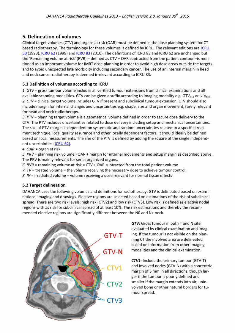
DAHANCA uses the following volumes and definitions for radiotherapy: GTV is delineated based on exami-nations, imaging and drawings. Elective regions are selected based on estimations of the risk of subclinical spread. There are two risk levels: high risk (CTV2) and low risk (CTV3). Low risk is defined as elective nodal regions with as risk for subclinical spread of at least 10%. The risk estimations and thereby the recom-mended elective regions are significantly different between the N0 and N+ neck.
GTV: Gross tumour in both T and N site evaluated by clinical examination and imag-ing. If the tumour is not visible on the plan-ning CT the involved area are delineated based on information from other imaging modalities and the clinical examination. CTV1: Include the primary tumour (GTV-T) and involved nodes (GTV-N) with a concentric margin of 5 mm in all directions, though lar-ger if the tumour is poorly defined and smaller if the margin extends into air, unin-volved bone or other natural borders for tu-mour spread.

DAHANCA Radiotherapy Guidelines 2013 – English version 2.0, January 30th 2015
7
CTV2: Contains CTV1 and the volume outside CTV1 that harbours the greatest risk for subclinical disease. It is defined as CTV1 with a concentrical margin of 5 mm in all directions. The margin may be less if it extends to air or surpasses natural barriers such as bone. The total geometrical margin from GTV to CTV”should not be larger than 12 mm. Furthermore, a disease specific high risk anatomical region is added. See below for details. For postoperative radiotherapy CTV2 contains the surgical bed after radical resection (both T- and N-position). CTV3: Contains CTV2 and regional elective lymph nodes without. The CTV3 definition is highly dependent on nodal status. N0 and N+ are treated as recommended in Gregoire 20031, Gregoire 20062, Gregoire3, re-spectively. For the N+ patients, the elective nodal regions are extended 2 cm cranial and caudal any patho-logical lymph nodes (GTV-N). The sternocleidomastoid muscle is included 2 cm above and below any patho-logical nodes in case of suspected muscle involvement. PTV1, PTV2, PTV3 (formerly PTV-T og PTV-E): Contains CTV with a set-up margin (SM) that may vary with field localisation, patient immobilisation and the use of IGRT (Image guided adaptive radiotherapy). It is recommended that all departments gather data for their respective SM. PTV can be further divided in sub-volumes, e.g. close to surfaces or with overlap with OAR and PRV.
5.3 Delineation guidelines
According to national legislation (Order no. 48 af 25. Jan. 1999 (Acceleratorbekendtgørelsen)) treatment planning must include:
delineation of target
delineation of organs at risk
determination of dose pr. fraction
determination of total dose for target
total dose for organs at risk A clear indication, dose prescription and target definition should be available before target delineation. It should be clearly described in medical records. The description could consist of a reference to guidelines or protocol. Example of workflow:
1. When present, GTV-T and GTV-N are delineated on CT scans in the dose planning system in coop-eration between oncologist and radiologist or nuclear medicine specialist. All available information, such as medical records, all available 2 and 3D imaging, tumour drawings, palpation and other in-formation discussed on a MDT, is used for target definition. If there is no macroscopic tumour, e.g. after surgery, the original tumour area is delineated based on previous clinical and imaging infor-mation.
1 Gregoire V et al. CT-based delineation of lymph node levels and related CTVs in the node-negative neck: DAHANCA,
EORTC, EORTEC, NCIC, RTOG consensus guidelines. Radiotherapy and Oncology 69 (2003) 227–236, 2003. 2 Grégoire V, Eisbruch A, Hamoir M, Levendag P. Proposal for the delineation of the nodal CTV in the node-positive and the post-operative neck. Radiother Oncol. 2006 Apr;79(1):15-20. 3 Grégoire V, Ang K, Budach W, Grau C, Hamoir M, Langendijk JA, Lee A, Le QT, Maingon P, Nutting C, O'Sullivan
B, Porceddu SV, Lengele B. Delineation of the neck node levels for head and neck tumors: A 2013 update.
DAHANCA, EORTC, HKNPCSG, NCIC CTG, NCRI, RTOG, TROG consensus guidelines. Radiother Oncol.
110:172-181,2014

DAHANCA Radiotherapy Guidelines 2013 – English version 2.0, January 30th 2015
8
2. CTV1 is generated from GTV by adding a isotropic margin of 5 mm, modified for bone, air or accord-ing to patient or disease specific considerations
3. CTV2 is generated by expanding CTV1 isotropic with 5 mm margin modified for bone, air or accord-
ing to patient or disease specific considerations 4. CTV3 is delineated according to atlases. Available on DAHANCA.dk. 5. Organs at risk are defined according to the treatment area and delineated according to atlases (Ap-
pendix 1). The spinal cord must always be delineated, and the brain stem should be delineated at least in all cases with a defined CTV3
6. Finally, targets and organs at risk should be visualized in beams eye view to identify target and OAR
irregularities and inconsistencies. The CTVs are then modified to represent clinical and biological relevant volumes.
7. The volumes are transferred to the dose planning process and considerations concerning bolus and
priorities are discussed with the dose planner. 8. Target definition and dose prescription used for the dose planning process is then formally ap-
proved and documented.
Delineation is performed on CT with contrast unless contraindications towards i.v. contrast. Ideally, in case of multiple imaging modalities PET/CT or MRI is performed in treatment position with fixation on a flat-top scanner bed. Otherwise imaging is co-registered with the planning CT or, imaging modalities are viewed 'side-by-side' with the planning CT. A contour can be delineated manually on all CT slices. Interpolation or automatic delineation can be performed, but all contours should be verified on all CT slices and adjusted.
6. Normal tissues
6.1 Atlas of relevant normal tissues
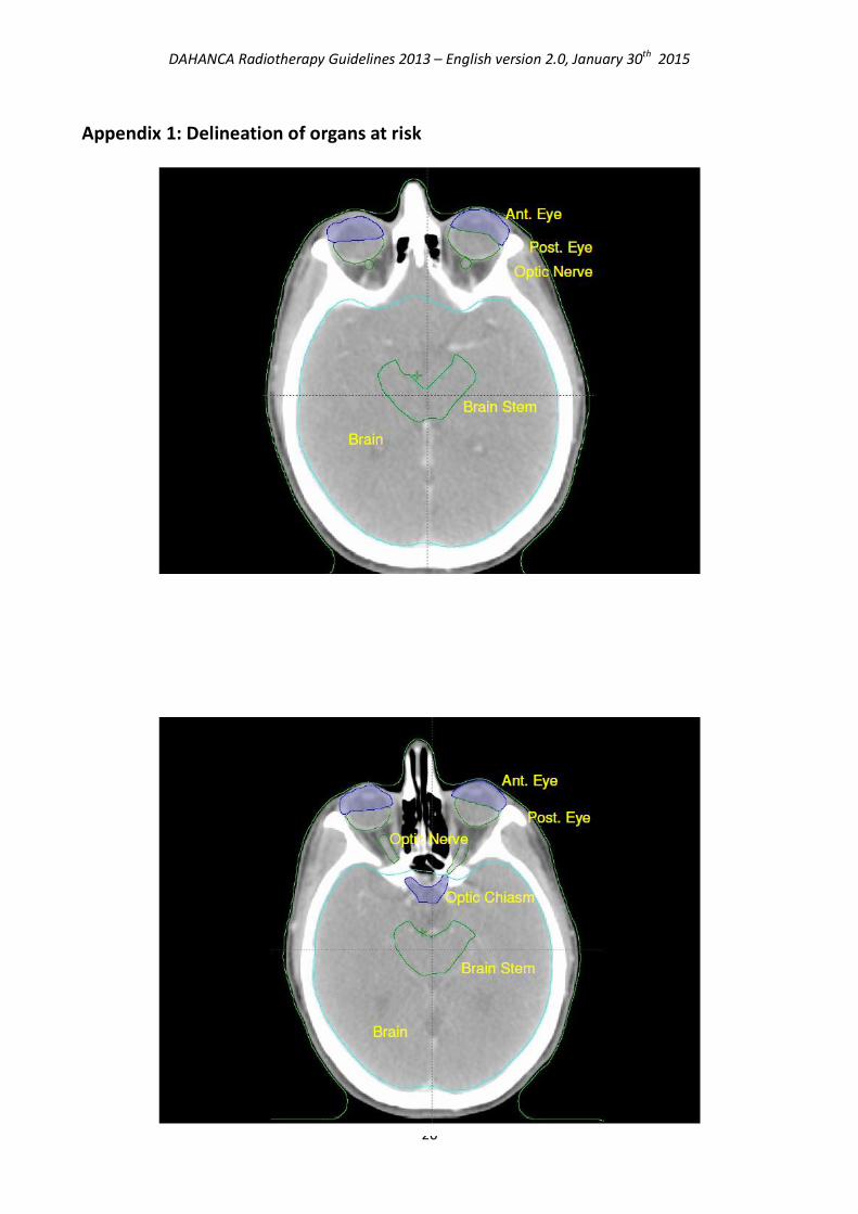
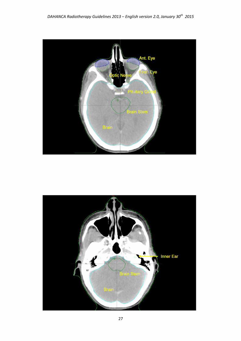
Knowledge on CT anatomy of normal tissues can be acquired from the literature. Typical delineation of
normal tissues is shown in appendix 1.
6.2 Dose volume constraints
Dose volume constraints relevant for head and neck radiotherapy are listed below. Data is mainly acquired
from studies of conventional radiotherapy of adults without concomitant chemotherapy. Normal tissue
tolerance can be different for other fractionation schedules and the table is only applicable for fractions
sizes of 2 Gy and below and not applicable for children or hypofractionation.
The nomenclature for normal tissues listed below is recommended. Suffixes for left / right should be ap-
plied. Dmax means D0,027 cm3 (3x3x3 mm3).

DAHANCA Radiotherapy Guidelines 2013 – English version 2.0, January 30th 2015
9
Structure Dose con-
straint
OAR [Gy]
Dose con-
straint PRV
[Gy]
Comments and organ definition
References
AB
SOLU
TE
Brain stem Dmax ≤ 54Gy Dmax ≤ 60Gy Treating ≤10 cm3 of OAR to a maximum
of 59 Gy results in a very low risk of
neurological damage. If it is necessary
for target coverage it can be done after
thorough patient information and con-
sent. Delineation: From the top of dens
(in order to avoid overdosing medulla)
to the bottom of 3rd ventricle (since
cranial border is difficult to identify on
CT).
Mayo et al. IJROBP
vol 76 (3) S36-S41,
2010
Spinal cord Dmax ≤ 45Gy Dmax ≤ 50Gy Risk for neurological damage is esti-
mated to 6 % for doses at 60 Gy. Lim-
ited overdosage may therefore be al-
lowed in order to achieve target cover-
age after thorough patient information
and consent. Delineation: Spinal cord
not spinal canal.
Kirkpatrick et al.
IJROBP 76 (3) s42-
9, 2010
MU
ST
Anterior eye (conjunctiva, lacrimal gland, cornea, iris)*
Dmax≤30Gy Dmax≤35Gy Even if constraints are not met for other parts of the optic pathways the anterior eye may be worth sparing in order to preserve the eye in situ. In case of se-vere dry eye syndrome the eye must often be removed. *The lenses have been removed from the list of OAR since it is contained in the anterior eye OAR and side effects may be treated.
Jeganathan et al. IJROBP 79 (3) 650-9, 2011 DAHANCA 2004
Chiasm and optic nerve
Dmax ≤ 54Gy Dmax≤ 60Gy Dmax ≤ 55 Gy leads to a very low risk of side effects. Doses above 60 Gy leads to an estimated risk of above 7%. Dose constraint can be violated in order to achieve target coverage after thorough patient information and consent.
Mayo et al IJROBP 76 (3) S28-35, 2010 RTOG0615
Posterior eye (retina)
Dmax≤ 45Gy Dmax≤ 50Gy Retinopathy is seen after doses as low as 30 Gy, and doses must be kept as low as possible. There is a volume effect and eg. Lateral retina can be spared sepa-rately.
DAHANCA 2004 Jeganathan et al. IJROBP 79 (3) 650-9, 2011

DAHANCA Radiotherapy Guidelines 2013 – English version 2.0, January 30th 2015
10
SHO
ULD
Cochlea Dmean ≤ 45Gy og D5% ≤ 55Gy
Cochlea is delineated (hypodense area in the temporal bone anterior of the internal auditory canal). Risk of clinical relevant hearing loss may be as high as 15% at mean doses of 47 Gy when using concomitant cisplatin.
Bhandare et al. IJROBP 76 (3) S pp S50-57, 2010; Chan et al IJROBP 73, (5) 1335-1342, 2009; Hichcock et al IJROBP 73 (3) 779-88, 2009
Parotid gland 1) Contralateral parotid: Dmean≤ 20Gy 2) Both parotids: Dmean
≤26Gy
A gradual reduction of function is seen from 10-40 Gy. Mean doses should be kept as low as possible.
DAHANCA 2004 Deasy et al IJROBP 76 (3) S58-63, 2010
Mandible Hotspots in the mandible should be avoided
Int J Radiat Oncol Biol Phys. 2010 April ; 76(5): 1333–1338
CA
N
Pituitary gland Dmean≤30Gy
No observed threshold. Hormonal
changes can occur after >30 Gy and may
require follow up by endocrinologist
Darzy et al. Pitui-tary (2009) 12;40-50
Brain Dmax≤ 60Gy At Dmax=72 Gy the risk of necrosis is 5% after 5 years. Cognitive disturbances may be seen after lower doses. The entire brain is delineated.
Lawrence et al. IJROBP vol 76 (3) S20-27, 2010
Submandibular gland
Dmean ≤ 35Gy The submandibular gland is a part of level Ib and should only be spared if level I or II are not parts of the target
Deasy et al IJROBP 76 (3) S58-63, 2010
Oral cavity Dmean ≤ 30Gy for non-involved oral cavity
Delineation: Mobile tongue, floor of mouth cheeks and hard palate.
RTOG 1016
Lips Dmean ≤ 20Gy RTOG 1016
Larynx Dmean≤44 Gy Delineation: larynx including aryte-
noidea from the hyoid bone to cricoid
cartilage
Rancanti et al. IJROBP 76 (3) s64-69, 2010
Thyroid gland Dmean<40 Gy No specific threshold has been identi-fied and the endpoint is uncertain in the literature. Thyroid stimulating hormone should be controlled at doses above constraint according to local guidelines
Garcia-Serra AJCO 28, (3) June 2005 p 255-8 Boomsma R&O 99(2011)1-5
Oesophagus Dmean ≤ 30Gy Delineation: Below the cricoid cartilage to the top of the manubrium
RTOG 1016
Absolute: Organs of critical importance that must be prioritized over target coverage as a rule.
Must: Serial organs that must be delineated but not necessarily prioritized over target coverage.
Should: Parallel organs with good evince for sparing or serial organs with severe side effects if damaged.
Can: Poor evidence, uncertain endpoints or manageable toxicity. Organs may be delineated according to
local guidelines/research projects. Swallowing structures may be included here.

DAHANCA Radiotherapy Guidelines 2013 – English version 2.0, January 30th 2015
11
7. Treatment planning Patients must be treated with photons or electrons. Dose intensity must be at least 0,1Gy/min in the clini-cal target area according to ICRU. Dose inside the PTV1 should ideally be homogeneous. Patient immobili-sation is compulsory.
7.1 Dose prescription
The prescribed dose for a target (CTV) is the mean dose.
7.2 Dose calculation
For photons mean dose of CTV1 is the highest prescribed dose. Dose to CTV2only (CTV2 minus CTV1) and
CTV3only (CTV3 minus CTV2) must be as close as achievable to the prescribed doses.
CTV1 must be covered with 95%-107% of the prescribed dose. CTV2 and CTV3 must be covered with 95% of
the prescribed doses. The 95% isodose curve for PTV1, PTV2 and PTV3 must be as close to the delineation
of PTV1, PTV2 and PTV3 respectively, as achievable.
Globally, a maximum of 1,8 cm3 is allowed to receive >107% of the prescribed dose to CTV1.
Dose calculation for photons must take differences in patient density into account. This applies to both primary and scattered radiation. For electrons the minimal dose for PTV must be 92,5% of the prescribed dose, and the maximal dose should be <107% of the prescribed dose. Dose calculation for electrons should preferably be based on density in-formation of the CT scanning, but for tumours close to the skin, a manual calculation may be performed.
7.3 Simultaneously Integrated Boost (SIB)
For simultaneously integrated boost, different doses are prescribed to different targets, but the number of fractions remains constant for all targets. Dose per fraction for the elective regions has therefore been in-creased from 46 Gy (2Gy/fx) to 50 Gy (1,5 Gy/fx) and 56 Gy (1,0 Gy/fx). See appendix 2.
7.4 Prioritization of treatment goals
The IMRT optimization algorithms and the dose planning systems need a prioritization of treatment goals. The prioritization listed below is recommended for maximal clinical benefit, but individual prioritization may differ according to patient wishes and the clinical situation. 1. Critical normal tissues, potentially lethal complication Spinal cord Brain stem 2. Target coverage GTV CTV1 3. Critical serial normal tissues Anterior eye Chiasm Posterior eye and optic nerve Cochlea

DAHANCA Radiotherapy Guidelines 2013 – English version 2.0, January 30th 2015
12
4. Target coverage PTV1 PTV2 PTV3 5. Sensitive normal tissues Brain Contralateral parotid Larynx Oesophagus Lips Oral cavity Submandibular gland Ipsilateral parotid gland Mandible Circumference Thyroid gland Pituitary gland
7.5 Good planning practice
This section contains some useful considerations for the treatment planning process. 7.5.1 Skin dose and build-up areas There is a risk of boosting areas close to the skin when using IMRT. The reason is that the PTV may extend to or even outside the skin. Fields extending such areas will induce build-up, build-down and lack of back-scatter in the volume according to field direction. This may cause a dose deficit in the volume and the IMRT dose optimization algorithm may compensate by increasing the dose to the skin compared to 3D conformal treatment. As such, the dose may be increased above what is necessary according to clinical experience, leading to undesirable skin toxicity. Multiple methods are available to avoid this effect. The chosen method often depends on the availability in the dose planning systems. Most centres use an ‘IMRT-PTV’ which is cropped under the skin surface by a few millimetres. This method should be applied with caution since the PTV margins must take uncertainties into consideration, also in the direction of the skin. 7.5.2 Field directions and collimator angles Unless rotational-IMRT is used, field directions should preferably be placed according to tumour extension and normal tissue localisation. Field divergence and isocentre placement must also be taken into account. By sensible choice of beam- and collimator angles, the IMRT algorithm easier achieves a higher conformity. A tongue and groove effect can arise if 0° or 180° is chosen as collimator angles. This is not necessarily taken into account by the dose planning system and by choosing different collimator angles; a higher de-gree of freedom is also achieved for the dose optimisation algorithm. 7.5.3 Unilateral radiotherapy IMRT may be applied for unilateral radiotherapy. As described above in chapter 7.5.2, field directions must be chosen carefully and entrance through the target areas are preferred, but contralateral field directions may sometimes be applied with good results. However, dose to the non-involved side RVR should be penal-ized in the dose optimisation (see chapter 7.6). Dose to contralateral OAR must be kept far below normal tolerance if possible.

DAHANCA Radiotherapy Guidelines 2013 – English version 2.0, January 30th 2015
13
7.5.4 Conformity index (CI) Conformity index (CI) could be used for quality assurance in order not to irradiate unnecessary large vol-umes. Different formulas exist4, and they have in common, that they calculate a number, which reflect how far the clinical plan is from the ideal theoretical one . Different formulas have different strengths and the most simple is the one of RTOG:
Conformity IndexRTOG =VRI / TV
VRI: The volume surrounded by a reference isodose curve (95% of prescribed dose). TV: target volume. The closer to unity, the better is the conformity. Together with the demands for target coverage by DAHANCA, the CI serves as an indication of the quality of the dose plan. 7.5.5 Overdosage of PTV2 and PTV3 With IMRT, target coverage and CI close to 1 is possible, but significant overdosage of PTV2 and PTV3 is frequent since PTV1 is often located inside PTV2 and PTV3. If the overdosage is not located in the proximity of PTV1, it should be considered if mean dose to PTV2 and PTV3 may be further reduced. 7.5.6 Dose constraints to normal tissues Dose constraints, recommended by DAHANCA, are mentioned in chapter 6. These limitations are products of what is thought to be an acceptable risk of side effects. However, it is important to remember that re-duction of dose often results in a clinically relevant reduction of risk even if the dose limit to a certain OAR is far exceeded (or hasn’t been reached). E.g. a reduction of the mean dose to the parotid from 50 Gy to 40 Gy leads to a reduction of the risk of side effects from 75% to 50% (Dijkema et al5) 7.5.7 Dose to RVR Apart from doses to targets and OARs, a Remaining Volume at Risk (RVR) may be defined as the volume not containing targets or OARs. Penalizing doses to this volume ensures that field directions far from the target have a lower weight. For example, the RVR may be used in order to avoid dose through the shoulders and minimize integral patient dose. RVR may also be used for reporting doses outside targets and OARs.
7.6 Biological dose planning
When data from radiobiological dose-response modelling is used for plan optimization, the term biological dose planning is used. The biological models can be incorporated into the dose planning system or the physically optimized plan can be evaluated using biological models. Target definition based on functional or molecular imaging is normally not considered a part of biological dose planning unless dose distribution inside the target is optimized according to imaging data. Most commercially available dose planning systems offer biological dose planning for optimization or evaluation. Models and parameters from the QUANTEC project, described by Marks6, are used in the com-mercial available systems. The parameters will probably change with increasing knowledge and therefore, model parameters are not a part of the present guidelines A large number of models exist and their description is above the scope of these guidelines. As long as physical dose constraints are observed for the critical normal tissues, biological optimisation can be used for the prioritization of non-critical normal tissues. After careful evaluation of the clinical situation and the validity of the models and its parameters, biological dose planning may aid the prioritization of target cov-erage and normal tissue sparing in case of very advanced cases or re-irradiation.
4 Feuvret et al Int. J. Radiation Oncology Biol. Phys., Vol. 64, No. 2, pp. 333–342, 2006.
5 Dijkema et al Int. J. Radiation Oncology Biol. Phys., Vol. 78, No. 2, pp. 449–453, 2010 6 LB Marks, IJROBP vol 76, No.3-S10-S19
DAHANCA Radiotherapy Guidelines 2013 – English version 2.0, January 30th 2015
14
8. Treatment
8.1 Image guidance
Patient positioning should be verified with 2 or 3D imaging according to local guidelines. Action levels and imaging frequency should be defined locally with respect to local PTV and PRV margins originating from measurements of random and systematic uncertainties in the whole process of treatment preparation and delivery7. The anatomical structures used for matching must be defined with respect to target localisation. For exam-ple, emphasis must be put on more cranial structures for nasopharyngeal tumours than for hypopharyngeal tumours. The ’region of interest’ (ROI) for the matching process must also take the extend of the elective areas into account. Match structures with low internal movements should be chosen, e.g. not the hyoid bone, but preferably the cervical spine. Soft tissue matching is often possible when CBCT scans are avail-able. The ROI must be chosen to ensure both target coverage and normal tissue sparing. Both automatic and manual match must be visually checked according to bone anatomy and visible soft tissue.
8.2 Re-planning
It should be continuously evaluated if patient anatomy and the effectiveness of the immobilisation device changes to a degree that may have significant implication for the dose distribution. In that case, a new CT scan with our without new immobilisation must be performed and dose distribution evaluated. If neces-sary, new targets and normal tissues must be delineated and a new treatment plan must be made. Evalua-tion during treatment, is based on regular CBCT’s and e.g. a CT half way through treatment if necessary. This is especially relevant in patients with large tumours.
9. Treatment prolongations
All fields must be treated at all fractions. Patients treated with 6 fractions per week must receive a fraction Monday to Friday and the sixth fraction should be given during the weekend or as an extra fraction Monday to Friday. An interval of 6 hours between fractions must always be respected. For patients receiving 10 fractions per week, two daily fractions with an interval of at least 6 hours is used. In case of treatment prolongations, the overall treatment time, from first to last fraction, should be main-tained if possible. The missing fraction(s) must be given as fast as possible and ideally be given within a week if clinically applicable. This can be done by delivering an extra fraction during weekends or on the day of a planned fraction (but 6 hours apart). Considering acute toxicity, treatment breaks must not be com-pensated with more than one extra fraction per week, and no more than 13 consecutive treatment days must be used. Furthermore, no more than 3 days of double fractionation must be used within 2 weeks for conventionally fraction sizes. To compensate longer treatment breaks, hyperfractionation and dose escalation could be worth consider-ing. See Dale et al8.
10. Quality assurance (QA)
Treatment methods must be quality assured and reported in clinical trials. QA can be divided into three steps:
Step 1: Preparation including writing guidelines, dose audits and delineation workshops.
7 M. van Herk: ”Errors and Margins in Radiotherapy”, Sem. Rad. Onc.14 (1), pp. 52-64 (2004).
8 Dale et al, Clin Oncol 14:382-393, 2002

DAHANCA Radiotherapy Guidelines 2013 – English version 2.0, January 30th 2015
15
Step 2: Daily QA: Technical QA of the performance of the accelerators, verification of delineation, dose plans and setup procedures.
Step 3: Follow-up on the given treatments, reporting, sampling and evaluation according to prede-fined criteria of minor and major deviations.
10.1 Preparation
Technical QA is described in the national legislation (‘Acceleratorbekendtgørelsen’9) and refers to ”Practical Guidelines for the Implementation of a Quality System in Radiotherapy” from the European Society for Therapeutic Radiology and Oncology, ”Comprehensive QA for Radiation Oncology”, Report of AAPM Radia-tion Therapy Committee Task Group 40, and ”Absorbed Dose Determination in Photon and Electron Beams”, Technical Report Series 398, from the International Atomic Energy Agency. Furthermore, the Danish healthcare Quality Programme (‘Den Danske Kvalitetsmodel (DDKM)’) describes that guidelines and instructions must be available for all aspects of any treatment in the Danish health care system, including the treatment of head and neck cancer. It will be described below how the correct treatment of head and neck cancer is ensured under the aus-pices of DAHANCA. Internal guidelines must exist in all centres to ensure adherence to the national guide-lines by DAHANCA. 10.1.1 Dose audit The path from CT scanning, dose planning and treatment delivery is complex. All steps must be verified. Nevertheless, transitions from one step to another may also introduce errors that may escape a stepwise QA. One way to assure all steps and transitions is a dose audit: A dose audit includes treating a standard-ized phantom according to specified guidelines to certain doses. Dose to the phantom is then measured and compared to the dose plan produced at the centre. It is recommended that an external dose audit is performed at least every 5 years under the auspices of the DAHANCA QA working group. 10.1.2 Delineation workshops The basis of dose planning is the delineation of the tumour and clinical target volume. Delineation guide-lines for the OAR and CTV contained in the present guidelines are aimed at increasing consistency and comparability between patients and centres. Nevertheless, no gold standard exists, and delineation praxis must be continuously evaluated. National delineation workshops will be held every 3 years under the aus-pices of the DAHANCA QA working group.
10.2 Daily quality assurance
Local guidelines must be available for daily QA. 10.2.1 Delineation verification and approval Delineation of targets and normal tissues must be approved by a trained specialist. Delineation must, as a rule, follow the guidelines of chapter 5, and deviations from the guidelines should be described in the medical records. 10.2.2 Dose planning verification and approval All dose plans must be verified by an independent dose planner. Prescribed dose and target coverage on all CT slices, as well as dose to the normal tissues, must be verified. See chapter 6 and 7. 10.2.3 Imaging calibration
9 Bekendtgørelse om elektronacceleratorer til patientbehandling med energier fra 1 MeV til og med 50 MeV
DAHANCA Radiotherapy Guidelines 2013 – English version 2.0, January 30th 2015
16
A procedure must be available on all centres for the calibration and QA of the localisation of imaging and treatment isocentre.
10.3 Follow up
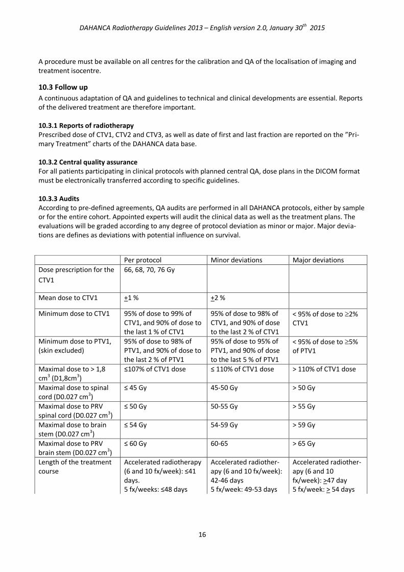
A continuous adaptation of QA and guidelines to technical and clinical developments are essential. Reports of the delivered treatment are therefore important. 10.3.1 Reports of radiotherapy Prescribed dose of CTV1, CTV2 and CTV3, as well as date of first and last fraction are reported on the ”Pri-mary Treatment” charts of the DAHANCA data base. 10.3.2 Central quality assurance For all patients participating in clinical protocols with planned central QA, dose plans in the DICOM format must be electronically transferred according to specific guidelines. 10.3.3 Audits According to pre-defined agreements, QA audits are performed in all DAHANCA protocols, either by sample or for the entire cohort. Appointed experts will audit the clinical data as well as the treatment plans. The evaluations will be graded according to any degree of protocol deviation as minor or major. Major devia-tions are defines as deviations with potential influence on survival.
Per protocol Minor deviations Major deviations
Dose prescription for the
CTV1
66, 68, 70, 76 Gy
Mean dose to CTV1 +1 % +2 %
Minimum dose to CTV1 95% of dose to 99% of CTV1, and 90% of dose to the last 1 % of CTV1
95% of dose to 98% of CTV1, and 90% of dose to the last 2 % of CTV1
< 95% of dose to 2% CTV1
Minimum dose to PTV1, (skin excluded)
95% of dose to 98% of PTV1, and 90% of dose to the last 2 % of PTV1
95% of dose to 95% of PTV1, and 90% of dose to the last 5 % of PTV1
< 95% of dose to 5% of PTV1
Maximal dose to > 1,8 cm3 (D1,8cm3)
≤107% of CTV1 dose ≤ 110% of CTV1 dose > 110% of CTV1 dose
Maximal dose to spinal cord (D0.027 cm3)
≤ 45 Gy 45-50 Gy > 50 Gy
Maximal dose to PRV spinal cord (D0.027 cm3)
≤ 50 Gy 50-55 Gy > 55 Gy
Maximal dose to brain stem (D0.027 cm3)
≤ 54 Gy 54-59 Gy > 59 Gy
Maximal dose to PRV brain stem (D0.027 cm3)
≤ 60 Gy 60-65 > 65 Gy
Length of the treatment course
Accelerated radiotherapy (6 and 10 fx/week): ≤41 days. 5 fx/weeks: ≤48 days
Accelerated radiother-apy (6 and 10 fx/week): 42-46 days 5 fx/week: 49-53 days
Accelerated radiother-apy (6 and 10 fx/week): >47 day 5 fx/week: > 54 days

DAHANCA Radiotherapy Guidelines 2013 – English version 2.0, January 30th 2015
17
11. Specific guidelines according to tumour site Oral Cavity Anatomy: The oral cavity includes buccal mucosa, gingiva, hard palate, anterior 2/3 of the tongue and floor of mouth. Lateral tumours are defined as tumours of the buccal mucosa, gingiva and retromolar trigone, with no involvement of contralateral nodes. Midline tumours are defined as tumours of the tongue, floor of mouth and hard palate and any tumours with involvement of these structures. Midline tumours have the propensity of bilateral nodal involvement. Drainage to the lymphatic system from the anterior tongue rarely spreads to level III and IV without involvement of proximal nodes. Primary treatment is described in the national guidelines (www.dahanca.dk). Shortly, the mainstay of treatment is surgery for operable tumours were a good functional and cosmetic result can be expected. Postoperative radiotherapy is added in case of non-radical surgery (R1 or R2) in N or T site, pN2-3 and or pT3-4, or any N stage with extracapsular extension (ECE). Target delineation is often greatly improved when the operating surgeon take part in the procedure. Radical Radiotherapy: CTV1: Primary tumour (GTV-T) and involved lymph nodes (GTV-N) with a concentrically isotropic margin of 5 mm. Larger margins should be used for ill-defined tumours and margins should be cropped for air and natural barriers such as bone, unless bone involvement is evident. CTV2: CTV1 with a concentrically isotropic margin of 5 mm. Margin should be cropped for air or natural barriers for tumour extension. CTV2 can be individually expanded to include high risk anatomical areas, e.g. the ipsilateral or whole tongue in case of tongue involvement or ipsilateral floor of mouth. CTV3: Midline tumours are treated with bilateral elective regions and lateral tumours with ipsilateral elec-tive regions. Elective nodal regions are:
N0: level I, II, III
N1-3: level I, II, III. Elective regions are extended at least 2 cm cranially and caudally of GTV-N. If ex-tension to nearby muscle involvement is suspected the entire muscle is included at least 2 cm above and below GTV-N
Postoperative radiotherapy: CTV1: Macroscopic tumour (R2), microscopically non-radical operated (R1) or areas of ECE, with a concen-trically isotropic margin of 5 mm. Larger margins should be used for ill-defined tumours and margins should be cropped for air and natural barriers such as bone, unless bone involvement is evident. CTV2: CTV1 with an isotropic concentrically margin of 5 mm. Margins could individually be enlarged to in-clude high risk regions and cropped for air and at natural barriers such as bone. If no CTV1 is present in case of radical surgery (R0), CTV2 is the operated area with a concentrically iso-tropic margin of 5 mm. If the indication for radiotherapy is nodal disease N2-N3, ECE or non-radical surgery in the N-site, the primary tumour bed (R0) is generally included as radiotherapy for recurrences are diffi-cult. CTV3: Midline tumours are treated with bilateral elective regions and lateral tumours with ipsilateral elec-tive regions.
DAHANCA Radiotherapy Guidelines 2013 – English version 2.0, January 30th 2015
18
Note: If the indication for radiotherapy is in the T-site alone, no elective nodal irradiation should be per-formed for pT1-2 Elective nodal regions are
pN0: level I, II, III. In case of involvement of macroscopic cranial nerve, the nerve is included to the base of scull.
pN1-3: level I, II, III. Elective regions are extended at least 2 cm cranially and caudally of GTV-N. If extension to nearby muscle involvement is suspected the entire muscle is included at least 2 cm above and below GTV-N.
Nasopharynx Anatomy: The nasopharynx is limited by choanea, base of scull, sphenoid sinus, posterior pharynx and soft palate. A diagnostic MRI should be present to aid delineation. CTV1: Primary tumour (GTV-T) and involved lymph nodes (GTV-N) with a concentrically isotropic margin of 5 mm. Larger margins should be used for ill-defined tumours and margins should be cropped for air and natural barriers such as bone, unless bone involvement is evident. . CTV2: CTV1 with an isotropic concentrically margin of 5 mm. Margins could individually be enlarged to in-clude high risk regions and cropped for air and at natural barriers such as bone. The entire nasopharyngeal mucosa, foramina of the base of scull and choana are included. In case of paranasal sinus involvement, the entire sinus is included. Other high risk areas may be included after individual considerations. CTV3: Elective nodal regions:
N0: bilateral level II, III, IV, V, retrostyloid and retropharyngeal nodes
N1-3: bilateral level II, III, IV, V, retrostyloid and retropharyngeal nodes. Elective regions are ex-tended at least 2 cm cranially and caudally of GTV-N. If extension to nearby muscle involvement is suspected the entire muscle is included at least 2 cm above and below GTV-N.
Oropharynx Anatomy: Oropharynx is limited by anterior faucail pillar, macroscopic taste buds (papillae vallatae), soft palate and the epiglottis. Oropharynx thereby includes posterior third of tongue, vallecula, tonsils, tonsillar pillars, posterior pharynx and soft palate. Tumours confined to the tonsillar fossa and tonsillar pillars are considered lateral tumours and should be treated with ipsilateral radiotherapy. Tumours arising in, or ex-tending to, the base of tongue, soft palate or posterior pharyngeal wall are considered midtline tumours and should be treated with bilateral elective irradiation. CTV1: Primary tumour (GTV-T) and involved lymph nodes (GTV-N) with a concentrically isotropic margin of 5 mm. Larger margins should be used for ill-defined tumours and margins should be cropped for air and natural barriers such as bone, unless bone involvement is evident. After diagnostic tonsillectomy, the tonsillar fossa and pillars are considered as the CTV1. The clinical exami-nation is very important in the evaluation of the extension to soft palate and especially the base of tongue. Base of tongue tumours are often difficult to depict on CT or MRI and it is often necessary to include large part of the base of tongue ion CTV1 or CTV2.
DAHANCA Radiotherapy Guidelines 2013 – English version 2.0, January 30th 2015
19
CTV2: CTV1 with an isotropic concentrically margin of 5 mm. Margins could individually be enlarged to in-clude high risk regions and cropped for air and at natural barriers such as bone. CTV2 can be individually expanded to include high risk areas such as the entire or ipsilateral base of tongue. CTV3: Bilateral or ipsilateral nodal regions according to midline involvement. See above. Elective nodal regions are:
N0: Level II, III. The retropharyngeal nodes are included in case of posterior pharyngeal wall in-volvement and level Ib is included in case of oral cavity involvement.
N1-3: Level II, III. Level IV on the side of nodal involvement. Elective regions are extended at least 2 cm cranially and caudally of GTV-N. If extension to nearby muscle involvement is suspected the en-tire muscle is included at least 2 cm above and below GTV-N. The retropharyngeal nodes are in-cluded in case of posterior pharyngeal wall involvement and level Ib is included in case of oral cav-ity involvement
Hypopharynx Anatomy: Hypopharynx is limited by oropharynx, larynx and oesophagus. The anterior wall includes aryte-noid cartilage and aryepiglottic fold til the lower cricoid cartilage. Pyriform sinus includes pharyngo-epiglottic fold and the upper extension of oesophagus. Laterally to the thyroid cartilage and medially from the hypopharyngeal surface of the aryepiglottic fold, arytenoid cartilage and cricoid cartilage. The hypo-pharyngeal posterior wall extends from a level through the hyoid bone (bottom of vallecula) to the lower border of the cricoid cartilage and from apex of one pyriform sinus to the other. CTV1: Primary tumour (GTV-T) and involved lymph nodes (GTV-N) with a concentrically isotropic margin of 5 mm. Larger margins should be used for ill-defined tumours and margins should be cropped for air and natural barriers such as bone, unless bone involvement is evident. CTV2: CTV1 with an isotropic concentrically margin of 5 mm. Margins could individually be enlarged to in-clude high risk regions and cropped for air and at natural barriers such as bone. CTV2 can be individually expanded to include high risk areas such as the entire or ipsilateral base of tongue. CTV3: Elective nodal regions are
N0: bilaterally level II, III, IV. Cranial part of level II can be excluded after individual consideration.
N1-3: bilaterally level II, III, IV. Elective regions are extended at least 2 cm cranially and caudally of GTV-N. If extension to nearby muscle involvement is suspected the entire muscle is included at least 2 cm above and below GTV-N. In case of subglottic or oesophageal involvement level VI is in-cluded.
Supraglottic larynx Anatomy: Supraglottic larynx includes larynx above the vocal folds i.e. the suprahyoid part of epiglottis (lin-gual and laryngeal surface above hyoid bone), aryepiglottic folds, infrahyoid epiglottis, ventricular folds and sinus of Morgagni. CTV1: Primary tumour (GTV-T) and involved lymph nodes (GTV-N) with a concentrically isotropic margin of 5 mm. Larger margins should be used for ill-defined tumours and margins should be cropped for air and natural barriers such as bone, unless bone involvement is evident.

DAHANCA Radiotherapy Guidelines 2013 – English version 2.0, January 30th 2015
20
CTV2 CTV1 with an isotropic concentrically margin of 5 mm. Margins could individually be enlarged to in-clude high risk regions and cropped for air and at natural barriers such as bone. CTV2 can be individually expanded to include high risk areas such as the laryngeal mucosa above the vocal cords. CTV3: Elective nodal regions:
N0: bilaterally level II and III.
N1-3: bilaterally level II and III. Level IV on the side of nodal involvement. Elective regions are ex-tended at least 2 cm cranially and caudally of GTV-N. If extension to nearby muscle involvement is suspected the entire muscle is included at least 2 cm above and below GTV-N
In case of subglottic or oesophageal involvement, level VI is included
The stoma is included in case of tracheostomy
Glottic larynx Anatomy: The region includes vocal cords, anterior and posterior commissure T1-2N0 CTV1: Primary tumour (GTV-T) and involved lymph nodes (GTV-N) with a concentrically isotropic margin of 5 mm. Larger margins should be used for ill-defined tumours and margins should be cropped for air and natural barriers such as bone, unless bone involvement is evident. Small tumours are usually difficult to visualize and the mucosa inside the thyroid cartilage (excluding the superior horns) is often delineated as CTV1 CTV2: CTV1 with an isotropic concentrically margin of 5 mm. Margins could individually be enlarged to in-clude high risk regions and cropped for air and at natural barriers such as bone. CTV2 can be individually expanded to include high risk areas. If CTV1 is defined as the mucosa inside the thyroid cartilage then CTV2=CTV1. CTV3: Differs between the following clinical scenarios
T1N0: No elective nodal irradiation.
T2N0: elective nodal irradiation can be omitted if the stage only depends on reduced vocal cord mobility.
All other T2N0 tumours require nodal irradiation as dependent on the areas of extension outside the cords. Most often level II and III with individual considerations of the benefit of irradiating the superior part of level II
In case of supraglottic extension, elective nodes should be irradiated according to recommenda-tions for that site
In case of subglottic or oesophageal involvement, level VI is included
The stoma is included in case of tracheostomy . T3-4N0 and all N+: CTV1: Primary tumour (GTV-T) and involved lymph nodes (GTV-N) with a concentrically isotropic margin of 5 mm. Larger margins should be used for ill-defined tumours and margins should be cropped for air and natural barriers such as bone, unless bone involvement is evident. Mucosa inside the thyroid cartilage should be included.

DAHANCA Radiotherapy Guidelines 2013 – English version 2.0, January 30th 2015
21
CTV2: CTV1 with an isotropic concentrically margin of 5 mm. Margins could individually be enlarged to include high risk regions and cropped for air and at natural barriers such as bone. CTV2 can be individually expanded to include high risk areas, e.g. supra- or subglottic larynx CTV3: Elective nodal regions
N0: bilaterally level II and III.
N1-3: bilaterally level II and III. Level IV on the side of nodal involvement. Elective regions are ex-tended at least 2 cm cranially and caudally of GTV-N. If extension to nearby muscle involvement is suspected the entire muscle is included at least 2 cm above and below GTV-N. In case of subglottic or oesophageal involvement, level VI is included
The stoma is included in case of tracheostomy
Subglottic larynx Anatomy: The region includes larynx below vocal cords CTV1: Primary tumour (GTV-T) and involved lymph nodes (GTV-N) with a concentrically isotropic margin of 5 mm. Larger margins should be used for ill-defined tumours and margins should be cropped for air and natural barriers such as bone, unless bone involvement is evident. CTV2: CTV1 with an isotropic concentrically margin of 5 mm. Margins could individually be enlarged to in-clude high risk regions and cropped for air and at natural barriers such as bone. CTV2 can be individually expanded to include high risk areas, e.g. glottic or supraglottic larynx. CTV3: Elective nodal regions
N0: bilaterally level III, IV, VI, and level II in case of supraglottic extension
N1-3: bilaterally level III, IV, VI, and level II in case of supraglottic extension. Elective regions are ex-tended at least 2 cm cranially and caudally of GTV-N. If extension to nearby muscle involvement is suspected the entire muscle is included at least 2 cm above and below GTV-N. In case of subglottic or oesophageal involvement, level VI is included
The stoma is included in case of tracheostomy
Postoperative radiotherapy after laryngectomy Elective nodal treatment can be performed using (chemo)irradiation in case of primary total laryngectomy. The target is individually defined by the multidisciplinary team. CTV1: Macroscopic tumour (R2), microscopically non-radical operated areas (R1) or areas of ECE, with a concentrically isotropic margin of 5 mm. Larger margins should be used for ill-defined tumours and margins should be cropped for air and natural barriers such as bone, unless bone involvement is evident. CTV2: CTV1 with an isotropic concentrically margin of 5 mm. Margins could individually be enlarged to in-clude high risk regions and cropped for air and at natural barriers such as bone. CTV2 can be individually expanded to include high risk areas. If no CTV1 is present in case of radical surgery (R0), CTV2 is the operated area with a concentrically iso-tropic margin of 5mm CTV3: Regional elective regions without margin as mentioned at the individual sites and the tracheostoma with 5 mm margin

DAHANCA Radiotherapy Guidelines 2013 – English version 2.0, January 30th 2015
22
Sinonasal Anatomy: The region includes nasal cavity behind the vestibule, maxillary sinus, ethmoid sinus, sphenoid sinus and frontal sinus. All areas are limited by bone except the anterior and posterior extend of the nasal cavity. Treatment is decided by national treatment guidelines (www.dahanca.dk). The mainstay of treatment is surgery for all patients with operable disease. Postoperative radiotherapy is indicated for all patients except T1 tumours after radical surgery. Often compromises between target coverage and dose to OAR are neces-sary. Radical radiotherapy CTV1: Primary tumour (GTV-T) and involved lymph nodes (GTV-N) with a concentrically isotropic margin of 5 mm. Larger margins should be used for ill-defined tumours and margins should be cropped for air and natural barriers such as bone, unless bone involvement is evident. CTV2: CTV1 with an isotropic concentrically margin of 5 mm. Margins could individually be enlarged to in-clude high risk regions and cropped for air and at natural barriers such as bone. Furthermore, the entire involved sinus(es) or ipsilateral nasal cavity is included as well as other high risk areas after individual con-sideration. CTV3: Elective nodal regions are:
N0: Nodal irradiation is only performed if the tumour extends to areas that normally elicit elective nodal irradiation: skin, palate, oral cavity or pharynx. In case of involvement of skin, oral cavity, pharynx minimal elective irradiation is given to level Ib, level II and often level III. In case of naso-pharyngeal involvement level IV, V, retropharyngeal and retrostyloid nodes are also included. Se-lective ipsilateral irradiation can be done in case of involvement of lateral structures only (e.g. gin-giva)
N1-3: Involved regions and regions as mentioned above. Elective regions are extended at least 2 cm cranially and caudally of GTV-N. If extension to nearby muscle involvement is suspected the entire muscle is included at least 2 cm above and below GTV-N. Selective ipsilateral irradiation can be done in case of involvement of lateral structures only (e.g. gingiva)
Postoperative radiotherapy CTV1: Macroscopic tumour (R2), microscopically non-radical operated areas (R1) or areas of ECE, with a concentrically isotropic margin of 5 mm. Larger margins should be used for ill-defined tumours and margins should be cropped for air and natural barriers such as bone, unless bone involvement is evident. CTV2: CTV1 with an isotropic concentrically margin of 5 mm. Margins could individually be enlarged to in-clude high risk regions and cropped for air and at natural barriers such as bone. Furthermore, the entire involved sinus(es) or ipsilateral nasal cavity is included as well as other high risk areas after individual con-sideration. If no CTV1 is present in case of radical surgery (R0), CTV2 is the operated area with a concentrically iso-tropic margin of 5mm and the entire involved sinus(es) or ipsilateral nasal cavity is included as well as other high risk areas after individual consideration. CTV3: Elective nodal regions are:

DAHANCA Radiotherapy Guidelines 2013 – English version 2.0, January 30th 2015
23
N0: Nodal irradiation is only performed if the tumour extends to areas that normally elicit elective nodal irradiation: skin, palate, oral cavity or pharynx. In case of involvement of skin, oral cavity, pharynx minimal elective irradiation is given to level Ib, level II and often level III. In case of naso-pharyngeal involvement level IV, V, retropharyngeal and retrostyloid nodes are also included. Se-lective ipsilateral irradiation can be done in case of involvement of lateral structures only (e.g. gin-giva)
N1-3: Involved regions and regions as mentioned above. Elective regions are extended at least 2 cm cranially and caudally of GTV-N. If extension to nearby muscle involvement is suspected the entire muscle is included at least 2 cm above and below GTV-N. Selective ipsilateral irradiation can be done in case of involvement of lateral structures only (e.g. gingiva)
Salivary gland Anatomy: Salivary gland tumours arise in the macroscopic glands (parotid, submandibular and sublingual glands) as well as the entire mucous membrane of the head and neck area, most often in the oral cavity. Salivary gland tumours are divided in low and high grade tumours based on histology. The treatment prin-ciples are determined by national guidelines (www.dahanca.dk). As a rule, surgery is performed as the pri-mary treatment of all operable tumours. Postoperative radiotherapy is recommended after non radical surgery of the T site (R1 or R2), T≥T3, N+, ECE, recurrences and high grade tumours irrespective of other risk factors. DAHANCA has divided salivary gland tumours in prognostic groups based on histology: Low grade: Acinic cell carcinoma, polymorphous low-grade adenocarcinoma, basal cell adenocarcinoma, epithelial-myoepithelial carcinoma, high and intermediate grade mucoepidermoid carcinoma, adenocarci-noma NOS (Not Otherwise Specified), well differentiated non- or minimally carcinoma in a pleomorph ade-noma, clear cell carcinoma NOS, sialoblastoma High grade: Adenoid cystic carcinoma, adenocarcinoma NOS, intermediate and poorly differentiated carci-noma in pleomorph adenoma with invasive depth of >1,5 mm, poorly differentiated mucoepidermoid car-cinoma, salivary duct carcinoma, primary squamous cell carcinomas, undifferentiated carcinoma (lym-phoepithelial carcinoma), large cell carcinoma, mucinous adenocarcinoma, oncocytic carcinoma, carcino-sarcomas, small cell carcinoma, myoepithilial carcinoma Radical Radiotherapy: CTV1: CTV1 should include the primary tumour (GTV-T) with a 5mm isotropic margin and include the entire macroscopic involved gland. CTV2: CTV1 with an isotropic concentrically margin of 5 mm. Margins could individually be enlarged to in-clude high risk regions and cropped for air and at natural barriers such as bone. CTV3: As a rule, selective ipsilateral regions are irradiated only. In case of involvement of midline structures both sides of the neck is irradiated
Parotid: level Ib + II + III
Submandibular: level Ia+ Ib + II + III
For all other glands the principles for the relevant region (often oral cavity) is applied. Elective re-gions are extended at least 2 cm cranially and caudally of any GTV-N If extension to nearby muscle involvement is suspected the entire muscle is included at least 2 cm above and below GTV-N.
Postoperative radiotherapy:
DAHANCA Radiotherapy Guidelines 2013 – English version 2.0, January 30th 2015
24
CTV1: Macroscopic tumour (R2), microscopically non-radical operated areas (R1) or areas of ECE, with a concentrically isotropic margin of 5 mm. Larger margins should be used for ill-defined tumours and margins should be cropped for air and natural barriers such as bone, unless bone involvement is evident CTV2: CTV1 with an isotropic concentrically margin of 5 mm. Margins could individually be enlarged to in-clude high risk regions and cropped for air and at natural barriers such as bone. If no CTV1 is present in case of radical surgery (R0), CTV2 is the operated area with a concentrically iso-tropic margin of 5mm. Furthermore, the entire salivary gland should always be included in the CTV2. CTV3: In case of pN0 no elective nodal irradiation is performed. As a rule, selective ipsilateral regions are irradiated only. In case of involvement of midline structures both sides of the neck is irradiated
Parotid: level Ib + II + III
Submandibular: level Ia+ Ib + II + III
For all other glands the principles for the relevant region (often oral cavity) is applied. Elective re-gions are extended at least 2 cm cranially and caudally of any GTV-N If extension to nearby muscle involvement is suspected the entire muscle is included at least 2 cm above and below GTV-N.
Neck metastasis from unknown primary (UP) Anatomy: Neck metastasis from unknown primary is defined as an undiagnosed primary tumour after thor-ough diagnostic procedures, at the beginning of treatment. Diagnostic procedures and treatment follows national guidelines (www.dahanca.dk). A distinction is made between squamous cell carcinomas and other histology’s. Neck nodes containing squamous cell carcinomas will often originate from the mucous membranes of the head and neck area. For other histology’s multiple sources of the metastasis exists. Some can be treated with curative intend, e.g. germ cell tumours, small cell lung cancer, and some are relative treatment resis-tant such as melanomas. Radiotherapy for squamous cell carcinomas An ipsilateral neck dissection is generally performed. All patients with pN2-3 disease, R1 or R2 resections and/or lymph nodes with extracapsular extension are candidates for postoperative radiotherapy. Generally, both regional lymph nodes as well as potential mucosal sources of the primary tumour should be irradiated. Elective mucosal irradiation is not indicated in case of other histology. Radiotherapy of the ipsi-lateral neck is difficult without irradiating the contralateral neck, and treatment of contralateral recur-rences will therefore be difficult. Bilateral nodal irradiation is therefore recommended. CTV1: Involved lymph nodes (GTV-N) or preoperative volume of nodes with insufficient margins (R1 and R2) or extracapsular extension, with a concentrically isotropic margin of 5 mm. Larger margins should be used for ill-defined tumours and margins should be cropped for air and natural barriers such as air or bone, unless bone involvement is evident. CTV2: CTV1 with an isotropic concentrically margin of 5 mm. Margins could individually be enlarged to in-clude high risk regions and cropped for air and at natural barriers such as bone. If no CTV1 is present in case of radical surgery (R0), CTV2 is the operated area with a concentrically iso-tropic margin of 5mm. Mucosal areas with extra high risk of harbouring the primary could be included in this volume /e.g. oropharyngeal mucosa in case of p16 positive tumours).

DAHANCA Radiotherapy Guidelines 2013 – English version 2.0, January 30th 2015
25
CTV3: The entire mucous membrane for 5 mm depth, in the pharynx and larynx, from the base of scull to below the cricoid cartilage, including base of tongue and tonsillary fossa. Elective regions include bilateral level II, III, IV. Level V is included if a nasopharyngeal primary is suspected. Elective regions are extended at least 2 cm cranially and caudally of any GTV-N. If extension to nearby muscle involvement is suspected the entire muscle is included at least 2 cm above and below GTV-N. Radiotherapy in other histology’s In case of adenocarcinoma therapy depends on the likely localisation of a primary. Localisations include salivary and thyroid glands, nasal cavity and paranasal sinuses, lungs, breast, gastrointestinal canal, uterus, ovary and prostate. Localisation, immunohistochemistry, serology and iodine scintigraphy may aid in the search of a primary and guide the treatment. In case of unknown primary after relevant diagnostics in-volved field irradiation to curative doses may be indicated, but elective nodal or mucosal irradiation is not recommended.
DAHANCA Radiotherapy Guidelines 2013 – English version 2.0, January 30th 2015
26
Appendix 1: Delineation of organs at risk
DAHANCA Radiotherapy Guidelines 2013 – English version 2.0, January 30th 2015
27

DAHANCA Radiotherapy Guidelines 2013 – English version 2.0, January 30th 2015
28

DAHANCA Radiotherapy Guidelines 2013 – English version 2.0, January 30th 2015
29
DAHANCA Radiotherapy Guidelines 2013 – English version 2.0, January 30th 2015
30
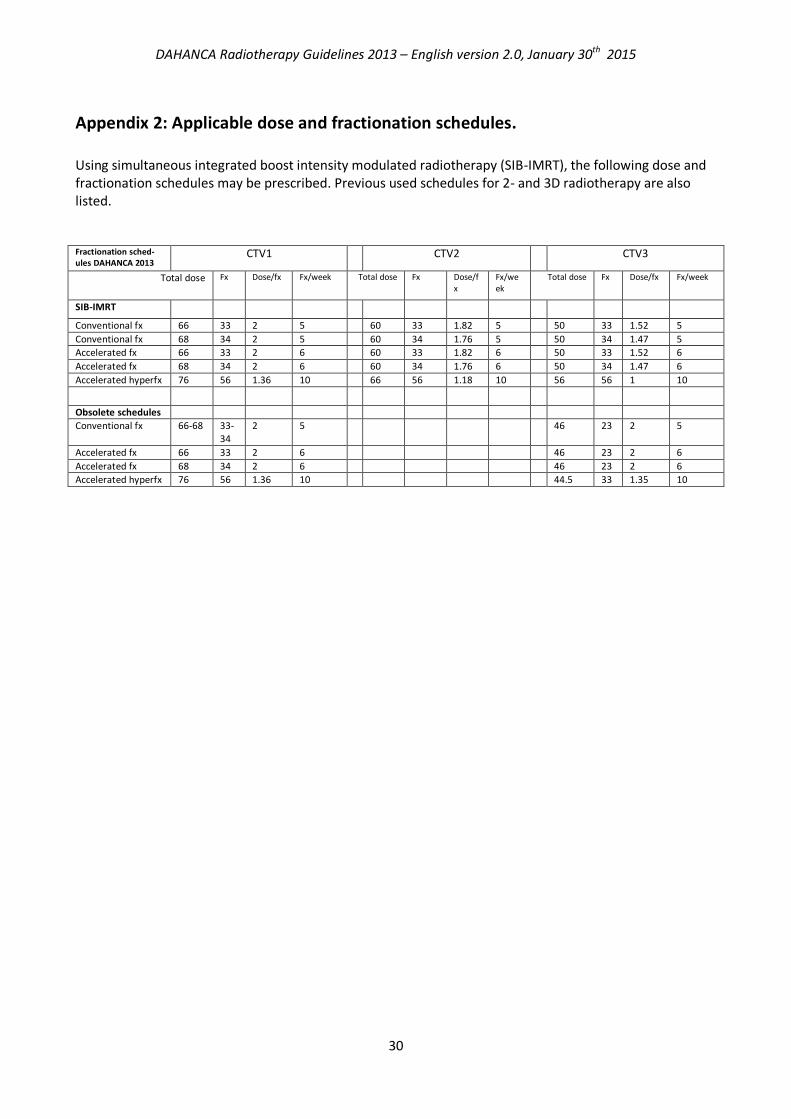
Appendix 2: Applicable dose and fractionation schedules.
Using simultaneous integrated boost intensity modulated radiotherapy (SIB-IMRT), the following dose and fractionation schedules may be prescribed. Previous used schedules for 2- and 3D radiotherapy are also listed. Fractionation sched-ules DAHANCA 2013
CTV1 CTV2 CTV3
Total dose Fx Dose/fx Fx/week Total dose Fx Dose/fx
Fx/week
Total dose Fx Dose/fx Fx/week
SIB-IMRT Conventional fx 66 33 2 5 60 33 1.82 5 50 33 1.52 5
Conventional fx 68 34 2 5 60 34 1.76 5 50 34 1.47 5
Accelerated fx 66 33 2 6 60 33 1.82 6 50 33 1.52 6
Accelerated fx 68 34 2 6 60 34 1.76 6 50 34 1.47 6
Accelerated hyperfx 76 56 1.36 10 66 56 1.18 10 56 56 1 10
Obsolete schedules
Conventional fx 66-68 33-34
2 5 46 23 2 5
Accelerated fx 66 33 2 6 46 23 2 6
Accelerated fx 68 34 2 6 46 23 2 6
Accelerated hyperfx 76 56 1.36 10 44.5 33 1.35 10