D r. Gamal Hassanain. Content Introduction Estimate of burn size Classification Management...
40
Presentation & Management of Burn Patients D r. Gamal Hassanain
-
Upload
harvey-bryant -
Category
Documents
-
view
213 -
download
0
Transcript of D r. Gamal Hassanain. Content Introduction Estimate of burn size Classification Management...
Presentation & Management of Burn
Patients
D r. Gamal Hassanain
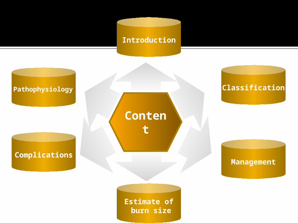
Content
Introduction
Estimate of burn size
Classification
ManagementComplications
Pathophysiology
Introduction
A burn is defined as a coagulative necrosis causing destruction of the epithelium.
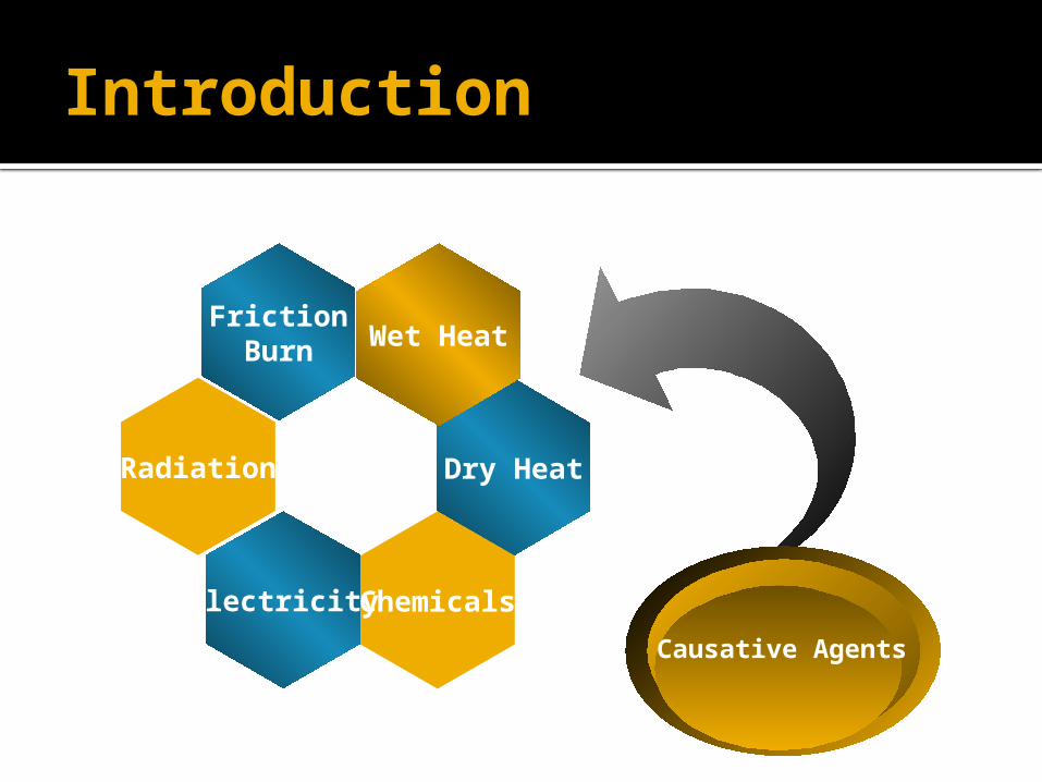
Introduction
Dry Heat
Wet Heat
ChemicalsElectricity
Causative Agents
FrictionBurn
Radiation
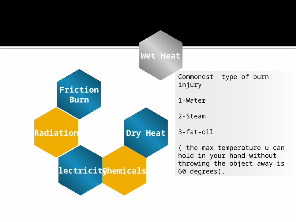
Wet Heat
Commonest type of burn injury
1-Water
2-Steam
3-fat-oil
( the max temperature u can hold in your hand without throwing the object away is 60 degrees).
Dry Heat
ChemicalsElectricity
FrictionBurn
Radiation
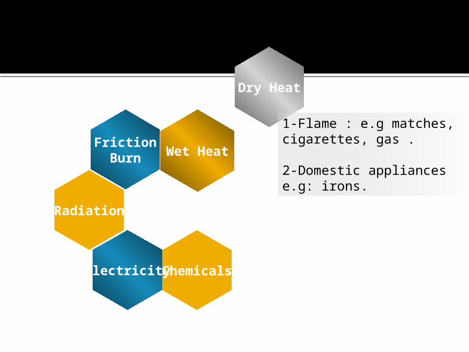
Dry Heat
1-Flame : e.g matches, cigarettes, gas .
2-Domestic appliances e.g: irons.
Wet Heat
ChemicalsElectricity
FrictionBurn
Radiation
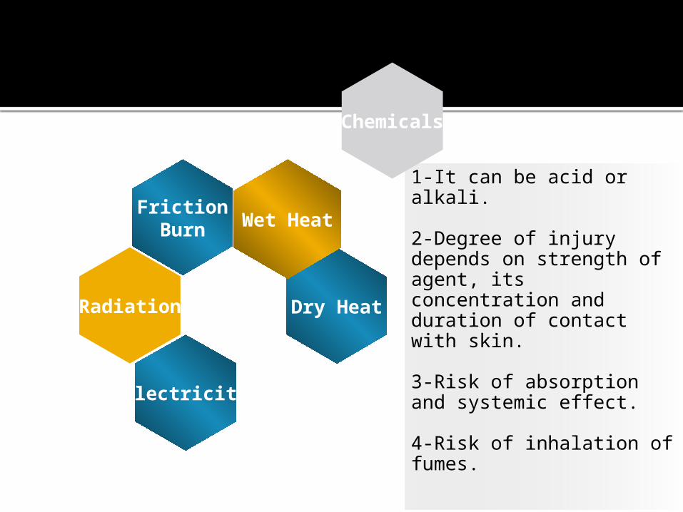
1-It can be acid or alkali.
2-Degree of injury depends on strength of agent, its concentration and duration of contact with skin.
3-Risk of absorption and systemic effect.
4-Risk of inhalation of fumes.
Chemicals
Dry Heat
Wet Heat
Electricity
FrictionBurn
Radiation
Acid Alkali
Coagulative necrosiswith a result of hard
tissue. The burns are clearly demarcated, dry and hard. Edema is mild
Lequifactive necrosispermitting the deeper
invasion of the tissue so the burns are deep with
marked edema
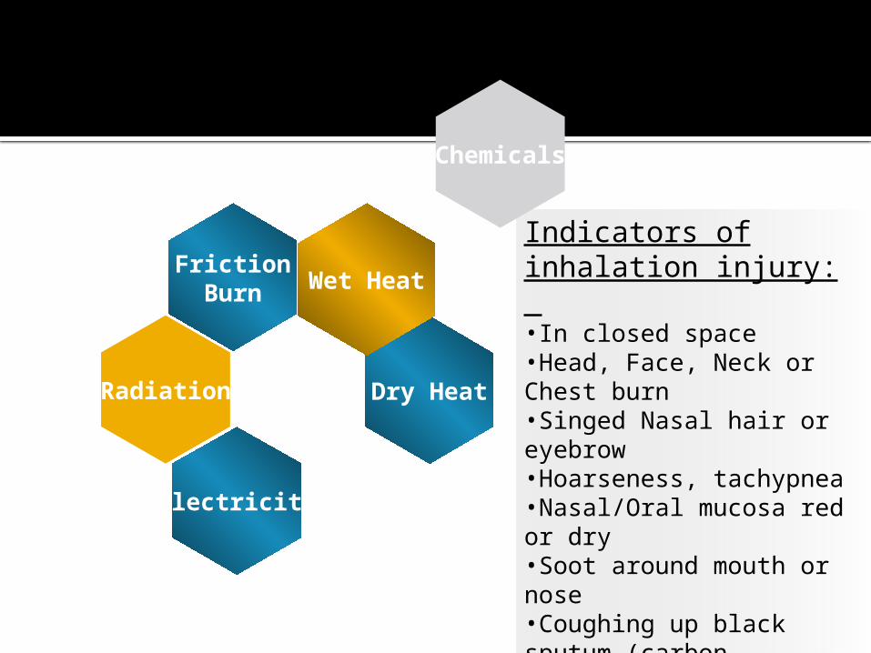
Indicators of inhalation injury: •In closed space •Head, Face, Neck or Chest burn•Singed Nasal hair or eyebrow •Hoarseness, tachypnea •Nasal/Oral mucosa red or dry•Soot around mouth or nose•Coughing up black sputum (carbon particle).
Chemicals
Dry Heat
Wet Heat
Electricity
FrictionBurn
Radiation
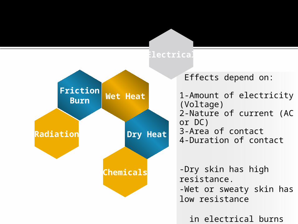
Effects depend on:
1-Amount of electricity (Voltage)2-Nature of current (AC or DC)3-Area of contact4-Duration of contact
-Dry skin has high resistance.-Wet or sweaty skin has low resistance
in electrical burns there is an entery wound (small) and an exit wound (large)
Electrical
Dry Heat
Wet Heat
Chemicals
FrictionBurn
Radiation
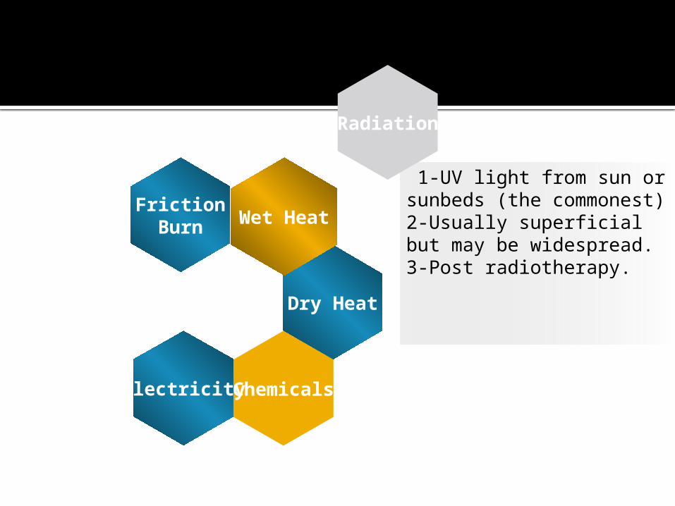
1-UV light from sun or sunbeds (the commonest)2-Usually superficial but may be widespread.3-Post radiotherapy.
Radiation
Dry Heat
Wet Heat
ChemicalsElectricity
FrictionBurn
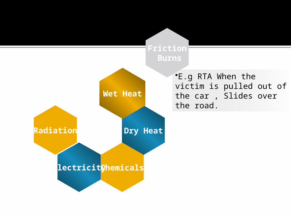
•E.g RTA When the victim is pulled out of the car , Slides over the road.
Friction Burns
Dry Heat
Wet Heat
ChemicalsElectricity
Radiation
Pathophysiology
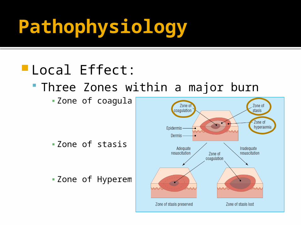
Local Effect: Three Zones within a major burn
▪ Zone of coagulation
▪ Zone of stasis
▪ Zone of Hyperemia
Pathophysiology
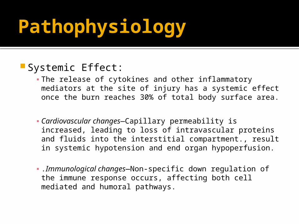
Systemic Effect:▪ The release of cytokines and other inflammatory
mediators at the site of injury has a systemic effect once the burn reaches 30% of total body surface area.
▪ Cardiovascular changes—Capillary permeability is increased, leading to loss of intravascular proteins and fluids into the interstitial compartment., result in systemic hypotension and end organ hypoperfusion.
▪ .Immunological changes—Non-specific down regulation of the immune response occurs, affecting both cell mediated and humoral pathways.
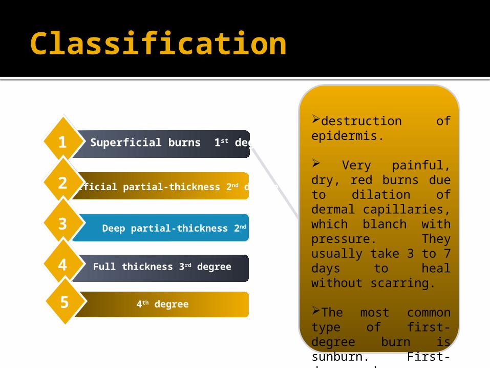
Classification
Superficial partial-thickness 2nd degree
Deep partial-thickness 2nd degree
11
22
33
44 Full thickness 3rd degree
Superficial burns 1st degree
55 4th degree
destruction of epidermis.
Very painful, dry, red burns due to dilation of dermal capillaries, which blanch with pressure. They usually take 3 to 7 days to heal without scarring.
The most common type of first-degree burn is sunburn. First-degree burns are limited to the epidermis, or upper layers of skin.
Classification
Superficial partial-thickness 2nd degree
Deep partial-thickness 2nd degree
11
22
33
44 Full thickness 3rd degree
Superficial burns 1st degree
55 4th degree
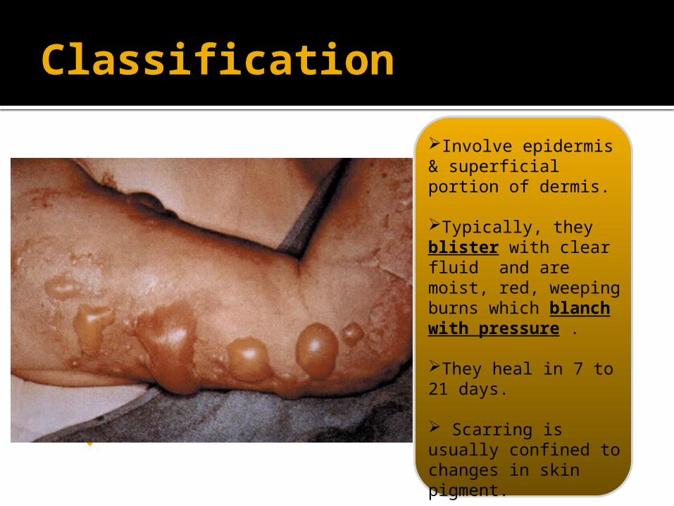
Involve epidermis & superficial portion of dermis. Typically, they blister with clear fluid and are moist, red, weeping burns which blanch with pressure .
They heal in 7 to 21 days.
Scarring is usually confined to changes in skin pigment.
Classification
Superficial partial-thickness 2nd degree
Deep partial-thickness 2nd degree
11
22
33
44 Full thickness 3rd degree
Superficial burns 1st degree
55 4th degree
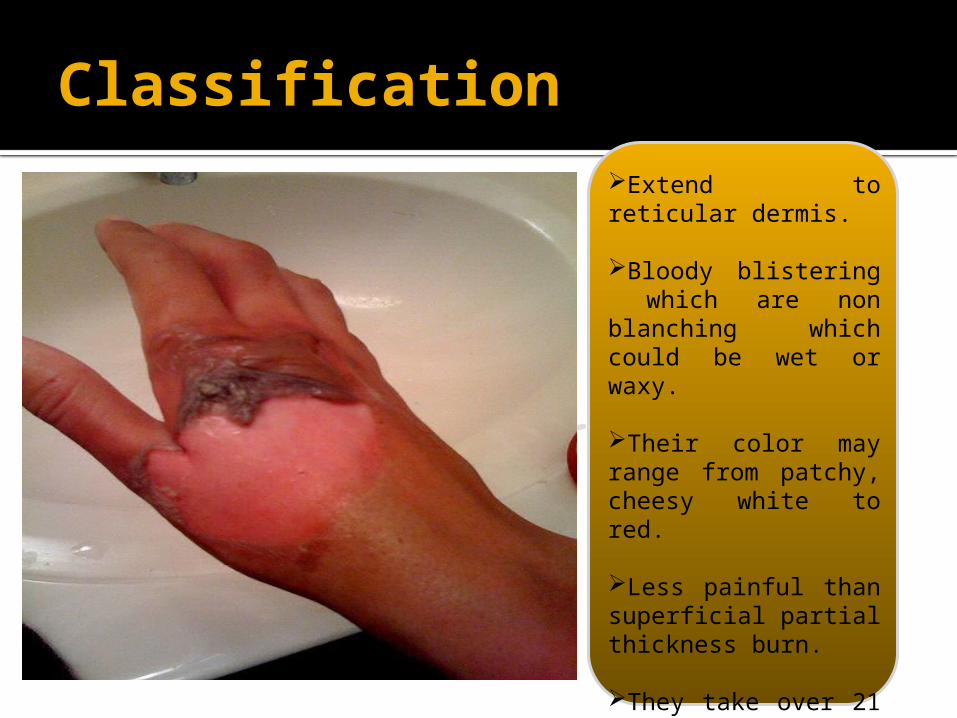
Extend to reticular dermis.
Bloody blistering which are non blanching which could be wet or waxy.
Their color may range from patchy, cheesy white to red.
Less painful than superficial partial thickness burn.
They take over 21 days to heal and scarring may be severe, May need grafting.
Classification
Superficial partial-thickness 2nd degree
Deep partial-thickness 2nd degree
11
22
33
44 Full thickness 3rd degree
Superficial burns 1st degree
55 4th degree
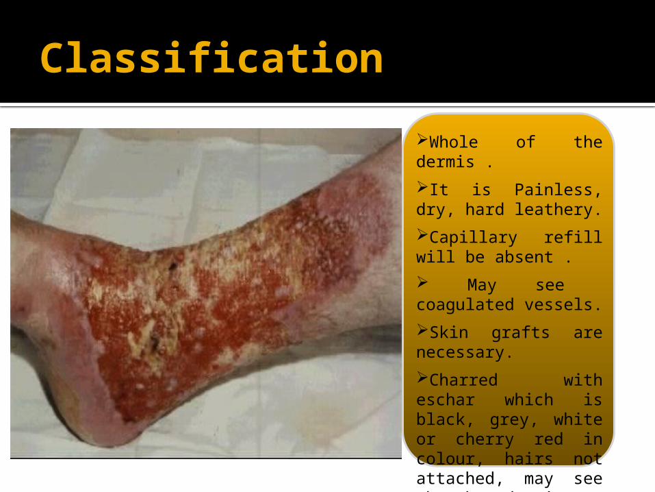
Whole of the dermis .
It is Painless, dry, hard leathery.
Capillary refill will be absent .
May see coagulated vessels.
Skin grafts are necessary.
Charred with eschar which is black, grey, white or cherry red in colour, hairs not attached, may see thrombosed veins.
Classification
Superficial partial-thickness 2nd degree
Deep partial-thickness 2nd degree
11
22
33
44 Full thickness 3rd degree
Superficial burns 1st degree
55 4th degree
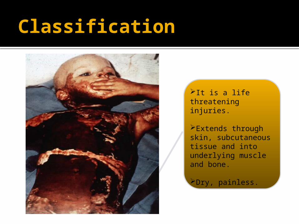
It is a life threatening injuries.
Extends through skin, subcutaneous tissue and into underlying muscle and bone.
Dry, painless.
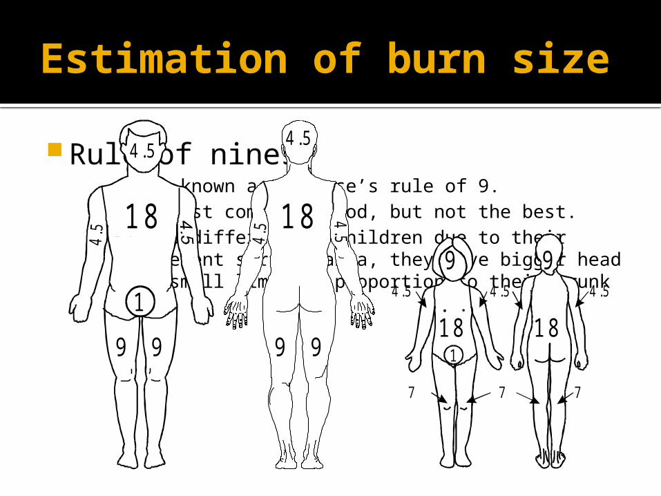
Rule of nines▪ Also known as Wallace’s rule of 9.▪ The most common method, but not the best.▪ It is different in children due to their different surface
area, they have bigger head and small limbs in proportion to their trunk
Estimation of burn size
18
4.5
9
1
9
4.5
184.5
4 .5
9 9
4.5
9 94 .5
7
4 .5
7
4 .5
7
18 181
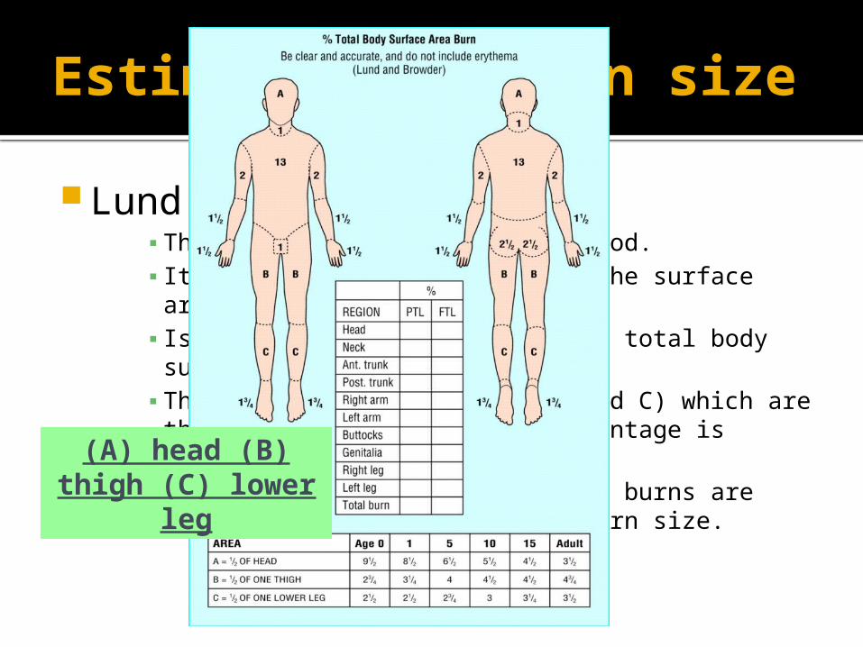
Estimation of burn size
Lund an Browder Chart▪ The best and most accurate method.▪ It considers the variation of the surface area
according to the age.▪ Is expressed as a percentage of total body surface
area.▪ There are 3 variables (A, B and C) which are the
areas that their size percentage is affected by growth.▪ Only partial and full thickness burns are included in
this estimate of burn size.
(A) head (B) thigh (C) lower
leg
Estimation of burn size
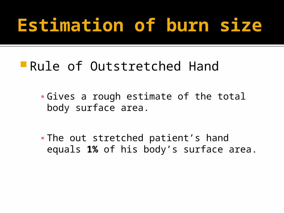
Rule of Outstretched Hand
▪ Gives a rough estimate of the total body surface area.
▪ The out stretched patient’s hand equals 1% of his body’s surface area.
Management
Resuscitation ABC’s a)Airway: ensure adequate airway.
b)Breathing: ▪ Circumferential burns of neck or chest may constrict breathing. ▪ Stridor or difficulty breathing indicates endotracheal intubation or ventilation . ▪ Prophylactic endotracheal/ nasotracheal intubation in case of:
inhalation Injury. supraglottic obstruction.extensive burns > 60%.deep facial burns. facial fracture. Closed head injury with
unconsciousness. c)Circulation:
Monitor : pulse, BP, failure to maintain adequate circulation may be followed by renal failure and eventually multi-organ failure.
Management
Hx The cause Time and place Age Any chronic illnesses, e.g. DM, HTN..etc Immunization for tetanus ( open
wounds), we give immunoglobulins for patients who have never been vaccinated
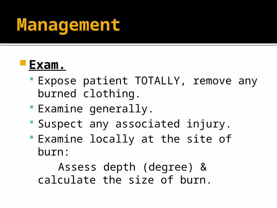
Management
Exam. Expose patient TOTALLY, remove any
burned clothing. Examine generally. Suspect any associated injury. Examine locally at the site of burn: Assess depth (degree) & calculate
the size of burn.
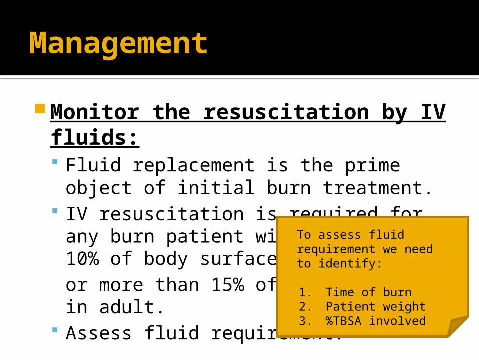
Management
Monitor the resuscitation by IV fluids: Fluid replacement is the prime object of
initial burn treatment. IV resuscitation is required for any burn
patient with; more than 10% of body surface in children or more than 15% of body surface in adult.
Assess fluid requirement.
To assess fluid requirement we need to identify:
1. Time of burn 2. Patient weight 3. %TBSA involved
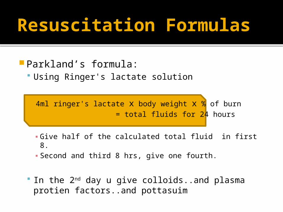
Parkland’s formula: Using Ringer's lactate solution
4ml ringer's lactate x body weight x % of burn = total fluids for 24 hours
▪ Give half of the calculated total fluid in first 8.▪ Second and third 8 hrs, give one fourth.
In the 2nd day u give colloids..and plasma protien factors..and pottasuim
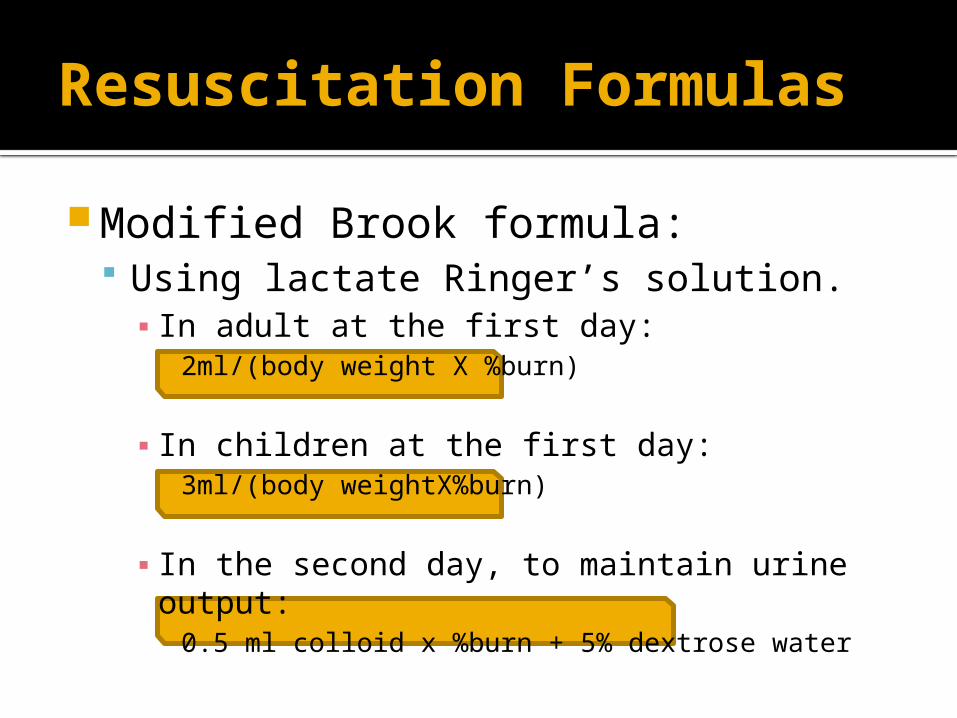
Resuscitation Formulas
Resuscitation Formulas
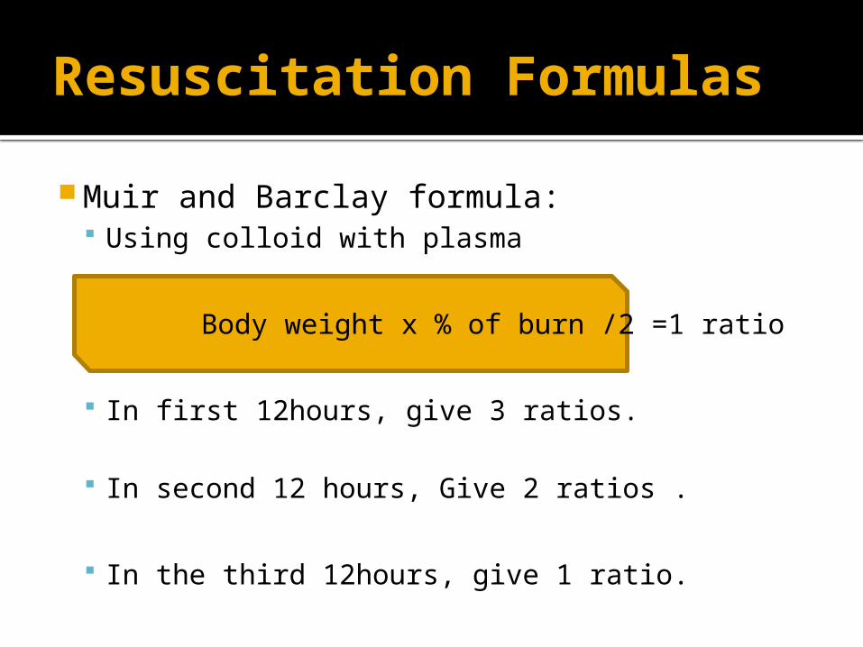
Muir and Barclay formula: Using colloid with plasma
Body weight x % of burn /2 =1 ratio
In first 12hours, give 3 ratios.
In second 12 hours, Give 2 ratios .
In the third 12hours, give 1 ratio.
Resuscitation Formulas
Modified Brook formula: Using lactate Ringer’s solution.▪ In adult at the first day:
2ml/(body weight X %burn)
▪ In children at the first day: 3ml/(body weightX%burn)
▪ In the second day, to maintain urine output:0.5 ml colloid x %burn + 5% dextrose water
Management
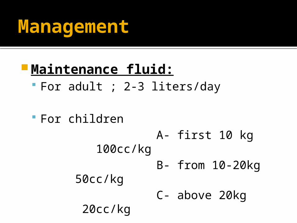
Maintenance fluid: For adult ; 2-3 liters/day
For children A- first 10 kg 100cc/kg B- from 10-20kg 50cc/kg C- above 20kg 20cc/kg
Management
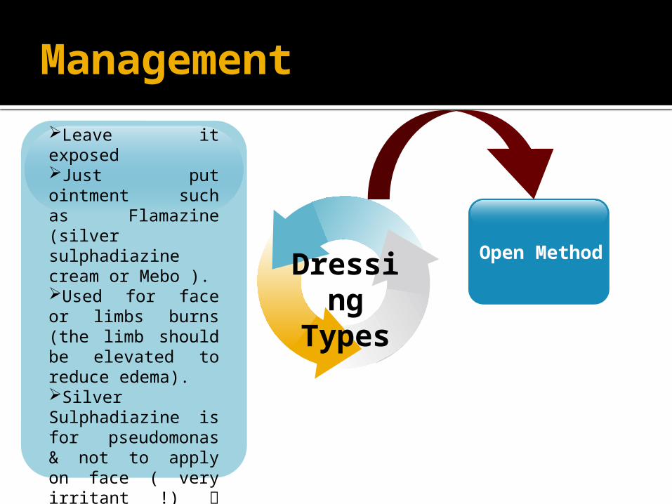
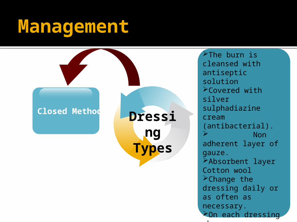
Dressing: The aim of the burn dressing is to keep
the wound clean and dry, and prevent infection
Two types.
Management
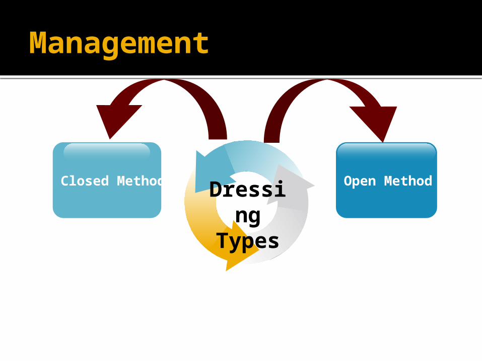
Closed Method Open MethodDressing
Types
Management
Dressing
Types
Leave it exposedJust put ointment such as Flamazine (silver sulphadiazine cream or Mebo ).Used for face or limbs burns (the limb should be elevated to reduce edema).Silver Sulphadiazine is for pseudomonas & not to apply on face ( very irritant !) use MEBO instead .Be careful for silver allergy( they will lose their skin).
Open Method
Management
Dressing
Types
The burn is cleansed with antiseptic solutionCovered with silver sulphadiazine cream (antibacterial). Non adherent layer of gauze.Absorbent layer Cotton woolChange the dressing daily or as often as necessary.On each dressing change, remove any loose tissue. Always use Closed dressing except : Face ,hand ,perineum.
Closed Method
Management
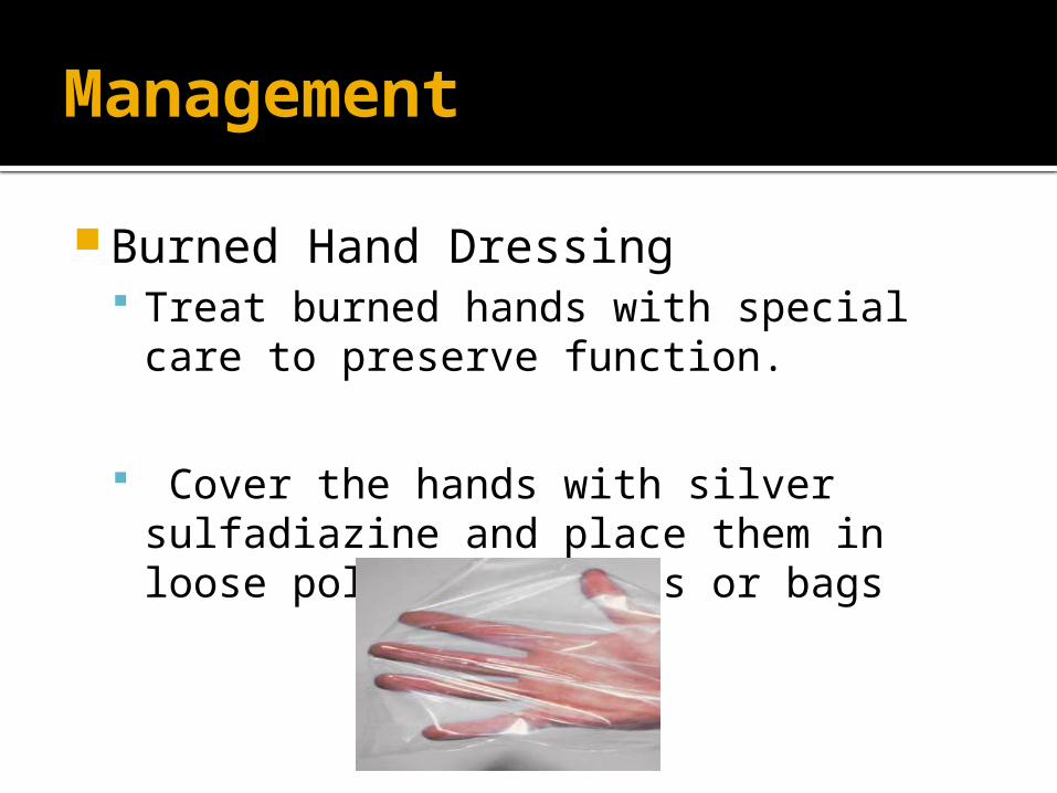
Burned Hand Dressing Treat burned hands with special care to
preserve function.
Cover the hands with silver sulfadiazine and place them in loose polythene gloves or bags
Management
Skin Graft Skin grafts are used in treating partial thickness and
full thickness burns Early surgical removal (excision or debridement) of
burned skin followed by skin grafting reduces the number of days in the hospital and usually improves the function and appearance of the burned area, especially when the face, hands, or feet are involved.
Role of grafting: Decrease evaporation & pain. Protects neurovascular tissue & tendons. Prevent facial desiccation & subsequent
infection. Prevent scarring ,contracture & deformity.
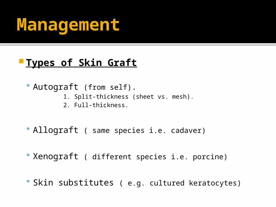
Management
Types of Skin Graft
Autograft (from self). 1. Split-thickness (sheet vs. mesh). 2. Full-thickness.
Allograft ( same species i.e. cadaver)
Xenograft ( different species i.e. porcine)
Skin substitutes ( e.g. cultured keratocytes)
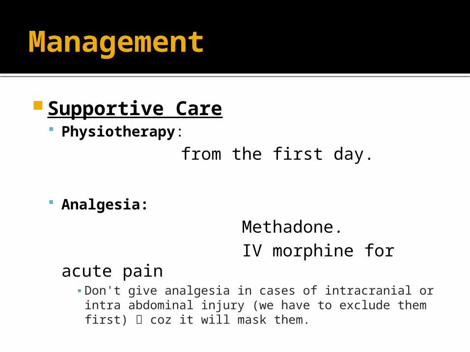
Management
Supportive Care Physiotherapy:
from the first day.
Analgesia:
Methadone. IV morphine for acute pain
▪ Don't give analgesia in cases of intracranial or intra abdominal injury (we have to exclude them first) coz it will mask them.
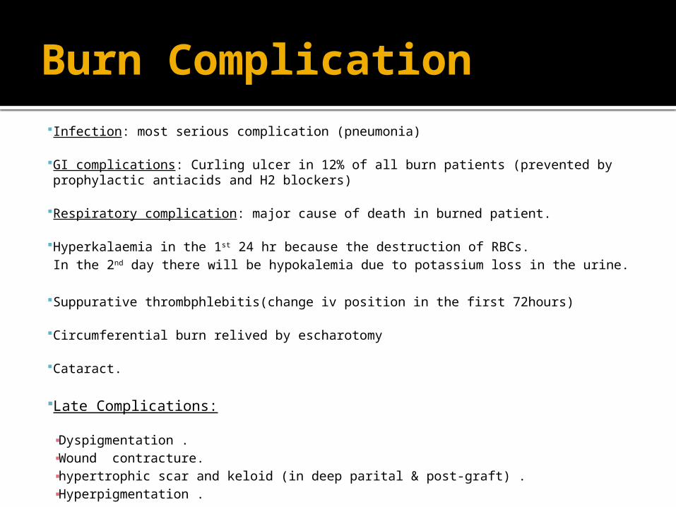
Burn Complication Infection: most serious complication (pneumonia)
GI complications: Curling ulcer in 12% of all burn patients (prevented by prophylactic antiacids and H2 blockers)
Respiratory complication: major cause of death in burned patient.
Hyperkalaemia in the 1st 24 hr because the destruction of RBCs.In the 2nd day there will be hypokalemia due to potassium loss in the urine.
Suppurative thrombphlebitis(change iv position in the first 72hours)
Circumferential burn relived by escharotomy
Cataract.
Late Complications:
▪ Dyspigmentation .▪ Wound contracture.▪ hypertrophic scar and keloid (in deep parital & post-graft) .▪ Hyperpigmentation .
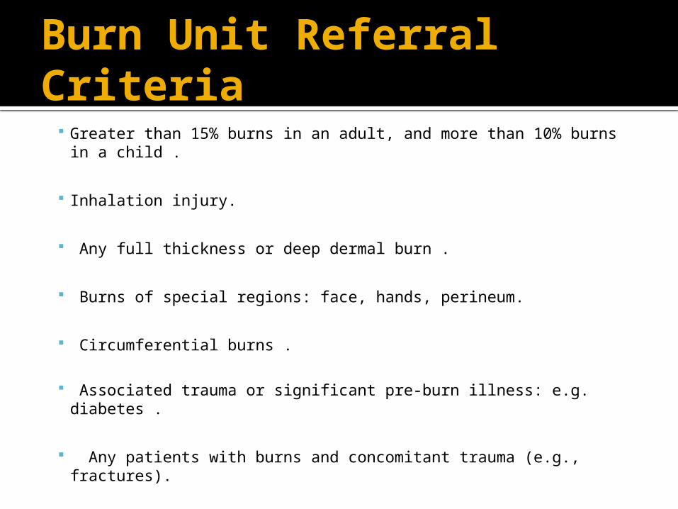
Burn Unit Referral Criteria
Greater than 15% burns in an adult, and more than 10% burns in a child .
Inhalation injury.
Any full thickness or deep dermal burn .
Burns of special regions: face, hands, perineum.
Circumferential burns .
Associated trauma or significant pre-burn illness: e.g. diabetes .
Any patients with burns and concomitant trauma (e.g., fractures).

Thank YouAny Questions