
D. Nitz and J. Siegel Am J Physiol Regulatory Integrative...
6
273:451-455, 1997. Am J Physiol Regulatory Integrative Comp Physiol D. Nitz and J. Siegel You might find this additional information useful... 11 other HighWire hosted articles, the first 5 are: This article has been cited by [PDF] [Full Text] [Abstract] , September 15, 2001; 21 (18): 7384-7391. J. Neurosci. Y.-Y. Lai, T. Kodama and J. M. Siegel Linked to Pontine Inhibition of Muscle Tone: An In Vivo Microdialysis Study Changes in Monoamine Release in the Ventral Horn and Hypoglossal Nucleus [PDF] [Full Text] [Abstract] , June 1, 2002; 22 (11): 4568-4576. J. Neurosci. J. Lu, A. A. Bjorkum, M. Xu, S. E. Gaus, P. J. Shiromani and C. B. Saper Eye Movement Sleep Selective Activation of the Extended Ventrolateral Preoptic Nucleus during Rapid [PDF] [Full Text] [Abstract] , July 1, 2002; 22 (13): 5282-5286. J. Neurosci. Peever and J. M. Siegel L. I. Kiyashchenko, B. Y. Mileykovskiy, N. Maidment, H. A. Lam, M.-F. Wu, J. John, J. Release of Hypocretin (Orexin) during Waking and Sleep States [PDF] [Full Text] [Abstract] , October 1, 2005; 94 (4): 2561-2574. J Neurophysiol S. Li, V. Varga, A. Sik and B. Kocsis Limbic System GABAergic Control of the Ascending Input From the Median Raphe Nucleus to the [PDF] [Full Text] [Abstract] R2041-R2049. , December 1, 2008; 295 (6): Am J Physiol Regulatory Integrative Comp Physiol J. John, L. Ramanathan and J. M. Siegel states in freely behaving rats Rapid changes in glutamate levels in the posterior hypothalamus across sleep-wake on the following topics: http://highwire.stanford.edu/lists/artbytopic.dtl can be found at Medline items on this article's topics Physiology .. Rapid-Eye-Movement Sleep Physiology .. Sleep Neuroscience .. Gaba Veterinary Science .. Raphe Nuclei Neuroscience .. Glycine Neuroscience .. Glutamate can be found at: Integrative and Comparative Physiology American Journal of Physiology - Regulatory, about Additional material and information http://www.the-aps.org/publications/ajpregu This information is current as of September 3, 2009 . http://www.the-aps.org/. the American Physiological Society. ISSN: 0363-6119, ESSN: 1522-1490. Visit our website at (monthly) by the American Physiological Society, 9650 Rockville Pike, Bethesda MD 20814-3991. Copyright © 2005 by biological organization, ranging from molecules to humans, including clinical investigations. It is published 12 times a year investigations that illuminate normal or abnormal regulation and integration of physiological mechanisms at all levels of publishes original The American Journal of Physiology - Regulatory, Integrative and Comparative Physiology on September 3, 2009 ajpregu.physiology.org Downloaded from
Transcript of D. Nitz and J. Siegel Am J Physiol Regulatory Integrative...
273:451-455, 1997. Am J Physiol Regulatory Integrative Comp PhysiolD. Nitz and J. Siegel
You might find this additional information useful...
11 other HighWire hosted articles, the first 5 are: This article has been cited by
[PDF] [Full Text] [Abstract]
, September 15, 2001; 21 (18): 7384-7391. J. Neurosci.Y.-Y. Lai, T. Kodama and J. M. Siegel
Linked to Pontine Inhibition of Muscle Tone: An In Vivo Microdialysis StudyChanges in Monoamine Release in the Ventral Horn and Hypoglossal Nucleus
[PDF] [Full Text] [Abstract], June 1, 2002; 22 (11): 4568-4576. J. Neurosci.
J. Lu, A. A. Bjorkum, M. Xu, S. E. Gaus, P. J. Shiromani and C. B. Saper Eye Movement Sleep
Selective Activation of the Extended Ventrolateral Preoptic Nucleus during Rapid
[PDF] [Full Text] [Abstract], July 1, 2002; 22 (13): 5282-5286. J. Neurosci.
Peever and J. M. Siegel L. I. Kiyashchenko, B. Y. Mileykovskiy, N. Maidment, H. A. Lam, M.-F. Wu, J. John, J.
Release of Hypocretin (Orexin) during Waking and Sleep States
[PDF] [Full Text] [Abstract], October 1, 2005; 94 (4): 2561-2574. J Neurophysiol
S. Li, V. Varga, A. Sik and B. Kocsis Limbic System
GABAergic Control of the Ascending Input From the Median Raphe Nucleus to the
[PDF] [Full Text] [Abstract]R2041-R2049.
, December 1, 2008; 295 (6):Am J Physiol Regulatory Integrative Comp PhysiolJ. John, L. Ramanathan and J. M. Siegel
states in freely behaving ratsRapid changes in glutamate levels in the posterior hypothalamus across sleep-wake
on the following topics: http://highwire.stanford.edu/lists/artbytopic.dtl
can be found at Medline items on this article's topics
Physiology .. Rapid-Eye-Movement Sleep Physiology .. Sleep Neuroscience .. Gaba Veterinary Science .. Raphe Nuclei Neuroscience .. Glycine Neuroscience .. Glutamate
can be found at: Integrative and Comparative PhysiologyAmerican Journal of Physiology - Regulatory,about Additional material and information
http://www.the-aps.org/publications/ajpregu
This information is current as of September 3, 2009 .
http://www.the-aps.org/.the American Physiological Society. ISSN: 0363-6119, ESSN: 1522-1490. Visit our website at (monthly) by the American Physiological Society, 9650 Rockville Pike, Bethesda MD 20814-3991. Copyright © 2005 by biological organization, ranging from molecules to humans, including clinical investigations. It is published 12 times a yearinvestigations that illuminate normal or abnormal regulation and integration of physiological mechanisms at all levels of
publishes originalThe American Journal of Physiology - Regulatory, Integrative and Comparative Physiology
on Septem
ber 3, 2009 ajpregu.physiology.org
Dow
nloaded from

GABA release in the dorsal raphe nucleus: role in the control of REM sleep
DOUGLAS NITZl AND JEROME SIEGEL2 Departments of lNeuroscience and 2Psychiatry, University of California, Los Angeles 90024; and lp2Veteran’s Affairs Medical Center, Sepulveda, California 91343
Nitz, Douglas, and Jerome Siegel. GABA release in the dorsal raphe nucleus: role in the control of REM sleep. Am. J. Physiol. 273 (Regulatory Integrative Comp. Physiol. 42): R451-R455, 1997.-The cessation of firing of serotonergic dorsal raphe neurons is a key controlling event of rapid eye movement (REM) sleep. We tested the hypothesis that this cessation of activity is due to y-aminobutyric acid (GABA) release using the in vivo microdialysis technique. We found that REM sleep is accompanied by a selective increase in GABA release, but not by a change in glutamate or glycine release in the dorsal raphe nucleus. Microinjection of the GABA agonist muscimol into the dorsal raphe increased REM sleep, although microperfusion of the GABA antagonist picro- toxin blocked REM sleep. These results implicate GABA release as a critical element in the production of the REM sleep state and in the control of discharge in serotonergic neurons across the sleep/wake cycle.
microdialysis; muscimol; picrotoxin; serotonin
THE SEROTONERGIC NEURONS of the brain stem dorsal raphe (DR) nucleus innervate multiple neocortical, hippocampal, diencephalic, and brain stem regions of the mammalian brain (3, 33, 37, 43). The activity of serotonergic neurons has been implicated in the control of shifts between states of sleep, nociception, cortical desynchronization, and normal and abnormal emo- tional states (38, 42). The greatest change in the discharge rate of DR cells occurs across the sleep/wake cycle. In the freely behaving cat, serotonergic neurons fire tonically at a rate of 3-6 Hz throughout waking and at l-2 Hz during slow-wave sleep (SWS). During rapid eye movement (REM) sleep, serotonergic neurons ex- hibit a near-complete cessation of activity (23,25).
The decreased activity of serotonergic neurons dur- ing REM sleep is accompanied by a decrease in seroto- nin release in several brain areas implicated in the production of sleep including the hypothalamus and pontine reticular formation (PRF) (15, 28, 44). Evi- dence from several laboratories suggests that the inac- tivity of DR serotonergic neurons disinhibits neuronal mechanisms responsible for the production of REM sleep. Thus blockade of DR neuronal activity by local cooling has been shown to induce REM sleep (6), and electrical stimulation of the DR blocks REM sleep (5). An increase in REM sleep time occurs with continuous perfusion of the DR with %hydroxy-2(di-n-propylamino)-
tetralin, a compound that inhibits the activity of DR serotonergic cells by activation of 5hydroxytryptamine la (5-HT1,) autoreceptors (29). The cessation of activity of serotonergic neurons is required for the generation of ponto-geniculo-occipital (PGO) spikes, a key phasic event of REM sleep (6, 18, 36). Recent evidence sug- gests that serotonin modulates the production of PGO spikes through actions in the amygdala (32) as well as through inhibition of brain stem cholinergic neurons, which are known to fire in conjunction with PGO waves (20,22,39).
The neurochemical mechanism underlying the cessa- tion of DR serotonergic unit activity during REM sleep is unknown. It is unlikely that serotonergic inhibition of DR serotonergic cells through an autoreceptor mech- anism is responsible, because the concentration of serotonin in the DR is lowest during REM sleep (28). In addition, data suggest that autoinhibitory mechanisms of serotonergic DR cells are significantly activated only during waking behaviors (8). Disfacilitation by removal of an excitatory noradrenergic or glutamatergic input could contribute to reduced discharge of DR units during sleep. However, serotonergic DR neurons ex- hibit pacemaker potentials in the anesthetized rat (1) and are unresponsive to excitatory input during REM sleep (23). Therefore, we hypothesized that the cessa- tion of activity in serotonergic DR neurons during REM sleep is mediated by active inhibition.
Both y-aminobutyric acid (GABA) and glycine po- tently inhibit serotonergic DR neurons (10, 11, 26). GABAergic and glycinergic neurons densely innervate the serotonergic neurons of the DR (9,12). A glycinergic mechanism mediates the characteristic muscle atonia of REM sleep through active inhibition of spinal moto- neurons (7). We hypothesized that GABA and/or glycine are selectively released on serotonergic neurons during REM sleep. We tested this hypothesis by developing techniques for the measurement of GABA and glycine release in the DR across sleep/wake states with in vivo microdialysis and high-performance liquid chromatog- raphy (HPLC).
METHODS
Adult mongrel cats served as subjects. Animals were anesthetized with pentobarbitol sodium (35 mg/kg ip). Screw electrodes were placed in the posterior orbit and sensorimotor cortex for the recording of eye movements and electroencepha-
R451
on Septem
ber 3, 2009 ajpregu.physiology.org
Dow
nloaded from
R452 GABA RELEASE IN THE RAPHE
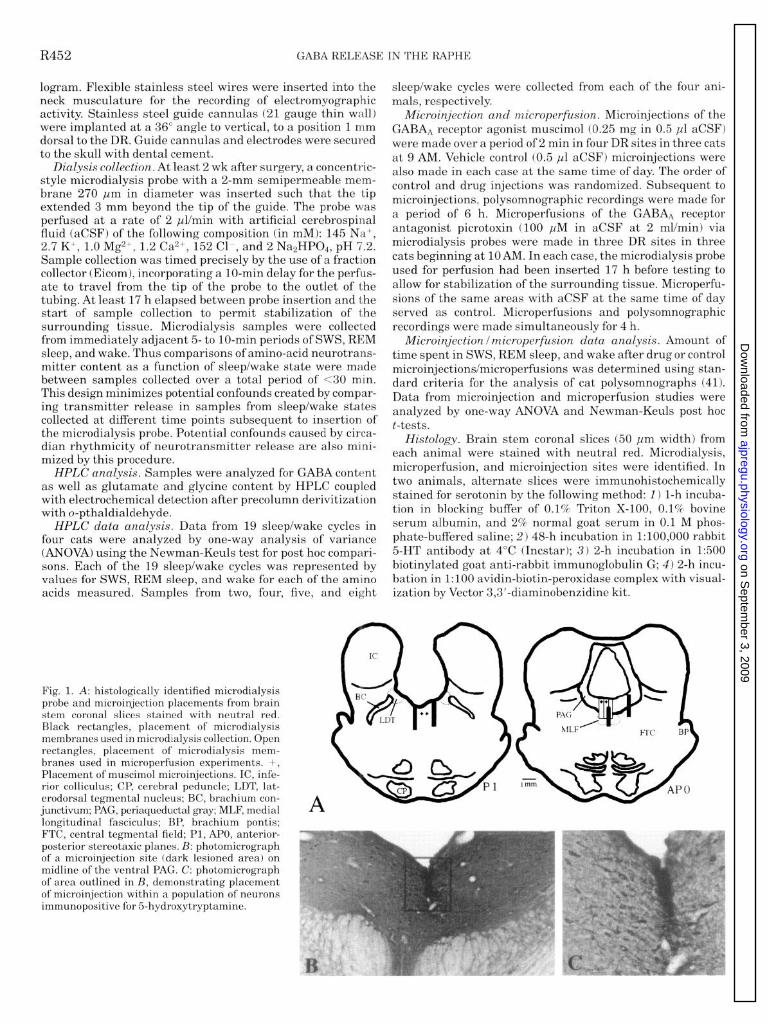
logram. Flexible stainless steel wires were inserted into the neck musculature for the recording of electromyographic activity. Stainless steel guide cannulas (21 gauge thin wall) were implanted at a 36” angle to vertical, to a position 1 mm dorsal to the DR. Guide cannulas and electrodes were secured to the skull with dental cement.
Dialysis collection. At least 2 wk after surgery, a concentric- style microdialysis probe with a 2-mm semipermeable mem- brane 270 pm in diameter was inserted such that the tip extended 3 mm beyond the tip of the guide. The probe was perfused at a rate of 2 pl/min with artificial cerebrospinal fluid (aCSF) of the following composition (in mM): 145 Na+, 2.7 K+, 1.0 Mg2+, 1.2 Ca2+, 152 Cl-, and 2 NasHPOb, pH 7.2. Sample collection was timed precisely by the use of a fraction collector (Eicom), incorporating a lo-min delay for the perfus- ate to travel from the tip of the probe to the outlet of the tubing. At least 17 h elapsed between probe insertion and the start of sample collection to permit stabilization of the surrounding tissue. Microdialysis samples were collected from immediately adjacent 5- to lo-min periods of SWS, REM sleep, and wake. Thus comparisons of amino-acid neurotrans- mitter content as a function of sleep/wake state were made between samples collected over a total period of ~30 min. This design minimizes potential confounds created by compar- ing transmitter release in samples from sleep/wake states collected at different time points subsequent to insertion of the microdialysis probe. Potential confounds caused by circa- dian rhythmicity of neurotransmitter release are also mini- mized by this procedure.
HPLC analysis. Samples were analyzed for GABA content as well as glutamate and glycine content by HPLC coupled with electrochemical detection after precolumn derivitization with o-pthaldialdehyde.
HPLC data analysis. Data from 19 sleep/wake cycles in four cats were analyzed by one-way analysis of variance (ANOVA) using the Newman-Keuls test for post hoc compari- sons. Each of the 19 sleep/wake cycles was represented by values for SWS, REM sleep, and wake for each of the amino acids measured. Samples from two, four, five, and eight
sleep/wake cycles were collected from each of the four ani- mals, respectively.
Microinjection and microperfusion. Microinjections of the GABAA receptor agonist muscimol (0.25 mg in 0.5 ~1 aCSF) were made over a period of 2 min in four DR sites in three cats at 9 AM. Vehicle control (0.5 ,~l aCSF) microinjections were also made in each case at the same time of day. The order of control and drug injections was randomized. Subsequent to microinjections, polysomnographic recordings were made for a period of 6 h. Microperfusions of the GABAA receptor antagonist picrotoxin (100 ,uM in aCSF at 2 mUmin) via microdialysis probes were made in three DR sites in three cats beginning at 10 AM. In each case, the microdialysis probe used for perfusion had been inserted 17 h before testing to allow for stabilization of the surrounding tissue. Microperfu- sions of the same areas with aCSF at the same time of day served as control. Microperfusions and polysomnographic recordings were made simultaneously for 4 h.
Microinjection lmicroperfusion data analysis. Amount of time spent in SWS, REM sleep, and wake after drug or control microinjections/microperfusions was determined using stan- dard criteria for the analysis of cat polysomnographs (41). Data from microinjection and microperfusion studies were analyzed by one-way ANOVA and Newman-Keuls post hoc t-tests.
Histology. Brain stem coronal slices (50 pm width) from each animal were stained with neutral red. Microdialysis, microperfusion, and microinjection sites were identified. In two animals, alternate slices were immunohistochemically stained for serotonin by the following method: 1) l-h incuba- tion in blocking buffer of 0.1% Triton X-100, 0.1% bovine serum albumin, and 2% normal goat serum in 0.1 M phos- phate-buffered saline; 2) 48-h incubation in l:lOO,OOO rabbit 5HT antibody at 4°C (Incstar); 3) 2-h incubation in 1:500 biotinylated goat anti-rabbit immunoglobulin G; 4) 2-h incu- bation in 1:lOO avidin-biotin-peroxidase complex with visual- ization by Vector 3,3’-diaminobenzidine kit.
Fig. 1. A: histologically identified microdialysis probe and microinjection placements from brain stem coronal slices stained with neutral red. Black rectangles, placement of microdialysis membranes used in microdialysis collection. Open rectangles, placement of microdialysis mem- branes used in microperfusion experiments. +, Placement of muscimol microinjections. IC, infe- rior colliculus; CP, cerebral peduncle; LDT, lat- erodorsal tegmental nucleus; BC, brachium con- junctivum; PAG, periaqueductal gray; MLF, medial longitudinal fasciculus; BP, brachium pontis; FTC, central tegmental field; Pl, APO, anterior- posterior stereotaxic planes. B: photomicrograph of a microinjection site (dark lesioned area) on midline of the ventral PAG. C: photomicrograph of area outlined in B, demonstrating placement of microinjection within a population of neurons immunopositive for 5hydroxytryptamine.
A
on Septem
ber 3, 2009 ajpregu.physiology.org
Dow
nloaded from
GABA RELEASE IN THE RAPHE R453
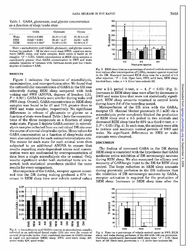
Table 1. GABA, glutamate, and glycine concentration as a function of sleep I wake state
GABA Glutamate Glycine
Wake 0.042 5 0.005 35.84 + 2.55 32.30 2 4.29 sws 0.049 5 0.007 36.42 2 4.57 34.05 t 4.80 REM 0.072 t 0.003* 33.98 5 2.03 33.89 5 4.11
Mean y-aminobutyric acid (GABA), glutamate, and glycine concen- tration (in pmol/pl) t SE for slow-wave sleep (SWS), rapid eye move- ment (REM) sleep, and wake samples. Each mean is based on 19 samples. *P < 0.001, GABA concentration in REM sleep samples is significantly greater than GABA concentration in SWS and wake samples (analysis of variance with Newman-Keuls post hoc t-tests, degrees of freedom = 2,54).
RESULTS
Figure 1 indicates the locations of microdialysis, microinjection, and microperfusion sites. We found that the extracellular concentration of GABA in the DR rose selectively during REM sleep compared with both waking and SWS (ANOVA, degrees of freedom 2,51, P < 0.001). GABA levels were similar during wake and SWS sleep. Overall, GABA concentration in REM sleep samples was found to be 47 and 71% greater than in SWS and wake samples, respectively. No significant differences in release of glutamate or glycine as a function of state were found. Table 1 lists the concentra- tions of the three compounds as a function of sleep/ wake state. Figure 2 depicts GABA content in microdi- alysis samples collected from an individual DR site over the course of several sleep/wake cycles. Mean values for GABA concentration as a function of sleep/wake state were also calculated for each animal/microdialysis site. The means for each state for each animal were then subjected to an additional ANOVA to ensure that results regarding state-dependent amino acid concen- tration were not confounded by overrepresentation of data from a single microdialysis site or animal. Only results significant under both statistical tests are re- ported; both statistical procedures yielded the same results for every comparison.
Microinjection of the GABA* receptor agonist musci- mol into the DR during waking produced a 67% in- crease in REM sleep time over vehicle control values
& 0.07
5 0.06 &I o . i3 0.05
2 0.04
$ 0.03
; 0.02
-g 0.01
a 0
Fig. 2. y-Aminobutyric acid (GABA) content in microdialysis samples collected in an individual dorsal raphe (DR) site over the course of several sleep/wake cycles. Note consistent increase in GABA release in rapid eye movement (REM) sleep. SWS, slow-wave sleep; AW, active wake; QW, quiet wake.
400 I I I
o-2 2-4 Hours post-injection
4-6
Fig. 3. REM sleep time as a percentage of control (vehicle microinjec- tion) values after microinjection of GABAA receptor agonist muscimol in the DR. Muscimol increased REM sleep time for a period of 2 h after injection. *P < 0.03. Open bars, SWS; solid bars, REM sleep; hatched bars, wake; 71 = 4. Error bars indicate SE.
over a 2-h period (t-test, n = 4, P c 0.03) (Fig. 3). Increases in REM sleep time were offset by decreases in SWS and wake time that were not statistically signifi- cant. REM sleep amounts returned to control levels during hours 3-6 of the recording period.
Microperfusion of the DR area with the GABAA receptor Cl- channel blocker picrotoxin (0.1 mM) via a microdialysis probe completely blocked the production of REM sleep over a 4-h period in two animals and decreased REM sleep time by 83% in a third (t-test, n = 3, P < 0.05) (Fig. 4). In each case, the animals were able to initiate and maintain normal periods of SWS and wake. No significant differences in SWS or wake amounts were observed.
DISCUSSION
The finding of increased GABA in the DR during REM sleep is consistent with the hypothesis that GABA mediates active inhibition of DR serotonergic neurons during REM sleep. We also examined the efficacy and necessity of GABAergic input to the DR for REM sleep control. The large reduction in REM sleep amounts during picrotoxin perfusion in the DR indicates that the inhibition of DR serotonergic neurons by GABAA receptor activation is required for the production of REM sleen. Increases in REM sleen time after the
180
160 T
140
“0 120
kJ 8
100
o 80 @ 60
40
20
0 b
T
sws REM WAKE
Fig. 4. Time (as a percentage of vehicle control) spent in SWS, REM sleep, and wake during perfusion of the DR with 100 pm picrotoxin. Picrotoxin significantly decreased REM sleep time. *P < 0.05. Open bars, aCSF; black bars, picrotoxin; IL = 3. Error bars indicate SE.
on Septem
ber 3, 2009 ajpregu.physiology.org
Dow
nloaded from

R454 GABA RELEASE IN THE RAPHE
microinjection of the GABA* receptor agonist muscimol are consistent with our hypothesis that the inhibition of DR serotonergic cells by a GABAergic mechanism is permissive to and facilitates REM sleep production.
Our microdialysis data suggest that GABAergic neu- rons, within or projecting to the DR, are selectively active during REM sleep. The location of neurons mediating the increased GABA release in the DR during REM sleep remains undetermined. Both intrin- sic and extrinsic sources of GABA innervation in the DR nucleus are possible origins of state-dependent GABA release. Fos immunolabeling indicates that small, nonserotonergic, possibly GABAergic, neurons of the DR are activated after the induction of a REM sleep- like state with microinjection of carbachol in the PRF (34,45). Aghajanian et al. (2) identified a population of nonserotonergic cells in the dorsal raphe that exhibited changes in firing opposite that of serotonergic cells in response to peripheral nerve stimulation. However, unit recording studies of the DR have not revealed units whose activity across the sleep/wake cycle paral- lel the changes we observed in GABA release. Thus GABA release selective to REM sleep could arise from small DR neurons not easily recorded with conven- tional extracellular recording techniques or from neu- rons external to the DR. There are GABAergic projec- tions to the DR from the preoptic and posterior hypothalamus, the periaqueductal gray, the midbrain and PRF, the peribrachial region, and the prepositus hypoglossal nucleus (9,12). It is likely that the source of state-dependent GABA release in the DR is caudal to the midbrain because DR units have been demon- strated to retain their REM-off pattern of firing in decerebrate cats (14).
Neurons discharging selectively during REM sleep have been identified in the dorsolateral PRF (35). A population of GABAergic cells has been identified in this same region (17). The dorsolateral PRF sends projections to the DR, some of which are GABAergic (4, 27). Together with the GABA release data presented herein, these findings suggest that some PRF REM-on neurons may be GABAergic.
REM sleep behavior disorder in humans is character- ized by fragmented REM sleep as well as a disruption of muscle atonia within REM sleep (24). It resembles a syndrome seen after lesions of the dorsolateral pons in the cat (13, 18). In contrast to normal REM sleep, serotonergic neurons are not completely silent during REM sleep in the REM sleep without atonia syndrome (40). REM sleep behavior disorder is successfully treated with clonazepam, a benzodiazepine that potentiates GABAA receptors and has been shown to decrease the use of serotonin in the mouse brain (30). This suggests that a disruption of the output of GABAergic PRF neurons projecting to the raphe nuclei could underlie this disorder of REM sleep.
Microiontophoretic application of the GABAA recep- tor antagonist bicuculline (Bit) reduces the decrease in DR unit discharge in SWS but not in REM sleep (21). We found no significant increase in GABA release in SWS. However, a small increase in the release of
GABA, possibly beyond the resolution of the microdialy- sis technique, might be sufficient to reduce DR unit discharge in SWS. This interpretation is consistent with the report by Lydic et al. (23) indicating greatly decreased response of DR cells to excitatory input in REM sleep compared with SWS, despite a relatively small decrease in discharge rate from SWS to REM sleep. Greatly increased levels of GABA release would not be reflected in DR unit discharge because DR discharge in REM sleep falls to zero from the already minimal SWS rates. Our microdialysis and microperfu- sion data provide strong evidence that GABA mediates the suppression of serotonergic unit discharge during REM sleep. Indeed, as suggested by Levine and Jacobs (21), the inability of iontophoresed Bit to reverse REM sleep cessation of DR unit discharge could be due to incomplete antagonism of DR GABAA receptors as a result of increased GABA release. The results of the present study are compatible with their hypothesis. This interpretation is also consistent with the blockade of REM sleep production that we observed with picro- toxin perfusion. The antagonism of GABAergic activity in the DR by picrotoxin would not be blocked by increased GABA concentration because picrotoxin acts at the GABAA receptor Cl- channel rather than compet- ing with GABA at the GABAA receptor binding site, as is the case for Bit (19).
In summary, we have demonstrated that GABA release in the DR is increased during REM sleep. Alterations in the production of REM sleep after the microinjection of a GABAA receptor agonist and antago- nist into the DR are consistent with the hypothesis that inhibition of serotonergic DR neurons by a GABAergic mechanism is a key controlling event of REM sleep. Our findings suggest that a group of REM-on neurons responsible for initiating REM sleep is GABAergic.
This research was supported by the Medical Research Service of the Department of Veteran’s Affairs and National Institutes of Health Grants NS-14610, NS-23724, and HL-41370.
These results have been reported in preliminary form (D. A. Nitz and J. M. Siegel. Wbrld Fed. Sleep Res. Sot. Abs. 24A: 49,1995).
Present address for D. Nitz: ARL Division of NSMA, University of Arizona, Life Sciences North Bldg., Suite 384, Tucson, AZ 85724.
Address for reprint requests: J. Siegel, Neurobiology Research, VAMC (151A3), 16111 Plummer St., Sepulveda, CA91343.
Received 21 January 1997; accepted in final form 1 April 1997.
REFERENCES
Aghajanian, G. K., and C. P. Vandermaelen. Intracellular recordings from serotonergic dorsal raphe neurons: pacemaker potentials and the effect of LSD. Brain Res. 238: 463-469,1982. Aghajanian, G. K., R. Y. Wang, and J. Baraban. Serotonergic and non-serotonergic neurons of the dorsal raphe: reciprocal changes in firing induced by peripheral nerve stimulation. Brain Res. 153: 169-175,1978. Azmitia, E. C., and M. Segal. An autoradiographic analysis of the differential ascending projections of the dorsal and medial raphe nuclei in the rat. J. Comp. Neurol. 179: 641-668,1978. Belin, M. F., M. Aguera, M. Tappaz, A. McRae-Degueurce, P. Bobillier, and J. F. Pujol. GABA-accumulating neurons in the nucleus raphe dorsalis and periaqueductal gray in the rat: a biochemical and radioautgraphic study. Brain Res. 170: 279- 297,1979.
on Septem
ber 3, 2009 ajpregu.physiology.org
Dow
nloaded from
GABA RELEASE IN THE RAPHE R455
5.
6.
7.
8.
9.
10.
11.
12.
13.
14.
15.
16.
17
18
19.
20.
21.
22.
23.
24.
25.
Benington, J. H., M. C. Woudenberg, and H. C. Heller. REM-sleep propensity accumulates during 2-h REM-sleep depri- vation in the rest period in rats. Neurosci. Lett. 180: 76-80,1994. Cespuglio, R., M. E. Gomez, E. Walker, and M. Jouvet. Effet du refroidissement et de la stimulation des noyaux du systemes du raphe sur les etats de vigilance chez le chat. Electroencepha- logr Clin. Neurophysiol. 3: 221-227,1979. Chase, M. H., and F. R. Morales. PrincipZes and Practice of SZeep Medicine, edited by M. Kryger, T. Roth, and W. C. Dement. New York: Saunders, 1989, p. 74-85. Fornal, C. A., W. J. Litto, C. W. Metzler, and F. Marrosu. Single unit responses of dorsal raphe neurons to 5-HTlA agonist and antagonist drug administration in behaving cats. J. Pharma- col. Exp. Ther. 270: 1345-1358,1994. Fort, P., P. H. Luppi, and M. Jouvet. Glycine-immunoreactive neurones in the cat brainstem reticular formation. Neuroreport 4: 1123-1126,1993. Gallager, D. W., and G. K. Aghajanian. Effect of antipsychotic drugs on the firing of dorsal raphe cells. II. Reversal by picro- toxin. Eur. J. Pharmacol. 39: 357-364,1976. Gallager, D. W., J. M. Lakoski, S. F. Gonsalves, and S. L. Rauch. Chronic benzodiazepine treatment decreases postsynap- tic GABA sensitivity. Nature 308: 74-77,1984. Harandi, RI., M. Aguera, H. Gamrani, M. Didier, M. Maitre, A. Calas, and M. F. Belin. Gamma-amino-butyric acid and 5-hydroxytryptamine interrelationship in the rat nucleus raphe dorsalis: combination of radioautographic and immunocytochemi- cal techniques at light and electron microscopy levels. Neurosci- ence 21:237-251,1987. Henley, K., and A. R. Morrison. Are-evaluation of the effects of lesions of the pontine tegmentum and locus coeruleus on phenom- ena of paradoxical sleep in the cat. Acta Neurobiol. Exp. (War& 34:215-232,1974. Hoshino, K., and 0. Pompeiano. Tonic inhibition of dorsal pontine neurons during the postural atonia produced by an anticholinesterase in the decerebrate cat. Arch. ItaL. Biol. 114: 310-340,1976. Iwakiri, H., K. Matsuyama, and S. Mori. Extracellular levels of serotonin in the medial pontine reticular formation in relation to sleep-wake cycle in cats: a microdialysis study. Neurosci. Res. 18:157-170,1993. Jacobs, B. L., L. 0. Wilkinson, and C. A. Fornal. The role of brain serotonin. A neurophysiologic perspective. Neuropsycho- pharmacology 3: 473-479,199O. Jones, B. E. Noradrenergic locus coeruleus neurons: their distant connections and their relationship to neighboring (includ- ing choline& and GABAergic) neurons of the central grey and reticular formation. Prog. Brain Res. 88: 15-30,199l. Jouvet, M., and J. F. Delorme. Supression du sommeil par le parachlorometamphetamine et la parachlorophenylalanine. C. R. SocBioZ. 160:2347-2350,1966. Kardos, J. The GABAA receptor channel mediated chloride ion translocation through the plasma membrane: new insights for 36C1- ion flux measurements. Synapse 13: 74-93,1993. Leonard, C. S., and R. Llinas. Serotonergic and cholinergic inhibition of mesopontine cholinergic neurons controlling REM sleep: an in vitro electrophysiological study. Neuroscience 59: 309-330,1994. Levine, E. S., and B. L. Jacobs. Neurochemical afferents controlling the activity of serotonergic neurons in the dorsal raphe nucleus: microiontophoretic studies in the awake cat. Neuroscience 12: 4037-4044,1992. Luebke, J. I., R. W. Greene, K. Semba, A. Kamondi, R. W. McCarley, and P. B. Reiner. Serotonin hyperpolarizes choliner- gic low-threshold burst neurons in the rat laterodorsal tegmental nucleus in vitro. Proc. NatZ. Acad. Sci. USA 89: 743-747,1992. Lydic, R., R. W. McCarley, and J. A. Hobson. Serotonin neurons andsleep. II. Time course of dorsal raphe discharge. Neurosci. Lett. 38: 35-40, 1983. Mahowald, M., and C. H. Schenck. Principles and Practice of SZeep Medicine, edited by M. Kryger, T. Roth, and W. C. Dement. New York: Saunders, 1989, p. 389-401. McGinty, D. J., and R. M. Harper. Dorsal raphe neurons: depression of firing during sleep in cats. Brain Res. 101: 569- 575,1976.
26.
27.
28.
29.
30.
31.
32.
33.
34.
35.
36.
37.
38.
39.
40.
41.
42.
43.
44.
45.
Pan, Z. Z., and J. T. Williams. GABA- and glutamate-mediated synaptic potentials in rat dorsal raphe neurons in vitro. J. Neurophysiol. 61: 719-726,1989. Peyron, C., and P. H. Luppi. GABAergic afferents to the rat dorsal raphe nucleus. Sot. Neurosci. Abstr. 20: 128.16,1994. Portas, C. M., and R. W. McCarley. Behavioral state related changes of extracellular serotonin concentrations in the dorsal raphe nucleus: a microdialysis study in the freely behaving cat. Brain Res. 648: 306-312,1994. Portas, C. M., M. Thakkar, D. Rainnie, and R. W. McCarley. Microdialysis perfusion of 8-hydroxy-2-(di-n-propylamino)tetra- lin (8-OHDPAT) in the dorsal raphe nucleus decreases serotonin release and increases rapid eye movement sleep in the freely moving cat. J. Neurosci. 16: 2820-2828,1996. Pratt, J., P. Jenner, E. H. Reynolds, and C. D. Marsden. Clonazepam induces decreased serotoninergic activity in the mouse brain. Neuropharmacology 18: 791-799,1979. Sanford, L. D., R. J. Ross, A. E. Seggos, A. R. Morrison, and W. A. Ball. Central administration of two 5-HT receptor agonists: effect on REM sleep initiation and PGO waves. PharmacoZ. Biochem. Behav. 49: 93-100,1994. Sanford, L. D., S. M. Tejani-Butt, R. J. Ross, and A. R. Morrison. Amygdaloid control of alerting and behavioral arousal in rats: involvement of serotonergic mechanisms. Arch. ItaL. BioZ. 134: 81-99,1995. Semba, K. Aminergic and choline& afferents to REM sleep induction regions of the pontine reticular formation in the rat. J. Comp. Neural. 330: 543-556,1993. Shiromani, P. J., M. Malik, S. Winston, and R. W. McCarley. Time course of Fos-like immunoreactivity associated with cholin- ergically induced REM sleep. J. Neurosci. 15: 3500-3508,1995. Siegel, J. Principles and Practice of Sleep Medicine (2nd ed.), edited by M. Kryger, T. Roth, and W. C. Dement. New York: Saunders, 1994, p. 125-144. Simon, R. P., M. D. Gershon, and D. C. Brooks. The role of the raphe nuclei in the regulation of ponto-geniculo-occipital wave activity. Brain Res. 58: 313-330,1973. Steininger, T. L., D. B. Rye, and B. H. Wainer. merent projections to the choline@ pedunculopontine tegrnental nucleus and adjacent midbrain extrapyramidal area in the albino rat. I. Retrograde tracing studies. J. Comp. NeuroZ. 321: 515-543, 1992. Steriade, M. Basic mechanisms of sleep generation. NeuroZogy 42: g-17,1992. Steriade, M., D. Pare, S. Datta, G. Oakson, and R. Curro Dossi. Different cellular types in mesopontine choline@ nuclei related to ponto-geniculo-occipital waves. J. Neurosci. 10: 2560- 2579,199o. . Trulson, M. E., B, L. Jacobs, and A. R. Morrison. Raphe unit activity during REM sleep in normal cats and in pontine lesioned cats displaying REM sleep without atonia. Brain Res. 226: 75-91,198l. Ursin, R., and B. Sterman. A Manual for Standardized Scoring of Sleep and Waking States in the Adult Cat. Los Angeles, CA: Brain Information Service, Brain Research Institute, UCLA, 1981. Vanderwolf, C., G. Baker, and C. Dickson. Serotonergic control of cerebral activity and behavior: models of dementia. Ann. NYAcad. Sci. 600: 366-382,199O. Vertes, R. P., and B. Kocsis. Projections of the dorsal raphe nucleus to the brainstem: PHA-L analysis in the rat. J. Comp. NeuroZ. 340: 11-26,1994. Wilkinson, L. O., S. B. Auerbach, and B. L. Jacobs. Extracel- lular serotonin levels change with behavioral state but not with pyrogen-induced hyperthermia. J. Neurosci. 11: 2732-2741, 1990. Yamuy, J., S. Sampogna, F. Lopez-Rodriguez, P. H. Luppi, F. R. Morales, and M. H. Chase. Fos and serotonin immunore- activity in the raphe nuclei of the cat during carbachol-induced active sleep: a double-labelling study. Neuroscience 67: 211-223, 1995.
on Septem
ber 3, 2009 ajpregu.physiology.org
Dow
nloaded from


![Am J Physiol Heart Circ Physiol 2011[1]](https://static.fdocuments.net/doc/165x107/577ce0031a28ab9e78b28109/am-j-physiol-heart-circ-physiol-20111.jpg)