D-Immunologic Detection of Microorganisms · 1 D-Immunologic Detection of Microorganisms...
14
1 D-Immunologic Detection of Microorganisms Immunologic methods take advantage of the specificity of antigen- antibody binding. For example, known antigens and antibodies are used as diagnostic tools in identifying microorganisms. In addition, serologic detection of a patient's immune response to infection, or antigenic or nucleic acid evidence of a pathogen in a patient's body fluids, is frequently useful. Immunologic methods are useful when the microorganism is difficult or impossible to isolate, or when a previous infection needs to be documented. SPECIMEN COLLECTION & PREPARATION 1. Collect 5ml whole blood samples aseptically from the patient. 2. Allow blood to clot(leave it at room teprature or in incubater or in waterbath). 3. Remove serum ,by centrifugation for 5 min., as soon as possible to prevent haemolysis. 4. Repeat centrifugation to make sure that the serum clear without fibrin(give fales result) 5. Store serum at 2-8°C for 48 hours before performing the test. For longer periods of time the serum must be frozen. A. Detection of microbial antigen with known antiserum: Capsular swelling reaction:e.g. serotypes of S. pneumoniae, H. influenzae type b, and Neisseria meningitidis groups A and C. Slide agglutination test: e.g. Salmonella species, can be identified by agglutination (clumping) of a suspension of bacterial cells on a microscopic slide. Agglutination occurs when a specific antibody directed against the microbial antigen is added to the suspension, causing cross-linking of the bacteria. B. Identification of serum antibodies: Detection in a patient's serum of antibodies that are directed against microbial antigens provides evidence for a current or past infection with a specific pathogen.
-
Upload
nguyenlien -
Category
Documents
-
view
225 -
download
0
Transcript of D-Immunologic Detection of Microorganisms · 1 D-Immunologic Detection of Microorganisms...

1
D-Immunologic Detection of Microorganisms
Immunologic methods take advantage of the specificity of antigen-
antibody binding. For example, known antigens and antibodies are used as
diagnostic tools in identifying microorganisms. In addition, serologic
detection of a patient's immune response to infection, or antigenic or nucleic
acid evidence of a pathogen in a patient's body fluids, is frequently useful.
Immunologic methods are useful when the microorganism is difficult or
impossible to isolate, or when a previous infection needs to be documented.
SPECIMEN COLLECTION & PREPARATION
1. Collect 5ml whole blood samples aseptically from the patient.
2. Allow blood to clot(leave it at room teprature or in incubater or in
waterbath).
3. Remove serum ,by centrifugation for 5 min., as soon as possible to
prevent haemolysis.
4. Repeat centrifugation to make sure that the serum clear without
fibrin(give fales result)
5. Store serum at 2-8°C for 48 hours before performing the test. For
longer periods of time the serum must be frozen.
A. Detection of microbial antigen with known antiserum:
Capsular swelling reaction:e.g. serotypes of S. pneumoniae, H. influenzae
type b, and Neisseria meningitidis groups A and C.
Slide agglutination test: e.g. Salmonella species, can be identified by
agglutination (clumping) of a suspension of bacterial cells on a microscopic
slide. Agglutination occurs when a specific antibody directed against the
microbial antigen is added to the suspension, causing cross-linking of the
bacteria.
B. Identification of serum antibodies:
Detection in a patient's serum of antibodies that are directed against
microbial antigens provides evidence for a current or past infection with a
specific pathogen.
2
Direct agglutination: to evaluate patients with fever of unknown origin,
or when a suspected organism is difficult or dangerous to culture in the
laboratory.e.g. Brucella .
Direct hemagglutination: Antibodies directed against red blood cells can
arise during the course of various infections.e.g. Epstein-Barr virus
,Treponema pallidum and Mycoplasma pneumoniae.
C. Other tests used to identify serum antigens or antibodies
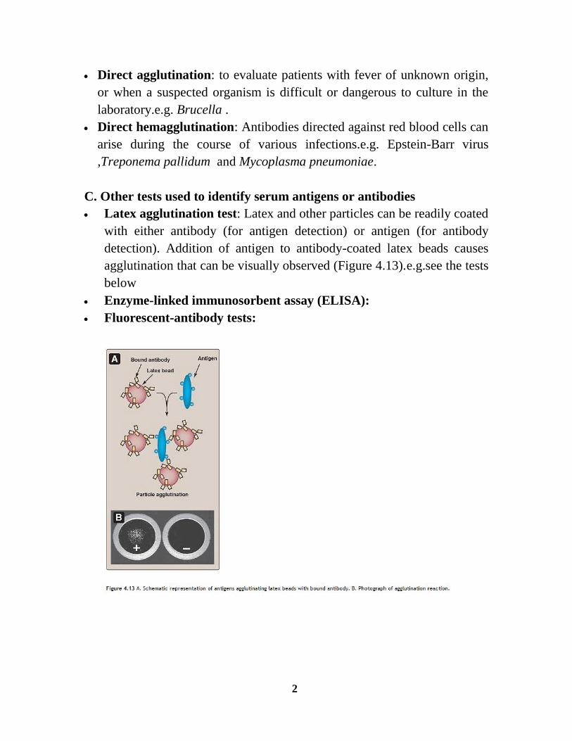
Latex agglutination test: Latex and other particles can be readily coated
with either antibody (for antigen detection) or antigen (for antibody
detection). Addition of antigen to antibody-coated latex beads causes
agglutination that can be visually observed (Figure 4.13).e.g.see the tests
below
Enzyme-linked immunosorbent assay (ELISA):
Fluorescent-antibody tests:
3
ASO-Titer
PRINCIPLE
Anti-Streptolysin O antibodies are produced during Streptococci pyogenes
infections, due to the presence of the Streptolysin O (SLO) liberated from
the bacteria.Information on the extent and degree of the infection can be
obtained from the latex agglutination of the antibodies serum level.
PROCEDURE
a) Qualitative method
1. Bring reagents and serum samples to roomtemperature.
2. Place one drop of undiluted serum onto a slide black area.
3. Add one drop of positive control and one drop of negative control in
separate circles.
4. Swirl the ASO-latex reagent gently before using and add one drop next to
the sample to be tested, one drop next to the negative and one drop next
to the positive.
5. Mix the drops with a stirrer, spreading them over the entire surface of the
circle. Use different stirrers for each sample.
6. Observe the presence or absence of agglutination within a period not
longer than 3 minutes.
b) Semi-quantitative method
Prepare serial two-fold dilutions in physiological saline and proceed for
each dilution as in the qualitative method. Then, observe the presence or
absence of agglutination. The approximate ASO concentration in serum
sample can be calculated by the following formula :
[ASO] (IU/ml)= highest positive dilution x 200 (as the reagent sensitivity is
200 IU/ml)
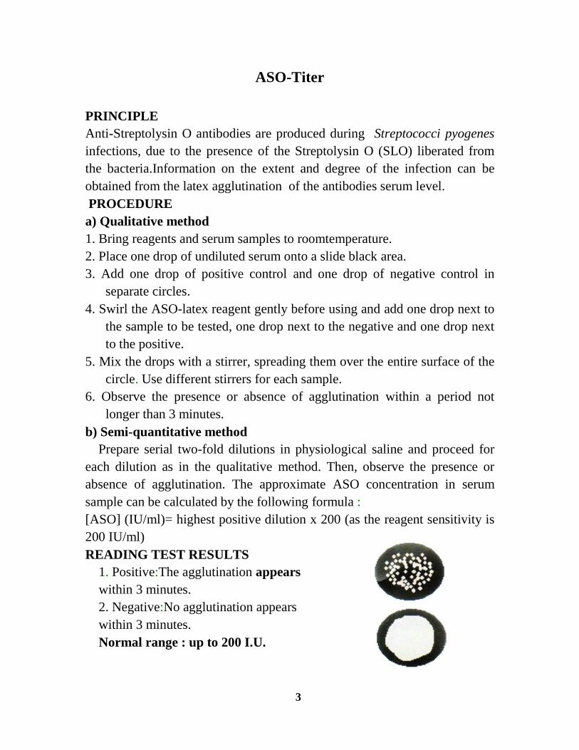
READING TEST RESULTS
1. Positive:The agglutination appears
within 3 minutes.
2. Negative:No agglutination appears
within 3 minutes.
Normal range : up to 200 I.U.

4
RA LATEX Test
Is a rapid agglutination procedure for semi-quantitative determination of
rheumatoid factors(rheumatoid arthritis) in human serum samples.
PRINCIPLE
Detecting rheumatoid factor using a suspension of fine plastic granules
coated with human gamma globulins which were agglutinated in the
presence of RA. The presence Or absence of a visible agglutination
indicates the presence or absence of RF in the samples tested.
PROCEDURE
Qualitative method
1. Allow each component to reach room temperature,
2. Gently shake the lalex reagent to disperse the particles.
3. Add one drop of the latex reagent using the dropper provided (40µl) to
each of the required circles of the agglutination slide.
4. Using the pipette stirrer provided, place a drop of undiluted serum onto a
circle of a test slide.
5. Spread the reagent and serum sample over the entire area of the test circle
using a separate stirrer for each sample. .
6. Gently tilt the test slide backwards and forwards approximately once
every two seconds for two minutes. Interpret results immediately after'2
minutes. Extended incubation may lead to false results. Positive and
negative controls should be included.
7. At the end of the test rinse the test slide with distilled water, dry and store
in a sealed bag.
5
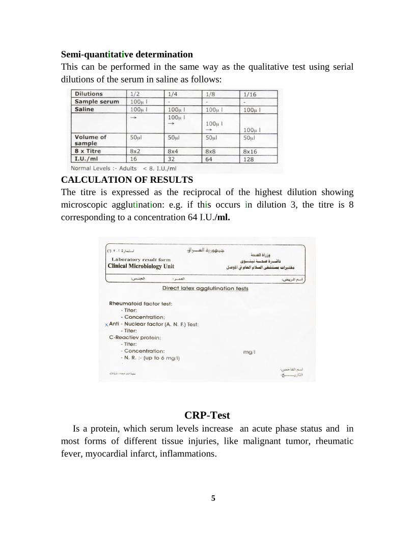
Semi-quantitative determination
This can be performed in the same way as the qualitative test using serial
dilutions of the serum in saline as follows:
CALCULATION OF RESULTS
The titre is expressed as the reciprocal of the highest dilution showing
microscopic agglutination: e.g. if this occurs in dilution 3, the titre is 8
corresponding to a concentration 64 I.U./ml.
CRP-Test
Is a protein, which serum levels increase an acute phase status and in
most forms of different tissue injuries, like malignant tumor, rheumatic
fever, myocardial infarct, inflammations.

6
PRINCIPLE
The CRP reagent is a suspension of polystyrene latex particles coated with
the gamma globulin fraction of antihuman CRPspecific serum. When CRP
is present in the sample, presence of agglutination indicates a content of
CRP equal or greater than 6 mg/ I, without previous sample dilution.
PROCEDURE
Qualitative method
1. Bring reagents and specimens to room temperature before use.
2. Place one drop (50µl) of the positive control on field 1 of the reaction
slide,Place one drop (50µl) of the negative control on field 2.Using a
pipette, place one drop (50µl) of each undiluted test specimen on
successive field.
3. Gently resuspend the Latex reagent and add one drop (50µl)to each test
field .Use a stirrer to spread reaction mixture over entire test field .Use
defferent stirrers for each sample.
4. Rotate the slide (80-100r.p.m.)for 2 minutes and read immediately under
direct light.
Semi-guantitative method
1. Bring reagents and specimens to room temperature before use.
2. Make serial two fold dilutions of the sample in saline.
3. Proceed for each dilution as in the qualitative method.
READING THE RESULT
A negative reaction is indicated by a uniform milky suspension with no
agglutination as observed with the negative control .
A positive reaction is indicated by any observable agglutination IN the
reaction mixture.The specimen reaction should be compared with the
positive control.
7
The titer, in the semi-quantitative method, is defined as the highest dilution
showing a positive result concentration will be reciprocal of positive reading
dilution x 6:
Normal Levels = Adults up to 6 mg/1
Rose Bengal Test
Brucella antigens are bacterial suspensions for use in slide agglutination
tests to detect the presence of bacterial antibody like agglutinins associated
with bacterial infection or previous exposure to a related organism. This test
is a screening procedure only to establish the presence or absence of
homologous antibody.
PRINCIPLE
Agglutinins combined with Brucella antigen (agglutinogen) under
controlled conditions is capable of causing agglutination. The Rose Bengal
stained Brucella antigen is used for the early detection of Brucella
agglutinins (Brucella abortus , B. melitensisand, B. suis).
PROCEDURE
1. Allow reagents and serum samples to reach roomtemperature for testing.
2. Shake the antigen bottle gently to insure a uniform suspension.
3. Place 50µl sample serum onto the selected ring ofthe slide.
4. Place one drop of the Rose Bengal antigen onto serum sample.
5. Mix serum sample with Rose Bengal antigen using stirring stick
6. Repeat these steps using the positive and negative controls instead of
serum sample.
7. Gently rock the slide for 2 minutes (automatic rotator can also be used)
8. Observe for agglutination after 4 minutes from beginning of shaking
.False positive results could appear if the test is read later than 2
minutes
RESULTS
Negative: No agglutination
Positive: Agglutination and the titer is 1/80 .
8
If positive, repeat the centrifugation of the sample serum and repeat the
procedure,if the result also positive we should know if the infection active
or inactive? by 2MET (2 merkapto ethanol broken IgM not IgG):
In tube,place 50µl sample serum+ 50µl 2ME reagent,incudate for 1
h..repeat procedure if positive=active infection(IgG)
if negative=inactive infection(IgM)
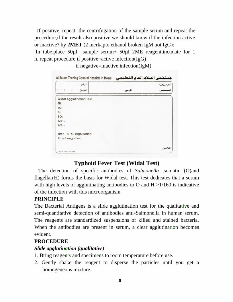
Typhoid Fever Test (Widal Test)
The detection of specific antibodies of Salmonella ,somatic (O)and
flagellar(H) forms the basis for Widal test. This test dedicates that a serum
with high levels of agglutinating antibodies to O and H >1/160 is indicative
of the infection with this microorganism.
PRINCIPLE
The Bacterial Antigens is a slide agglutination test for the qualitative and
semi-quantitative detection of antibodies anti-Salmonella in human serum.
The reagents are standardized suspensions of killed and stained bacteria.
When the antibodies are present in serum, a clear agglutination becomes
evident.
PROCEDURE
Slide agglutination (qualitative)
1. Bring reagents and specimens to room temperature before use.
2. Gently shake the reagent to disperse the particles until you get a
homogeneous mixture.

9
3. Place 50 µl of undiluted serum and 1 drop of Positive and Negative
controls onto separate circles of the slide.
4. Add a drop (50 µsl) of the Reagent next to the drop of serum.
5. Mix both drops spreading them over the full surface of the circle
6. Rotate the slide manually or with mechanical shaker (80 - 100 rpm)
during 1minute.
RESULTS
Slide agglutination method
Examine microscopically the presence or absence of clumps within 1
minute, after removing the slide from the rotator comparing the test results
with the control sera. . If a reaction is found ,the titre is 1/160 and establish
the titer by a tube test.
Pregnancy Test (PT)
Human chorionic gonadotropin(hCG) is a glycoprotein hormone
secreted by the developing placenta shortly after fertilization.The appearance
of hCG in the urine and serum soon after conception and its subsequent rise
in concentration during early gestational growth, make it an excellent marker
for the early detection of pregnancy .The Pregnancy Test is a rapid ,high
sensitive,specific, qualitative test used to detect the presence of hCG in urine
or serum.
PRINCIPLE
The sample is applied to the card and reacts initially with the specific,
anti-hCG conjugate on the test membrane. This mixture moves along the
membrane, by capillary action, and reacts with a specific anti-hCG in the
test region. If hCG is present in the sample, the result is the formation of a
colored band in the test region. If there is no hCG in the sample, the area
will remain white. The sample continues to flow to the control region and
forms a pink to purple color, indicating the test is working and the result is
valid.
SAMPLE COLLECTION
URINE: Collect specimen in a clean, dry glass or plastic container.It is not
necessary to obtain a first morning specimen,except in watery urine,
however concentrations of hCG may be higher in this specimen. In cause of
10
turbidity or bloody urine , it shoud be centrifuged then procedure done.The
sample can be refrigerated up to 72 hours prior to testing. A refrigerated
sample must be allowed to warm to room temperature and mixed before
testing.
PROCEDURES
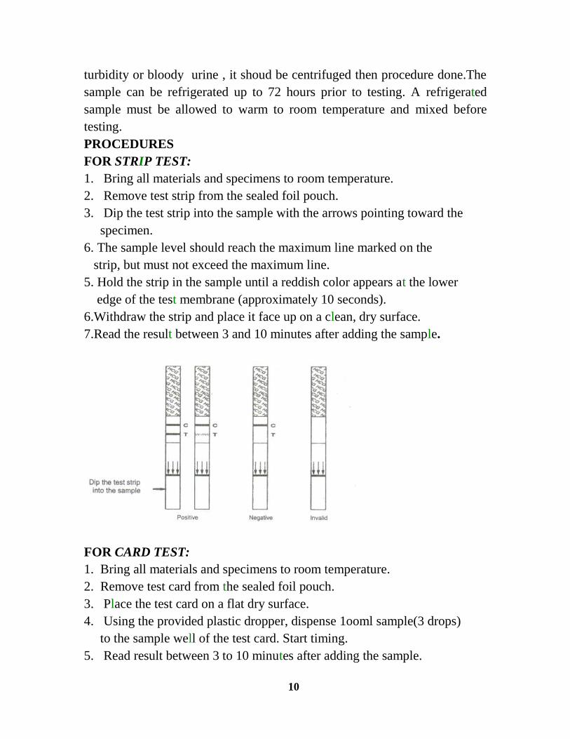
FOR STRIP TEST:
1. Bring all materials and specimens to room temperature.
2. Remove test strip from the sealed foil pouch.
3. Dip the test strip into the sample with the arrows pointing toward the
specimen.
6. The sample level should reach the maximum line marked on the
strip, but must not exceed the maximum line.
5. Hold the strip in the sample until a reddish color appears at the lower
edge of the test membrane (approximately 10 seconds).
6.Withdraw the strip and place it face up on a clean, dry surface.
7.Read the result between 3 and 10 minutes after adding the sample.
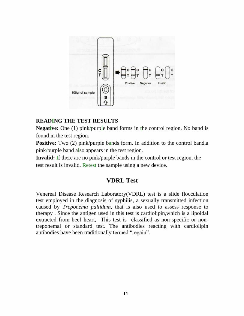
FOR CARD TEST:
1. Bring all materials and specimens to room temperature.
2. Remove test card from the sealed foil pouch.
3. Place the test card on a flat dry surface.
4. Using the provided plastic dropper, dispense 1ooml sample(3 drops)
to the sample well of the test card. Start timing.
5. Read result between 3 to 10 minutes after adding the sample.
11
READING THE TEST RESULTS
Negative: One (1) pink/purple band forms in the control region. No band is
found in the test region.
Positive: Two (2) pink/purple bands form. In addition to the control band,a
pink/purple band also appears in the test region.
Invalid: If there are no pink/purple bands in the control or test region, the
test result is invalid. Retest the sample using a new device.
VDRL Test
Venereal Disease Research Laboratory(VDRL) test is a slide flocculation
test employed in the diagnosis of syphilis, a sexually transmitted infection
caused by Treponema pallidum, that is also used to assess response to
therapy . Since the antigen used in this test is cardiolipin,which is a lipoidal
extracted from beef heart, This test is classified as non-specific or non-
treponemal or standard test. The antibodies reacting with cardiolipin
antibodies have been traditionally termed “regain”.
12
Principle:
Patients suffering from syphilis produce antibodies that react with
cardiolipin antigen in a slide flocculation test, which are read using a
microscope. It is not known if the antibodies that react with cardiolipin
are produced against some lipid component of
Treponema pallidum or as a result of tissue injury following infection.
Procedure:
Patients’ serum is inactivated by heating at 56 o C for 30 minutes in a water
bath to remove non-specific inhibitors (such as complement). The test can be
performed both qualitatively and quantitatively. Those tests that are reactive
by qualitative test are subjected to quantitative test to determine the antibody
titres.
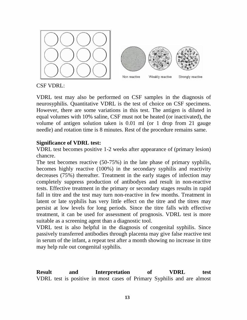
Qualitative test:
0.05 ml of inactivated serum is taken into one well. 1/60 th ml (or 1 drop
from 18 gauge needle) of the cardiolipin antigen is then added with the help
of a syringe (unbeveled) to the well and rotated at 180 rpm for 4 minutes.
Every test must be accompanied with known reactive and non-reactive
controls. The slide is then viewed
under low power objective of a microscope for flocculation. The reactive
and non-reactive controls are looked first to verify the quality of the antigen.
Depending on the size the results are graded as weakly reactive (W) or
reactive (R). Reactive samples are then subjected to quantitative test.
Qualitative test:
this is performed to determine the antibody titres. The serum is doubly
diluted in saline from 1in 2 to 1:256 or more. 0.05 ml of each dilution is
taken in the well and 1/60 ml of antigen is added to each dilution and rotated
in a rotator. The results are then checked under the microscope. The highest
dilution showing flocculation is
considered as reactive titre.
13
CSF VDRL:
VDRL test may also be performed on CSF samples in the diagnosis of
neurosyphilis. Quantitative VDRL is the test of choice on CSF specimens.
However, there are some variations in this test. The antigen is diluted in
equal volumes with 10% saline, CSF must not be heated (or inactivated), the
volume of antigen solution taken is 0.01 ml (or 1 drop from 21 gauge
needle) and rotation time is 8 minutes. Rest of the procedure remains same.
Significance of VDRL test:
VDRL test becomes positive 1-2 weeks after appearance of (primary lesion)
chancre.
The test becomes reactive (50-75%) in the late phase of primary syphilis,
becomes highly reactive (100%) in the secondary syphilis and reactivity
decreases (75%) thereafter. Treatment in the early stages of infection may
completely suppress production of antibodyes and result in non-reactive
tests. Effective treatment in the primary or secondary stages results in rapid
fall in titre and the test may turn non-reactive in few months. Treatment in
latent or late syphilis has very little effect on the titre and the titres may
persist at low levels for long periods. Since the titre falls with effective
treatment, it can be used for assessment of prognosis. VDRL test is more
suitable as a screening agent than a diagnostic tool.
VDRL test is also helpful in the diagnosis of congenital syphilis. Since
passively transferred antibodies through placenta may give false reactive test
in serum of the infant, a repeat test after a month showing no increase in titre
may help rule out congenital syphilis.
Result and Interpretation of VDRL test
VDRL test is positive in most cases of Primary Syphilis and are almost
14
always positive in Secondary Syphilis. It has a very good sensitivity for
syphilis, except in late Tertiary Syphilis form.
The titer of reagin antibodies decreases with effective treatment, so VDRL
test can be used to determine the treatment response of Syphilis.
A positive test result may mean have syphilis. If the test is positive, the next
step is to confirm the results with fluorescent treponemal antibody-
absorption (FTA-ABS) test, T. pallidum hemagglutination assays (TPHA),
and the microhemagglutination assay (MHA-TP) which is a more specific
syphilis test.
False positive VDRL test result
Reagin antibodies may be produced in response to nontreponemal diseases of
an acute and chronic nature in which tissue damage occurs such as:
1. Leprosy
2. Hepatitis B
3. Infectious Mononucleosis
4. HIV
5. Certain types of pneumonia
6. Malaria
7. Systemic lupus erythematosus
8. Rheumatic fever
9. Rheumatoid arthritis
10. Tuberculosis