Cytoskeleton changes and impaired motility of monocytes at modelled low gravity
8
Summary. Investigations performed in space have shown that gravity changes affect important cellular mechanisms like proliferation, differ- entiation, genetic expression, cytoskeletal architecture, and motility in lymphocytes, monocytes, and other mammalian cells. In particular, a dramatic depression of the mitogenic in vitro activation of human pe- ripheral blood lymphocytes was observed at low gravity. The hypothesis of the present work is that a reduced interaction between T lymphocytes and monocytes, essential for the second signalling pathway, might be one of the reasons for the observed depression of the in vitro activation of human lymphocytes. Cell motility and with it a continuous rearrange- ment of the cytoskeletal network within the cell is essential for cell-to- cell contacts. Whereas nonactivated lymphocytes in suspension are highly motile at low gravity, no data are available so far on the motility of adherent monocytes. It thus can be argued that impaired monocyte locomotion and cytoskeletal changes could be responsible for a reduced interaction of monocytes with T lymphocytes. In this study, the locomo- tion ability of J-111 cells, an adherent monocyte cell line, attached to colloidal gold particles on coverslips and exposed to modelled low grav- ity in the random positioning machine was found to be severely reduced compared with that of controls and the structures of actin, tubulin, and vinculin were affected. Keywords: Monocyte; Locomotion; Cytoskeleton; Microgravity; Ran- dom positioning machine. Introduction Remarkable findings in space and gravitational biology have shown that mammalian cells subjected to modelled low- gravity conditions on ground as well as to spaceflight condi- tions in dedicated space missions and in several sounding-rocket flights, are showing alterations in their structure and function (Cogoli and Cogoli-Greuter 1997, Lewis 2002). Cells of the immune system are in particular the most se- verely affected by the space environment. Especially for T lymphocytes, it is known that exposure of cells in culture to actual or modelled low gravity is often accompanied by a major inhibitory effect, remarkably reducing their mitogenic activation process (Cogoli et al. 1984) and severely altering growth rate, cytokine production, gene expression, cy- toskeletal structures, and motility (for reviews, see Cogoli 1993, 1997; Cogoli and Cogoli-Greuter 1997; Lewis 2002; Cogoli-Greuter 2004). Furthermore, it has been demon- strated that human lymphocytes are forced to apoptosis in modelled low gravity, through a pathway based on calcium- dependent 5-LOX activation, mitochondrial membrane dis- ruption, and cytochrome c release, followed by caspase activation and cell death (Maccarrone et al. 2003). The mechanism of T-cell activation is very complex and is based on three distinct pathways (Crabtree and Clipstone 1994). Interaction between T cells and the antigen-present- ing cells is the first step in the signal transduction, whereas in a next step a costimulatory signal is delivered most probably by accessory cells (usually monocytes) via B7/CD28 interaction (Geppert et al. 1990). Leukocytes are highly motile cells. Their locomotion is crucial not only for recruiting cells into inflammatory sites but also for their migration and for the contact interactions of antigen-bearing accessory cells with lymphocytes in in- duction of the immune response. The locomotor capacity of lymphocytes varies with their activation status, whereas monocytes have acquired locomotor capacity during differ- entiation (Wilkinson 1987). Leukocytes undergoing migra- Protoplasma (2006) 229: 243–249 DOI 10.1007/s00709-006-0210-2 PROTOPLASMA Printed in Austria Cytoskeleton changes and impaired motility of monocytes at modelled low gravity M. A. Meloni 1 , G. Galleri 1 , P. Pippia 1 , and M. Cogoli-Greuter 2, * 1 Dipartimento di Scienze Fisiologiche, Biochimiche e Cellulari, Universita di Sassari, Sassari 2 Space Biology Group, Swiss Federal Institute of Technology, Zürich Received September 4, 2005; accepted November 2, 2005; published online December 16, 2006 © Springer-Verlag 2006 * Correspondence and reprints: Space Biology Group, Swiss Federal Institute of Technology, Technopark, Technoparkstrasse 1, 8005 Zürich, Switzerland. E-mail: [email protected]
-
Upload
m-a-meloni -
Category
Documents
-
view
212 -
download
0
Transcript of Cytoskeleton changes and impaired motility of monocytes at modelled low gravity

Summary. Investigations performed in space have shown that gravitychanges affect important cellular mechanisms like proliferation, differ-entiation, genetic expression, cytoskeletal architecture, and motility inlymphocytes, monocytes, and other mammalian cells. In particular, adramatic depression of the mitogenic in vitro activation of human pe-ripheral blood lymphocytes was observed at low gravity. The hypothesisof the present work is that a reduced interaction between T lymphocytesand monocytes, essential for the second signalling pathway, might beone of the reasons for the observed depression of the in vitro activationof human lymphocytes. Cell motility and with it a continuous rearrange-ment of the cytoskeletal network within the cell is essential for cell-to-cell contacts. Whereas nonactivated lymphocytes in suspension arehighly motile at low gravity, no data are available so far on the motilityof adherent monocytes. It thus can be argued that impaired monocytelocomotion and cytoskeletal changes could be responsible for a reducedinteraction of monocytes with T lymphocytes. In this study, the locomo-tion ability of J-111 cells, an adherent monocyte cell line, attached tocolloidal gold particles on coverslips and exposed to modelled low grav-ity in the random positioning machine was found to be severely reducedcompared with that of controls and the structures of actin, tubulin, andvinculin were affected.
Keywords: Monocyte; Locomotion; Cytoskeleton; Microgravity; Ran-dom positioning machine.
Introduction
Remarkable findings in space and gravitational biology haveshown that mammalian cells subjected to modelled low-gravity conditions on ground as well as to spaceflight condi-tions in dedicated space missions and in severalsounding-rocket flights, are showing alterations in their
structure and function (Cogoli and Cogoli-Greuter 1997,Lewis 2002).
Cells of the immune system are in particular the most se-verely affected by the space environment. Especially for Tlymphocytes, it is known that exposure of cells in culture toactual or modelled low gravity is often accompanied by amajor inhibitory effect, remarkably reducing their mitogenicactivation process (Cogoli et al. 1984) and severely alteringgrowth rate, cytokine production, gene expression, cy-toskeletal structures, and motility (for reviews, see Cogoli1993, 1997; Cogoli and Cogoli-Greuter 1997; Lewis 2002;Cogoli-Greuter 2004). Furthermore, it has been demon-strated that human lymphocytes are forced to apoptosis inmodelled low gravity, through a pathway based on calcium-dependent 5-LOX activation, mitochondrial membrane dis-ruption, and cytochrome c release, followed by caspaseactivation and cell death (Maccarrone et al. 2003).
The mechanism of T-cell activation is very complex andis based on three distinct pathways (Crabtree and Clipstone1994). Interaction between T cells and the antigen-present-ing cells is the first step in the signal transduction, whereasin a next step a costimulatory signal is delivered mostprobably by accessory cells (usually monocytes) viaB7/CD28 interaction (Geppert et al. 1990).
Leukocytes are highly motile cells. Their locomotion iscrucial not only for recruiting cells into inflammatory sitesbut also for their migration and for the contact interactionsof antigen-bearing accessory cells with lymphocytes in in-duction of the immune response. The locomotor capacity oflymphocytes varies with their activation status, whereasmonocytes have acquired locomotor capacity during differ-entiation (Wilkinson 1987). Leukocytes undergoing migra-
Protoplasma (2006) 229: 243–249DOI 10.1007/s00709-006-0210-2 PROTOPLASMA
Printed in Austria
Cytoskeleton changes and impaired motility of monocytes at modelled low gravity
M. A. Meloni1, G. Galleri1, P. Pippia1, and M. Cogoli-Greuter2,*
1 Dipartimento di Scienze Fisiologiche, Biochimiche e Cellulari, Universita di Sassari, Sassari2 Space Biology Group, Swiss Federal Institute of Technology, Zürich
Received September 4, 2005; accepted November 2, 2005; published online December 16, 2006© Springer-Verlag 2006
* Correspondence and reprints: Space Biology Group, Swiss FederalInstitute of Technology, Technopark, Technoparkstrasse 1, 8005 Zürich,Switzerland.E-mail: [email protected]

tion, activation, and cell–cell interaction develop a polarizedmorphology (Sanchez-Madrid and del Pozo 1999) with theformation of two functionally and morphologically distinctpoles: the leading edge and the uropod. This involves a reor-ganisation of the cytoskeletal network with a collapse of thevimentin system, which retracts into the uropod (Brownet al. 2001). During locomotion, the cytoskeletal structuresare subjected to repeated cycles of reassembly processes.
Cell–cell interaction and aggregate formation are also im-portant means of cell communication and signal delivery inthe mitogenic in vitro activation of human T lymphocytesextensively studied in real and modelled low gravity. Nonac-tivated peripheral blood lymphocytes in suspension werefound to be motile at low gravity. They displayed an au-tonomous motion in random directions and often changedtheir morphology from round shaped to a polarized form(Cogoli-Greuter et al. 1998). Lymphocytes activated at lowgravity with the mitogen concanavalin A formed aggregates(Cogoli-Greuter et al. 1996), although they contained fewercells than the comparable aggregates in the 1 g control onground. Aggregate formation was observed in real time byvideo microscopy. The mean velocity of single cells outsidethe aggregates was significantly higher at low gravity com-pared with the ground control. Changes in cell activationand signal transduction as well as cell movements and ag-gregate formation may be related to changes in the cy-toskeleton. In fact, marked alterations in the structure of theintermediate filaments of vimentin (Cogoli-Greuter et al.1998) as well as in the microtubule network (Lewis et al.1998) were observed in Jurkat cells – a T cell line – after ex-posure to low gravity.
Since suspended T lymphocytes were found to be highlymotile at microgravity even in the absence of the mitogen, itcan be argued that an impaired motility of human mono-cytes acting as accessory cells could hinder the delivery ofthe costimulatory signal to activate the B7/CD28 pathwayand thus could be one of the reasons of the loss of T-cell ac-tivation at low gravity. This is supported by the findings thata costimulation of CD3-activated cells by CD28 antibodiesin modelled low gravity results in a normal T-cell activation(Vadrucci et al. 2006). In this research, we studied the motil-ity of and cytoskeletal changes in human monocytes J-111subjected to modelled low-gravity conditions, using the ran-dom positioning machine.
Material and methods
Cell line and cell culture
J-111 is a monocyte/macrophage cell line derived from human acute mono-cytic leukemia, obtained from the Istituto Zooprofilattico Sperimentale
della Lombardia e dell’Emilia Romagna, Brescia, Italy. This cell line wasfound to have a HeLa profile by DNA fingerprinting, to display a good ad-hesion capacity and a certain extent of epithelial morphological polymor-phism related to different functional and metabolic status of the cell.
Cells cultured from the frozen stocks were utilised at passage levelthree to eight for all tests. The cells were grown in RPMI 1640 medium(Glutamax; GIBCO) containing 10% fetal calf serum (GIBCO), 20 mMHEPES, 5 mM sodium bicarbonate, and 50 �g of gentamycin per ml andwere subcultured every 3 days using 0.25% trypsin–EDTA.
Cytoskeletal structures and motility were studied both at 1 g and mod-elled low-gravity conditions, using the random positioning machine(RPM) (Dutch Space) as earth-based model of spaceflight, keeping sam-ples under continuous rotation at 60 rpm. The time-averaged gravita-tional vector acting on these samples is reduced to about 10�2 g.
Cytoskeleton staining
Analysis of the cytoskeletal structures were performed by an indirect im-munofluorescence technique.
J-111 cells were seeded onto chamber slides (Lab-Tek products) at1.5 � 104 cells per ml and incubated at 37 °C for 24 h. Chamber slides,completely fluid-filled, were positioned on the RPM at 37 °C for 1 h and24 h, respectively.
Ground 1 g controls, not subjected to rotation, were placed onto themachine supporting static frame in order to subject them to the same vi-brational stress.
Subsequently all samples were fixed in 4% paraformaldehyde at 4 °Cfor 30 min.
After extensive washing and cell permeabilization with Triton X-100(Sigma) 10% in phosphate-buffered saline (PBS) for 3 min, fluorescentstaining was performed by exposing the slides to a monoclonal antibodyagainst �-tubulin (diluted 1 : 100 in bovine serum albumin [BSA]–PBSwithout Ca�� and Mg�� [PBSO] from Sigma) and to a monoclonal anti-body against vinculin (diluted 1 : 100 in BSA-PBS from Sigma), both at37 °C for 1 h in a moist chamber. After washing in PBSO and PBS, re-spectively, a second layer of fluorescein isothiocyanate-conjugated goatanti-mouse gamma globulins (diluted 1 : 150 in BSA-PBSO or BSA-PBS, respectively) was applied for 45 min at room temperature in thedark. For cytochemical labelling for filamentous actin (F-actin), cellswere stained with 5 �g of fluorescent phalloidin-tetramethylrhodamineisothiocyanate conjugated solution in PBS with 1% dimethyl sulfoxideat 37 °C for 15 min.
Nuclei were stained with 100 ng of 4�,6-diamidino-2-phenylindole hy-drochloride per ml in PBS for 6 min.
Slides were rinsed in PBS and mounted with immune mount (Shandon)and the cytoskeletal components were observed by fluorescence-invertedmicroscopy.
Microscopic analysis
Cytoskeletal structures were observed by fluorescence-inverted mi-croscopy (Olympus) using a magnification of �400 with oil immersion.
Migration tracks were visualised by a bright-field illumination differ-ential interference contrast (Nomarski) microscope.
Video data were collected using an Olympus charge-coupled-device FView II Image camera coupled to the Analysis software to calculate thecell displacement. Tracks were counted for each experiment, from 1 gcontrols and samples exposed to modelled low gravity.
Locomotion assay
The locomotion, i.e., the displacement of cells on an artificial substrate, hasbeen successfully quantified in the past by different authors (Burk 1973,DiPasquale 1975). Also a quantitative assay for the motility phenomena inspreading animal cells in culture, which are associated with large surface
244 M. A. Meloni et al.: Cell motility and cytoskeleton at low gravity
extensions, i.e., lamellipodia and filopodia (microspikes), has been de-scribed (Albrecht-Buehler and Lancaster 1976). Freshly suspended cells,plated on top of a gold particle-coated coverslip, produce various surfaceprotrusions and remove the particles within a ring around each cell. The re-action of the spreading cell microspikes upon contact to colloidal gold par-ticles leads to a centripetal transport of particles to the cell body during thevery early stages of cell spreading and to a cleaned area around each cell(Albrecht-Buehler and Goldman 1976). This phenomenon can be used toquantify the motile activity of such protrusions under various extracellularconditions. In fact, during their spreading the cells begin to move whilecleaning more particles out of their way. The particles around the cells aremostly cleaned out by surface protrusions during the first hour after platingand become partly internalised (phagocytosed) and partly accumulated onthe cell surface in big clumps without being phagocytosed (Albrecht-Buehler and Lancaster 1976). Therefore, the movement of such cellsreflects a surface movement rather than a cytoplasmic movement (Al-brecht-Buehler and Yarnell 1973). In order to distinguish locomotion onplain surfaces from this combination of phagocytosis and cellular displace-ment, we better indicate this phenomenon as “phagokinetics” and the parti-cle-free tracks conveniently visualised as “phagokinetic tracks”.
In approaching the problem of quantifying the motile behaviour ofmonocytes in modelled low gravity (or the phenomena of the motile ac-tions of surface protrusions) we plated J-111 cells onto chamber slidescoated with colloidal gold (Sigma) according to Albrecht-Buehler andLancaster (1976).
Exposure of the cells to gold particles (1.45 mM AuCl4H) had no ob-vious toxic effects as proven by preliminary viability tests (trypan bluedye exclusion test). J-111 cells showed normal growth and spreading ongold-coated coverslips.
Gold coating
Chamber slides were incubated with BSA (10 mg/ml of tridistilled H2O) atroom temperature for 10 min, then quickly washed with 100% ethanol andincubated at 85 °C for 10 min. Subsequently, 5 ml of the gold suspensionwas added at 60–80 °C, and after 45 min of incubation, the chamber slideswere washed in normal salt solution (113 mM NaCl, 3 mM KCl, 1 mMMgCl2, 15 mM Na-phosphate, pH 7.8, in tridistilled H2O). Chamber slideswere filled with 3 ml of culture medium and kept at 37 °C until needed forthe cell inoculation. J-111 cells were plated at 1.5 � 104 cells per ml andincubated at 37 °C for 4–5 h to allow them to adhere.
Cells attached to coated chamber slides were exposed to modelled lowgravity on the RPM for 1 h and 24 h and then fixed in 4% paraformalde-hyde at 4 °C for 30 min. A simultaneous 1 g control was performed asdescribed above.
Statistical analysis
After grouping displacement ranks, the frequency percentages (percent-age of cells showing a displacement in the distinct groups 1–11) andstandard deviation have been calculated. Data were analysed by one-wayanalysis of variance following the rank sum test by the Sigma Stat pro-gram. The data from at least 3 independent experiments monitoring 50cells per experiment are presented. Statistical significance was acceptedat the P � 0.0001 level.
Results
Cytoskeletal architecture
Cytoskeletal structures of J-111 cells at 1 g conditionsshowed a well organised network, whereas severe alter-ations in their structure were observed after 1 h of expo-
sure to modelled low gravity in the RPM. Moreover it hasbeen observed that the actin and tubulin network starts toreorganise to a normal structure after 24 h of modelledlow gravity.
Microfilaments: F-actin
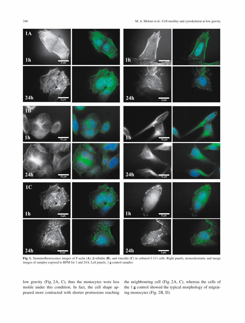
In the 1 g controls, F-actin filaments appeared abundantand well organised into cytosolic bundles and in the elon-gated and extended filopodia (Fig. 1A, left panels). Con-versely, the F-actin network of J-111 cells exposed tomodelled low gravity showed a remarkable decrease inthe filamentous biopolymer density and the actin stressfibres appeared localised like continuous subplasmaticbundles, both after 1 h and 24 h (Fig. 1A, right panels). Inparticular, an initial reorganisation of the actin networkwas observed after 24 h exposure to modelled low gravity(Fig. 1A, left panels). This reorganisation of actin mightrepresent an adaptative mechanism and might have rele-vance in the process of cell adaptation to gravitational un-loading.
Microtubules: �-tubulin
�-Tubulin showed a perinuclear position with reduction inaborisation, losing the radial disposition, even after 1 h ofmodelled low gravity in the RPM (Fig. 1B, right panels),in contrast to 1 g controls, where they appeared radiatingfrom the microtubule-organising centre to the plasmamembrane (Fig. 1B, left panels).
Vinculin
The anchor protein vinculin appeared as focal contactslinking actin filaments to the plasma membrane in the 1 gcontrols (Fig. 1C, left panels), whereas samples exposedfor 1 and 24 h to modelled low gravity showed vinculinproteins not evenly spread but thickened close to the cellmembrane as globular clusters (Fig. 1C, right panels).
Cell motility
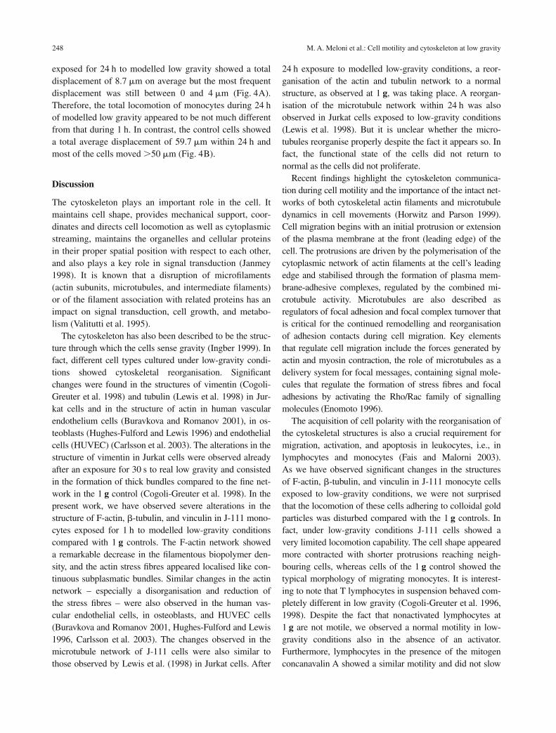
The microscopic analysis revealed migration tracks ofmonocytes at 1 g similar to those described in the litera-ture (Horwitz et al. 1999). A normal pattern of cell migra-tion was observed on gold particle-coated chamber slidesof ground samples. Areas around cells appeared com-pletely cleaned out of gold particles partly internalised in-side the cells or accumulated to the surface of the cells(Fig. 2B). Conversely, very short migration tracks wereobserved after both 1 and 24 h of exposure to modelled
M. A. Meloni et al.: Cell motility and cytoskeleton at low gravity 245
246 M. A. Meloni et al.: Cell motility and cytoskeleton at low gravity
low gravity (Fig. 2A, C), thus the monocytes were lessmotile under this condition. In fact, the cell shape ap-peared more contracted with shorter protrusions reaching
the neighbouring cell (Fig. 2A, C), whereas the cells ofthe 1 g control showed the typical morphology of migrat-ing monocytes (Fig. 2B, D).
Fig. 1. Immunofluorescence images of F-actin (A), �-tubulin (B), and vinculin (C) in cultured J-111 cells. Right panels, monochromatic and mergeimages of samples exposed to RPM for 1 and 24 h. Left panels, 1 g control samples

Cell migration tracks were analysed quantitatively bythe Analysis software (Olympus). Data obtained demon-strated a remarkable difference in the locomotion abilityof the cells even after 1 h of modelled low gravity in theRPM when compared to 1 g controls. Cells exposed to
modelled low gravity for 1 h moved on average by 6 �mand the most frequent displacement was between 0 and4 �m (Fig. 3A), whereas 1 g controls showed a displace-ment of 14.9 �m on average and the most frequent dis-placement was between 10 and 14 �m (Fig. 3B). Cells
M. A. Meloni et al.: Cell motility and cytoskeleton at low gravity 247
Fig. 3. Displacement frequencies of J-111 cells on gold particle-coatedchamber slides after 1 h of exposure to modelled low gravity (RPM) (A)and to 1 g (B). Displacement rank: 1, 0–4 �m; 2, 5–9 �m; 3, 10–14 �m;4, 15–19 �m; 5, 20–24 �m; 6, 25–29 �m; 7, 30–34 �m; 8, 35–39 �m; 9,40–44 �m; 10, 45–49 �m; 11, �50 �m. The results are the average fromat least 3 independent experiments with 50 cells for each experiment
Fig. 2 A–D. Migration tracks on gold particle-coated chamber slides of J-111 cells observedby bright-field illumination differential in-terference contrast (Nomarski) microscopy.A Very short migration tracks were observedafter 24 h of exposure to modelled low grav-ity (RPM). This is more evident under highermagnification (C). B and D Control sam-ples showing areas around cell completelycleaned out of gold particles and with typicalmorphology of migrating cell (arrow)
Fig. 4. Displacement frequencies of J-111 cells on gold particle-coatedchamber slides (Y axis) after 24 h of exposure to modelled low gravity(RPM) (A) and to 1 g (B). For definitions of displacement ranks see leg-end of Fig. 3. The results are the average from at least 3 independent ex-periments with 50 cells for each experiment
exposed for 24 h to modelled low gravity showed a totaldisplacement of 8.7 �m on average but the most frequentdisplacement was still between 0 and 4 �m (Fig. 4A).Therefore, the total locomotion of monocytes during 24 hof modelled low gravity appeared to be not much differentfrom that during 1 h. In contrast, the control cells showeda total average displacement of 59.7 �m within 24 h andmost of the cells moved 50 �m (Fig. 4B).
Discussion
The cytoskeleton plays an important role in the cell. Itmaintains cell shape, provides mechanical support, coor-dinates and directs cell locomotion as well as cytoplasmicstreaming, maintains the organelles and cellular proteinsin their proper spatial position with respect to each other,and also plays a key role in signal transduction (Janmey1998). It is known that a disruption of microfilaments(actin subunits, microtubules, and intermediate filaments)or of the filament association with related proteins has animpact on signal transduction, cell growth, and metabo-lism (Valitutti et al. 1995).
The cytoskeleton has also been described to be the struc-ture through which the cells sense gravity (Ingber 1999). Infact, different cell types cultured under low-gravity condi-tions showed cytoskeletal reorganisation. Significantchanges were found in the structures of vimentin (Cogoli-Greuter et al. 1998) and tubulin (Lewis et al. 1998) in Jur-kat cells and in the structure of actin in human vascularendothelium cells (Buravkova and Romanov 2001), in os-teoblasts (Hughes-Fulford and Lewis 1996) and endothelialcells (HUVEC) (Carlsson et al. 2003). The alterations in thestructure of vimentin in Jurkat cells were observed alreadyafter an exposure for 30 s to real low gravity and consistedin the formation of thick bundles compared to the fine net-work in the 1 g control (Cogoli-Greuter et al. 1998). In thepresent work, we have observed severe alterations in thestructure of F-actin, �-tubulin, and vinculin in J-111 mono-cytes exposed for 1 h to modelled low-gravity conditionscompared with 1 g controls. The F-actin network showeda remarkable decrease in the filamentous biopolymer den-sity, and the actin stress fibres appeared localised like con-tinuous subplasmatic bundles. Similar changes in the actinnetwork – especially a disorganisation and reduction ofthe stress fibres – were also observed in the human vas-cular endothelial cells, in osteoblasts, and HUVEC cells(Buravkova and Romanov 2001, Hughes-Fulford and Lewis1996, Carlsson et al. 2003). The changes observed in themicrotubule network of J-111 cells were also similar tothose observed by Lewis et al. (1998) in Jurkat cells. After
24 h exposure to modelled low-gravity conditions, a reor-ganisation of the actin and tubulin network to a normalstructure, as observed at 1 g, was taking place. A reorgan-isation of the microtubule network within 24 h was alsoobserved in Jurkat cells exposed to low-gravity conditions(Lewis et al. 1998). But it is unclear whether the micro-tubules reorganise properly despite the fact it appears so. Infact, the functional state of the cells did not return tonormal as the cells did not proliferate.
Recent findings highlight the cytoskeleton communica-tion during cell motility and the importance of the intact net-works of both cytoskeletal actin filaments and microtubuledynamics in cell movements (Horwitz and Parson 1999).Cell migration begins with an initial protrusion or extensionof the plasma membrane at the front (leading edge) of thecell. The protrusions are driven by the polymerisation of thecytoplasmic network of actin filaments at the cell’s leadingedge and stabilised through the formation of plasma mem-brane-adhesive complexes, regulated by the combined mi-crotubule activity. Microtubules are also described asregulators of focal adhesion and focal complex turnover thatis critical for the continued remodelling and reorganisationof adhesion contacts during cell migration. Key elementsthat regulate cell migration include the forces generated byactin and myosin contraction, the role of microtubules as adelivery system for focal messages, containing signal mole-cules that regulate the formation of stress fibres and focaladhesions by activating the Rho/Rac family of signallingmolecules (Enomoto 1996).
The acquisition of cell polarity with the reorganisation ofthe cytoskeletal structures is also a crucial requirement formigration, activation, and apoptosis in leukocytes, i.e., inlymphocytes and monocytes (Fais and Malorni 2003).As we have observed significant changes in the structuresof F-actin, �-tubulin, and vinculin in J-111 monocyte cellsexposed to low-gravity conditions, we were not surprisedthat the locomotion of these cells adhering to colloidal goldparticles was disturbed compared with the 1 g controls. Infact, under low-gravity conditions J-111 cells showed avery limited locomotion capability. The cell shape appearedmore contracted with shorter protrusions reaching neigh-bouring cells, whereas cells of the 1 g control showed thetypical morphology of migrating monocytes. It is interest-ing to note that T lymphocytes in suspension behaved com-pletely different in low gravity (Cogoli-Greuter et al. 1996,1998). Despite the fact that nonactivated lymphocytes at1 g are not motile, we observed a normal motility in low-gravity conditions also in the absence of an activator.Furthermore, lymphocytes in the presence of the mitogenconcanavalin A showed a similar motility and did not slow
248 M. A. Meloni et al.: Cell motility and cytoskeleton at low gravity
down within the 3 days needed for activation, as describedfor cells at 1 g. The fact that T lymphocytes are motile inlow gravity in the presence of the mitogen may also be thereason that aggregate formation was observed. On the otherhand, it can be speculated that no monocytes are in theseaggregates and thus no contact between lymphocytes andmonocytes – essential for the delivery of the second activa-tion signal – is occurring in low gravity. This could be oneof several reasons for the observed depression of the mito-genic lymphocyte activation.
As described for other cell types (Buravkova andRomanov 2001), our results on the motility of J-111 cellsclearly revealed that modelled low gravity affects their ca-pacity for locomotion. Buravkova and Romanov (2001)also observed changes in the actin, especially a disorgani-sation and reduction of stress fibres, i.e., disturbances sim-ilar to those observed by us. The importance of an intactand dynamic cytoskeletal network for cell movement hasbeen recently pointed out by Horwitz and Parson (1999).On the basis of our results and those of others, it can thusbe speculated that the impaired motility of adherentmonocytes in low gravity might be due to the disruptionof the cytoskeletal network.
Acknowledgments
This work was supported by a grant of the Italian Space Agency. Wethank M. A. Camboni and A. G. Campus for their invaluable technicalassistance.
References
Albrecht-Buehler G, Goldman RD (1976) Microspike-mediated particletransport towards the cell body during early spreading of 3T3 cells.Exp Cell Res 97: 329–339
Albrecht-Buehler G, Lancaster RM (1976) A quantitative description ofthe extension and retraction of surface protrusions in spreading 3T3mouse fibroblasts. J Cell Biol 71: 370–382
Albrecht-Buehler G, Yarnell MM (1973) A quantitation of movement ofmarker particles in the plasma membrane of 3T3 mouse fibroblasts.Exp Cell Res 78: 59–66
Brown MJ, Hallam JA, Colucci-Guyon E, Shaw S (2001) Rigidity of cir-culating lymphocytes is primarily conferred by vimentin intermediatefilaments. J Immunol 166: 6640–6646
Buravkova LB, Romanov YA (2001) The role of cytoskeleton in cellchanges under condition of simulated microgravity. Acta Astronaut48: 647–650
Burk RR (1973) A factor from a transformed cell line that affects cellmigration. Proc Natl Acad Sci USA 70: 369–372
Carlsson SIM, Bertilaccio TS, Ballabio E, Maier JAM (2003) Endothe-lial stress by gravity unloading: effects on cell growth and cytoskeletalorganization. Biochim Biophys Acta 1642: 173–179
Cogoli A (1993) The effect of hypogravity and hypergravity on cells ofthe immune system. J Leukoc Biol 54: 259–268
Cogoli A (1997) Signal transduction in T lymphocytes in microgravity.ASGSB Bull 10(2): 5–16
Cogoli A, Cogoli-Greuter M (1997) Activation and proliferation of lym-phocytes and other mammalian cells in microgravity. Adv Space BiolMed 6: 33–79
Cogoli A, Tschopp A, Fuchs-Bislin P (1984) Cell sensitivity to gravity.Science 225: 228–230
Cogoli-Greuter M (2004) Effect of gravity changes on the cytoskeletonin human lymphocytes. ASGSB Bull 17(2): 27–37
Cogoli-Greuter M, Meloni MA, Sciola L, Spano A, Pippia P, Monaco G,Cogoli A (1996) Movements and interactions of leukocytes in micro-gravity. J Biotechnol 47: 279–287
Cogoli-Greuter M, Spano A, Sciola L, Pippia P, Cogoli A (1998) Influ-ence of microgravity on mitogen binding, motility and cytoskeletonpatterns of T lymphocytes and Jurkat cells – experiments on soundingrocket. Jpn J Aerospace Environ Med 35: 27–39
Crabtree GR, Clipstone NA (1994) Signal transmission between theplasma membrane and the nucleus of T lymphocytes. Annu RevBiochem 63: 1045–1083
DiPasquale A (1975) Locomotory activity of epithelial cells in culture.Exp Cell Res 94: 191–215
Enomoto T (1996) Microtubule disruption induces the formation of actinstress fibers and focal adhesions in cultured cells: possible involve-ment of the rho signal cascade. Cell Struct Funct 21: 317–326
Fais S, Malorni W (2003) Leukocyte uropod formation and membrane/cytoskeleton linkage in immune interactions. J Leukoc Biol 73:556–563
Geppert TD, Davis LS, Gur H, Wacholtz MC, Lipsky PE (1990) Accessorycell signals involved in T-cell activation. Immunol Rev 117: 5–66
Horwitz AR, Parson JT (1999) Cell migration – movin’ on. Science 286:1102–1104
Hughes-Fulford M, Lewis ML (1996) Effects of microgravity on os-teoblast growth activation. Exp Cell Res 224: 103–109
Ingber D (1999) How cells (might) sense microgravity. FASEB J 13Suppl: S3–S15
Janmey PA (1998) The cytoskeleton and cell signalling: component lo-calization and mechanical coupling. Physiol Rev 78: 763–781
Lewis ML (2002) The cytoskeleton, apoptosis, and gene expression in Tlymphocytes and other mammalian cells exposed to altered gravity.Adv Space Biol Med 8: 77–128
Lewis ML, Reynolds JL, Cubano LA, Hatton JP, Lawless BD, PiepmeierEH (1998) Spaceflight alters microtubules and increases apoptosis inhuman lymphocytes (Jurkat). FASEB J 12: 1007–1018
Maccarrone M, Battista N, Meloni MA, Bari M, Galleri G, Pippia P,Cogoli A, Finazzi-Agrò A (2003) Creating conditions similar to thosethat occur during exposure of cells to microgravity induces apoptosisin human lymphocytes by 5-lipoxygenase-mediated mitochondrial un-coupling and cytochrome c release. J Leukoc Biol 73: 472–481
Sanchez-Madrid F, del Pozo MA (1999) Leukocyte polarization in cellmigration and immune interactions. EMBO J 18: 501–511
Vadrucci S, Henggeler D, Lovis P, Lambers B, Cogoli A (2006) Ef-fects of vector-averaged gravity on the response to different stimu-latory signals in T-cells. In: Warmbein B (ed) Proceedings of the9th European Symposium on Life Sciences Research in Space and26th Annual International Gravitational Physiology Meeting. Con-ference proceeding, SP-585. European Space Agency PublicationsDivision, Noordwijk
Valitutti S, Dessing M, Aktories K, Gallati H, Lanzavecchia A (1995)Sustained signalling leading to T cell activation results from pro-longed T cell receptor occupancy. Role of T cell actin cytoskeleton. JExp Med 181: 577–584
Wilkinson PC (1987) Leukocyte locomotion: behavioural mechanismsfor accumulation. J Cell Sci Suppl 8: 104–119
M. A. Meloni et al.: Cell motility and cytoskeleton at low gravity 249

Verleger: Springer-Verlag GmbH, Sachsenplatz 4–6, 1201 Wien, – Herausgeber: Dr. P. Nick, Institut für Biologie, Universität Karlsruhe, Kaiserstraße 2, 76128 Karlsruhe, Bundesrepublik Deutschland. – Redaktion: Sachsenplatz 4–6, 1201 Wien. – Satz und Umbruch: Thomson Press (India) Ltd., Chennai. – Druck:
Holzhausen Druck und Medien GmbH, Holzhausenplatz 1, 1140 Wien, – Verlagsort: Wien. – Herstellungsort: Wien. – Printed in Austria