
土浦三高学校案内2015 最終Š£ \ K [ = ¯ ¯ \ \ y ^ K ' [ = å£ ¢ å£ å . å£ ¢ å£ Í å£ ¢ å£ å£ ~ å£
Upload
leonard-mckenzieCategory
view
218download
0
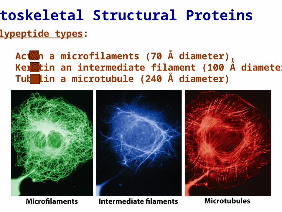
Cytoskeletal Structural ProteinsPolypeptide types:
Actin a microfilaments (70 Å diameter),Keratin an intermediate filament (100 Å diameter)Tubulin a microtubule (240 Å diameter)

Actin Microfilament Assembly
F-actin (filamentous) and G-actin (globular)
Polarity with ATP-binding end the negative (-) end
Greater addition on the positive (+) end
Single subunit

Microfilament Treadmilling

Crawling Cells via Microfilament Treadmilling
Upturned Leading edge
Highly Dense Highly Dense Leading edgeLeading edge
Cytoskeletal Structural ProteinsPolypeptide types:
Actin a microfilaments (70 Å diameter),Keratin an intermediate filament (100 Å diameter)Tubulin a microtubule (240 Å diameter)

Intermediate Filaments
Basic structural unit: dimer of α-helices that wind around each other – coiled coil
Includes soft keratins that define internal structures and hard keratins of skin, hair and claws
Exclusively structural proteins
Dead epidermal skin Dead epidermal skin cells mostly keratincells mostly keratin

Residue Arrangement in a Coiled Coil StructureSeven amino acid pseudo repeat with positions 1 and 4 hydrophobic residues
Hydrophobic strip along the side of the helix

Intermediate Filament Dimer Model
Human hair

Special Intermediate Filament for Large Animals: Collagen
Triple helix extracellular matrix
Holds cells, organs and bones together
Rich in Gly (30%), Pro (30%) and hydroxyproline (30%)
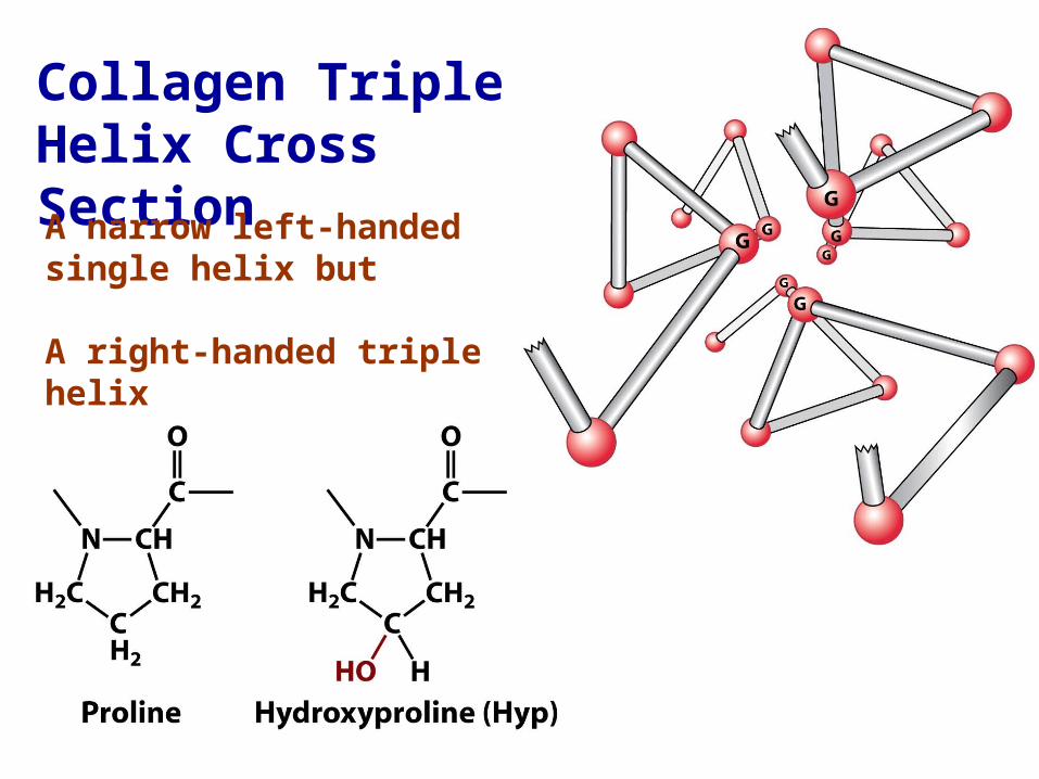
Collagen Triple Helix Cross Section
A narrow left-handed single helix but
A right-handed triple helix
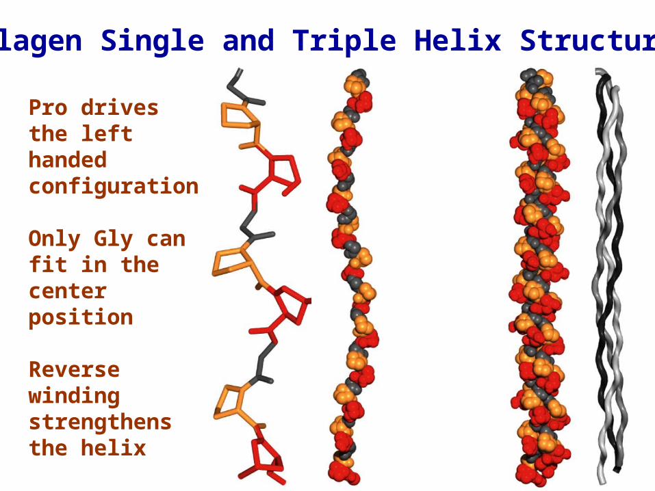
Collagen Single and Triple Helix Structure
Pro drives the left handed configuration
Only Gly can fit in the center position
Reverse winding strengthens the helix
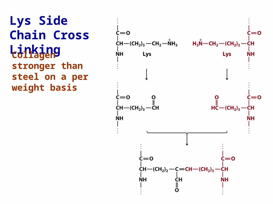
Lys Side Chain Cross Linking
Collagen stronger than steel on a per weight basis

Cytoskeletal Structural ProteinsPolypeptide types:
Actin a microfilaments (70 Å diameter),Keratin an intermediate filament (100 Å diameter)Tubulin a microtubule (240 Å diameter)
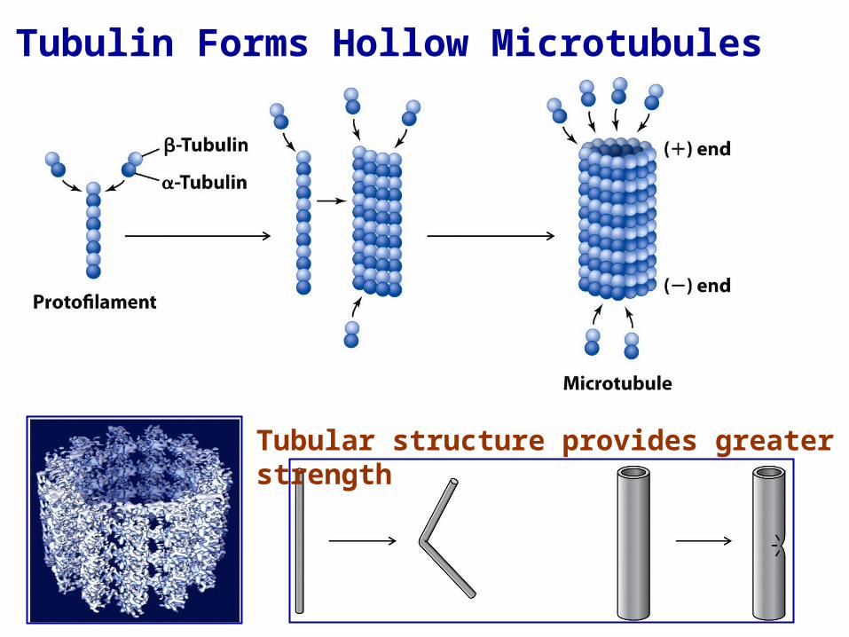
Tubulin Forms Hollow Microtubules
Tubular structure provides greater strength
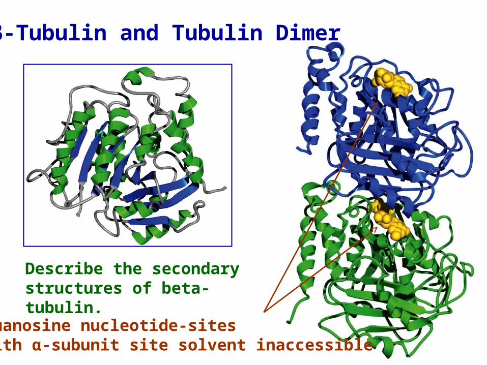
β-Tubulin and Tubulin Dimer
Describe the secondary structures of beta-tubulin.
Guanosine nucleotide-sites with α-subunit site solvent inaccessible

Microtubules Function in Cell Division
Draws chromosomes apart before cell division
Polar structurethat grows morerapidly at the (+)end
Microtubule treadmilling can occur

Drug-Mediated Microtubule DepolymerizationColchicine binds between the alpha and beta subunits

Drug-Mediated Microtubule Polymerization
Taxol stabilizes beta-tubulin subunits preventing depolymerization
Blocks rapidly dividing cells such as tumor cells

Motor Protein
Thick Filament
Actin interacts with the thick filament of myosin causing muscle contraction
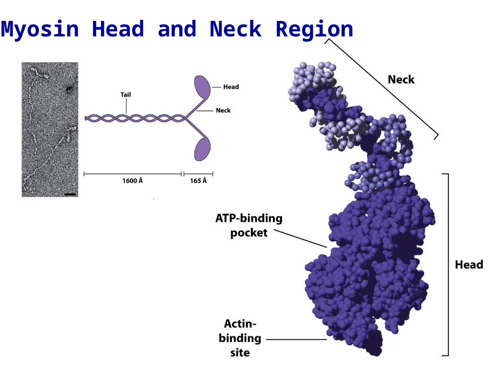
Myosin Head and Neck Region

Binding of ATP to Myosin Releases Myosin from Actin

ATP Hydrolysis
Causes a myosin conformational shift and
Increases myosin binding to actin
Myosin binds to actin farther alongthe thin filament

Myosin binding to actin causes a release of phosphate and ADP
Power Stroke

Chapter 5 Problems:
1, 3, 5, 7, 9, 11, 13, and 17, 20, 25, 51


