CYTOLOGY: What every general practitioner...
30
CYTOLOGY: What every general practitioner should know (… and every future specialist) 16 April 2014 Dr C Crause Pathologist and Senior Lecturer Department of Anatomical Pathology University of Pretoria/NHLS
Transcript of CYTOLOGY: What every general practitioner...

CYTOLOGY:
What every general
practitioner should know
(… and every future specialist)
16 April 2014
Dr C Crause
Pathologist and Senior Lecturer
Department of Anatomical Pathology
University of Pretoria/NHLS

What is cytology?
• Study of single cells
– Pap smears
– Sputum
– Fluids (pleural, pericardial, ascitis and urine)
– FNA/FNAB of preferably solid lesions
– Please not blood, bone marrow aspirates or trephines
– Please not chunks of tissue
• Histology
– This is where chunks of tissue belong

Why cytology?
SAFE• Simple
• Accurate
• Fast
• Economic

How (method in detail)
Technique of fine needle aspiration (FNA)
• The skin is cleaned with antiseptic
• The sterile 22G needle with an attached 10 or 20ml syringe is inserted into the lesion with the dominant hand while the mass is stabilised with the non-dominant hand.
• The syringe is retracted to produce and maintain a negative pressure of about 5ml.
• The needle is moved in various directions to sample cells from different areas of the mass while maintaining a negative pressure.
• When only the nub of the syringe is filled with aspirate material, the needle is withdrawn while maintaining a negative pressure until the subcutaneous tissue is reached. The piston of the syringe is then released to equalize the pressure.
• The needle is withdrawn and then completely disconnected from the syringe.
• The syringe is filled with air and reconnected to the needle.
• The material is then expressed at the frosted end of the glass slide.

Fixing the specimen on the slide• A second glass slide is placed face down parallel to the bottom slide
and both slides pulled apart in separate directions.• One of the slides is fixed immediately using spray fixative the other
slide is left to air dry. The slides should be clearly marked as to which is fixed and which is air dried.
• Please label all slides in pencil with the patients name and hospital number.
• The aerosol spray fixative must be held 17-22cm from the glass slide.
• The glass slide should be held horizontally while spraying with fixative. (If the slide is held at an angle, the material may be sprayed off the slide).
• The expiry date on the bottle of spray should be checked regularly.

Method of FNA

Types of specimens
• Pap smear
• FNAB/FNA
• Fluids

The request form
• Junk in, junk out ….
• Patient details– Name, surname, sex, age, hospital number
• Clinician details– Name, surname, MP number, telephone number
• Correct date
• Complete clinical history– Please keep it relevant
• Topography – where was the specimen taken from
• Any urgent specimens – please arrange with laboratory

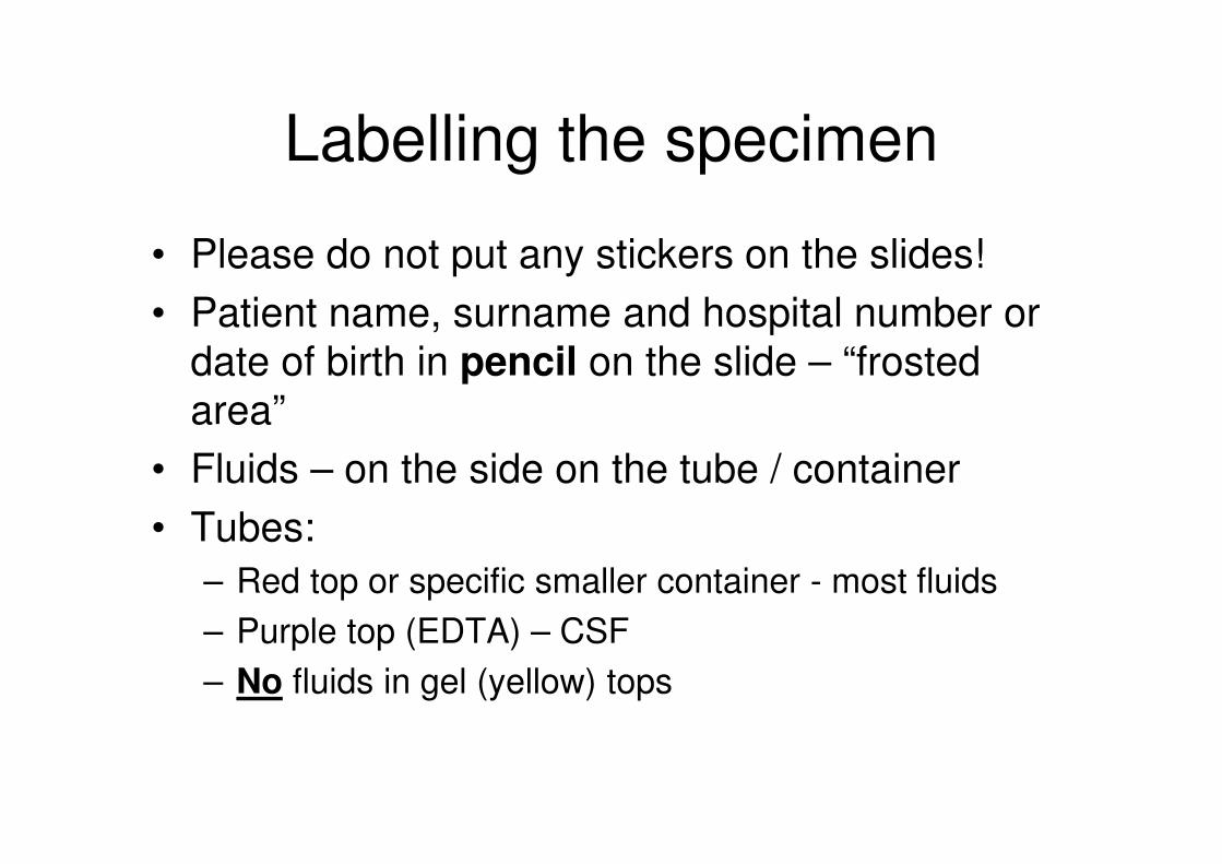
Labelling the specimen
• Please do not put any stickers on the slides!
• Patient name, surname and hospital number or
date of birth in pencil on the slide – “frosted
area”
• Fluids – on the side on the tube / container
• Tubes:
– Red top or specific smaller container - most fluids
– Purple top (EDTA) – CSF
– No fluids in gel (yellow) tops

Pap smears
• Papanicalou smear
• Currently in use to stain these smears
• Useful in diagnosis of:– Infections (HPV, HSV, CMV, Candida, TB…)– In situ malignancy (LSIL and HSIL)– Invasive malignancy (Squamous cell carcinoma)
• Please don’t wipe vigorously before taken the smear
• First pap smear then biopsy (if necessary)
• Please don’t put in the sun to dry
• Immediate fixation (spraycyte)
• Please don’t cover with another slide
• Only representative when endocervical cells present or lesion noted

Sputums
• Please ask your friendly physiotherapist for help (take after morning physio)
• Please take before breakfast
• Often multiple sputums required before final diagnosis (minimum 5)
• Please differentiate between bronchial washings and sputum
• Indications:– Malignancy
– Infective conditions (Tuberculosis)

Fluids
• Ascitis, Pleural and Pericardial
• Indicate topography
• Please send immediately and mark separately for cytology
• Malignancy vs Reactive conditions

Urine
• Midstream if possible
• Catheter if known patient with cervix carcinoma
• Fresh specimen
• Indications:
– Cystitis
– Malignancy
– Bilharzia

CSF
• Indication:
– Malignancy only!!! (ALL and SRBCT/Germ cell
tumours in children)
– No cases of meningitis – microbiology/virology
welcomes them with open arms!
• Separate tube (please not 5ml, 1ml is enough)
• Within 1 hour after obtaining the specimen
• Please contact the laboratory and arrange that
this specimen is received and prepared in time

FNA/FNAB
• Spread evenly
• Spray immediately
• Maximum 4 slides– Two spray fixed
– Two air dried
• Please don’t cover the slides
• Two sites:– Two separate forms (4 slides / site)
– Breast (left and right)
– Axilla and inguinal area

Important sites
• Salivary gland– Benign vs. Malignant
– Inflammatory conditions
• Breast– Benign vs. Malignant
• Thyroid– MNG, Hyperplastic nodules, reactive conditions and
lesions – benign and malignant
• Lymph node– Reactive, TB, malignancy (lymphoma and metastatic
carcinoma)

Good sites for FNA
• Breast
• Lung
• Thyroid
• Lymph nodes
• Salivary glands

Bad sites for FNA
• Soft tissue
• Intra-abdominal organs

Ugly sites for FNA
• Skin
• Bone
• Cartilage

What happens to the specimen
• Transport
• Registration
• Prepared – laboratory technician
• Screened – cytotegnologist
• Rescreened/Reviewed – pathologist
• Diagnosis entered
• Authorized
• Available on Labtrak


Poor quality
• No cells
• Only blood
• Degenerated
• Crush artifact
• Too thick
• The lesion was missed

Figure 1: Cryptococcus neoformans in a lymph node

Figure 2: Cryptococcus neoformans on CSF

Figure 3: Squamous cell carcinoma on sputum

Figure 4: Dominent nodule in a multinodular goitre

Figure 5: Pleomorphic adenoma of the parotid gland

Liquid based cytology

Cell blocks