Cytogenetics: Karyotypes and Chromosome Aberrations Chapter 6.
63
Cytogenetics: Karyotypes and Chromosome Aberrations Chapter 6
-
Upload
leo-knight -
Category
Documents
-
view
228 -
download
2
Transcript of Cytogenetics: Karyotypes and Chromosome Aberrations Chapter 6.

Cytogenetics: Karyotypes and Chromosome Aberrations
Chapter 6

6.2 The Human Chromosome Set
The number and appearance of chromosomes in the nucleus of an organism is an important characteristic
Chromosome analysis is a powerful and useful technique in human genetics Human chromosomes exist in pairs, with most cells having
23 homologous pairs, or 46 chromosomes. This is the diploid, 2n, number
Eggs and sperm (gametes), contain only one copy of each chromosome, which is haploid, or n number.
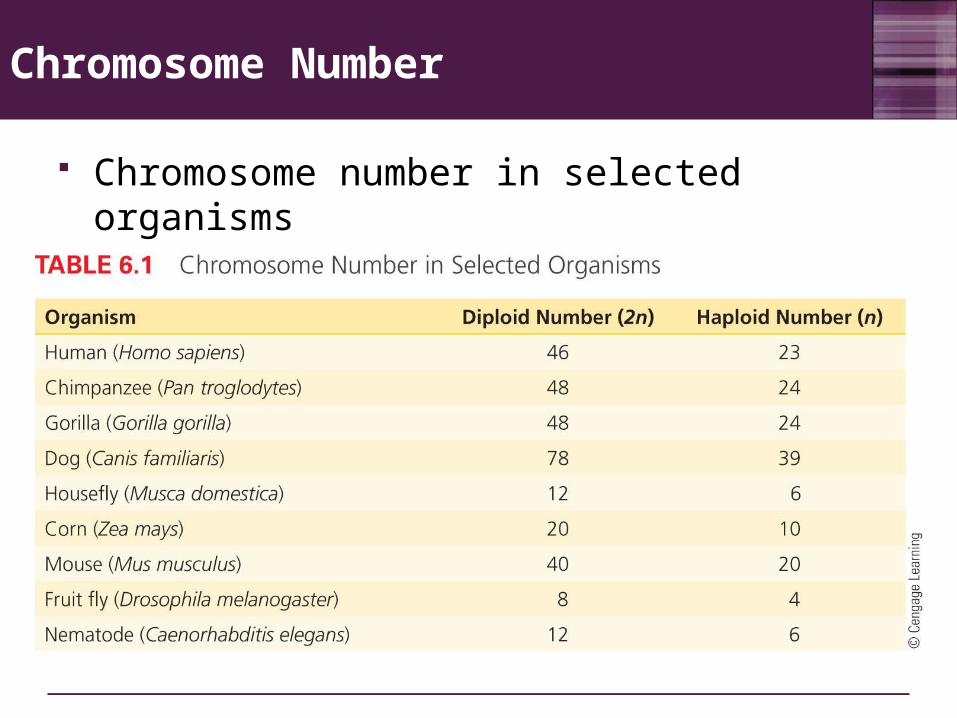
Chromosome Number
Chromosome number in selected organisms

Chromosome Shape
As chromosomes condense and become visible during cell division, certain structural features can be recognized
Centromere A region of a chromosome to which microtubule fibers
attach during cell division Its location gives a chromosome its characteristic
shape Metacentric - A chromosome that has a centrally
placed centromere Submetacentric - A chromosome whose centromere
is placed closer to one end than the other Acrocentric - A chromosome whose centromere is
placed very close to, but not at, one end

Metaphase Chromosomes
Chromosomes are identified by size, centromere location, and banding pattern
Metacentric Submetacentric Acrocentric
Short arm (p)
Satellite
p Centromere
pStalk
Long arm (q)
q q
3 17 21

Human Chromosomes
Replicated chromosomes at metaphase consist of sister chromatids joined by a single centromere

Types of Chromosomes
Sex chromosomes In humans, the X and Y chromosomes are involved in
sex determination. These have different sizes and shapes
Autosomes Chromosomes other than the sex chromosomes In humans, chromosomes 1 to 22 are autosomes
Human chromosomes are analyzed by construction of karyotypes Karyotype - A complete set of chromosomes from a
cell that has been photographed during cell division and arranged in a standard sequence

6.3 Making a Karyotype
Allows any region to be identified by a descriptive address (chromosome number, arm, region, and band)
Different stains and dyes produce banding patterns specific to each chromosome
Karyotypes reveal variations in chromosomal structure and number 1959: Discovery that Down syndrome is caused
by an extra copy of chromosome 21 Chromosome banding and other techniques can
identify small changes in chromosomal structure

A Human Karyotype

Karyogram: Chromosome Banding Patterns
1 23
4 5 6
7 8 9 10 11 12
13 14 1516 17 18
19 20 21 22 YX

System of Naming Chromosome Bands
Band
6Region 5
3 4321
2 2
Arm 31
p 112
q 11 2
123
2 45
3 21
443
12
1
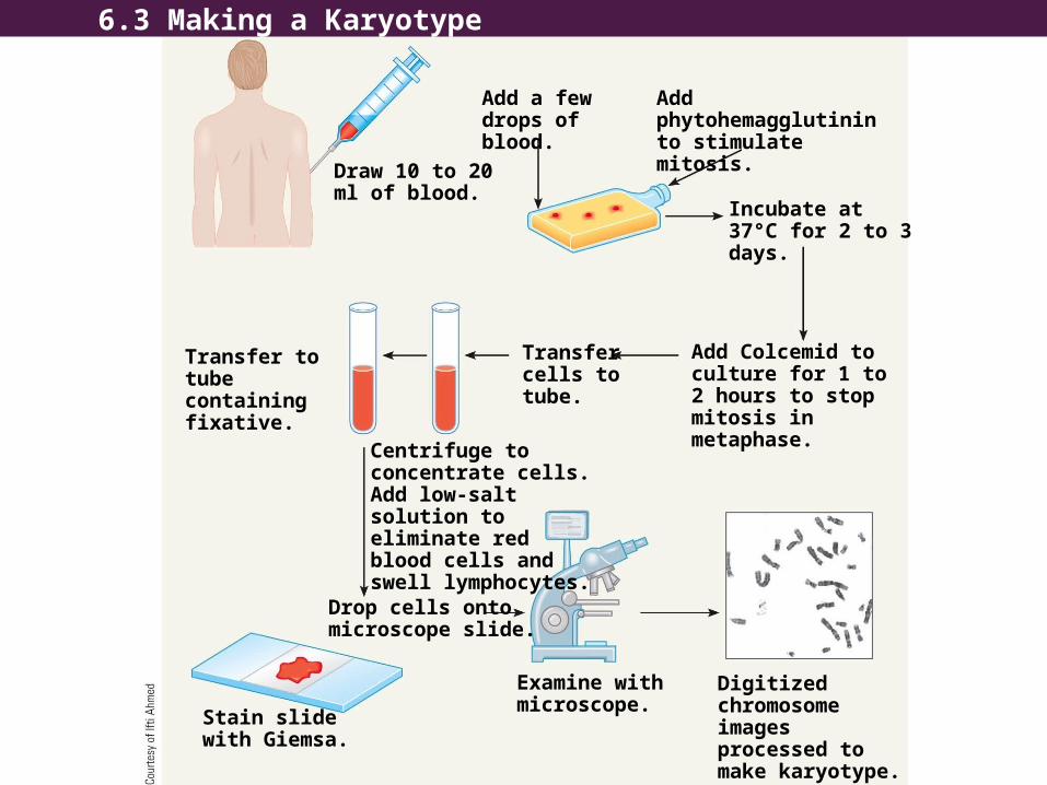
Add a few drops of blood.
Add phytohemagglutinin to stimulate mitosis.
Draw 10 to 20 ml of blood.
Incubate at 37°C for 2 to 3 days.
Transfer to tube containing fixative.
Transfer cells to tube.
Add Colcemid to culture for 1 to 2 hours to stop mitosis in metaphase.Centrifuge to
concentrate cells. Add low-salt solution to eliminate red blood cells and swell lymphocytes.
Drop cells onto microscope slide.
Examine with microscope.
Digitized chromosome images processed to make karyotype.
Stain slide with Giemsa.
6.3 Making a Karyotype

ANIMATION: Karyotype preparation
To play movie you must be in Slide Show ModePC Users: Please wait for content to load, then click to play
Mac Users: CLICK HERE

Metaphase Chromosomes (a) Arranged Into a Karyotype (b)

Information Obtained from a Karyotype
1. Number of chromosomes
2. Sex chromosome content
3. Presence or absence of individual chromosomes
4. Nature and extent of large structural abnormalities

Four Common Chromosome Staining Procedures
Banding technique Appearance of chromosomes
G-banding — Treat metaphase spreads with trypsin, an enzyme that digests part of chromosomal protein. Stain with Giemsa stain. Observe banding pattern with light microscope.
Darkly stained G bands.
Q-banding — Treat metaphase spreads with the chemical quinacrine mustard. Observe fluorescent banding pattern with a special ultraviolet light microscope.
Bright fluorescent bands upon exposure to ultraviolet light; same as darkly stained G bands.

Four Common Chromosome Staining Procedures
R-banding — Heat metaphase spreads at high temperatures to achieve partial denaturation of DNA. Stain with Giemsa stain. Observe with light microscope.
Darkly stained R bands correspond to light bands in G-banded chromosomes. Pattern is the reverse of G-banding.
Banding technique Appearance of chromosomes
C-banding — Chemically treat metaphase spreads to extract DNA from the arms but not the centromeric regions of chromosomes. Stain with Giemsa stain and observe with light microscope.
Darkly stained C band centromeric region of the chromosome corresponds to region of constitutive heterochromatin.
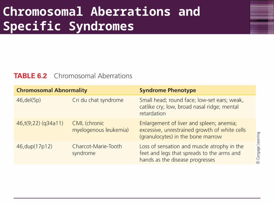
Chromosomal Aberrations and Specific Syndromes

Chromosome Painting
New techniques using fluorescent dyes generate unique patterns for each chromosome

Obtaining Cells for Chromosome Studies
Any nucleus can be used to make karyotype Lymphocytes, skin cells, cells from biopsies,
tumor cells Sampling cells before birth
Amniocentesis Chorionic villus sampling (CVS)

Amniocentesis
A method of sampling the fluid surrounding the developing fetus by inserting a hollow needle and withdrawing suspended fetal cells and fluid A needle is inserted through the abdominal and uterine
walls (avoiding placenta and fetus) into the amniotic sac surrounding the fetus.
10-30mL of fluid is withdrawn – contains cells shed from the skin, respiratory tract, and urinary tract of the fetus
Cell are collected by centrifugation and grown in a lab Used in diagnosing fetal genetic and developmental
disorders Usually performed in the sixteenth week of pregnancy

Amniocentesis
Removal of about 20 ml of amniotic fluid containing suspended cells that were sloughed off from the fetus
A few biochemical analyses with some of the amniotic fluid
Centrifugation
Quick determination of fetal sex and analysis of purified DNA
Fetal cells
Biochemical analysis for the presence of alleles that cause many different metabolic disorders
Growth for several days in culture medium
Karyotype analysis(a)

Amniocentesis
Restricted to certain circumstances:• Mother is over age 35 – risk of chromosome
abnormalities dramatically increases after 35• Mother has a child with a chromosomal aberration• Either parent carries one or more structurally abnormal
chromosomes• Mother is a carrier of X-linked biochemical disorder that
cannot otherwise be diagnosed prenatally and is willing to abort if the fetus is male
• When couple has had a number of previous miscarriages or unexplained fertility problems

Chorionic Villus Sampling (CVS)
A method of sampling fetal chorionic cells (fetal tissue that forms part of the placenta) by inserting a catheter through the vagina or abdominal wall into the uterus Used in diagnosing biochemical and cytogenetic
defects in the embryo Usually performed in the eighth or ninth week of
pregnancy Both amniocentesis and CVS can be coupled
with genomic DNA testing for prenatal diagnosis of mutant alleles.

Chorionic Villus SamplingChorionic
villi
Developing placenta
Ultrasound to monitor procedure
Developing fetus
Bladder
Uterus Chorion
CatheterAmniotic cavity
Rectum
(a)

ANIMATION: Amniocentesis and CVS
To play movie you must be in Slide Show ModePC Users: Please wait for content to load, then click to play
Mac Users: CLICK HERE

Exploring Genetics: Noninvasive Prenatal Diagnosis
Methods are being investigated to isolate fetal cells that can pass into the mother’s bloodstream, and cell-free fetal DNA (cff DNA) for genetic testing

6.4 Variations in Chromosome Number
Changes in chromosome number or chromosome structure can cause genetic disorders
A karyotype can detect A change in chromosomal number A change in chromosomal arrangement

Changes in Chromosome Number
Polyploidy A chromosomal number that is a multiple (3n or
4n) of the normal haploid chromosomal number Aneuploidy
A chromosomal number that is not an exact multiple of the haploid number
Involves the gain or loss of a single chromosome Monosomy – loss of a single chromosome (2n-1) Trisomy – gain of a single chromosome (2n+1)

Polyploidy
Triploidy A chromosomal number that is three times the haploid
number, having three copies of all autosomes and three sex chromosomes
Found in 15-18% of all miscarriages Approximately 75% of all cases of triploidy are 69,XYY and
have two sets of paternal chromosomes Triploid newborns have multiple abnormalities including
enlarged head, fused fingers and toes, and malformations of the mouth, eyes, and genitals
Tetraploidy A chromosomal number that is four times the haploid
number, having four copies of all autosomes and four sex chromosomes
Found in 5% of all miscarriages but is extremely rare in live births

A Triploid Karyotype

A Triploid Infant

Causes of Aneuploidy
Nondisjunction The failure of homologous chromosomes to separate
properly during anaphase in meiosis. Two cell divisions in meiosis, and nondisjunction can
occur in either the first or second, with different genetic consequences:
In meiosis I – all gametes will be abnormal and carry either both members of a chromosomal pair or neither member of the pair
In meiosis II – produces two normal haploid cells and two abnormal cells, one with an extra copy of a chromosome and one missing a chromosome.

ANIMATION: Nondisjunction
To play movie you must be in Slide Show ModePC Users: Please wait for content to load, then click to play
Mac Users: CLICK HERE

Nondisjunction in Meiosis Leads to Aneuploidy
Extra chromosome (n + 1)
Nondisjunction
Extra chromosome (n + 1)
Missing chromosome (n − 1)
Missing chromosome (n − 1)Meiosis I
Meiosis II Gametes
(a)
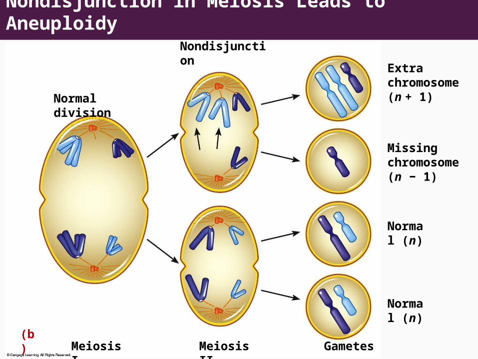
Nondisjunction in Meiosis Leads to AneuploidyNondisjunction
Extra chromosome (n + 1)Normal division
Missing chromosome (n − 1)
Normal (n)
Normal (n)
Meiosis I Meiosis II Gametes(b)

Effects of Monosomy and Trisomy
Autosomal monosomy is a lethal condition Eliminated early in development (spontaneous
abortion)
Some autosomal trisomies are relatively common Most result in spontaneous abortion Three types can result in live births (13, 18, 21)

Trisomies in Spontaneous Abortions
7.5Survey of 4,088
spontaneous abortions
5
4
3
2
Per
cen
tag
e o
f tr
iso
mie
s
1
1 2 3 4 5 6 7 8 9 10 11 12 13 14 15 16 17 18 19 20 21 22
Chromosome number

Trisomy 13: Patau Syndrome (47,+13)
A lethal condition - 1 in 10,000 births Half of all affected individuals die in the first month.
Facial malformations, eye defects, extra fingers or toes, feet with large protruding heels
Internally – malformations of the brain and nervous system and congenital heart defects
Parents of children with trisomy 13 are older (average 32 years old) than parents who have normal children

Trisomy 18: Edwards Syndrome (47,+18)
A lethal condition - 1 in 11,000 births Average survival – 2-4 months
80% are females Small at birth, grow very slowly, mentally retarded,
clenched fists (with second and fifth fingers overlapping the third and fourth fingers and malformed feet), heart malformations – heart failure or pneumonia are usual causes of death
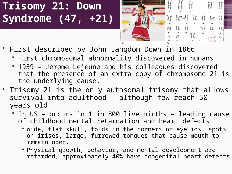
Trisomy 21: Down Syndrome (47, +21)
First described by John Langdon Down in 1866 First chromosomal abnormality discovered in humans 1959 – Jerome Lejeune and his colleagues discovered that
the presence of an extra copy of chromosome 21 is the underlying cause.
Trisomy 21 is the only autosomal trisomy that allows survival into adulthood – although few reach 50 years old In US – occurs in 1 in 800 live births – leading cause of
childhood mental retardation and heart defects Wide, flat skull, folds in the corners of eyelids, spots on irises,
large, furrowed tongues that cause mouth to remain open. Physical growth, behavior, and mental development are
retarded, approximately 40% have congenital heart defects

6.6 Risks for Autosomal Trisomy
The causes of autosomal trisomy are unknown
Factors that have been proposed include:
Genetic predisposition Exposure to radiation Viral infection Abnormal hormone levels
Maternal age is the leading risk factor for trisomy 94% of nondisjunctions occur
in the mother

Why is Maternal Age a Risk Factor?
1. Meiosis is not completed until ovulation Primary oocytes are formed early in embryonic development
and enter meiotic prophase I well before birth. However, meiosis I stops and is not completed until ovulation,
so oocytes produces at age 40 have been in meiosis I for over 40 years.
During this time, intracellular events or environmental agents may increase risk of nondisjunction, resulting in aneuploidy
2. Maternal selection – embryo-uterine interactions normally result in the miscarriage of chromosomally abnormal embryos. Embryo-uterine interactions become less effective as women
age

6.7 Aneuploidy of the Sex Chromosomes
More common than autosomal aneuploidy 1 in 400 for males and 1 in 650 for females
Can involve both X and Y chromosomes A balance is needed for normal development
At least one copy of the X chromosome is required for development
Increasing numbers of X or Y chromosomes causes progressively greater disturbances in phenotype and behavior

Turner Syndrome (45,X) Monosomy of the X chromosome that results in female
sterility. Short, wide-chested, with rudimentary ovaries and puffiness
of the hands and feet May have an aortic constriction, but no mental retardation 1 in 10,000 female births Females require two X chromosomes for normal
development and growth patterns

Klinefelter Syndrome (47, XXY)
Individuals (males) have some fertility problems but few additional symptoms 1 in 1,000 male births 60% of cases – maternal nondisjunction

XYY Syndrome (47,XYY)
1965 – cytogenetic survey of 197 males imprisoned for violent and dangerous antisocial behavior – 9 were XYY 7 of the 9 were of subnormal intelligence Affected individuals are usually taller than normal and
some, but not all, have personality disorders Frequency of XYY in penal and mental institutions is
significantly higher than in the population at large Some defendants have attempted to use their XYY
karyotype as legal defense in criminal trials Vast majority of XYY males lead socially normal lives

6.8 Structural Changes in Chromosomes
Changes in the structure of chromosomes Deletion – loss of a chromosome part Duplication – extra copies of chromosome parts Translocation – transfer of a chromosome part to
another, nonhomologous chromosome Inversion – order of chromosome segments is reversed

ANIMATION: Translocation
To play movie you must be in Slide Show ModePC Users: Please wait for content to load, then click to play
Mac Users: CLICK HERE

ANIMATION: Deletion
To play movie you must be in Slide Show ModePC Users: Please wait for content to load, then click to play
Mac Users: CLICK HERE

Structural Changes in Chromosomes
Deletion of segment F
(a) Deletion

Structural Changes in Chromosomes
(b) Duplication

Deletions
Involve loss of chromosomal material Associated with several genetic disorders
Cri du chat syndrome Prader-Willi syndrome

Caused by a deletion in the short arm of chromosome 5 and occurs in 1 in 20,000 to 1 in 50,000 births Associated with an array of malformations, the most
characteristic of which is an infant cry that resembles a meowing cat due to defects in the larynx
Cri du chat syndrome

Translocations
Two major types:1. Reciprocal translocation – two nonhomologous
chromosomes exchange genetic parts No genetic information gained or lost from the cell, but
genes are moved to new chromosomal locations. In some cases, there are no phenotypic effects, and the
translocation is passed through a family for generations.
2. Robertsonian translocations – can produce genetically unbalanced gametes with duplicated or deleted chromosomal segments that can result in embryonic death or abnormal offspring
About 5% of Down Syndrome cases involve this

14 21Robertsonian translocation
14/21 Translocation carrier
Normal cell
Meiosis and gamete formation
Normal gamete Fertilization
PhenotypeTranslocation
carrierNormal Translocation
Down syndrome
Monosomy 21 lethal
Trisomy 14q lethal
Monosomy 14 lethal
Chromosome number 45 46 46 45 46 45
Robertsonian Translocation

6.8 Consequences of Aneuploidy?
Aneuploidy is the leading cause of reproductive failure in humans Results in miscarriages and birth defects
Aneuploidy also is associated with many cancers Especially leukemia

6.10 Other Forms of Chromosome Changes
Uniparental disomy (UPD) - A condition in which both copies of a chromosome are inherited from a single parent Can occur in meiosis or in mitotic divisions after fertilization. Associated with several genetic diseases
Females affected with rare X-linked disorders, such as hemophilia
Father-to-son transmission of rare, X-linked disorders where the mother is homozygous for the normal allele
Children affected with rare autosomal recessively inherited disorders where only one parent is heterozygous
Autosomal recessive disorders (Prader-Willi syndrome, Angelman syndrome) – deletions in the long arm of chromosome 15 or by UPD
If both copies of chromosome 15 are inherited from the mother, the child will have Prader-Willi syndrome
If both copies of chromosome 15 are inherited from the father, the child will have Angelman syndrome
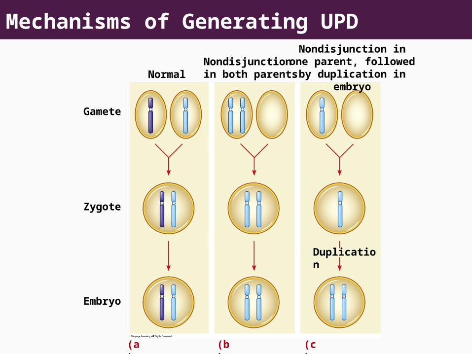
Mechanisms of Generating UPDNondisjunction in one
parent, followed by duplication in embryo
Nondisjunction in both parentsNormal
Gamete
Zygote
Duplication
Embryo
(a) (b) (c)

6.10 Other Forms of Chromosome Changes (contd.)
Copy number variation A particular gene or chromosomal region is present in
multiple copies The relationship between copy number variation in the PMP22
gene and the symptoms of Charcot-Marie-Tooth Syndrome A duplication encodes a protein involved in making a sheath
surrounding nerve cells. Duplication disrupts the production of this protective sheath and
causes an autosomal dominant disease Normal individuals – two copies of region – one on each
homologue CMT – three copies of this chromosome region, two on one
homologue, and one on the other

Human Diseases Associated with Copy Number Variants

6.10 Other Forms of Chromosome Changes (contd.)
Fragile sites Appear as gaps or breaks in chromosomes• Over 100 fragile sites have been identified in the human
genome.• Regions susceptible to breakage• One fragile site on the X chromosome is associated
with a common form of mental retardation in males know as Fragile X Syndrome (Martin-Bell Syndrome)

Fragile Sites on the X Chromosome
FRAX BFRAX C
FRAX F
FRAX AFRAX D
(a)FRAX E