Cytogenetic map of common bean (Phaseolus vulgaris L.) et al. Cytog...Cytogenetic map of common bean...
16
Cytogenetic map of common bean (Phaseolus vulgaris L.) Artur Fonsêca & Joana Ferreira & Tiago Ribeiro Barros dos Santos & Magdalena Mosiolek & Elisa Bellucci & James Kami & Paul Gepts & Valérie Geffroy & Dieter Schweizer & Karla G. B. dos Santos & Andrea Pedrosa-Harand Received: 28 January 2010 / Revised: 12 March 2010 / Accepted: 28 March 2010 / Published online: 7 May 2010 # The Author(s) 2010. This article is published with open access at Springerlink.com Abstract A cytogenetic map of common bean was built by in situ hybridization of 35 bacterial artificial chromosomes (BACs) selected with markers mapping to eight linkage groups, plus two plasmids for 5S and 45S ribosomal DNA and one bacteriophage. Together with three previously mapped chromosomes (chromo- somes 3, 4, and 7), 43 anchoring points between the genetic map and the cytogenetic map of the species are now available. Furthermore, a subset of four BAC clones was proposed to identify the 11 chromosome pairs of the standard cultivar BAT93. Three of these BACs labelled more than a single chromosome pair, indicating the presence of repetitive DNA in their inserts. A repetitive distribution pattern was observed for most of the BACs; for 38% of them, highly repetitive pericentromeric or subtelomeric signals were Chromosome Research (2010) 18:487–502 DOI 10.1007/s10577-010-9129-8 Responsible Editor: Hans de Jong. Electronic supplementary material The online version of this article (doi:10.1007/s10577-010-9129-8) contains supplementary material, which is available to authorized users. A. Fonsêca : J. Ferreira : T. R. B. dos Santos : K. G. B. dos Santos : A. Pedrosa-Harand Laboratory of Plant Cytogenetics, Department of Botany, Federal University of Pernambuco, Recife, PE 50670-420, Brazil M. Mosiolek : D. Schweizer : A. Pedrosa-Harand (*) Department of Chromosome Biology, University of Vienna, 1030 Vienna, Austria e-mail: [email protected] M. Mosiolek : D. Schweizer Gregor Mendel Institute of Molecular Plant Biology, Austrian Academy of Sciences, 1030 Vienna, Austria E. Bellucci Dipartimento di Scienze Ambientali e delle Produzioni Vegetali, Università Politecnica delle Marche, 60131 Ancona, Italy E. Bellucci National Institute of Agricultural Botany, Cambridge CB3 0LE, UK J. Kami : P. Gepts Department of Plant Sciences/MS1, Section of Crop and Ecosystem Sciences, University of California, Davis, CA 95616-8780, USA V. Geffroy Institut de Biotechnologie des Plantes, UMR-CNRS 8618, INRA, Université Paris Sud, 91405 Orsay, France V. Geffroy Unité Mixte de Recherche de Génétique Végétale, Institut National de la Recherche Agronomique, 91190 Gif-sur-Yvette, France
Transcript of Cytogenetic map of common bean (Phaseolus vulgaris L.) et al. Cytog...Cytogenetic map of common bean...

Cytogenetic map of common bean (Phaseolus vulgaris L.)
Artur Fonsêca & Joana Ferreira & Tiago Ribeiro Barros dos Santos &
Magdalena Mosiolek & Elisa Bellucci & James Kami & Paul Gepts &
Valérie Geffroy & Dieter Schweizer & Karla G. B. dos Santos &
Andrea Pedrosa-Harand
Received: 28 January 2010 /Revised: 12 March 2010 /Accepted: 28 March 2010 /Published online: 7 May 2010# The Author(s) 2010. This article is published with open access at Springerlink.com
Abstract A cytogenetic map of common bean wasbuilt by in situ hybridization of 35 bacterial artificialchromosomes (BACs) selected with markers mappingto eight linkage groups, plus two plasmids for 5S and45S ribosomal DNA and one bacteriophage. Togetherwith three previously mapped chromosomes (chromo-somes 3, 4, and 7), 43 anchoring points between thegenetic map and the cytogenetic map of the species are
now available. Furthermore, a subset of four BACclones was proposed to identify the 11 chromosomepairs of the standard cultivar BAT93. Three of theseBACs labelled more than a single chromosome pair,indicating the presence of repetitive DNA in theirinserts. A repetitive distribution pattern was observedfor most of the BACs; for 38% of them, highlyrepetitive pericentromeric or subtelomeric signals were
Chromosome Research (2010) 18:487–502DOI 10.1007/s10577-010-9129-8
Responsible Editor: Hans de Jong.
Electronic supplementary material The online version of thisarticle (doi:10.1007/s10577-010-9129-8) contains supplementarymaterial, which is available to authorized users.
A. Fonsêca : J. Ferreira : T. R. B. dos Santos :K. G. B. dos Santos :A. Pedrosa-HarandLaboratory of Plant Cytogenetics, Department of Botany,Federal University of Pernambuco,Recife, PE 50670-420, Brazil
M. Mosiolek :D. Schweizer :A. Pedrosa-Harand (*)Department of Chromosome Biology,University of Vienna,1030 Vienna, Austriae-mail: [email protected]
M. Mosiolek :D. SchweizerGregor Mendel Institute of Molecular Plant Biology,Austrian Academy of Sciences,1030 Vienna, Austria
E. BellucciDipartimento di Scienze Ambientali e delle ProduzioniVegetali, Università Politecnica delle Marche,60131 Ancona, Italy
E. BellucciNational Institute of Agricultural Botany,Cambridge CB3 0LE, UK
J. Kami : P. GeptsDepartment of Plant Sciences/MS1,Section of Crop and Ecosystem Sciences,University of California,Davis, CA 95616-8780, USA
V. GeffroyInstitut de Biotechnologie des Plantes, UMR-CNRS 8618,INRA, Université Paris Sud,91405 Orsay, France
V. GeffroyUnité Mixte de Recherche de Génétique Végétale,Institut National de la Recherche Agronomique,91190 Gif-sur-Yvette, France

observed. These distribution patterns corresponded topericentromeric and subtelomeric heterochromatinblocks observed with other staining methods. Alto-gether, the results indicate that around half of thecommon bean genome is heterochromatic and thatgenes and repetitive sequences are intermingled in theeuchromatin and heterochromatin of the species.
Keywords fluorescent in situ hybridization (FISH) .
bacterial artificial chromosome (BAC) . physical map .
repetitive DNA sequences . Fabaceae
AbbreviationsBAC Bacterial artificial chromosomeCMA Chromomycin A3DAPI 4′,6-diamidino-2-phenylindoleFISH Fluorescent in situ hybridizationFITC Fluorescein isothiocyanateLG Linkage groupRAPD Random amplification of polymorphic DNArDNA Ribosomal DNASDS Sodium dodecyl sulfateSSC Saline–sodium citrate
Introduction
Common bean (Phaseolus vulgaris L.) is the mosteconomically important species of the genus Phaseo-lus and the primary dietary protein source for severalpopulations, mainly in Latin America and Africa(Evans 1986). In order to assist common beanbreeding, several tools have been developed for thisspecies, including genetic maps (Vallejos et al. 1992;Nodari et al. 1993; Adam-Blondon et al. 1994; Freyreet al. 1998; Hougaard et al. 2008) and bacterialartificial chromosome (BAC) libraries (Vanhoutenand Mackenzie 1999; Kami et al. 2006; Gepts et al.2008). In 2003, an international consortium namedPhaseomics was created in an effort to accelerate theimprovement of common bean (Broughton et al.2003). One of the aims of this initiative was toestablish a cytogenetic-based physical map for thisspecies.
Cytogenetic maps of different plant species havebeen developed in fluorescent in situ hybridization(FISH) experiments using BAC clones as probes(Jiang and Gill 2006). This approach is especially
recommended for constructing maps of species witha small genome because the large proportion ofrepetitive DNA in larger genomes may hamper themapping of BAC clones to unique genomic positions(Jiang and Gill 1996; Dong et al. 2000; Islam-Faridiet al. 2002; Kim et al. 2005b; Pedrosa et al. 2002).Such maps are often associated with genetic andcontig maps, and may be useful during whole-genome sequencing projects, either helping to eval-uate the size of the putative remaining gaps (Chenget al. 2001) or helping to decide which BACs areeuchromatic and potentially gene rich and thusshould be sequenced (Young et al. 2005; Peters etal. 2009).
Common bean is a small-genome species selectedfor whole-genome sequencing (Gepts et al. 2005), andaccession G19833 is currently being sequenced by agroup of US laboratories (Scott Jackson, personalcommunication). Its chromosomes (2n=22) are small(around 2 µm) and have similar morphologies,hindering a detailed cytogenetic characterization byclassic methods. Since FISH was first applied to itsmitotic chromosomes, major advances have, however,been obtained (Moscone et al. 1999; Pedrosa et al.2003; Pedrosa-Harand et al. 2006). A preliminaryFISH analysis has suggested a low general correlationbetween genetic and physical distances when linkagegroups (LGs) and chromosomes are compared,indicating the necessity to establish a more detailedphysical map (Pedrosa et al. 2003). Recently,Pedrosa-Harand et al. (2009) published a BAC FISHmapping for three ‘BAT93’ common bean chromo-somes (chromosomes 3, 4, and 7, according to thestandard common bean nomenclature proposed byPedrosa-Harand et al. 2008), which reinforced the lowcorrelation between genetic and physical distances inthis crop.
In the present study, we extended this analysis tocomplete the chromosomal map of common bean.For this purpose, BACs from a genomic library fromthe Mesoamerican genotype BAT93 (Kami et al.2006) were selected for the remaining eight chro-mosome pairs (chromosomes 1, 2, 5, 6, 8, 9, 10, and11) and mapped by FISH. The results were com-pared to the corresponding genetic maps (Vallejos etal. 1992; Hougaard et al. 2008) and correlated to thedistribution of different repetitive sequences on eachchromosome.
488 A. Fonsêca et al.

Materials and methods
Plant material
Seeds from the P. vulgaris Mesoamerican breedingline BAT93 were obtained from the germplasm bankof the International Center for Tropical Agriculture(CIAT; Cali, Colombia).
Chromosome preparation and fluorochrome staining
Root tips obtained from germinated seeds werepretreated with 2 mM 8-hydroxyquinoline for 18 hat 10°C, fixed in ethanol–acetic acid (3:1 vol/vol),and stored in fixative at −20°C for up to severalweeks. Somatic chromosome preparation, selection ofslides, chromomycin A3 (CMA)/4′,6-diamidino-2-phenylindole (DAPI) staining, and destaining forFISH were performed in accordance with Cabral etal. (2006).
Pachytene chromosome spreads were prepared asdescribed previously, except that flower buds werefixed without pretreatment and digested in 2% (wt/vol)cellulase Onozuka R-10 (Serva), 1% (wt/vol) pecto-lyase (Sigma-Aldrich), and 1% (wt/vol) cytohelicase(Sigma-Aldrich) for 2 h at 37°C, and meiocytes werewashed twice for 5 min in ice-cold 0.01 M citric acid–sodium citrate buffer (pH 4.8) and left in distilled waterovernight at 4°C before dissection.
DNA probes
The probe D2, a 500-bp fragment containing 5Sribosomal DNA (rDNA) from Lotus japonicus(Pedrosa et al. 2002), and the probe R2, a 6.5-kbfragment of an 18S–5.8S–25S rDNA repeat unit fromArabidopsis thaliana (Wanzenböck et al. 1997), wereused to localize 5S and 45S rDNA sites, respectively.
BAC clones were selected by screening high-density BAC filters from a BAT93 HindIII genomiclibrary (Kami et al. 2006) using genetically mappedmarkers (the common bean genomic plasmid cloneBng), as described by Pedrosa et al. (2003) andPedrosa-Harand et al. (2009). A second group ofBAC clones was selected from the same library usingthe legume marker Leg (Hougaard et al. 2008), as willbe described later. Finally, one λ bacteriophage,SJ19.12, obtained after screening of a JaloEEP558
genomic library with a nucleotide-binding site probe(Ferrier-Cana et al. 2003), was also included in thisanalysis. Bacteriophage SJ19.12 mapped at one endof LG B10, distal to marker D1476, in the vicinity ofthe PROD15-680 random amplification of poly-morphic DNA (RAPD) marker (Geffroy et al. 2000).
BAC and plasmid DNA were isolated using thePlasmid Mini Kit (Qiagen), whereas DNA Nucleo-bond AX columns (Macherey-Nagel) were used forbacteriophage isolation, both following the manufac-turers' instructions. All selected clones were labelledby nick translation (Invitrogen or Roche Diagnostics)with digoxigenin-11-dUTP (Roche Diagnostics),Cy3-dUTP (5-amino-propargyl-2′-deoxyuridine 5′-triphosphate coupled to red cyanine fluorescent dye;GE), or SpectrumGreen-dUTP (Vysis).
Screening of BAC library with Leg
The Laboratory of Gene Expression, Department ofMolecular Biology, University of Aarhus, Denmark,provided 13 Leg primer pairs developed using theapproach described by Fredslund et al. (2005, 2006a,b) and 17 Leg sequences of P. vulgaris BAT93(Hougaard et al. 2008) for the LG of interest. Usingthese sequences, we designed primer pairs withPrimer3 software (Rozen and Skaletsky 2000; http://frodo.wi.mit.edu/). After selecting 17 low-copy orsingle-copy Leg probes by Southern blot or dot blotanalysis, we radioactively screened the BAC libraryin accordance with Kami et al. (2006), with minormodifications, using two (of six) filters for each of thethree pools of five to six probes. In order to assigneach clone to the corresponding probe, we extractedBAC DNA following a standard alkaline lysisplasmid miniprep protocol and amplified it with theLeg primer pairs.
Dot blot analysis for detection of BACs containingrepetitive DNA
Denatured BAC DNA corresponding to Bng133 andBng152 from chromosome 5 was dot blotted onto aNylon membrane (Hybond-N+; GE) and submittedto hybridization with the C0t−1 fraction [where C0 isthe initial concentration of single-stranded DNA (inmol/L), and t is the reannealing time (in s)] of P.vulgaris genomic DNA as probe, isolated in accor-
Cytogenetic map of common bean 489

dance with Zwick et al. (1997). The probe waslabelled with digoxigenin using the Dig high-primeDNA labelling kit (Roche Diagnostics). The mem-brane was hybridized overnight with probe DNA inDig Hyb hybridization buffer (Roche Diagnostics) at37°C. After hybridization, the membranes werewashed twice in 2× saline–sodium citrate (SSC)buffer and 0.1% sodium dodecyl sulfate (SDS) for5–15 min, and in 0.5× SSC buffer and 0.1% SDS for15 min at 68°C. The detection was performed usinganti-DIG alkaline phosphatase conjugate (RocheDiagnostics) and the chemiluminescent substrateCDP-Star (Roche Diagnostics), according to themanufacturer's instructions. Signals were capturedon an X-ray ECL film (GE).
Fluorescence in situ hybridization
The FISH procedure applied to both mitotic andmeiotic chromosomes was essentially the same aspreviously described (Pedrosa et al. 2002). Mitotic andmeiotic preparations were denatured for 5 min at 75°Cand for 3 min at 73°C, respectively. The P. vulgarisC0t−100 fraction was added in 20-fold to 400-foldexcess to the hybridization mix to block repetitivesequences when necessary. Digoxigenin-labelledprobes were detected with 0.4 μl of sheep anti-digoxigenin conjugated with fluorescein isothiocyanate(FITC; Roche Diagnostics) and with 0.7 μl of donkeyanti-sheep/goat conjugated with FITC (Serotec) in 3%bovine serum albumin in phosphate-buffered saline.Reprobing of slides for localization of different DNAsequences in the same cell was performed, in accor-dance with Heslop-Harrison et al. (1991, 1992), up tofour times. When a repetitive probe was used in aprevious hybridization, reprobing of slides was per-formed after the chromosomal DNA had been dena-tured with 100 µl of 50% formamide in 2× SSC bufferat 75°C for 5 min, dehydrated in an ice-cold ethanolseries, and air dried.
Data analysis
Photographs were taken in an epifluorescence LeicaDMLB microscope equipped with a Cohu charge-coupled device video camera using the Leica QFISHsoftware. For final processing, images were super-imposed and artificially colored using the AdobePhotoshop software version 10.0 and adjusted for
brightness and contrast only. Chromosomes werenamed and oriented according to the standard com-mon bean nomenclature (Freyre et al. 1998; Pedrosa-Harand et al. 2008).
Measurements
Relative chromosome size and arm ratio werecalculated based on measurements of the chromo-some and arm lengths of at least five mitoticmetaphases. The centromere was determined bythe presence of DAPI+ bands at this position afterFISH. The “measurement” tool of Adobe Photoshopwas used for all size estimations, including the sizeof repetitive BAC signals and 45S rDNA clusters inrelation to chromosome size. The size of DAPI+
bands generated after FISH was measured from themore conspicuous and reproducible terminal andpericentromeric bands, and for major nucleolarorganizer regions on chromosomes 9 and 10(excluding the distended region). For each of thesethree categories, the total measured value wascompared to the total genome size obtained bymeasuring all chromosome lengths. Neither thebrightness nor the contrast of these pictures wasadjusted for measurements in order to avoid possibledistortions in signal extension.
In order to establish the relative position of eachclone (single-copy BACs, bacteriophage, and rDNAsites) along the chromosomes, we selected at least 15clear hybridization signals. In this case, the softwareImage Tool 3.0 was used for measurements after thecontrast and the brightness of the pictures had beenadjusted with Adobe Photoshop 10.0. To calculate theposition of the signals, we took the followingmeasurements: (1) the distance between the oppositetelomere and the center or the start (45S rDNA) of thesignal; (2) the distance between both telomeres(which gives the total chromosomal length), byprolonging the previous measurement until the closesttelomere; and (3) the ratio between the first measure-ment and the second measurement, to determine therelative position of the signal along the chromatid.The top of the short arm was conventionally deter-mined as 0, and the bottom of the long arm wasconventionally determined as 1. Assignment of aclone to a specific chromosome arm was confirmedby reprobing the slides with a previously mappedclone.
490 A. Fonsêca et al.

Results
In order to achieve a high genome coverage for theeight unmapped common bean chromosomes (chro-mosomes 1, 2, 5, 6, 8, 9, 10, and 11), we used 74 Bngmarkers (Vallejos et al. 1992) and 17 single-copy Leg(Hougaard et al. 2008) mapped to the correspondingLGs for screening the BAT93 BAC library (Kami etal. 2006). Following the two different approachesdescribed previously, we identified 82 BACs for 23Bng markers and 39 BACs for 11 Leg, making a totalof 121 BACs corresponding to 34 markers (Supple-mentary Table 1). For the remaining markers used, nocorresponding BAC could be identified.
In the present article, 35 of the above selectedBACs (Table 1)—plus BAC gF11 (previously selectedfor LG B8 by Melotto et al. 2004), one bacteriophage(SJ19.12, mapped to LG B10), and two plasmids(containing 5S and 45S rDNA sequences)—werehybridized in situ. The other BACs listed in Supple-mentary Table 1 were not used for FISH because theywere selected with the same markers or mappedgenetically very close to localized clones. Consider-ing all the 36 BACs hybridized in the present articleand 30 BACs hybridized previously (Pedrosa-Harandet al. 2009), making a total of 66 BACs used forconstructing a cytogenetic map for common bean, weobserved that only 39 (59%) showed unique localizedsignals in just one chromosome pair; however, forhalf of them (18 BACs), the use of C0t−1 or C0t−100blocking DNA in the hybridization mix (20-fold to100-fold more concentrated than the probe) wasnecessary in order to eliminate labelling of dispersedrepetitive sequences and to obtain unique signals.Although selected with single-copy markers, 25BACs (38%) showed highly repetitive signals, pre-dominantly pericentromeric or subtelomeric, in allchromosomes and could not be mapped. The remain-ing two BACs did not give a signal.
Many attempts to block pericentromeric and sub-telomeric sequences present in repetitive BACs—from using C0t genomic fractions as general blockerto using a specific blocker (khipu satellite) forsubtelomeric BACs—were performed (David et al.2009; K.G.B. dos Santos, unpublished data).However, none of the attempts was successful (datanot shown), possibly because the proportion of theserepetitive sequences in the genome is too high.Nevertheless, it was possible to remove subtelomeric
signals (but not the pericentromeric ones) from slides,making them useful for rehybridization with otherprobes.
Hybridization signals of 15 BACs, showing prac-tically the same pattern in the pericentromeric regionof all chromosomes and corresponding approximatelyto 34% of the chromosome complement length,coincided with the CMA+ bands generated afterCMA/DAPI staining (Fig. 1a, b), except for bandscorresponding to 45S rDNA sites. After the FISHprocedure, small DAPI+ bands were sometimes alsovisible at the centromeric region of mitotic chromo-somes, corresponding to 12% of the chromosomecomplement length (Fig. 1c). However, no BAC usedin this work showed a similar centromeric hybridiza-tion pattern. These centromeric DAPI+ bands possiblycorrespond to centromeric repeats not interspersedwith single-copy sequences and, therefore, not isolat-ed by our screening. On the other hand, terminalDAPI+ bands, generated after FISH, coincided eitherwith major 45S rDNA sites or with regions labelledby five subtelomeric BACs (Fig. 1f), corresponding to5% and 9% of the chromosome complement length,respectively. Considering both CMA+ and DAPI-after-FISH bands as indicative of constitutive hetero-chromatin (see "Discussion"), we estimate that 48% ofthe common bean somatic karyotype is heterochromat-ic (5% of rDNA, 9% of other subtelomeric blocks,and 34% of pericentromeric blocks). Although thisfraction is visibly enriched in repetitive sequences,it is also populated by single-copy sequences, andlikely by genes, since single-copy markers are presentin the inserts of BACs showing heterochromatindistribution.
Although single-copy sequences are generallymore efficient for chromosome identification (Figs. 2,3, 4), BACs showing repetitive hybridization patternswere very informative in this study. With only onesingle-copy BAC (BAC 177I19 for chromosome 8)and three repetitive BACs [one pericentromeric (BAC12M3), one subtelomeric (BAC 63H6), and oneshowing a repeat block in chromosome 7 (BAC255F18)], it was possible to identify each chromo-somal pair of ‘BAT93’ through differences in signalintensity and localization, combined with chromo-some size. A BAC for chromosome 5 (36H21)confirmed its correct identification by the four-BACpool probe (Fig. 3e–g). The rDNA-bearing chromo-somes (chromosomes 6, 9, and 10) could be alterna-
Cytogenetic map of common bean 491

tively distinguished by the distribution of theirrepetitive sequences (Fig. 4).
Eighteen selected BACs plus BAC gF11 (selectedwith the SAS13 marker from the anthracnose resis-
tance locus C0−4) and bacteriophage SJ19.12 weremapped to the eight chromosomes analyzed in thepresent study (Table 2 and Figs. 2, 3, 4). On average,two clones were mapped per chromosome, but only
Table 1 List of mapped markers used for screening BAC clones and the general pattern of hybridization of the selected BACs afterFISH with or without blocking DNA
LGa Markerb BAC clone FISH pattern without blocking DNA FISH pattern with blocking DNA
B1/H Bng41 221F15 Unique + weakly scattered Unique 20� C0t � 100ð ÞBng171 38C24 Unique –
Bng173 257L12 Unique –
B2/D 4-Gm 21N14 Disperse proximally Unique 80� C0t � 100ð ÞBng45 225P10 Unique –
Bng57 14F2 Subtelomeric Subtelomeric 100� C0t � 100ð Þ18D16 Disperse proximally + one terminal block Disperseproximally 20� C0t � 1ð Þ
Bng174 92I7 Pericentromeric –
L120 82K2 No signal –
L188 127F19 Unique –
L224 17P14 Subtelomeric –
B5/E Bng49 36H21 Disperse Unique 70� C0t � 100ð ÞBng133 230M2 Pericentromeric –
Bng152 193O2 Pericentromeric –
Bng161 103P12 Pericentromeric Pericentromeric 400� C0t � 100ð ÞB6/G Bng95 121F5 Unique –
Bng177 143N10 Pericentromeric –
Bng202 18B15 Unique –
L56 260H1 Pericentromeric –
B8/F Bng58 169G16 Disperse Unique 50� C0t � 100ð ÞBng96 234P20 Subtelomeric Subtelomeric 50� C0t � 100ð ÞBng138 177I19 Unique –
B9/K Bng2 224I16 Unique + weakly scattered Unique 50� C0t � 100ð ÞL159 123O22 Pericentromeric Pericentromeric (50× C0t − 100)
L206 37P17 No signal –
L207 163I7 Unique + weakly scattered Unique 20� C0t � 100ð ÞB10/I Bng200 173P6 Unique + weakly scattered Unique 20� C0t � 100ð Þ
Bng218 63H6 Subtelomeric –
81A17 Pericentromeric –
L177 119E19 Subtelomeric –
B11/J Bng1 25D1 Disperse Unique 60� C0t � 100ð ÞBng25 255F18 Unique + one terminal block –
Bng112 179N14 Unique –
Bng187 66N11 Pericentromeric –
L220 127J2 Unique + weak subtelomeric –
(–) Not analyzed.a LGs D, E, and so on, defined by Vallejos et al. (1992), corresponding to LGs B2, B5, and so on from the common bean core map(Freyre et al. 1998), as indicated previously.bBng markers were mapped by Vallejos et al. (1992) on the Florida map, while markers 4-Gm and Leg were mapped by Hougaard etal. (2008) on another mapping population.
492 A. Fonsêca et al.

one BAC (36H21) could be mapped to chromosome5. After dot blot analysis using the C0t−1 genomefraction as probe, no other BAC identified for thischromosome was selected for FISH, since all showedstrong hybridization signals indicative of the presenceof highly repetitive DNA in its insert (data notshown). For chromosome 1, two BACs were colo-calized on the distal region of the long arm (Fig. 2a).In order to have a higher-resolution mapping of thecolocalized BACs, we used pachytene chromosomes,providing evidence that BAC 257L12 is moreterminally located (Fig. 2a, bottom insert).
Based on previous (Pedrosa-Harand et al. 2009)and present data, an idiogram was built for commonbean (Fig. 5). P. vulgaris cv. BAT93 has threemetacentric chromosomes (chromosomes 4, 5, and8), seven submetacentric chromosomes (chromo-somes 1, 2, 3, 7, 9, 10, and 11), and one acrocentricchromosome (chromosome 6), according to the
morphology classification of dos Santos Guerra(1986). Chromosomes 6 and 10 have both 5S and45S rDNA sites. An additional 45S rDNA site ispresent on chromosome 9. Pericentromeric repetitivesequences, identified by pericentromeric BACs, werepresent on all chromosomes as very evident blocks,except on chromosome 6, which labelled a smallregion, and chromosome 9, with an even smaller one.Subtelomeric repetitive sequences, present in BACswith this hybridization pattern, were visualized on allchromosomes, except for chromosome 9. Chromo-somes 1, 4, 5, 7, 8, 10, and 11 presented subtelomericsites on both arms, although some with different sizes,while chromosomes 2, 3, and 6 presented only onesite each, but with different sizes.
Four BACs were mapped to chromosome 11, twoto each arm. BAC 225F18, localized proximally onthe short arm, showed an additional prominent signalterminally on the long arm of chromosome 7
Fig. 1 Repetitive-rich regions visualized on mitotic chromo-somes of common bean. a CMA banding pattern (45S rDNAloci on chromosomes 9 and 10 are indicated by arrowheads). bIn situ hybridization of BAC 12M3, labelled with Cy3-dUTPfluorochrome in the same cell as (a), showing a pericentromericpattern corresponding to CMA+ pericentromeric bands. cDAPI+ bands after FISH. d–f In situ hybridization of repetitive
BACs in the same cell. d DAPI counterstaining (chromosomenumbers are indicated). e BAC 12M3, labelled with aSpectrumGreen-dUTP fluorochrome, showing a pericentro-meric pattern. f BAC 63H6, labelled with Cy3-dUTP, showinga subtelomeric pattern. Chromosomes are counterstained withDAPI and visualized in gray, except in (f). Bar in (f) represents2.5 µm
Cytogenetic map of common bean 493

(Fig. 3e), corresponding to a new repetitive DNAsequence (T.R.B. dos Santos et al., manuscript inpreparation). While the distance between the DNAmarkers corresponding to BACs 225F18 and 179N14corresponds to almost 50% of the LG size, thephysical distance between them only represents 8%of the total chromosomal length. Similarly, thephysical distance between BACs 163I7 and 224I16,from the long arm of chromosome 9, was equivalentto 36% of the total chromosomal length, but theestimated genetic distance between their DNAmarkers probably corresponds to ca. 90% of the LG.These data suggest a higher recombination rate alongthese chromosome arms. On the other hand, acomparison of genetic and physical distances betweenBng25 (225F18) and Bng1 (25D1) from chromosome11 revealed suppression of recombination in thepericentromeric region.
Discussion
In this article, we present the mapping of theremaining eight chromosome pairs of common beanusing 20 clones that gave unique sequences with orwithout blocking DNA, complementing the previousmapping of chromosomes 3, 4, and 7 (Pedrosa-Harand et al. 2009). We also propose a set of fourBAC clones that can be used to recognize allchromosomes of the complement of BAT93, aidingfuture mapping. This is the third cytogenetic map of alegume species, after L. japonicus (Pedrosa et al.2002) and Medicago truncatula (Kulikova et al.2001), and the first for a tropical legume. Because ofits proximity to other economically important speciesfrom the phaseoloids clade such as soybean andcowpea (Gepts et al. 2005), it will be useful for furthersyntenic studies in the group, especially considering
Fig. 2 In situ hybridization of genetically assigned BACs tocommon bean mitotic and pachytene chromosomes. a BACsmapped to chromosome 1: 257L12 (pink), 38C24 (blue), and221F15 (yellow). The top and middle inserts show eachhomologous chromosome twofold enlarged with individualsignals of BACs 257L12 (pink) and 38C24 (blue), respectively.
The bottom insert shows both BACs on pachytene chromo-somes (bar, 1 µm). b BACs mapped to chromosome 2: 225P10(pink) and 127F19 (yellow). c BAC mapped to chromosome 5:36H21 (pink). d BACs mapped to chromosome 6: 18B15 (pink)and 121F5 (yellow). Chromosomes are counterstained withDAPI and visualized in gray. Bar in (d) represents 2.5 µm
494 A. Fonsêca et al.

the tetraploid nature of soybean (Shoemaker et al.2006; Schlueter et al. 2008).
The common bean cytogenetic map presented hereis integrated into the core linkage map of the species(Freyre et al. 1998) through BACs selected usingmarkers from the Florida map (Vallejos et al. 1992)and the recently developed map using comparativeanchor-tagged sequence loci (Hougaard et al. 2008).Except for chromosome 5, for which only one
anchoring point could be established, LGs wereassigned and oriented in respect to chromosome shortand long arms, confirming the orientation of LGs andchromosomes recently proposed (Pedrosa-Harand etal. 2008). LG I (Vallejos et al. 1992) corresponds tothe short arm and part of the long arm of chromosome10 only because the BAC corresponding to theterminal Bng200 marker mapped to the middle ofthe long arm. Although the long arm has a large
Fig. 3 In situ hybridizationof genetically assigned andrepetitive clones to commonbean mitotic chromosomes.a BACs mapped to chro-mosome 8: 169G16 (yellow)and 177I19 (pink). b BACsmapped to chromosome 9:163I7 (pink) and 224I16(yellow). c Clones mappedto chromosome 10: SJ19.12(pink) and 173P6 (yellow). dBACs mapped to chromo-some 11: 25D1 (pink) and179N14 (yellow). e–g Insitu hybridization of repeti-tive and single-copy BACsin the same cell. e DAPIcounterstaining. f BACclones: 63H6 (subtelomeric;orange), 12M3 (pericentro-meric; green), 225F18(chromosomes 7 and 11;blue), and 177I19 (chromo-some 8; pink). Identificationof chromosome 5 was con-firmed by hybridizing with36H21 (yellow). g Karyo-gram where all 11 chromo-some pairs are identified.Chromosomes are counter-stained with DAPI andvisualized in gray. Barrepresents 2.5 µm in (f)and 2 µm in (g)
Cytogenetic map of common bean 495

496 A. Fonsêca et al.

terminal 45S rDNA cluster, the position of the clonesmapped to this chromosome indicates that LG I doesnot have good coverage. Indeed, more terminalmarkers were mapped to the corresponding LG 10
(Hougaard et al. 2008), including an rDNA locus(Freyre et al. 1998) and an RAPD marker PROD15680
(Geffroy et al. 2000), in the vicinity of which thebacteriophage SJ19.12 was mapped. LGs G and K, onthe other hand, apparently only correspond to the longarms of chromosomes 6 and 9, respectively. In bothcases, short arms seem to be composed mainly of 45SrDNA. The 45S rDNA locus on chromosome 6 hasbeen observed in all accessions of P. vulgarisinvestigated so far (Moscone et al. 1999; Pedrosa etal. 2003; Pedrosa-Harand et al. 2006). Variations inthe size of this cluster were correlated with the armand chromosome sizes. Indeed, although chromosome6 is acrocentric and is the smallest chromosome in‘BAT93,’ it is metacentric and the largest in ‘Saxa’
Genetic map Cytogenetic map
Marker Positiona Clone Meanb n Standard deviation
Chromosome 1/H
Bng41 0.15 BAC 221F15 0.23 15 0.03
Bng171 0.96 BAC 38C24 0.91 15 0.03
Bng173 0.98 BAC 257L12 0.91 15 0.04
Chromosome 2/D
4-Gm – BAC 21N14 0.71 15 0.05
L188 – BAC 127F19 0.71 15 0.06
Bng45 1.00 BAC 225P10 0.93 15 0.02
Chromosome 5/E
Bng49 0.54 BAC 36H21 0.65 15 0.05
Chromosome 6/G
Bng95 0.33 BAC 121F5 0.61 15 0.05
Bng202 1.00 BAC 18B15 0.85 15 0.04
Chromosome 8/F
SAS13 – BAC gF11 0.08 15 0.02
Bng138 0.08 BAC 177I19 0.19 15 0.03
Bng58 0.80 BAC 169G16 0.92 15 0.03
Chromosome 9/K
L207 – BAC 163I7 0.56 15 0.02
Bng2 1.00 BAC 224I16 0.92 15 0.02
Chromosome 10/I
Bng200 1.00 BAC 173P6 0.53 15 0.02
– Phage SJ19.12 0.59 15 0.05
Chromosome 11/J
Bng112 0.00 BAC 179N14 0.08 15 0.03
Bng25 0.51 BAC 255F18c 0.25 15 0.04
Bng1 0.79 BAC 25D1 0.67 15 0.04
L220 – BAC 127J2 0.87 15 0.04
Table 2 Genetic locationsof markers and the physicallocations of their associatedBACs on the respectiveLGs and mitotic metaphasechromosomes
a The position of a geneticmarker on the genetic mapis indicated as a percentageof the total LG length, cal-culated from data presentedby Vallejos et al. (1992).Markers 4-Gm, Leg, andSAS13 were mapped onother populations; thus,their approximate geneticposition is indicated in theidiogram only.b The position of BACsalong the chromosome isindicated as a percentageof total chromosome length,with the telomere of theshort arm indicated as 0.00and with the telomere of thelong arm indicated as 1.00.c A stronger additionalsignal was mapped to chro-mosome 7 at position 0.89±0.04 (n=15).
Fig. 4 Physical localization of BAC clones to P. vulgaris‘BAT93’ mitotic chromosomes counterstained with DAPI(gray). Subtelomeric (63H6) and pericentromeric (12M3)repetitive BACs are shown in orange and green, respectively,and were isolated from the same cell in order to show therelative intensity of the signals among chromosomes. Uniqueclones (yellow, blue, and pink) are ordered according to theirdistribution along arms (from top to bottom). The 5S (red) and45S (green) rDNA loci are also shown. Chromosomes 3, 4, and7 were not included here because they had been previouslymapped (Pedrosa-Harand et al. 2009). Bar on top represents5 µm
R
Cytogenetic map of common bean 497

(Moscone et al. 1999; present results). Chromosome 9also seems to carry a conserved rDNA locus(Pedrosa-Harand et al. 2006), but its morphologyhad not been correctly established before. It wasinitially considered to be metacentric (Moscone et al.1999; Pedrosa et al. 2003), but its centromere wasprobably misidentified due to lack of a conspicuousprimary constriction. The presence of pericentromericrepetitive sequences adjacent to the terminal rDNAcluster and the occasional observation after FISH ofthe DAPI staining of positive dots in the same regionindicate that this chromosome is submetacentric in‘BAT93’ and probably in all accessions of the species.
The order of markers in the LG was in completeagreement with the order of the corresponding BACsalong the chromosomes. For chromosome 1, two BACclones 38C24 and 257L12, corresponding toBng171 andBng173, respectively, colocalized on mitotic chromo-somes, but could be ordered after mapping on pachytenechromosomes. BAC 257L12 mapped more terminallythan 38C24, corresponding to the more terminalposition of Bng173 relative to Bng171 in the LG. TheseBACs were adjacent to each other in pachytenechromosomes, confirming their close physical proximity
and explaining the difficulty in ordering these markerswith high confidence on the genetic map (Vallejos et al.1992). The map of chromosome 11 confirmed thesuppression of recombination in extended pericentro-meric chromosome regions observed previously(Pedrosa-Harand et al. 2009): less than a quarter of theLG length corresponded to more than half of thechromosome size. The mapping of heterochromatinalong chromosomes indicated that suppression ofrecombination correlates with the presence of prominentpericentromeric heterochromatic blocks (see Fig. 5).
The pericentromeric heterochromatin was definedby the repetitive distribution pattern of BAC 12M3,which gave pericentromeric signals, and the colocal-ization of these regions with the bright CMA+ bandsafter CMA/DAPI staining (see also Zheng et al.1993). The BAC clone that mapped more proximallyon the long arm of chromosome 11, BAC 25D1, andtwo other BACs (36H21 and 173P6) mapped topericentromeric heterochromatin and needed theaddition of C0t−100 blocking DNA to give single-copy signals. Blocking was, however, also necessaryfor some BACs that mapped to euchromatin. Theoccurrence of many BACs containing repetitive
Size in µµm 2,14 2,11 2,22 2,06 1,89 1,7 2,25 2,34 2,23 2,4 2,14
Arm ratio 2,05 2,90 1,85 1,47 1,05 5,19 1,69 1,43 2,60 2,68 1,81
Size in Mpb 58,09 57,14 60,12 55,88 51,36 46,16 61,19 63,62 60,39 65,02 58,03
Size in cM 73,3 85,8 94,6 104 85,5 73,6 104,7 78 56,2 70,6 60,7
Mpb/cM ratio 0,79 0,67 0,64 0,54 0,60 0,63 0,58 0,82 1,07 0,92 0,96
5S rDNAPericentromeric BACs Subtelomeric BACs 45S rDNASingle-copy BACs BAC 255F18Overlaped BACs
221F15
257L12/
21N14127F19/
225P10
147K17
199D13
91K16
221J10
26B20
86K9
36H21
45S
121F5
18B15
5S
144D16
33M20
267K20/255F18
gF11
177I19
169G16
45S
163I7
224I1645S
173P6
SJ19.12
5S
38C24
1 3 42 5 6 7 8 9 10 11
179N14
255F18
25D1
127J2
Fig. 5 Idiogram of common bean in comparison to its geneticlinkage map established by Vallejos et al. (1992). The positionof Leg on LGs from the Florida map is tentatively indicated byvertical bars and based on the position of those markers in thecore map (Hougaard et al. 2008). The idiogram shows therelative chromosome length, position of centromeres, distribu-tion of pericentromeric and subtelomeric heterochromatin,
rDNA loci, and position of the mapped clones. The sizes ofthe subtelomeric blocks revealed by BAC 63H6 are onlyapproximate. All other sizes and positions are based onmeasurements (see "Materials and Methods"). For chromo-somes 3, 4, and 7, only a few BACs mapped by Pedrosa-Harand et al. (2009) are indicated. Chromosome 10 was usedfor normalizing chromosome and LG lengths
498 A. Fonsêca et al.

sequences, although all had been selected with single-copy markers, indicates that unique sequences arefrequently interspersed with repetitive sequences,confirming the less compartmentalized nature of thecommon bean genome (Pedrosa-Harand et al. 2009).In complete agreement with this hypothesis, sequenc-ing of the B4 resistance gene cluster in BAT93,combined with FISH experiments, revealed that low-copy genes were interspersed with repetitive sequen-ces (Geffroy et al. 2009; David et al. 2010).
The high proportion of repetitive sequences inter-mingled with single-copy sequences in euchromatin andheterochromatin is not the only prominent feature of therelatively small common bean genome (approximately600 Mb; Arumuganathan and Earle 1991). Half (48%)of its genome was estimated to be heterochromaticbased on the proportion of rDNA (5% of the genome),subtelomeric blocks (9%), and pericentromeric blocks(34%) on mitotic chromosomes. This value may beunderestimated because small regional heterochromaticregions are not detectable by the cytological proce-dures used (Houben et al. 2003), but it correlates wellwith estimates of the proportion of repetitive sequence(49.3%; Schlueter et al. 2008) and the proportion ofmethylated DNA (60%), obtained by methyl filtrationof common bean (BAT93) DNA (P. Gepts, unpub-lished data). It is, however, larger than previousestimates for the heterochromatin content of the species(approximately 10% and 21%), which were based oncentromeric DAPI+ bands after FISH (Moscone et al.1999) and C-bands (Zheng et al. 1991), respectively. Itis also considered high when compared to estimates forother species based on measurements of heterochro-matin revealed by C-banding or CMA/DAPI staining(see, for example, Guerra 1993). Furthermore, it issimilar to the estimated heterochromatin content fromsorghum, which has a larger genome (818 Mb; Kimet al. 2005a), although smaller than estimates based onpachytene-condensed regions fromM. truncatula (60%of 560–580 Mb; Kulikova et al. 2004). Interestingly,the pericentromeric heterochromatin of the species canbe clearly subdivided into two different domains: thepericentromeric region, corresponding to BAC 12M3distribution, CMA+ bands, probably the stronglycondensed pachytene regions (Pedrosa-Harand, unpub-lished data), and the C-bands described in previouspublications (Zheng et al. 1991; Moscone et al. 1999),and the centromeric region, revealed by DAPI aftersome FISH experiments and corresponding to only
about 12% of the chromosome length. None of thehybridized BACs selected with genetically mappedmarkers showed a “centromeric” distribution, possiblydue to the lack of single-copy dispersed sequenced inthis domain. We hypothesize that the centromericregions may be composed mainly of tandemly repeatedsequences, while the pericentromeric regions surround-ing the centromere are composed of dispersedrepetitive sequences interspersed with single-copysequences, leading to distinct stainability after differentbanding procedures of these two heterochromaticdomains. Supporting this hypothesis, subcloning andsequencing of pericentromeric BACs identifiedretroelement-like sequences in their inserts (K.G.B.dos Santos et al., manuscript in preparation). Thismarked difference is, however, not clear in thecentromere structure of other plant species studied sofar, such as Arabidopsis, rice, and maize, because thecentromeres of these species are composed of inter-mingled satellite repeats, centromere-specific retro-transposons, and genes (Nagaki et al. 2004; Ma et al.2007; Yan and Jiang 2007; Gill et al. 2008). Therefore,it is possible that the proportion of these different typesof sequences, rather than their presence or absence,defines these different chromatin domains.
It is worth noting that the bands obtained withDAPI after the FISH procedure are different from theCMA+ bands that, because of their extension, prob-ably correspond to the previously reported C-bands(Zheng et al. 1991, 1993; Moscone et al. 1999).DAPI-after-FISH bands have been considered aslargely equivalent to C-bands in common bean andother species (Moscone et al. 1996, 1999; Barros eSilva and Guerra 2009), but we demonstrate that theycorrespond to only a small fraction of the pericentro-meric heterochromatin in common bean. Consequent-ly, estimates of heterochromatin content basedexclusively on DAPI-after-FISH bands should beconsidered with caution. A combination of differentapproaches is more likely to reveal the heterochro-matin of a species in its totality and complexity.
We demonstrate that the genome of common beanis composed of 52% euchromatin enriched withsingle-copy sequences interspersed with dispersedrepeats, 31% of pericentromeric heterochromatin(22%, excluding 12% of centromeric heterochroma-tin) and subtelomeric (9%) heterochromatin enrichedwith dispersed and tandem repeats interspersed withsingle-copy genetically mapped sequences, and 17%
Cytogenetic map of common bean 499
of rDNA (5%) and centromeric (12%) heterochroma-tin composed of repeats probably organized in tandemand largely devoid of low-copy-number genes. Thetandem arrangement of the repeats in the subtelomericheterochromatin of common bean has been demon-strated (David et al. 2009). This organization hasimplications for the strategy defined for sequencing thecommon bean genome. In practice, only the latterrelatively small fraction (17%) could be left asideduring a genome sequencing endeavor, consideringthat most of the heterochromatin also contains genesthat should be sequenced. Therefore, a whole-genomeapproach, such as the selected whole-genome shotgunsequencing strategy (Scott Jackson, personal commu-nication), is more appropriate. We should also antici-pate the difficulty of dealing with a high proportion ofrepetitive sequences in the common bean genome(Schlueter et al. 2008) and possibly the need for furtherBAC FISH mapping for ordering contigs or estimatinggaps, as performed in tomato (Peters et al. 2009).Furthermore, this organization suggests that the het-erochromatin should be considered as a heterogeneouschromosomal domain (see Chang et al. 2008) even in aspecies with a small genome.
Acknowledgements We thank Alba Torres (CIAT) for provid-ing the seeds, Dr. Maeli Melotto (University of Texas at Arlington)for BAC gF11, Dr. C. Eduardo Vallejos (University of Florida) forthe Bng clones, and the Laboratory of Gene Expression,Department of Molecular Biology, University of Aarhus, for theLeg primers and sequences. A.F. and T.R.B. dos Santos weresupported by grants from Conselho Nacional de Desenvolvi-mento Científico e Tecnológico, Brazil. J.F. and K.G.B. dosSantos were supported by grants from Fundação de Amparo àCiência e Tecnologia do Estado de Pernambuco, Brazil. Thework was partially supported by the Gregor Mendel Institute ofMolecular Plant Biology, Austria, and by Conselho Nacional deDesenvolvimento Científico e Tecnológico, Brazil. Developmentof BAC libraries was funded by the US Department ofAgriculture Cooperative State Research, Education, and Exten-sion Service National Research Initiative Plant Genome.
Open Access This article is distributed under the terms of theCreative Commons Attribution Noncommercial License whichpermits any noncommercial use, distribution, and reproduction inanymedium, provided the original author(s) and source are credited.
References
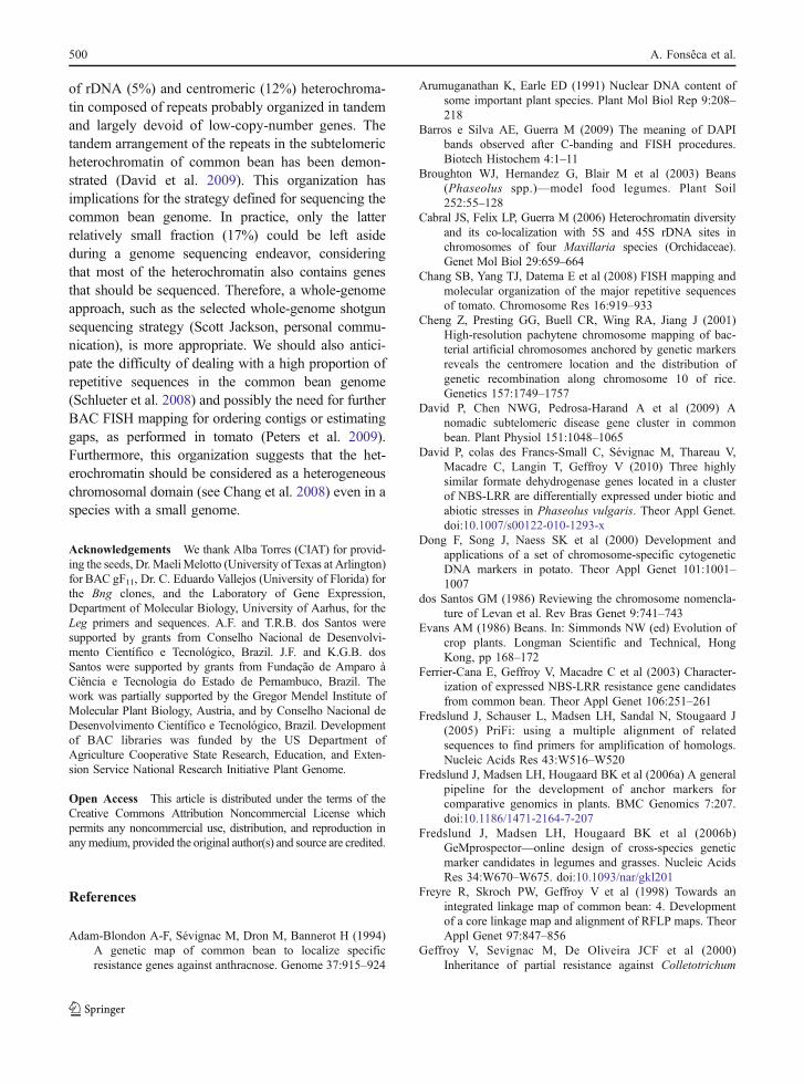
Adam-Blondon A-F, Sévignac M, Dron M, Bannerot H (1994)A genetic map of common bean to localize specificresistance genes against anthracnose. Genome 37:915–924
Arumuganathan K, Earle ED (1991) Nuclear DNA content ofsome important plant species. Plant Mol Biol Rep 9:208–218
Barros e Silva AE, Guerra M (2009) The meaning of DAPIbands observed after C-banding and FISH procedures.Biotech Histochem 4:1–11
Broughton WJ, Hernandez G, Blair M et al (2003) Beans(Phaseolus spp.)—model food legumes. Plant Soil252:55–128
Cabral JS, Felix LP, Guerra M (2006) Heterochromatin diversityand its co-localization with 5S and 45S rDNA sites inchromosomes of four Maxillaria species (Orchidaceae).Genet Mol Biol 29:659–664
Chang SB, Yang TJ, Datema E et al (2008) FISH mapping andmolecular organization of the major repetitive sequencesof tomato. Chromosome Res 16:919–933
Cheng Z, Presting GG, Buell CR, Wing RA, Jiang J (2001)High-resolution pachytene chromosome mapping of bac-terial artificial chromosomes anchored by genetic markersreveals the centromere location and the distribution ofgenetic recombination along chromosome 10 of rice.Genetics 157:1749–1757
David P, Chen NWG, Pedrosa-Harand A et al (2009) Anomadic subtelomeric disease gene cluster in commonbean. Plant Physiol 151:1048–1065
David P, colas des Francs-Small C, Sévignac M, Thareau V,Macadre C, Langin T, Geffroy V (2010) Three highlysimilar formate dehydrogenase genes located in a clusterof NBS-LRR are differentially expressed under biotic andabiotic stresses in Phaseolus vulgaris. Theor Appl Genet.doi:10.1007/s00122-010-1293-x
Dong F, Song J, Naess SK et al (2000) Development andapplications of a set of chromosome-specific cytogeneticDNA markers in potato. Theor Appl Genet 101:1001–1007
dos Santos GM (1986) Reviewing the chromosome nomencla-ture of Levan et al. Rev Bras Genet 9:741–743
Evans AM (1986) Beans. In: Simmonds NW (ed) Evolution ofcrop plants. Longman Scientific and Technical, HongKong, pp 168–172
Ferrier-Cana E, Geffroy V, Macadre C et al (2003) Character-ization of expressed NBS-LRR resistance gene candidatesfrom common bean. Theor Appl Genet 106:251–261
Fredslund J, Schauser L, Madsen LH, Sandal N, Stougaard J(2005) PriFi: using a multiple alignment of relatedsequences to find primers for amplification of homologs.Nucleic Acids Res 43:W516–W520
Fredslund J, Madsen LH, Hougaard BK et al (2006a) A generalpipeline for the development of anchor markers forcomparative genomics in plants. BMC Genomics 7:207.doi:10.1186/1471-2164-7-207
Fredslund J, Madsen LH, Hougaard BK et al (2006b)GeMprospector—online design of cross-species geneticmarker candidates in legumes and grasses. Nucleic AcidsRes 34:W670–W675. doi:10.1093/nar/gkl201
Freyre R, Skroch PW, Geffroy V et al (1998) Towards anintegrated linkage map of common bean: 4. Developmentof a core linkage map and alignment of RFLP maps. TheorAppl Genet 97:847–856
Geffroy V, Sevignac M, De Oliveira JCF et al (2000)Inheritance of partial resistance against Colletotrichum
500 A. Fonsêca et al.

lindemuthianum in Phaseolus vulgaris and co-localizationof quantitative trait loci with genes involved in specificresistance. Mol Plant–Microbe Interact 13:287–296
Geffroy V, Macadré C, David P et al (2009) Molecular analysisof a large subtelomeric NBS-LRR family in two represen-tative genotypes of the major gene pools of Phaseolusvulgaris. Genetics 181:405–419
Gepts P, Beavis WD, Brummer EC et al (2005) Legumes as amodel plant family. Genomics for food and feed. Report ofthe Cross-Legume Advances through Genomics Confer-ence. Plant Physiol 137:1228–1235
Gepts P, Aragão FJL, Barros ED et al (2008) Genomics ofPhaseolus beans, a major source of dietary protein andmicronutrients in the tropics. In: Moore PH, Ming R (eds)Genomics of tropical crop plants. Springer, Berlin,pp 113–143
Gill N, Hans CS, Jackson S (2008) An overview of plantchromosome structure. Cytogenet Genome Res 120:194–201
Guerra M (1993) High amount of heterochromatin in a tropicaltree species: Genipa americana L. (Rutaceae). Cytologia58:427–442
Heslop-Harrison JS, Schwarzacher T, Anamthawat-Jónsson Ket al (1991) In situ hybridization with automated chromo-some denaturation. Technique 3:109–115
Heslop-Harrison JS, Harrison GE, Leitch IJ (1992) Reprobingof DNA:DNA in situ hybridization preparations. TIGG8:372–373
Houben A, Demidov D, Gernand D et al (2003) Methylation ofhistone H3 in euchromatin of plant chromosomes dependson basic nuclear DNA content. Plant J 33:967–973
Hougaard BK, Madsen LH, Sandal N et al (2008) Legumeanchor markers link syntenic regions between Phaseolusvulgaris, Lotus japonicus, Medicago truncatula andArachis. Genetics 179:2299–2312
Islam-Faridi MN, Childs KL, Klein PE et al (2002) Amolecular cytogenetic map of sorghum chromosome: 1.Fluorescence in situ hybridization analysis with mappedbacterial artificial chromosomes. Genetics 161:345–353
Jiang J, Gill BS (1996) Current status and potential offluorescence in situ hybridization in plant genome map-ping. In: Paterson AH (ed) Genome mapping in plants. RGLandes Company, Georgetown, pp 127–135
Jiang J, Gill BS (2006) Current status and the future offluorescence in situ (FISH) in plant genome research.Genome 49:1057–1068
Kami J, Poncet V, Geffroy V, Gepts P (2006) Development offour phylogenetically-arrayed BAC libraries and sequenceof the APA locus in Phaseolus vulgaris. Theor Appl Genet112:987–998
Kim J-S, Islam-Faridi MN, Klein PE et al (2005a) Comprehen-sive molecular cytogenetic analysis of sorghum genomearchitecture: distribution of euchromatin, heterochromatin,genes and recombination in comparison to rice. Genetics171:1963–1976
Kim J-S, Klein PE, Klein RR et al (2005b) Molecularcytogenetic maps of Sorghum linkage groups 2 and 8.Genetics 169:955–965
Kulikova O, Gualtieri G, Geurts R et al (2001) Integration ofthe FISH pachytene and genetic maps of Medicagotruncatula. Plant J 27:49–58
Kulikova O, Geurts R, Lamine M et al (2004) Satellite repeats inthe functional centromere and pericentromeric heterochro-matin of Medicago truncatula. Chromosoma 113: 276–283
Ma J, Wing RA, Bennetzen JL, Jackson SA (2007) Plantcentromere organization: a dynamic structure with con-served functions. Trends Genet 23:134–139
Melotto M, Coelho MF, Pedrosa-Harand A et al (2004) Theanthracnose resistance locus C0–4 of common bean islocated on chromosome 3 and contains putative diseaseresistance-related genes. Theor Appl Genet 109:690–699
Moscone EA, Matzke MA, Matzke JM (1996) The use ofcombined FISH/GISH in conjunction with DAPI counter-staining to identify chromosomes containing transgeneinserts in amphidiploid tobacco. Chromosoma 105:231–236
Moscone EA, Klein F, Lambrou M, Fuchs J, Schweizer D (1999)Quantitative karyotyping and dual-color FISHmapping of 5Sand 18S–25S rDNA probes in the cultivated Phaseolusspecies (Leguminosae). Genome 42:1224–1233
Nagaki K, Cheng Z, Ouyang S et al (2004) Sequencing of a ricecentromere uncovers active genes. Nat Genet 36:138–145
Nodari RO, Tsai SM, Gilbertson RL, Gepts P (1993) Towards anintegrated linkage map of common bean: 2. Development ofan RFLP-based linkage map. Theor Appl Genet 85:513–520
Pedrosa A, Sandal N, Stougaard J, Schweizer D, Bachmair A(2002) Chromosomal map of the model legume Lotusjaponicus. Genetics 161:1661–1672
Pedrosa A, Vallejos CE, Bachmair A, Schweizer D (2003)Integration of common bean (Phaseolus vulgaris L.) linkageand chromosomal maps. Theor Appl Genet 106:205–212
Pedrosa-Harand A, de Almeida CCS, Mosiolek M et al (2006)Extensive ribosomal DNA amplification during Andeancommon bean (Phaseolus vulgaris L.) evolution. TheorAppl Genet 112:924–933
Pedrosa-Harand A, Porch T, Gepts P (2008) Standard nomen-clature for common bean chromosomes and linkagegroups. Annu Rep Bean Improv Coop 51:106–107
Pedrosa-Harand A, Kami J, Gepts P et al (2009) Cytogeneticmapping of common bean chromosomes reveals a lesscompartmentalized small-genome plant species. Chromo-some Res 17:405–417
Peters SA, Datema E, Szinay D et al (2009) Solanumlycopersicum cv. Heinz 1706 chromosome 6: distributionand abundance of genes and retrotransposable elements.Plant J 58:857–869
Rozen S, Skaletsky HJ (2000) Primer3 on the WWW for generalusers and for biologist programmers. In: Krawetz S, MisenerS (eds) Bioinformatics methods and protocols: methods inmolecular biology. Humana, Totowa, NJ, pp 365–386
Schlueter JA, Goicoechea JL, Collura K et al (2008) BAC-endsequence analysis and a draft physical map of the commonbean (Phaseolus vulgaris L.) genome. Trop Plant Biol1:40–48
Shoemaker RC, Schlueter J, Doyle JJ (2006) Paleopolyploidyand gene duplication in soybean and other legumes. CurrOpin Plant Biol 9:104–109
Vallejos CE, Sakiyama NS, Chase CD (1992) A molecularmarker-based linkage map of Phaseolus vulgaris L.Genetics 131:733–740
Vanhouten W, Mackenzie S (1999) Construction and charac-terization of a common bean bacterial artificial chromo-some library. Plant Mol Biol 40:977–983
Cytogenetic map of common bean 501

Wanzenböck E-M, Schöfer C, Schweizer D, Bachmair A(1997) Ribosomal transcription units integrated viaT-DNA transformation associate with the nucleolus anddo not require upstream repeat sequences for activity inArabidopsis thaliana. Plant J 11:1007–1016
Yan H, Jiang J (2007) Rice as a model for centromere andheterochromatin research. Chromosome Res 15:77–84
Young ND, Roe BA, Town CD, Cannon SB, Sato S, Tabta S(2005) Sequencing the genespaces of Medicago truncatulaand Lotus japonicus. Plant Physiol 137:1174–1181
Zheng J, Masashi M, Uchiyama H,Morikawa H, Tanaka R (1991)Giemsa C-banding patterns in several species of PhaseolusL. and Vigna Savi, Fabaceae. Cytologia 56:459–466
Zheng JY, Nakata M, Irifune K, Tanaka R, Morikawa H (1993)Fluorescent banding pattern analysis of eight taxa ofPhaseolus and Vigna in relation to their phylogeneticrelationships. Theor Appl Genet 87:38–43
Zwick MS, Hanson RE, McKnight TD et al (1997) A rapidprocedure for isolation of C0t–1 DNA from plants.Genome 40:138–142
502 A. Fonsêca et al.