
Cytoarchitectonic subdivisions in the subtectal midbrain of the lizard Gallotia galloti
25
Journal of Neurocytology 29, 569–593 (2000) Cytoarchitectonic subdivisions in the subtectal midbrain of the lizard Gallotia galloti CARMEN D ´ IAZ 1,* , CARMEN YANES 2 , CARMEN-MAR ´ IA TRUJILLO 2 and LUIS PUELLES 1,† 1 Department of Morphological Sciences, Faculty of Medicine, University of Murcia, 30100 Murcia, Spain; 2 Unit of Cell Biology, Faculty of Biology, University of La Laguna, 38071 La Laguna, Canary Islands, Spain Received 5 June 2000; revised 2 October 2000; accepted 12 October 2000 Summary Contemporary study of molecular patterning in the vertebrate midbrain is handicapped by the lack of a complete topological map of the diverse neuronal complexes differentiated in this domain. The relatively less deformed reptilian midbrain was chosen for resolving this fundamental issue in a way that can be extrapolated to other tetrapods. The organization of midbrain centers was mapped topologically in terms of longitudinal columns and cellular strata on transverse, Nissl-stained sections in the lizard Gallotia galloti. Four columns extend along the whole length of the midbrain. In dorsoventral order: 1) the dorsal band contains the optic tectum, surrounded by three ventricularly prominent subdivisions, named griseum tectale, intermediate area and torus semicircularis, in rostrocaudal order; 2) a subjacent region is named here the lateral band, which forms the ventral margin of the alar plate and also shows three rostrocaudal divisions; 3) the basal band forms the basal plate or tegmentum proper; it appears subdivided into medial and lateral parts: the medial part contains the oculomotor and accessory efferent neurons and the medial basal part of the reticular formation, which includes the red nucleus rostrally; the lateral part contains the lateral basal reticular formation, and includes the substantia nigra caudally; 4) the median band contains the ventral tegmental area, representing the mesencephalic floor plate. The alar regions (dorsal and lateral) show an overall cellular stratification into periventricular, central and superficial strata, with characteristic cytoarchitecture for each part. The lateral band contains two well developed superficial nuclei, one of which is commonly misidentified as an isthmic formation. The basal longitudinal subdivisions are simpler and basically consist of periventricular and central strata. Introduction Recent embryological and molecular advances on the isthmic organizer and on dorsoventral patterning have thrown new light on some position-dependent causal aspects of midbrain structure (see Discussion). It is widely assumed that early gene patterns and diverse gradients of morphogenetic molecules establish two- dimensional developmental fields within which spe- cific neuronal populations and sets of axonal connec- tions eventually develop as tridimensional complexes. Such studies aim to unravel the emerging rules of midbrain dorsoventral and anteroposterior patterning. However, experience in this field indicates that fre- quently the power of these approaches is handicapped by insufficient understanding of the topological struc- ture and cytoarchitectonic diversity of the vertebrate midbrain, irrespective of previous efforts to systematize available data. For instance, it is not clear whether the midbrain shows transverse anteroposterior subdivi- * Present address: Faculty of Medicine, University of Castilla-La Mancha, 02071 Albacete, Spain. † To whom correspondence should be addressed. sions and we lack a modern model of midbrain organi- zation, which may help both the predictions and deduc- tions made when interpreting gene functions during midbrain morphogenesis, and the comparative study of the differential evolution of midbrain structure in the diverse vertebrate lineages (i.e., distinction of con- served versus evolved features). We aimed to provide a solution to this issue by re- examining in detail the morphology and apparent em- bryonic derivation of all detectable cytoarchitectonic el- ements of a reptilian midbrain. For practical reasons, we chose the lizard Gallotia galloti. The reptilian midbrain offers advantages for an inquiry on basic structural pat- terns, due to its simpler construction and limited topo- logical deformation, as compared, for example, with the midbrain of birds or mammals (Senn, 1979). On the other hand, its intermediate range of cytoarchitectural differentiation, cell migration and cell aggregation in 0300–4864 C 2000 Kluwer Academic Publishers
-
Upload
carmen-diaz -
Category
Documents
-
view
212 -
download
0
Transcript of Cytoarchitectonic subdivisions in the subtectal midbrain of the lizard Gallotia galloti
Journal of Neurocytology 29, 569–593 (2000)
Cytoarchitectonic subdivisions in the subtectalmidbrain of the lizard Gallotia gallotiCARM E N DIAZ 1,∗ , CARMEN YANES 2, CARMEN-MAR IA TRUJILLO 2
a n d L UIS P UELLES 1,†
1 Department of Morphological Sciences, Faculty of Medicine, University of Murcia, 30100 Murcia, Spain; 2 Unit of Cell Biology, Facultyof Biology, University of La Laguna, 38071 La Laguna, Canary Islands, Spain
Received 5 June 2000; revised 2 October 2000; accepted 12 October 2000
Summary
Contemporary study of molecular patterning in the vertebrate midbrain is handicapped by the lack of a complete topologicalmap of the diverse neuronal complexes differentiated in this domain. The relatively less deformed reptilian midbrain waschosen for resolving this fundamental issue in a way that can be extrapolated to other tetrapods. The organization of midbraincenters was mapped topologically in terms of longitudinal columns and cellular strata on transverse, Nissl-stained sections inthe lizard Gallotia galloti. Four columns extend along the whole length of the midbrain. In dorsoventral order: 1) the dorsal bandcontains the optic tectum, surrounded by three ventricularly prominent subdivisions, named griseum tectale, intermediate areaand torus semicircularis, in rostrocaudal order; 2) a subjacent region is named here the lateral band, which forms the ventralmargin of the alar plate and also shows three rostrocaudal divisions; 3) the basal band forms the basal plate or tegmentum proper;it appears subdivided into medial and lateral parts: the medial part contains the oculomotor and accessory efferent neurons andthe medial basal part of the reticular formation, which includes the red nucleus rostrally; the lateral part contains the lateralbasal reticular formation, and includes the substantia nigra caudally; 4) the median band contains the ventral tegmental area,representing the mesencephalic floor plate. The alar regions (dorsal and lateral) show an overall cellular stratification intoperiventricular, central and superficial strata, with characteristic cytoarchitecture for each part. The lateral band contains twowell developed superficial nuclei, one of which is commonly misidentified as an isthmic formation. The basal longitudinalsubdivisions are simpler and basically consist of periventricular and central strata.
Introduction
Recent embryological and molecular advances on theisthmic organizer and on dorsoventral patterning havethrown new light on some position-dependent causalaspects of midbrain structure (see Discussion). It iswidely assumed that early gene patterns and diversegradients of morphogenetic molecules establish two-dimensional developmental fields within which spe-cific neuronal populations and sets of axonal connec-tions eventually develop as tridimensional complexes.Such studies aim to unravel the emerging rules ofmidbrain dorsoventral and anteroposterior patterning.However, experience in this field indicates that fre-quently the power of these approaches is handicappedby insufficient understanding of the topological struc-ture and cytoarchitectonic diversity of the vertebratemidbrain, irrespective of previous efforts to systematizeavailable data. For instance, it is not clear whether themidbrain shows transverse anteroposterior subdivi-
∗ Present address: Faculty of Medicine, University of Castilla-La Mancha, 02071 Albacete, Spain.† To whom correspondence should be addressed.
sions and we lack a modern model of midbrain organi-zation, which may help both the predictions and deduc-tions made when interpreting gene functions duringmidbrain morphogenesis, and the comparative studyof the differential evolution of midbrain structure inthe diverse vertebrate lineages (i.e., distinction of con-served versus evolved features).
We aimed to provide a solution to this issue by re-examining in detail the morphology and apparent em-bryonic derivation of all detectable cytoarchitectonic el-ements of a reptilian midbrain. For practical reasons, wechose the lizard Gallotia galloti. The reptilian midbrainoffers advantages for an inquiry on basic structural pat-terns, due to its simpler construction and limited topo-logical deformation, as compared, for example, withthe midbrain of birds or mammals (Senn, 1979). On theother hand, its intermediate range of cytoarchitecturaldifferentiation, cell migration and cell aggregation in
0300–4864 C© 2000 Kluwer Academic Publishers

570 DIAZ, YANES, TRUJILLO and PUELLES
the mantle zone provides characteristics not generallyavailable in anamniotes. It is thus one aim of this reportto explore how far a purely cytoarchitectural analysiscan help to define midbrain subdivisions which may beof general relevance in tetrapods and may be taken asthe end result of midbrain molecular patterning.
The adult reptilian midbrain is delimited conven-tionally by coronal planes passing through the poste-rior commissure and behind the trochlear nerve root,without express reference to embryological data (tenDonkelaar, 1998). It is widely accepted that the mid-brain is subdivided dorsoventrally into tectal and sub-tectal or tegmental portions, which possibly implies alongitudinal subdivision into alar and basal columnslike in other vertebrates (Palmgren, 1921; Bergquist,1953, 1954; Kuhlenbeck, 1975). However, the absenceof a sulcus limitans in the midbrain of reptilian em-bryos and adults handicaps the usual procedure forlocating the alar-basal boundary. Various maps of thereptilian mesencephalon in fact do not draw such aboundary (Frederikse, 1931; Senn, 1968, 1979; Cruce &Nieuwenhuys, 1974; ten Donkelaar et al., 1987). Fromthe developmental point of view sketched above, itshould be of interest to know which parts of the sub-tectum may be attributed respectively to the alar orbasal longitudinal columns, and what is their respec-tive structural complexity. There is also ambiguity inthe literature on which neural centers actually developwithin the midbrain vesicle and thus are properly called“mesencephalic”, as well as on what is their respectivelocation and mutual relations within the midbrain.
The subtectum is reported to contain some very char-acteristic grisea, like the torus semicircularis, the ocu-lomotor complex, the nucleus ruber and the substantianigra, which are recognized as mesencephalic by allauthors. There are also some less well defined cytoar-chitectural entities, like the “torus semicircularis parslaminaris” or the “nucleus profundus mesencephali”,whose localization by different authors is not unani-mous. Other grisea are often expressly or implicitlyincluded in the mesencephalon, even though there isgood separate evidence that they belong either to thepretectum or to the isthmus (e.g., the trochlear, isthmicand interpeduncular nuclei, which develop in the isth-mus and the n.lentiformis mesencephali, n. of the basaloptic root and n.fasciculi longitudinalis medialis, whichdevelop in the pretectum; see Discussion). Finally, thereare a number of blank (uncharted) spaces in publishedmaps of the reptilian and other vertebrate midbrains.On comparison with histological preparations, thesecorrespond to well populated but still scarcely inves-tigated areas of the subtectum. It was accordingly ouraim to provide a full description of neuronal groupsfound strictly within the embryological limits of thelizard midbrain, classifying them consistently accord-ing to longitudinal, rostrocaudal and radial position.Examination of these points in a reptile is relevant for
the understanding of changes in midbrain structure inother reptiles, and more generally in tetrapods.
The literature records three embryologically-basedapproaches which might help to resolve questionson the detailed structural organization of the mesen-cephalon in reptiles and other vertebrates. In the firstplace, Palmgren (1921) firmly established the rostraland caudal embryological boundaries of the mesen-cephalon in various vertebrates. His conclusions werecorroborated by Rendahl (1924), Vaage (1969, 1973),Keyser (1972) and others (i.e.: Puelles & Martınez-de-la-Torre, 1987, Puelles et al., 1987). Palmgren (1921) alsoput forward a rather detailed scheme for the longi-tudinal subdivision of the subtectum, distinguishingin it four columns (dorsal, lateral, medial and ven-tral) and several smaller parcellations. His materialincluded cartilaginous and teleost fishes, amphibians,birds and mammals, but very few reptilian specimens.Unfortunately, Palmgren’s seminal contribution on lon-gitudinal parcellation of the vertebrate midbrain waslargely disregarded by subsequent workers (the onlyexception known to us is Rendahl’s 1924 study of thedeveloping chick diencephalon, in which he corrobo-rated Palmgren’s longitudinal columns and extendedthem into the forebrain). The simpler schema preferredby Bergquist (1953), for example, divides longitudi-nally the subtectum only into dorsal and ventral ar-eas. Bergquist and Kallen (1953a,b, 1954), who, togetherwith Vaage (1969), conceived a rostrocaudal division ofthe midbrain into two segments, named the two sub-tectal columns ventrolateral and ventral areas, respec-tively. Most authors, however, conceive a poorly struc-tured midbrain tegmentum.
Separately, Senn (1970, 1979) developed a midbrain-based general conception of stratification in the reptil-ian brain. His periventricular, central and superficiallayers are inspired in tectal stratification (Ramon, 1896;Huber & Crosby, 1933). Senn’s studies highlighted theanalysis of stratification as a useful tool for understand-ing the changing structure of the brain wall along the ra-dial dimension. Actually, a general radial subdivision ofthe neural tube into three strata was also conventionallycontemplated by previous embryological workers (e.g.,stratum cellulare internum/intermedium/superficialeof Palmgren, 1921; Rendahl, 1924).
Finally, Nieuwenhuys (1974) developed a procedurefor topological representation of neural formationsupon a two-dimensional map, irrespective of their dif-ferent adult radial positions. This consists basically inprojecting graphically the elements to be mapped intothe ventricular contour, using the deformed internalradial lines revealed by radial glia and/or blood ves-sels as guides. Theoretically, this graphic operation vi-sualizes the nuclei close to the place where they orig-inated. Topological maps of the midbrain in a turtleand a lizard are available (Cruce & Nieuwenhuys, 1974;ten Donkelaar & Nieuwenhuys, 1979).

Cytoarchitectonics of lizard subtectum 571
The present investigation reexamines the structuralsubdivision of the adult mesencephalon of the lizardGallotia galloti, using jointly the interpretational sys-tems offered in the work of Palmgren (1921), Senn(1970, 1979) and Nieuwenhuys (1974). This led usto search for transverse segmental boundaries, lon-gitudinal columns and radially stratified grisea. Ourstudy emphasizes subtectal structure, since the optictectum has been described extensively (Ramon, 1896;Senn, 1968, 1979; see for reviews Northcutt, 1984; tenDonkelaar, 1998).
Methods
Eight young lizards (Gallotia galloti) were used for this study.Some of these animals (n = 3) were deeply anesthetized withether, perfused with 4% phosphate-buffered paraformalde-hyde, and then postfixed overnight. The others (n = 5) wereperfused with phosphate-buffered saline and the brains wereextracted for in vitro HRP-labelling experiments (not de-scribed in this paper; protocol as in Diaz et al., 1992a). Thesespecimens were immersion-fixed overnight after 24–36 hoursin the culture medium. All the brains were stripped ofmeninges. After cryprotection in 30% sucrose, the fixed brainswere serially sectioned in a cryostat in sagittal (n = 4), hori-zontal (n = 1) and transverse (n = 3) planes. Sections werecut at 10 µm from the perfused brains and at 50 µm fromthe immersion-fixed brains. All sections were stained withacetate-buffered cresyl violet (in the experimental cases, as acounterstain to the HRP reaction). Other Gallotia brains sec-tioned in paraffin, stained with the luxol-fast-blue myelin pro-cedure and counterstained with cresyl-violet were availablefor comparison.
A series of selected transverse 50µm-thick sections throughone midbrain served to prepare a detailed atlas of cytoarchi-tectonic subdivisions, and to obtain a topological midbrain re-construction according to Nieuwenhuys (1974), in which thediverse boundaries were projected along curved radial linesonto the ventricular surface. The nomenclature used in thisreport follows Huber and Crosby (1926, 1933) and Senn (1970,1979) in the cell strata, while the names of cell groups and fibertracts follow in general the conventional terminology (Cruce& Nieuwenhuys, 1974; ten Donkelaar & Nieuwenhuys, 1979;ten Donkelaar, 1998), although we introduced some changesaccording to the embryological origin of the cell masses (i.e.,excluding those nuclei which are isthmic or pretectal). Sev-eral cell groups and other features are first identified in thesubtectum of the lizard Gallotia and new terms are proposedfor them.
Results
BOUNDARIES OF THE MESENCEPHALON
The embryological and comparative definition of mid-brain limits given by Palmgren (1921) agrees with recentdata on gene expression (Rubenstein & Puelles, 1994).Notably, the isthmo-mesencephalic boundary is nowaccepted to coincide with the caudal boundary of ex-pression of the gene Otx-2 in the midbrain (Millet et al.,
1996) and the pretecto- or diencephalo-mesencephalicboundary coincides with the caudal boundary of ex-pression of the gene Pax-6 in the diencephalon (Puschelet al., 1992; MacDonald et al., 1994; Stoykova et al., 1996;Mastick et al., 1997; Warren & Price, 1997; Puelles et al.,2000). Corresponding embryological evidence for theseboundaries in the lizard Gallotia galloti was obtainedby Trujillo and Puelles (unpublished). These transverseboundaries are best observed in sagittal sections, sincetheir topography is modified during development bythe progressive bending of the longitudinal axis of thebrain at the cephalic flexure.
The rhombo-mesencephalic (or isthmo-mesencep-halic) limit is marked in the mantle zone by a clearcutgap (gap “z”, or fissura rhombo-mesencephalica ofPalmgren, 1921) that separates clearly the neighbor-ing neuronal populations and the fibers coursing trans-versely in both region (Fig. 1a–c). Dorsally, the gappasses behind the external bulge of the torus semicir-cularis, just in front of the trochlear nerve decussation(Fig. 1a); dorsolaterally it courses between the magno-cellular and rostral isthmic nuclei (Im, Ir; Fig. 1b; thelatter actually lies in the midbrain and therefore is notisthmic; see this issue resolved for the chick in Vaage,1973; Puelles & Martınez-de-la-Torre, 1987; Marın andPuelles, 1994). Ventrally, the gap divides the oculomo-tor nucleus from the trochlear nucleus (Fig. 1a) and ex-tends behind the substantia nigra and in front of therostralmost raphe nucleus, surfacing behind the oculo-motor root, just in front of the interpeduncular nucleus(Fig. 1a–c). This boundary is also clearly marked at theventral midline by the end of the palisade-like floor-plate raphe of the rhombencephalon at the rostral endof the fovea isthmi; rostral to this, the median radial gliacells suddenly change into the less conspicuous floor-plate cells of the midbrain and diencephalon.
The diencephalo-mesencephalic (or pretecto-mesen-cephalic) limit is less obvious cytoarchitecturally, be-cause mesencephalic grisea are juxtaposed to thepretectal ones without a separating cell-poor gap.However, embryological evidence (Palmgren, 1921;Rendahl, 1924; Bergquist, 1953, 1954; Vaage, 1969;Trujillo & Puelles, unpublished) suggests a plane pass-ing just behind and parallel to the posterior commis-sure, dorsally, and just behind the nucleus of the basaloptic root, ventrally, as the most appropriate boundary(Fig. 1a and b). This plane lies slightly in front of theoculomotor nerve root.
These topologically transverse limits of the midbrainconverge basally (as seen in sagittal sections), due to theinflexion of the axis at the cephalic flexure and the dis-proportionate growth of the optic tectum dorsally. Thisgives to the mesencephalon a wedge shape (Fig. 1c). Theoblique topography of these boundaries in the adultbrain explains the tendency of many authors study-ing cross-sections to incorporate erroneously isthmicor pretectal cell masses into the midbrain.

572 DIAZ, YANES, TRUJILLO and PUELLES

Cytoarchitectonics of lizard subtectum 573
The ventricular cavity of the mesencephalon is sub-divided into a caudal narrow “aqueductal” portion, co-inciding with the region occupied by the torus semicir-cularis, and a rostral enlarged portion, which displayswide lateral recesses inside the optic lobes (Fig. 1a).
LONGITUDINAL, TRANSVERSAL AND RADIALSUBDIVISIONS: OBSERVATIONS IN TRANSVERSESECTIONS
Our cytoarchitectonic analysis of the subtectum inGallotia galloti is presented in a series of sectioning lev-els, illustrated in photographs (Figs. 2–6) and line draw-ings (Fig. 7). These sections (and also others interca-lated in between) served to draw a topological map ofthe midbrain grisea (Fig. 8b), using the procedure ofNieuwenhuys (1974). There are various graphical co-nundrums caused by the invaginated ventricular reliefat the rostral and caudal poles of the mesencephalon,as well as by its global wedge shape, which necessarilyleads to successive changes in the topological sectionplane (oblique to transverse to oblique). We thereforeonly claim to provide a tentative solution in the map-ping of our Fig. 8b. However, we think that this recon-struction, and the more schematic version in Fig. 8a,correctly visualizes the relative connectedness of the di-verse longitudinal and transverse regions, as revealedby their rather distinct cytoarchitectural properties.
We found there are at least two alar longitudinalparts of the midbrain, named here dorsal and lateralbands. The optic tectum is predominant in the dor-sal band, though it is complemented rostrally, ventrallyand caudally by the griseum tectale, the intermediatearea and the torus semicircularis, respectively. The “lat-eral band” represents the topological ventral marginof the alar plate (Figs. 2–8); curiously, this band ex-pands dorsalward close to the isthmo-mesencephalicboundary, where it approaches the roof plate just cau-dal to the torus semicircularis (see schema in Fig. 8a).This laterocaudal midbrain domain contains the so-called nucleus isthmi rostralis (Sereno, 1985; Sereno& Ulinski, 1987). Since the latter term is topographi-cally wrong—proper “isthmic” nuclei lie outside themidbrain—and this may be confusing for causal devel-opmental analysis, we propose to rename it “laterocau-dal mesencephalic nucleus” (LC; Figs. 2–4, 7a–e, and 8aand b). The alar mesencephalon is thus built by the gri-
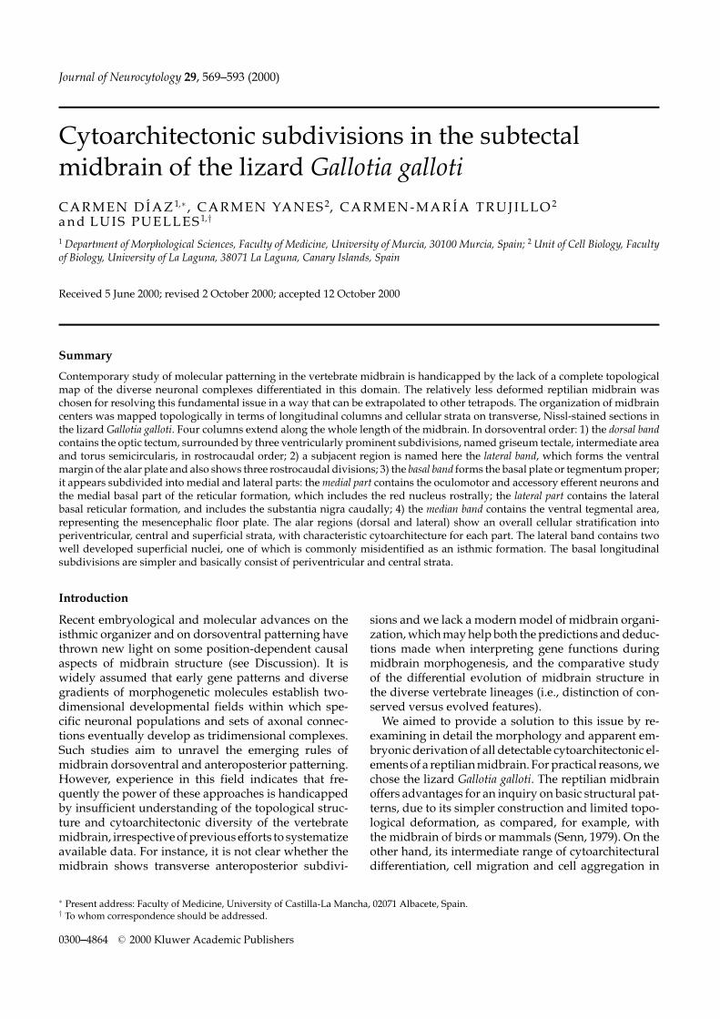
Fig. 1. General layout of the lizard midbrain. (a, b) Microphotographs of two sagittal sections from different young Gallotializards, showing the location of the caudal and rostral transverse boundaries of the mesencephalon (dash lines), as well asseveral midbrain formations and tracts. Rostral is to the right. The bar = 200 µm. The section in (a) is myelin-stained and (b)shows a Nissl-stained section. A cell- and myelin-poor gap marks the limit between the isthmus and the midbrain (a, b). Notethat some myelinated tracts run parallel to the transverse boundaries, as is the case of the posterior commissure (cp), or thecrossed tectobulbar tract (cp,ctb; in a, and in the inset); the fibers of the longitudinal bundles follow the bent brain longitudinalaxis (a). (c) Schematic drawing illustrating the relevant transverse boundaries and general topographic relationships of somemidbrain centers. The posterior commissure, longitudinal fibers and oculomotor/trochlear nerves are shaded in gray. Thesection levels illustrated in Figs. 2–6, whose interpretation is also collected in Fig. 7a–j, are indicated.
seum tectale/tectum/toral complex and an underlyinglateral band with distinct structure. On the other hand,the mesencephalic tegmentum proper appears subdi-vided longitudinally into a basal band (basal plate) anda median band (floor plate) (Fig. 8a and b). These aredescribed in more detail below.
Dorsal bandThe optic tectum will not be described here. We concen-trate instead on the other formations within this band.These are characterized by protruding more or less intothe ventricle, in contrast with the evaginated tectum.We distinguish at least three different grisea along thisbulge: the torus semicircularis, the intermediate areaand the griseum tectale (Figs. 1 and 8). These are nextdescribed in caudorostral order.
The torus semicircularis is more massive caudome-dially, where it replaces the tectum at the dorsal midline.This portion bulges outwards behind the caudal poleof the optic tectum, forming an inferior-colliculus-likestructure (TS; Figs. 1a, 2a, and 7a). The torus also pro-trudes into the ventricular cavity, where the right andleft tori semicirculares are partly fused together (Figs. 2aand b and 7a and b). Structurally, the caudomedial partof the torus is formed by a much enlarged, sphericalperiventricular stratum, that is separated from a thinlypopulated central/superficial stratum by the myeli-nated fibers of the mesencephalic trigeminal tract andthe intertoral commissure (Figs. 1a and 2a and b). Theperiventricular stratum is divided into a thin, pale-staining, deep lamina (continuous with tectal layer 3;terminology of Senn, 1979) and a bilayered outer cellmass. The inner layer of the latter has a neuropile withthinly myelinated fibers, terminal arborizations andsome interspersed small cells, while the outer layer hasslightly larger and more numerous basophilic cells andseems comparable to the paratorus described in Natrix(Senn, 1979) (Figs. 1a and 2a and b). This bilayer is glob-ally continuous with tectal layer 5. A thick fibrillar layer2, containing some thin myelinated fibers, and a thinplexiform layer 4 are also present within the caudo-medial torus (Fig. 1a). However, the neuropile of thetoral layer 4 is characteristically separated from the ad-jacent tectal layer 4 by a small transitional zone in whichthe cellular laminae 3 and 5 are fused together (arrow;Figs. 3b and 4a).
574 DIAZ, YANES, TRUJILLO and PUELLES

Cytoarchitectonics of lizard subtectum 575
576 DIAZ, YANES, TRUJILLO and PUELLES

Cytoarchitectonics of lizard subtectum 577
578 DIAZ, YANES, TRUJILLO and PUELLES

Cytoarchitectonics of lizard subtectum 579
The torus semicircularis complex thins out graduallyas it extends lateroventrally under the tectum (topolog-ically ventralwards–see Fig. 8a), the chief change beinga drastic reduction in its surface extent, probably asa result of the circumferential expansion of the adja-cent tectum (Figs. 3a and b, 4a, and 7c–e). The toralstratum centrale simultaneously expands with a retic-ular appearance, laxer than the tectal central stratum(Figs. 2b, 3a and b, 4a, and 7b–e). The periventricularcell layer 3 becomes thicker and more densely popu-lated in the lateroventral torus. The torus layer 4 neu-ropil remains separated from the tectal plexiform layer4 by a short portion in which layers 3 and 5 are fusedtogether (marked by an arrow in the figures). The layer4 neuropil diminishes progressively in size in the ros-tralmost sections (arrowhead; Figs. 3a and b and 4a).The periventricular outer cellular lamina (layer 5) alsobecomes gradually poorer in cells and less overtly bilay-ered, and can be distinguished from the adjacent layer5 of the tectum by its lower cell density (Figs. 3a and band 4a).
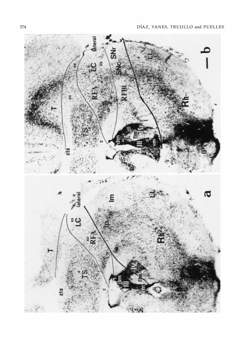
The intermediate area is found laterally in the middleof the midbrain and may be conceived as a transitionalregion between the torus and the griseum tectale (IA;Figs. 4b, 5a and b, 7f–h, and 8a and b). It differs fromthe torus semicircularis and the optic tectum in variousaspects: the periventricular stratum is modified, with athicker plexiform layer 2, a more dense and basophiliccellular layer 3, practically no layer 4 neuropile, and thecellular layer 5 is much reduced in thickness and celldensity, to the point that both its cellular laminae seemcontinuous with tectal layer 3, instead of with layer5 (IA; Fig. 4b). Additionally, the IA stratum centralekeeps the typical shape and position adjacent to thecentral tectal stratum that was described for the torus,but shows a number of large-sized neurons in its deeperpart (open arrow; Fig. 4b). Such cells are not present inthe stratum centrale of the torus semicircularis.
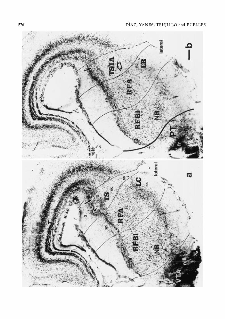
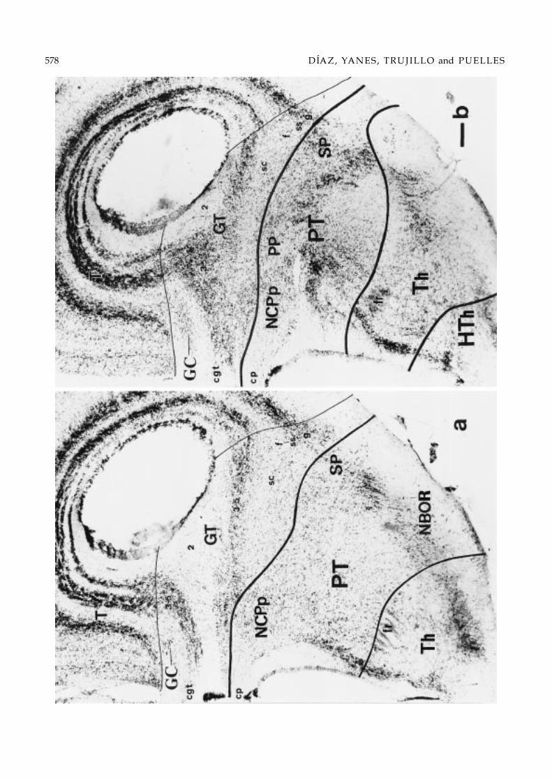
The griseum tectale is the rostral end of the dorsalband. It extends dorsomedially up to the midbrain roofplate, where it replaces the tectum at the rostral con-striction separating the optic lobe and the pretectum(GT; Figs. 5b, 6a and b, and 8). The griseum tectale is astratified complex that protrudes into the rostral part of
Figs. 2–6. Series of 10 selected transverse 50 µm-thick sections through the midbrain of Gallotia galloti (see section levels inFig. 1c). This brain received a HRP deposit in the rostral interpeduncular nucleus at the level of Fig. 2, which mainly labeledascending axons into the diencephalic floor and scarce neurons in the midbrain reticular formation; moreover, label difusedinto the oculomotor nerve root, which serendipituously served to label precisely the oculomotor nucleus (Figs. 2 and 3).The order of the sections within the series is caudorostral. The microphotographs show one side of the Nissl-counterstainedsections, dorsal being oriented upwards. Thick lines drawn in mark the transverse boundaries of the midbrain, either withthe rhombencephalon (Rh), caudally (Figs. 2a and 3b), or the pretectum (PT), rostrally (Figs. 4b–6b). Note that caudal sectionsintersect obliquely the isthmomesencephalic limit, showing isthmic structures underneath it. Likewise, sections rostral tothe oculomotor root intersect obliquely the pretecto-mesencephalic limit, showing various diencephalic grisea underneathit. The thin lines trace the boundaries between the observed midbrain longitudinal subdivisions. Each longitudinal columnwithin the mesencephalon is subdivided in periventricular (sp), central (sc) and, eventually, superficial (ss) strata (Senn, 1979).Bar = 100 µm.
the midbrain ventricle (GT; Figs. 5b, 6a, and 7h and i).Laterally, its superficially compressed shape is similarto that of the rostrolateral torus semicircularis and inter-mediate area (Fig. 6a and b), but its lamination patternis different. It has a reduced periventricular stratumthat is limited to a rather thick plexiform layer 2 and asingle compact cellular layer 3 (these form the stratumgriseum et fibrosum periventriculare of the GT), thuslacking the cell-sparse layer 5 observed in the interme-diate area (compare Figs. 6a and 4b). Its central stra-tum displays an inner stratum album centrale with fewneurons and an outer, more populated stratum griseumcentrale, as occurs in the tectum. Myelinated fibers inthe stratum album centrale cross the dorsal midline ina “commissure of the griseum tectale” that lies in be-tween the posterior commissure and the tectal commis-sure (cgt; Figs. 1a, 6a and b, and 7i and j; see also Perez-Santana et al., 1996). The stratum griseum centrale ispopulated by small and medium sized neurons; a par-ticular condensation of similar cells found dorsomedi-ally near the cgt may be defined as a separate ‘griseumcommissurale’ (GC; Figs. 1a, 6a and b, and 7i and j). Thegriseum tectale also has a superficial stratum or stratumgriseum et fibrosum superficiale (sgfs) that is simplerthan the tectal sgfs. It consists of an inner, scarcely pop-ulated plexiform superficial layer, and an outer cellularsuperficial layer with medium sized neurons placed be-low a subpial axonal stratum (Figs. 5b and 6a and b).The latter two laminae of the GT are traversed by op-tic tract fibers; these laminae are not present over thegriseum commissurale. The area occupied by the gri-seum commissurale expands superficially as it extendsdorsomedially towards the cgt commissure; here it hasmaximal superficial area and even bulges a little out-wards medial to the border of the optic tract and caudalto the posterior commissure. This mimics the externalbulge of the torus semicircularis at the caudal pole ofthe mesencephalon in a minor size range (Figs. 1a, 5b,6a and b, and 7h–j).
Lateral bandThis distinct longitudinal band corresponds essen-tially to Palmgren’s (1921) lateral column, with some

580 DIAZ, YANES, TRUJILLO and PUELLES

Cytoarchitectonics of lizard subtectum 581
modifications. According to our observations, the lat-eral band represents a layered complex whose typi-cal structure runs longitudinally all along the mesen-cephalon, just below the dorsal band, but it also extendsdorsally behind the caudomedial torus formation. Thatis, the lateral band represents the margin of the mes-encephalic alar plate both with regard to the subjacenttegmentum (basal plate) and the isthmo-mesencephalicboundary (Fig. 8a and b). Like the dorsal band, the lat-eral band can be roughly subdivided in larger caudaland rostral sectors, with a small transitional zone inbetween.
Caudally, the lateral band first appears just caudalto the external torus bulge, separating it from isthmicand cerebellar centers. As occurs generally in the alarplate (Senn, 1979), the lateral region consists of periven-tricular, central and superficial strata. The periventric-ular stratum is formed by a sizeable deep plexiformlayer and a dense cellular plate (lateral; Fig. 2a). Thelatter is distinctly separated from the paler-stainingperiventricular stratum of the torus semicircularis bya thin cell-poor gap. The lateral central stratum is athick intermediate layer, populated by a mixture ofsmall-, medium- and large-sized neurons, which appar-ently includes the vaguely defined “n.profundus mes-encephali” (Cruce & Nieuwenhuys, 1974). This inter-mediate layer may be conceived as forming the caudalpart of an alar reticular formation (RFA). The overlyingsuperficial stratum is formed by a magnocellular nu-cleus which lies immersed in a subpial fiber tract, theventral tectothalamic tract. This superficial nucleus isknown as “nucleus isthmi (magnocellularis) rostralis”(Sereno & Ulinski, 1987), though it obviously formswithin the mesencephalon. To avoid confusion andproperly indicate its topography, we propose to nameit ‘laterocaudal mesencephalic nucleus’ (LC; Fig. 2a).
In sections rostral to the point where the caudal lat-eral band first contacts the mesencephalic basal plate,its periventricular cell plate has strongly basophilic cellsand a neuropile populated by few small neurons ap-pears deep to this cell plate (lateral; Figs. 2b, 3a and b,and 4a). There are also numerous large neurons in thecorresponding ventral part of the lateral central layeror alar reticular formation (RFA; Figs. 2b and 3a and b).The laterocaudal mesencephalic nucleus ends in an en-larged rostroventral portion (LC; Figs. 3a and b and7c and d) and, after a brief transition occupied by lessdistinct, smaller neurons, is substituted in more ros-
Fig. 7. (a–j) Line drawings of the 10 transverse sections photographed in Figs. 2–6. The location of the transverse sections isindicated in Fig. 1c. The shaded areas represent regions found within the transverse limits of the midbrain. The main distinctnuclei or compact cellular laminae of the subtectum were left unshaded for easier localization. The four longitudinal bandsof distinct structure are labeled in (e) only, for clarity, but are uniformly delimited one from another by continuous thin lines.The dash lines show secondary subdivisions. The basic periventricular, central and superficial strata are labelled as sp, sc andss, respectively. The sublaminae of the toral periventricular stratum are indicated with the usual numbering system (Ramon,1896). Transverse intersegmental boundaries of the midbrain and diencephalon are drawn as thicker black lines.
tral sections by another compact superficial nucleus,which also contains slightly smaller neurons. We calledit the laterorostral mesencephalic nucleus. This extendsto the rostral limit of the lateral band at the mesen-cephalic border with the pretectum (LR; Figs. 4b, 5aand b, and 7f–h).
Basal plateThe analysis of this structural region is most affected bythe wedge-shape deformation of the mesencephalon.Thus, in our selection of transverse sections onlyone of them represents integrally the midbrain basalplate, passing at the level of the oculomotor nerveroot (Fig. 4a; see section level in Fig. 1c). More caudalsections intersect obliquely the isthmo-mesencephalicboundary, thus apparently showing various isthmicstructures “ventrally” (Figs. 2, 3, and 7a–d). Simi-larly, sections rostral to the oculomotor root intersectobliquely the pretecto-mesencephalic limit and showvarious basal diencephalic grisea apparently “ventral”to it (Figs. 4b and 7f; compare with Fig. 1c). FollowingPalmgren (1921), we divided the basal plate into medialand lateral regions.
The topological mapping indicates that the medial-most basal nuclei are the oculomotor nucleus, the acces-sory nucleus of Edinger-Westphal and the small-cellednucleus of Darkschewitsch (Fig. 8b). These form a cau-dorostrally slightly overlapping series of periventric-ular cell masses, with the somatomotor III nerve neu-rons being caudalmost (III; Figs. 2a and b, 3a and b,and 7a–d), the accessory nucleus lying in an interme-diate position (EW; Figs. 3a and b, 4a, and 7c–e) andthe n.Darkschewitsch rostralmost (D; Figs. 4b and 7f).Ventral to these periventricular grisea, that is, in thecorresponding central stratum, there is the fasciculuslongitudinalis medialis (flm) and the medial reticulartegmental region (RFBm). The latter is characterizedby a dispersed neuronal population, except in the ros-tral half of the mesencephalon, where the magnocel-lular red nucleus appears (NR; Figs. 4a and b, 5a, and7e–g). Lateral to this medial tegmental region there is alateral basal tegmentum. This shows a rather compactperiventricular cell plate and a reticular central stratumextending radially to the ventrolateral mesencephalicsurface (RFBl; Figs. 2b, 3a and b, 4a and b, and 5a). Themidbrain part of the substantia nigra, a migrated cellpopulation (Medina et al., 1994), appears superficially

582 DIAZ, YANES, TRUJILLO and PUELLES

Cytoarchitectonics of lizard subtectum 583
within the caudal part of this tegmental region, in theimmediate neighborhood of the isthmo-mesencephalicboundary and behind the oculomotor root fibers (SN;Figs. 2b, 3a and b, and 7b–d).
It is difficult to visualize rostrocaudal subdivisions ofthe basal plate in cross-sections, but such subdivisionsare suggested by our sagittal myelin-stained sections.These show that the fan-shaped, crossed tectobulbartract (ctb; Fig. 1a and inset) collects ventrally into a com-pact bundle at the center of the midbrain tegmentum,thus dividing the caudal part of the basal plate from therostral one. In many sections this gives the impressionof a third, ctb-related, intermediate partition of the mes-encephalic basal reticular formation (Fig. 1a and inset;Fig. 1b). A tripartite constitution is also supported byconnectivity data (Dıaz, 1991). We therefore postulatecaudal, intermediate and rostral distinct regions of thebasal plate; these are held to be aligned with the threecorresponding regions of the alar plate.
Median bandWe distinguish this very small territory (Figs. 4a and 8b)as a separate longitudinal entity for coherence withdevelopmental studies. These suggest a retarded hete-rochronic pattern of neurogenesis in a band adjacent toor overlapping the mesencephalic midline raphe (andcontinuing rostrally into the hypothalamus). This floor-plate-related band is therefore histogenetically distin-guishable from the typically precocious basal plate(paramedian band of Puelles et al., 1987). A clearcutderivative of the median band in the midbrain is theventral tegmental area, a ventral aggregate of smallneurons which partially migrate subpially lateral to theoculomotor root (VTA; Figs. 4a and 7e; Dıaz & Puelles,1992a,b; Medina et al., 1994). The VTA is only foundrostral to the interpeduncular nucleus.
Discussion
In the present report we studied the internal organi-zation of the subtectum of the lizard Gallotia galloti,exploring the usefulness of tridimensional topologi-cal characterization of grisea in the antero-posterior,
Fig. 8. (a) Simplified flatmap diagram of topological relationships in the lizard midbrain. The thick black lines represent thetransverse rostral and caudal boundaries of the midbrain. Each area represents a tridimensional histogenetic complex extendingfrom the ventricle to the pial surface of the midbrain, irrespective of whether in some cases we indicated specific nuclei lyingat a particular depth level. In this schema only topological relationships are respected and the relative sizes of the regions havebeen normalized to the possible proportions at early embryonic stages (no morphogenetic deformation of the midbrain vesicle).(b) Topological flatmap of midbrain structures (commissures and distinct grisea), represented as projected along curved radial-glial lines onto the graphically reconstructed ventricular surface. Note midbrain boundaries (thick black lines) deformed intoa wedge shape. This map is based on the sections shown in Figs. 2–7, plus other 7 intercalated sections (positions shown asthin lines in the bottom ruler). Whereas size relationships theoretically are more realistic than in (a), the differential extent ofthe ventricular surface, particularly in the caudal midbrain (where midline fusion of the toral protrusions may have furtherdecreased the available surface), possibly leads to underrepresentation of the nuclei and tectal portions associated to the torussemicircularis. The positions of several commissures in the brain roof are indicated.
dorso-ventral and radial dimensions. In the followingsections, the delimitation approach used, the resultingsubdivisions and the specific midbrain cell populationsidentified will be discussed.
MIDBRAIN DELIMITATION
One of the problems we addressed was to reexaminewhat should be regarded as mesencephalic and whatnot, consistent with the best available embryologicalinformation. We adopted Palmgren’s (1921) definitionof the midbrain boundaries, which was based on thecomparison of series of embryonic and adult brains ofseveral vertebrates. We corroborated that a cell-poorgap formed at the isthmo-mesencephalic boundaryneatly separates the isthmic derivatives from the mid-brain. Fate maps performed at early stages in the chickshow that the initial mesencephalic vesicle includes theprospective midbrain and a part of the prospective isth-mocerebellum (although the prospective boundary isalready clearly marked by the caudal limit of Otx-2expression; Millet et al., 1996; Puelles et al., 1996b).Morphological changes subsequent to anteroposteriorpatterning induced by the isthmic organizer and theresulting differential growth give rise to transverse re-gions that contribute separately to the mature midbrainand to the isthmic and cerebellar structures (Vaage,1973; Puelles & Martınez-de-la-Torre, 1987; Martınez& Alvarado-Mallart, 1989; Hallonet et al., 1990; Marın& Puelles, 1994; Millet et al., 1996; Puelles et al., 1996b;Joyner, 1996). Vaage (1973) convincingly showed thatall components of the alar isthmic nuclei in the chickoriginate from the rostralmost rhombencephalon, ex-cept the nucleus isthmi principalis pars magnocellu-laris, which has a mesencephalic origin; this nucleusclearly corresponds to the laterocaudal mesencephalicnucleus in the lizard (rostral isthmic nucleus of Sereno& Ulinski, 1987). Vaage’s conclusions on isthmic nu-clei were experimentally corroborated by Marın andPuelles (1994), whose data also located the rostralmostraphe nuclei and the interpeduncular nucleus in theisthmus. Likewise, the trochlear nucleus arises fromthe rhombencephalic isthmus (Vaage, 1973; Muller &O’Rahilly, 1988; Lumsden & Keynes, 1989; Noden, 1991;

584 DIAZ, YANES, TRUJILLO and PUELLES
Gilland & Baker, 1993; Fritzsch et al., 1995; Bass & Baker,1997).
The pretecto-mesencephalic boundary poses a sim-ilar problem. Often the pretectum has been taken asa “transitional area” between the diencephalon andthe mesencephalon in reptiles (Huber & Crosby, 1926;Butler & Northcutt, 1973; Senn, 1979; Smeets et al.,1986a), or has been divided into “mesencephalic” and“diencephalic” parts (Kuhlenbeck, 1973). The tenta-tive postulation by His (1893) of a diencephalo- mes-encephalic boundary that passed across the posteriorcommissure and close to the mammillary bodies inhuman embryos probably was determinant for thisviewpoint, since it was widely used without any ex-perimental support in the subsequent comparative neu-roanatomical literature. This is also the original sourcefor the idea that the midbrain tegmentum (basal plate)extends far rostral to its alar plate into the mammil-lary area, thus forming the so-called prerubral tegmen-tum (Kuhlenbeck, 1973, 1975; Senn, 1979). Curiously,His (1893) intended to provide only a very tentative,provisional reference until more data accrued. In con-trast, Palmgren (1921), Rendahl (1924) and Vaage (1969)embryologically redefined the rostral boundary of themidbrain as passing just behind the posterior commis-sure and parallel to its fibers; this view has been cor-roborated by various additional embryologic evidence(Keyser, 1972; Puelles et al., 1987; Puelles & Trujillo, un-published). Moreover, this boundary correlates with re-cent molecular data on the caudal limit of the expres-sion domain of the gene Pax-6 in several vertebrates(Puschel et al., 1992; MacDonald et al., 1994; Stoykovaet al., 1996; Mastick et al., 1997; Warren & Price, 1997;Puelles et al., 2000).
According to these definitions, a few pretectal nucleioften are incorrectly assigned to the mesencephalon.For instance, the nucleus lentiformis mesencephali is lo-cated superficially within the juxtacommissural/com-missural pretectum, rostral to the griseum tectale (seeButler & Northcutt, 1973; Smeets et al., 1986a). The pre-tectal tegmental area lies rostral to the red nucleus andincludes the nucleus of the basal optic root and the nu-cleus of the medial longitudinal fasciculus, which of-ten are considered part of the mesencephalic tegmen-tum (see i.e. Kuhlenbeck, 1973; Cruce & Nieuwenhuys,1974; Schwab, 1979; ten Donkelaar, 1998). It has beenshown several times that this tegmental area devel-ops within the caudal diencephalon of vertebrates(Palmgren, 1921; Rendahl, 1924; Keyser, 1972; Puelleset al., 1987, 1996a; Pombal & Puelles, 1999).
LONGITUDINAL, TRANSVERSAL AND RADIALSUBDIVISIONS
The structure of the adult brain reflects how it was builtduring the embryonic period, though sometimes in anon overt way. The neuroepithelium is specified molec-
ularly into primary histogenetic subdivisions which arelongitudinal (i.e., roof, alar, basal and floor plates) ortransversal (neuromeres; Puelles & Rubenstein, 1993;Rubenstein & Puelles, 1994; Puelles, 1995). Each pri-mary parcellation becomes subdivided into smaller his-togenetic fields by primary and secondary anteroposte-rior and dorsoventral regionalization effects, which aresometimes dependent on specific organizer regions. Forinstance, the basal plate owes its existence to ventraliz-ing inductive effects arising from the axial mesodermand the neural floor plate (Placzek et al., 1993; Yamadaet al., 1993; Tanabe et al., 1995; Marti et al., 1995), whereasthe alar plate is affected by dorsalizing effects stem-ming from the roof plate and early non-neural ecto-derm (Liem et al., 1995, 1997). Tectal, toral and isthmicstructures are known to be set in place in their mu-tual anteroposterior relationships by the isthmic orga-nizer, which forms at the isthmo-mesencephalic bound-ary (Martınez & Alvarado-Mallart, 1990; Gardner &Barald, 1991; Martınez et al., 1991; Nakamura & Itasaki,1992; Marın & Puelles, 1994; see also the reviews byAlvarado-Mallart, 1993; Martınez et al., 1995; Puelleset al., 1996b). Each histogenetically distinct area gen-erates a characteristic sequence of radially migratingyoung neurons, and further differentiation, layeringand trophic stabilization of these elements gives riseto the specific morphological and functional popula-tions observed in the adult. Insofar as the positionof neuronal derivatives in the mantle zone remainsundisturbed, covering the respective matrix areas inthe neuroepithelium, the topology of cell populationsfound in the adult can be assumed to reflect regional-ization events which took place during development.This assumption needs checking for eventual tangen-tial migration of some neurons, which might compli-cate the analysis (Puelles & Martınez-de-la-Torre, 1987;Martınez et al., 1992).
In the present study, we found evidence for distinctlongitudinal, transversal and radial midbrain subdivi-sions in the lizard Gallotia. The roof plate, the alar andbasal subdivisions (the dorsal and lateral bands, plusthe medial and lateral subdivisions of the basal plate),and the median floor-plate band can therefore be un-derstood as alternative dorsoventral histogenetic fatesof the midbrain neuroepithelium. These fates proba-bly are positionally dependent on the crossed dorsaliz-ing and ventralizing mechanisms cited above. On theother hand, the main transverse boundaries of the mid-brain, and the differential histologic structure found atrostral, intermediate and caudal levels of most of thelongitudinal components (Fig. 8b) may be explainedas anteroposterior patterning effects possibly influ-enced by their relative distance from the isthmic orga-nizer (known to induce at least caudal midbrain struc-tures in the chick; see Martınez & Alvarado-Mallart,1990; Gardner & Barald, 1991; Martınez et al., 1991;Nakamura & Itasaki, 1992; Marın & Puelles, 1994;

Cytoarchitectonics of lizard subtectum 585
reviews by Alvarado-Mallart, 1993, Martınez et al.,1995; & Puelles et al., 1996b) and/or from the dienc-ephalo-mesencephalic boundary. There is only circum-stantial evidence that the latter may function also asa secondary organizer (Martınez et al., 1992; Marın &Puelles, 1994).
An intriguing observation is the dorsal expansion ofthe caudal part of the lateral band along the isthmo-mesencephalic border, separating the toral complexfrom the isthmus proper. Some published evidence andwork in course indicates that there are some mouse andchick developmental genes whose expression patternduplicates this peculiar fate distribution—i.e., Nkx-2.2and Sim-1 (Puelles & Rubenstein, 1993; Fan et al., 1996;Puelles, E. & Puelles, L. unpublished), but a causal con-nection can not be proposed at present.
Longitudinal subdivisionsWe found four main longitudinal cytoarchitectoniccolumns in the lizard midbrain. These largely corre-spond to the dorsal, lateral, medial and ventral bandsdetected by Palmgren (1921), but we chose to namethem more descriptively, consistent with recent devel-opmental literature, as dorsal, lateral, basal and medianbands.
The dorsal and lateral bands constitute the alar plate,while the basal and median bands constitute the mesen-cephalic tegmentum proper. Using this definition, thealar plate correlates in dorsoventral position with theisthmic nuclei and the sensory columns of the romben-cephalic alar plate (cochlear, vestibular and somatosen-sorial/visceral fields), caudally, and with the alar di-encephalon (pretectum, dorsal and ventral thalami),rostrally. We corroborated also Rendahl’s (1924) find-ing that Palmgren’s (1921) dorsal and lateral bands con-tinue from the midbrain into the caudal diencephalon;this suggests the existence of common dorsoventralpatterning mechanisms, in agreement with the rele-vant data on gene functions cited above. The basaland median bands of the midbrain are also caudallycontinuous with the somato-/visceromotor columnsof the hindbrain basal and floor plates, comprisingmotor nuclei, reticular formation and raphe nuclei.The mesencephalic tegmentum contacts rostralwardsthe diencephalic tegmentum (i.e., prerubral or pretec-tal tegmentum, posterior tuberculum and retromam-millary region), with which similarity resides at leastin the development of catecholaminergic populations(Puelles & Medina, 1994; Puelles & Verney, 1998).
Our postulate that the midbrain alar/basal boundarylies between the lateral band and the lateral part of thebasal tegmentum is tentative and needs corroborationby means of molecular markers (for instance, the alarplate marker genes Pax3/Pax7, not yet cloned for thelizard). Puelles and Rubenstein (1993) and Shimamuraet al. (1995) suggested that the linear expression of the
gene Nkx-2.2 in the midbrain and diencephalon mayapproximate the location of the alar/basal boundary.Recent detailed analysis of Nkx-2.2 in chick embryos(Puelles et al., 1999) suggests however that this gene isexpressed in a three-dimensional band across the mid-brain neuroepithelium and mantle layer, which proba-bly overlaps with the equivalent of our present lateralpart of the basal plate.
In the columnar approach, the sulcus limitans ofHis (1893) is widely identified as the delimiting land-mark between the alar and the basal territories ofthe hindbrain and midbrain (His, 1893, 1904; Herrick1910, 1917; Kuhlenbeck, 1973), but it is an unreliablelandmark (Rothig, 1923; Nieuwenhuys & Bodenheimer,1966; Keyser, 1972). Moreover, the sulcus limitans, evenwhen it is visible, lacks exact correlation with gene-expression domains (Bulfone et al., 1993). In the Testudotopological map, Cruce and Nieuwenhuys (1974) couldnot follow the sulcus limitans from the hindbrain intothe midbrain.
Although in our cytoarchitectonic analysis of thispoint we have not found a definitive answer, we thinkthat the characteristic pluristratified structure of the lat-eral band places it in the same side of morphospace asthe dorsal band, whose alar nature seems undisputable.The lateral periventricular stratum is only subtly differ-ent from that seen at the dorsal band. It is to be noted,though, that the lateral central stratum, and perhapsalso the laterorostral and laterocaudal mesencephalicnuclei, which form its superficial stratum, have an ap-pearance that has favoured in the past their inclusionin the reticular formation (sometimes as nucleus pro-fundus mesencephali; i.e., Schwab, 1979; Wang et al.,1983). This does not pose any particular difficulty as re-gards classification of the lateral band as an alar deriva-tive, since the lateralmost (dorsal) part of the hindbrainreticular formation in mammals also clearly belongs tothe alar plate, as indicated by its radial position un-derneath the trigeminal sensory column (Paxinos &Watson, 1986; Paxinos et al., 1990; Voogd et al., 1998).
The idea of an alar midbrain portion distinct fromthe tectum and the torus semicircularis was consideredby others besides Palmgren (1921), including Cruceand Nieuwenhuys (1974), who saw their ’nucleus pro-fundus mesencephali rostralis and caudalis’ (probablyequivalent to our lateral central stratum) as a rostral ex-tension of the special somatic sensory zone of the alarrhombencephalon (see also comments on the “tegmen-tum sensorium”, on page 2147 of Nieuwenhuys et al.,1998).
Our dorsal band corresponds to the dorsal columndescribed by Palmgren (1921) in fishes, amphibians,birds and mammals, and by Rendahl (1924) in thechick embryo. However, these authors did not iden-tify the intermediate area nor the griseum tectale. Thedorsal mesencephalic column of Bergquist and Kallen(1953) instead seems to correspond with the dorsal

586 DIAZ, YANES, TRUJILLO and PUELLES
band plus the lateral band, which indicates that we alsoagree with them on the alar/basal boundary. Our dataagree basically with the schema proposed by Puelleset al. (1994) for reptiles, as regards relative positions ofthe griseum tectale, tectum and toral complex (com-pare their Fig. 14—or the copy in Nieuwenhuys et al.,1998—with our Fig. 4). However, we are still uncertainwhich alar subdivision in the lizard may correspondto the avian intercollicular area, since we have twocandidates, namely the intermediate area of the dorsalband and the intermediate part of the lateral band. Thecholinergic terminal neuropil identified in a potentiallizard intercollicular area by Medina et al. (1993) is com-parable in location to the periventricular stratum of ourlateral band at an intermediate area level (compare theirFigs. 4n and 15B with our Figs. 2f–h and 1, respectively).Further analysis will be needed, nevertheless, keep-ing in mind other potential markers (Martınez-de-la-Torre, 1985; Robles, 1995).
On the other hand, the somatosensory recipient areaof the reptilian midbrain, identified by Foster and Hall(1978) as ‘area intercollicularis’ in Iguana iguana, alsoidentified previously by Ebbesson (1967) in Tupinam-bis nigropunctatus, clearly does not have a topologicalposition comparable to the avian ‘intercollicular area’,as defined by Puelles et al. (1994). Foster and Hall’s(1978) area intercollicullaris constitutes basically a cau-dolateral portion of the central stratum of the torussemicircularis, comparable to the avian spinorecipient‘preisthmic superficial area’(see Puelles et al., 1994). Thesame can be said of the ‘midbrain somatosensory area’identified by Pritz and Stritzel (1989, 1990) in Caimancrocodilus.
In agreement with Palmgren (1921), we divided thebasal band longitudinally in lateral and medial parts,although the agreement is not complete. Our lateralbasal tegmentum does correspond roughly with hismedial column (Palmgren’s term ‘medial’ is confus-ing, since the intended meaning was ‘intermediate’,as seems clear from the context). However, our me-dial basal tegmentum, including the oculomotor andred nuclei, only corresponds with the lateral part of hisventral column, whereas our median band, where theventral tegmental area arises, corresponds to the medialpart of Palmgren’s ventral column.
The basal reticular formation of the midbrain hasremained poorly defined in reptilian cytoarchitecturalstudies, particularly because the often cited NFLM nu-cleus actually belongs to the pretectal tegmentum. At arhombencephalic level, the reptilian reticular formationwas subdivided in medial and lateral parts; the medialpart was held to extend into the midbrain, while thelateral reticular formation was thought to be restrictedto the hindbrain (Cruce & Nieuwenhuys, 1974; tenDonkelaar et al., 1987). In contrast, Newman and Cruce(1982) extended both portions of their superior reticulardomain into the mesencephalon, thus apparently agree-
ing with our observations. Contrarily, the lateral andmedial midbrain reticular formations of Reiner et al.(1984) correlate with our alar and basal reticular for-mations, respectively. Our concept of an alar reticularformation added to medial and lateral basal parts ofthe midbrain reticular formation, suggests a commonstructural pattern with the dorsal, intermediate and me-dial parts of the hindbrain reticular formation, as dis-tinguished by Paxinos and Watson (1986), Paxinos et al.(1990) and Voogd et al. (1998) in mammals.
Various connectivity data show a more complexstructure of the reptilian midbrain reticular formationthan has been rendered by the cytoarchitectural studiesso far (see i.e. Hoogland, 1982; Welker et al., 1983; Bruce& Butler, 1984; Kunzle & Schnyder, 1984; Sereno, 1985;Diaz, 1991; ten Donkelaar, 1998). This suggests that thisextensive area should be a subject for more detailedstudies.
The median band (floor plate of Kingsbury, 1920,1922, 1930; Vaage, 1969) originally was not recognizedin the midbrain (i.e., Rendahl, 1924; Vaage, 1969; Keyser,1972; Kuhlenbeck, 1973). However, histochemical andgene expression data subsequently have shown that amodified sort of floor plate (divided into a median epi-chordal strip and paramedian bands) extends all alongthe mesencephalon and caudal diencephalon in verte-brates (though it is less massive than that found in thehindbrain); this “epichordal strip” ends just behindthe mammillary region (Newgreen et al., 1981; Wallace,1982; Teitelman et al., 1983; Puelles et al., 1987; Matsuiet al., 1990; Puelles, 1995; Shimamura et al., 1995; Verneyet al., submitted).
Transversal subdivisionsOur results suggest an internal rostrocaudal subdivi-sion of the lizard midbrain that had not been reportedpreviously. The caudorostral series of three sensorygrisea of the dorsal band (GT, T + IA, TS) is correlativelyaligned with three less distinct transverse regions eachin the lateral and basal bands (see Figs. 1 and 8). Theintermediate transverse subdivision is an area of scarcecells, which is selectively crossed by the dorsoventralfibers of the tectobulbar tract. Our transversal subdivi-sion of the lizard midbrain therefore does not coincidewith Vaage’s (1969, 1973) proposal of two mesomeresin the midbrain. His descriptive evidence supportingsuch a neuromeric subdivision is tenuous in the lightof recent chick fate-mapping experiments mentionedabove, indicating that caudal parts of the early mid-brain vesicle contribute later to isthmocerebellar struc-tures (this argument affects as well the generalizedmidbrain schema proposed by Nieuwenhuys (1998),where the isthmus—without the corresponding cere-bellar part—is appended to the midbrain). Whateverinterpretation is given to Vaage’s (1969, 1973) ‘atrophic’m2 area, it would seem that only his m1 neuromere
Cytoarchitectonics of lizard subtectum 587
contains prospective midbrain material. Our tripartiterostrocaudal subdivision occurs thus wholly insideVaage’s m1. Further research may resolve whether thesepartitions represent three mesomeres or just secondarytransverse parts within a single mesomere. The func-tional meaning of the internal transverse subdivision ofthe midbrain remains to be studied. Preliminary datafrom HRP injections at different rostrocaudal levels ofthe fore-, mid- and hindbrain basal or median bandsin Gallotia reveal that some of these transverse internalsubdivisions can be labelled specifically depending onthe site of label injection (Diaz, 1991; Dıaz & Puelles,unpublished data; see also Diaz et al., 1999).
Radial subdivisionsWe also reported here on radial subdivisions in thelizard midbrain, basically following the brain strati-fication system and terminology of Senn (1970). Themost complex radial structure was found in the alarplate. Each histogenetic areal compartment displayed acharacteristic variant of the fundamental three-layeredschema (periventricular, central and superficial strata).The torus semicircularis has a predominant develop-ment of its periventricular stratum, as was alreadypointed out by Senn (1970, 1979), in contrast to the optictectum, where central and superficial strata are betterdeveloped. Nevertheless, individual toral cellular andplexiform laminae can be unequivocally related to theneighboring tectal laminae, independently of their par-ticular thickness or cellular density, as revealed by thefeasibility of using Ramon’s (1896) tectal layer numer-ation system (Senn, 1970, 1979). The same can be saidfor the intermediate area and the griseum tectale, thelatter showing a tectum-like, more equilibrated devel-opment of the three basic strata, as compared with thetorus. The periventricular stratum of these last two re-gions, together with that of the lateral band, is indis-tinctly referred to in the literature as “laminar portionof the torus semicircularis” (ten Donkelaar, 1998). Wesuggest that this term is confusing due to the differ-ences between the areas encompassed within it, and itarbitrarily incorporates into the toral complex regionswhich seem to have a completely different nature (notethat GT is retinorecipient; Martınez-de-la-Torre, 1985;Medina, 1990, and the intercollicular area homolog—ifwe follow the definition issued by Puelles et al., 1994—is related to vocalization rather than to hearing). Wethus propose that use of this term be discontinued.
An issue raised here that we are unable to resolve ishow far stratification and lamination similarities de-pend on development of similar classes of neurons,which would interact under different local conditionsto generate the particular histic phenotypes observed.The only alar midbrain area that has been studied inminimally sufficient detail as regards neuronal typol-ogy is the optic tectum.
Radial positioning of neurons in the basal plate issimpler, as also underlined by Senn (1979). There areperiventricular elements that include the oculomotorneurons, and migrated central stratum elements thatcontribute to the basal part of the reticular formation,including the rostrally placed red nucleus. The partlysuperficial position of the substantia nigra neurons maybe strictly due to radial migration, too, though a po-tential contribution of some cells migrated tangentiallyfrom the ventral tegmental area cannot be discarded onthe basis of our data (see Puelles & Medina, 1994).
CELL POPULATIONS
Contrary to the idea of Cruce and Nieuwenhuys (1974)of the midbrain tegmentum, defined as “a continu-ous zone of diffuse gray” in which “individualized cellmasses can be distinguished but . . .no complete parcel-lation is possible”, we presented here a more detailed“parcellation” of the lizard tegmentum. This serves tofill in the relevant blank spaces that usually appearin the published maps of the midbrain in vertebrates(i.e., reptiles: Cruce & Niewenhuys, 1974; birds: Stokeset al., 1974; Karten & Hodos, 1967; mammals: Paxinos& Watson, 1986). We focus the following discussion onthe less well-known cell groups.
Dorsal bandIn addition to the tectum and the torus semicircularis,we found two other laminated grisea in the dorsal band,the intermediate area and, rostrally, the griseum tec-tale. The intermediate area is still poorly understood.More research is needed to examine the possibility thatit may relate to a vocal center variously identified as“dorsomedial nucleus” or intercollicular area in birds(see Puelles et al., 1994).
The laminated griseum tectale was previously iden-tified in Caiman sclerops (Reperant, 1975), Vipera aspis(Reperant & Rio, 1976) and several other reptiles includ-ing Gallotia galloti (Martınez-de-la-Torre, 1985; Medinaet al., 1993, 1994), based on its identical topography com-pared to the avian griseum tectale (Puelles & Zabala,1982; Puelles et al., 1988; Gamlin & Cohen, 1988a,b).The reptilian griseum tectale receives retinal afferents(Reperant et al., 1978; Medina, 1990; Medina & Smeets,1992; Medina et al., 1993), as in birds (Puelles & Zabala,1982; Gamlin & Cohen, 1988a). De Lange (1913) in-cluded this area within his nucleus lentiformis mes-encephali. In mammals, the posterior pretectal nu-cleus was compared to the griseum tectale of reptilesand birds due to its similar position caudal to theposterior commissure and rostral to the superior col-liculus, its histochemical features and its connectiv-ity (Martınez-de-la-Torre, 1985; Caballero-Bleda, 1988;Caballero-Bleda et al., 1992; Gamlin & Cohen, 1988b).A griseum tectale formation has been distinguished aswell in Rana and Xenopus frogs (Puelles et al., 1996a;

588 DIAZ, YANES, TRUJILLO and PUELLES
Milan & Puelles, 2000). Whereas the amphibian GTcomplex remains largely periventricular, the reptilianGT develops periventricular, central and superficialstrata, and the avian and mammalian GT homologslargely constitute superficially migrated formations.
Lateral bandThe magnocellular laterocaudal mesencephalic nucleuswas apparently misidentified as nucleus profundusmesencephali in several reptiles (Foster & Hall, 1978;Wang et al., 1983; Wolters et al., 1985; ten Donkelaaret al., 1987), if we accept Cruce and Nieuwenhuys’(1974) mapping, that locates nucleus profundus (asthe name indicates) at a deeper locus, correspondingto our lateral intermediate stratum, or alar part ofthe midbrain reticular formation. Dacey and Ulinski(1986) apparently identified the conspicuous LC nu-cleus as ’a caudal part of the nucleus of the tectothala-mic tract plus the mesencephalic isthmi nucleus’ in thesnake Thamnophis sirtalis. Sereno (1985) and Sereno andUlinski (1987) identified it as “nucleus isthmi, pars mag-nocellularis rostralis” in the turtle Pseudemys scripta.However, it clearly belongs to the midbrain, as occurswith its avian counterpart, the magnocellular isthmicnucleus (Vaage, 1973; Puelles & Martınez-de-la-Torre,1987), both being located rostral to the trochlear nu-cleus and the trochlear decussation. This nucleus hasreciprocal connections with the optic tectum in rep-tiles (Kunzle & Schnyder, 1984; Sereno & Ulinski, 1987;Perez-Santana, 1993) and birds (Puelles, unpublishedobservations; see Fig. 3 in Martınez & Puelles, 1989).
The superficial laterorostral mesencephalic nucleusis a practically unknown cell group, which may cor-respond to the nucleus confusingly called ‘profundusmesencephali rostrolateralis’ by Sereno (1985). This au-thor described in the turtle Pseudemys scripta a cau-dorostral sequence of nuclei sandwiched between thesensorially specialized dorsal mesencephalic griseaand the tegmental structures. These seem to corre-spond to our two superficial lateral nuclei (see hisFigs. 2, 3, and 12). We were led to propose newnames for them—laterorostral and laterocaudal mesen-cephalic nuclei—because we think that the terms foundin the literature for these superficial nuclei are ex-tremely confusing and easily lead to misidentification.It should be interesting to explore the possible homol-ogy of the laterorostral mesencephalic nucleus with the‘torus lateralis’ formation found so prominently in themidbrain of teleosts, with a similar topological position(Palmgren, 1921; Nieuwenhuys et al., 1998).
The periventricular stratum of the lateral band isa badly studied locus in most vertebrates. The mam-malian counterpart perhaps might be found in the lat-eral portion of the periacueductal gray. In lizards, it maycorrespond at least in part to the torus semicircularis,pars laminaris, since ten Donkelaar and Nieuwenhuys
(1979) and others described this area as restricted to “thecompact periventricular cell layer.” In other lizards,the nucleus laminaris of the torus semicircularis re-ceives spinal afferents (Kunzle & Woodson, 1982; tenDonkelaar et al., 1987) while the central nucleus of thetorus semicircularis relates to the auditory pathway.
Median bandA restricted concept of the ventral tegmental area (VTA)as a floor plate derivative was previously defined inGallotia (Martınez-de-la-Torre, 1985; Puelles & Medina,1994). This view considers the VTA proper as a reducedmedian small-celled group which lies medial to the ocu-lomotor nerve roots rostral to the isthmic interpedun-cular nucleus, and partially migrates tangentially lat-eral to the oculomotor roots (Dıaz & Puelles, 1992a,b;Medina et al., 1994). The VTA receives bilateral inner-vation from the habenula (Dıaz & Puelles, 1992a) and afew of its cells are habenulopetal, being perhaps compa-rable to the interfascicular nucleus of the VTA in rats.The catecholaminergic VTA described in reptiles hasa much broader extension and may partially overlapwith our substantia nigra (see Smeets, 1994; Medinaet al., 1994). On the other hand, a VTA-like formation ex-tends outside the midbrain itself, into the diencephalicprerubral tegmentum, posterior tuberculum and retro-mammillary areas (Smeets, 1994; Puelles & Medina,1994). The similarity of the midbrain VTA with the morerostral analogues probably results from the operationof similar inductive effects (Ericson et al., 1995; Hyneset al., 1995a,b).
Abbreviations
cer cerebellumcgt commissure of griseum tectalecp posterior commissurectb crossed tectobulbar tractcte commissure of optic tectumcto commissure of the torus semicircularisD nucleus of DarkschewitschDien diencephalonEW accessory oculomotor nucleus
of Edinger-Westphalflm medial longitudinal fasciculusfr retroflex fasciculusGC griseum commissuraleGP pretectal geniculate nucleusGT griseum tectale (tectal gray)HTh hypothalamusIA intermediate areaIII oculomotor nucleusIIIdl oculomotor nucleus, dorsolateral partIIIdm oculomotor nucleus, dorsomedial partIm magnocellular isthmic nucleusIP interpeduncular nucleus

Cytoarchitectonics of lizard subtectum 589
Ip parvicellular isthmic nucleusIr rostral isthmic nucleusIIIvm oculomotor nucleus, ventromedial partIV trochlear nucleusl lateral part of basal bandLC laterocaudal mesencephalic nucleusLL lateral lemniscal nucleusLR laterorostral mesencephalic nucleusm medial part of basal bandmtt mesencephalic trigeminal tractNBOR nucleus of the basal optic rootNCPm magnocellular nucleus of the posterior
commissureNCPp parvocellular nucleus of the posterior
commissureNFLM nucleus of the medial longitudinal fasciculusNR red nucleusnIII oculomotor nervenIV trochlear nervePP principal pretectal nucleusPT pretectumRFA alar reticular formationRFB basal reticular formationRFBl basal reticular formation, lateral partRFBm basal reticular formation, medial partRh Rhomb, rhombencephalonsc central stratumSN substantia nigraSNc substantia nigra, pars compactaSNr substantia nigra, pars reticulataSP subpretectal nucleussp periventricular stratumsp2-5 layers 2–5 of the periventricular stratumss superficial stratumss-g stratum griseum superficialess-f stratum fibrosum superficialeT optic tectumTh thalamusTS torus semicircularisVTA ventral tegmental area
Acknowledgments
Work supported by Spanish CICYT Agency grantsPB93-1137 and PB98-0397 to L.P. and a Spanish Min-istry of Education Post-doctoral contract to C.D.
References
ALVARADO-MALLART, R. M. (1993) Fate and potentiali-ties of the avian mesencephalic/metencephalic neuroep-ithelium. Journal of Neurobiology 24, 1341–1355.
BASS, A. H. & BAKER, R. (1997) Phenotypic specificationof hindbrain rhombomeres and the origins of rhythmiccircuits in vertebrates. Brain, Behavior and Evolution 50,3–16.
BELEKHOVA, M. G., ZHARSKAJA, V. D.,KHACHUNTS, A. S., GAIDAENKO, G. V. &TUMANOVA, N. L. (1985) Connections of themesencephalic, thalamic and telencephalic auditorycenters in turtles. Some structural bases for audioso-matic interrelations. Journal fur Hirnforschung 26, 127–152.
BERGQUIST, H. (1953) On the development of diencephalicnuclei and certain mesencephalic relations in Lepidochelysolivacea and other reptiles. Acta Zoologica (Stockholm) 34,155–190.
BERGQUIST, H. (1954) Ontogenesis of diencephalic nuclei invertebrates. A comparative study. Kungliska FysiografiskaSallskapets (Lund) Handlingar 6, 1–34.
BERGQUIST, H. & KALLEN, B. (1953a) Studies on topog-raphy of the migrations areas in the vertebrate brain. ActaAnatomica 17, 353–369.
BERGQUIST, H. & KALLEN, B. (1953b) On the develop-ment of the neuromeres to migration areas in the verte-brate central tube. Acta Anatomica (Basel) 18, 65–73.
BERGQUIST, H. & KALLEN, B. (1954) Notes on the earlyhistogenesis and morphogenesis of the central nervoussystem in vertebrates. Journal of Comparative Neurology100, 627–660.
BRUCE, L. L. & BUTLER, A. B. (1984) Telencephalic con-nections in lizards. I. Projections to cortex. Journal of Com-parative Neurology 229, 585–601.
BULFONE, A., PUELLES, L., PORTEUS, M. H.,FROHMAN, M. A., MARTIN, G. R. & RUBENSTEIN,J. L. R. (1993) Spatially restricted expression of Dlx-1,Dlx-2 (Tes-1), Gbx-2, and Wnt-3 in the embryonic day 12.5mouse forebrain defines potential transverse and longi-tudinal segmental boundaries. Journal of Neuroscience 13,3155–3172.
BUTLER, A. B. & NORTHCUTT, R. G. (1973) Architectonicstudies of the diencephalon of Iguana iguana (Linnaeus).Journal of Comparative Neurology 149, 439–462.
CABALLERO-BLEDA, M. (1988) Region alar del diencefaloy mesencefalo en el conejo: Quimioarquitectonıa deAChE y NADH-diaforasa, como contribucion a suneuroanatomıa comparada. Ph.D. dissertation, Univer-sidad de Murcia, Murcia, Spain.
CABALLERO-BLEDA, M., FERNANDEZ, B. & PUELLES,L. (1992) The pretectal complex of the rabbit: Distributionof acetylcholinesterase and reduced nicotinamide ade-nine dinucleotide diaphorase activities. Acta Anatomica144, 7–16.
CRUCE, W. L. R. & NIEUWENHUYS, R. (1974) The cellmasses in the brain stem of the turtle Testudo hermanni;A topographical and topological analysis. Journal of Com-parative Neurology 156, 277–306.
DACEY, D. M. & ULINSKI, P. S. (1986) Optic tectum of theeastern garter snake, Thamnophis sirtalis. V. Morphologyof brainstem afferents and general discussion. Journal ofComparative Neurology 245, 423–453.
DE LANGE, S. J. (1913) Das Zwischenhirn und das Mittelhirnder Reptilien. Folia Neurobiologica 7, 67–138.
DIAZ, C. (1991) Contribucion al estudio de la conec-tividad intersegmental diencefalica: Algunas pobla-ciones del parencefalo posterior en Gallotia galloti.Ph.D. dissertation, Universidad de La Laguna, Tenerife,Spain.
590 DIAZ, YANES, TRUJILLO and PUELLES
DIAZ, C. & PUELLES, L. (1992a) In-vitro HRP-labeling ofthe fasciculus retroflexus in the lizard Gallotia galloti.Brain, Behavior and Evolution 39, 305–311.
DIAZ, C. & PUELLES, L. (1992b) Afferent connections ofthe habenula complex in the lizard Gallotia galloti. Brain,Behavior and Evolution 39, 312–324.
DIAZ, C., PEREZ-SANTANA, L., MARTINEZ-DE-LA-TORRE, M. & PUELLES, L. (1999) Diencephalic neu-ronal populations projecting axons into the basal plate inthe lizard Gallotia galloti. European Journal of Morphology37, 130–133.
EBBESSON, S. O. E. (1967) Ascending axon degenerationfollowing hemisection of the spinal cord in the tegu lizard(Tupinambis nigropunctatus). Brain Research 5, 178–206.
ERICSON, J., MUHR, J., PLACZEK, M., LINTS, T.,JESSELL, T. M. & EDLUND, T. (1995) Sonic hedgehoginduces the differentiation of ventral forebrain neurons:A common signal for ventral patterning within the neu-ral tube. Cell 81, 747–756.
FAN, C. H., KUWANA, E., BULFONE, A., FLETCHER,C. F., COPELAND, N. G., JENKINS, N. A., CREWS,C., MARTINEZ, S., PUELLES, L., RUBENSTEIN,J. L. R. & TESSIER-LAVIGNE, M. (1996) Expressionpatterns of two murine homologs of Drosophila single-minded suggest possible roles in embryonic patterningand in the pathogenesis of Down syndrome. Molecularand Cellular Neuroscience 7, 1–16.
FOSTER, R. E. & HALL, W. C. (1978) The organization ofcentral auditory pathways in a reptile, Iguana iguana. Jour-nal of Comparative Neurology 178, 783–832.
FREDERIKSE, A. (1931) The lizard’s brain. An Investigationon the Histological Structure of the Brain of Lacerta vivipara.C.C.Callensbach, Nijkerk.
FRITZSCH, B., NICHOLS, D. H., ECHELARD, Y. &MCMAHON, A. P. (1995) Development of midbrain andanterior hindbrain ocular motoneurons in normal andWint-1 knockout mice. Journal of Neurobiology 27, 457–469.
GAMLIN, P. D. R. & COHEN, D. H. (1988a) Projections ofthe retinorecipient pretectal nuclei in the pigeon (Columbalivia). Journal of Comparative Neurology 269, 18–46.
GARDNER, C. A. & BARALD, K. F. (1991) The cellularenvironment controls the expression of engrailed-likeprotein in the cranial neuroepithelium of quail-chickchimeric embryos. Development 113, 1037–1048.
GILLAND, E. & BAKER, R. (1993) Conservation of neu-roepithelial and mesodermal segments in the embryonicveretebrate head. Acta Anatomica 148, 110–123.
HALLONET, M. R., TEILLET, M. & LE DOUARIN, N.M. (1990) A new approach to the development of thecerebellum provided by the quail-chick marker system.Development 108, 19–31.
HERRICK, J. C. (1910) The morphology of the forebrain inamphibia and reptilia. Journal of Comparative Neurology20, 413–547.
HERRICK, J. C. (1917) The internal structure of the mid-brain and thalamus of the Necturus. Journal of ComparativeNeurology 28, 215–348.
HIS, W. (1893) Vorschlage zur Einteilung des Gehirns.Archiv fur Anatomie und Entwicklungsgeschichte (Leipzig)17, 172–179.
HIS, W. (1904) Die Entwicklung des menschlichen Gehirnswahrend der ersten Monate. Leipizg: S. Hirzel Verlag.
HOOGLAND, P. V. (1982) Brainstem afferents to the thala-mus in a lizard, Varanus exanthematicus. Journal of Com-parative Neurology 210, 152–162.
HUBER, G. C. & CROSBY, E. C. (1926) The nuclei and fiberpaths in the brain of the American alligator. Journal of Com-parative Neurology 48, 1–225.
HUBER, G. C. & CROSBY, E. C. (1933) The reptilian optictectum. Journal of Comparative Neurology 57, 57–163.
HYNES, M., PORTER, J. A., CHIANG, C., CHANG, D.& TESSIER-LAVIGNE, M. (1995a) Induction of mid-brain dopaminergic neurons by Sonic hedgegog. Neuron15, 35–44.
HYNES, M., POUSEL, K., TESSIER-LEVIGNE, M. &ROSENTHAL, A. (1995b) Control of neuronal diversityby the floor plate: Contact-mediated induction of mid-brain dopaminergic neurons. Cell 80, 95–102.
JOYNER, A. L. (1996) Engrailed, Wnt and Pax genes regulatemidbrain-hindbrain development. Trends in Genetics 12,15–20.
KARTEN, H. J. & HODOS, W. (1967) A Stereotaxic Atlas of theBrain of Pigeon (Columba livia). Baltimore: John HopkinsPress.
KEYSER, A. (1972) The development of the diencephalon ofthe Chinese hamster. An investigation of the vality ofthe criteria of subdivision of the brain. Acta Anatomica 83(Suppl. 59), 1–178.
KINGSBURY, B. F. (1920) The extent of the floor-plate of Hisand its significance. Journal of Comparative Neurology 32,113–135.
KINGSBURY, B. F. (1922) The fundamental plan of the ver-tebrate brain. Journal of Comparative Neurology 34, 461–491.
KINGSBURY, B. F. (1930) The developmental significanceof the floor-plate of the brain and spinal cord. Journal ofComparative Neurology 50, 177–207.
KUHLENBECK, H. (1973) The Central Nervous System of Ver-tebrates, Vol. 3 (II). Berlin: S. Karger A.G.
KUHLENBECK, H. (1975) The Central Nervous System of Ver-tebrates, Vol. 4. Berlin: S. Karger A.G.
KUNZLE, H & WOODSON, W. (1982) Mesodiencephalicand other target regions of ascending spinal projectionsin the turtle, Pseudemys scripta elegans. Journal of Compar-ative Neurology 212, 349–364.
KUNZLE, H. & SCHNYDER, H. (1984) Do retinal and spinalprojections overlap within the turtle thalamus?. Neuro-science 10, 161–168.
LIEM, K. F. JR., TREMML, G. & JESSEL, T. M. (1997) Arole for the roof plate and its resident TGF beta-relatedproteins in neuronal patterning in the dorsal spinal cord.Cell 91, 127–138.
LIEM, K. F. JR., TREMML, G. ROELINK, H. & JESSEL,T. M. (1995) Dorsal differentation of neural plate cellsinduced by BMP-mediated signals from epidermal ecto-derm. Cell 82, 969–979.
LUMSDEN, A. & KEYNES, R. (1989) Segmental patternsof neuronal development in the chick hindbrain. Nature337, 424–428.
MACDONALD, R., XU, Q., BARTH, K. A., MIKKOLA, I.,HOLDER, N., FJOSE, A., KRAUSS, S. & WILSON,S. W. (1994) Regulatory gene expression boundaries de-marcate sites of neuronal differentiation in the embryoniczebrafish forebrain. Neuron 13, 1039–1053.
Cytoarchitectonics of lizard subtectum 591
MARIN, F. & PUELLES, L. (1994) Patterning of the embry-onic avian midbrain after experimental inversions: A po-larizing activity from the isthmus. Developmental Biology163, 19–37.
MARTI, E., BUMCROT, D. A., TAKADA, R. &MCMAHON, A. P. (1995) Requirement of 19K form ofsonic hedgehog for induction of distinct ventral cell typesin CNS explant. Nature 375, 322–325.
MARTINEZ, S. & PUELLES, L. (1989) Avian nucleus isthmiventralis projects to the contralateral optic tectum. BrainResearch 481, 181–184.
MARTINEZ, S. & ALVARADO-MALLART, R. M. (1989)Rostral cerebellum originates from the caudal portionof the so-called “mesencephalic” vesicle: A study usingchick-quail chimeras. European Journal of Neuroscience 1,549–560.
MARTINEZ, S. & ALVARADO-MALLART, R. M. (1990)Expression of the homeobox chick-en gene in chick/quailchimeras with inverted mes-metencephalic grafts. Devel-opmental Biology 139, 432–436.
MARTINEZ, S., PUELLES, L. & ALVARADO-MALLART, R. M. (1992) Tangential neuronal migrationin the avian tectum: Cell type identification and map-ping of regional differences with quail/chick homotopictransplants. Developmental Brain Research 66, 153–163.
MARTINEZ, S., WASSEF, M. & ALVARADO-MALLART,R. M. (1991) Induction of a mesencephalic phenotypein the 2 day-old chick prosencephalon is preceded bythe early expression of the homeobox gene en. Neuron 6,971–981.
MARTINEZ, S., MARIN, F., NIETO, M. A. & PUELLES,L. (1995) Induction of ectopic engrailed expressionand fate change in avian rhombomeres: Intersegmen-tal boundaries as barrier. Mechanisms of Development 51,289–303.
MARTINEZ-DE-LA-TORRE, M. (1985) Estructura del mes-encefalo y diencefalo en aves y reptiles: Aportaciones auna sıntesis en la busqueda de homologıas. Ph.D. disser-tation, Universidad de Murcia, Murcia, Spain.
MASTICK, G. S., DAVIS, N. M., ANDREW, G. L. &EASTER, S. S. JR. (1997) Pax-6 functions in boundaryformation and axon guidance in the embryonic mouseforebrain. Development 124, 1985–1997.
MATSUI, Y., ZSEBO, K. M. & HOGAN, B. L. M. (1990)Embryonic expression of a hematopoietic growth factorencoded by the SI locus and the ligand for c-kit. Nature347, 667–669.
MEDINA, L. (1990) Est dio ontogenetico e inmun hstoquımico de los centros visuales en el lagarto Gallo-tia galloti. Ph.D. dissertation, Universidad de La Laguna,Spain.
MEDINA, L. & SMEETS, W. J. A. J. (1992) Cholinergic,monoaminergic and peptidergic innervation of the pri-mary visual centers in the brain of the lizards Gekkogecko and Gallotia galloti. Brain, Behavior and Evolution 40,157–181.
MEDINA, L., PUELLES, L. & SMEETS, W. J. A. J. (1994)Development of catecholamine systems in the brain ofthe lizard Gallotia galloti. Journal of Comparative Neurology350, 41–62.
MEDINA, L., SMEETS, W. J. A. J., HOOGLAND, P. V.& PUELLES, L. (1993) Distribution of cholinacetyltrans-
ferase immunoreactivity in the brain of the lizard Gallotiagalloti. Journal of Comparative Neurology 331, 261–285.
MILAN, F. J. & PUELLES, L. (2000) Patterns of calre-tinin, calbindin and tyrosine-hydroxylase expression areconsistent with the prosomeric map of the frog dien-cephalon. Journal of Comparative Neurology 419, 96–121.
MILLET, S., BLOCH-GALLEGO, E., SIMEONE, A. &ALVARADO-MALLART, R. M. (1996) The caudal limitof Otx-2 gene expression as a marker of the mid-brain/hindbrain boundary: A study using in situ hy-bridization and chick/quail homotopic grafts. Develop-ment 122, 3785–3797.
MULLER, F. & O’RAHILLY, R. (1988) The development ofthe human brain, incluiding the longitudinal zoning inthe diencephalon at stage 15. Anatomy and Embryology179, 55–71.
NAKAMURA, H. & ITASAKI, N. (1992) Expression of en inthe prosencephalon heterotopically transplanted into themesencephalon. Development, Growth and Differentiation34, 387–391.
NEWGREEN, D. F., ALLAN, I. J., YOUNG, H. M. &SOUTHWELL, B. R. (1981) Accumulation of exogenouscatecholamines in the neural tube and non-neural tis-sue of the early fowl embryo. Correlation with morpho-genetic movements. Roux’s Archives 190, 320–330.
NEWMAN, D. B. & CRUCE, W. R. L. (1982) The organiza-tion of the reptilian brainstem reticular formation: A com-parative study using Nissl and Golgi techniques. Journalof Morphology 173, 325–349.
NIEUWENHUYS, R. (1974) Topological analysis of the brainstem: A general introduction. Journal of Comparative Neu-rology 156, 255–276.
NIEUWENHUYS, R. (1998) Morphogenesis and generalstructure. In The Central Nervous System of Vertebrates,Vol. 1. (edited by NIEUWENHUYS, R., TEN
DONKELAAR H. J. & NICHOLSON, C.) pp. 158–228.Berlin: Springer.
NIEUWENHUYS, R. & BODENHEIMER, T. S. (1966) Thediencephalon of the primitive bony fish Polypterus in thelight of the problem of homology. Journal of Morphology118, 415–450.
NIEUWENHUYS, R., TEN DONKELAAR, H. J. &NICHOLSON, C. (1998) The meaning of it all. In TheCentral Nervous System of Vertebrates, Vol. 3. (editedby NIEUWENHUYS, R., TEN DONKELAAR, H. J &NICHOLSON, C.) pp. 2135–2195. Berlin: Springer.
NODEN, D. M. (1991) Vertebrate craniofacial development:The relation between ontogenetic process and morpho-logical outcome. Brain, Behavior and Evolution 38, 190–225.
NORTHCUTT, R. G. (1984) Anatomical organization of theoptic tectum of reptiles. In Comparative Neurology of theOptic Tectum (edited by VANEGAS, H.) pp. 547–600.New York: Plenum.
PALMGREN, A. (1921) Embryological and morphologicalstudies on the midbrain and cerebellum of vertebrates.Acta Zoologica (Stockholm) 2, 1–94.
PAXINOS, G. & WATSON, C. (1986) The Rat Brain in Stereo-taxic Coordinates. Sidney: Academic Press.
PAXINOS, G., TORK, I., HALLIDAY, G. & MEHLER,W. R. (1990) Human homologues to brainstem nu-clei identified in other animals as revealed by acetyl-cholinesterase activity. In The Human Nervous System

592 DIAZ, YANES, TRUJILLO and PUELLES
(edited by PAXINOS, G.) pp. 149–202. San Diego: Aca-demic Press.
PEREZ-SANTANA, L. (1993) Aportaciones a la conectivi-dad del techo optico del lagarto Gallotia stehlini desdela perspectiva segmentaria. Ph.D. dissertation, Universi-dad de Las Palmas de Gran Canaria, Gran Canaria, Spain.
PEREZ-SANTANA, L., MARTINEZ-DE-LA-TORRE, M.,LORO, J. F. & PUELLES, L. (1996) Intertectal commis-sural projection in the lizard Gallotia stehlini: Origin andmidline topography. Journal of Comparative Neurology 366,360–369.
PLACZEK, M., JESSEL, T. M. & DODD, J. (1993) Induc-tion of floor plate differentiation by contact-dependent,homeogenetic signals. Journal of Comparative Neurology117, 205–218.
POMBAL, M. A. & PUELLES, L. (1999) A prosomeric mapof the lamprey forebrain based on calretinin immunocy-tochemistry, Nissl stain and ancillary markers. Journal ofComparative Neurology 414, 391–422.
PRITZ, M. B. &. STRITZEL, M. E. (1989) Reptilian so-matosensory midbrain: Identification based on inputfrom the spinal cord and dorsal column nucleus. Brain,Behavior and Evolution 33, 1–14.
PRITZ, M. B. & STRITZEL, M. E. (1990) Thalamic projec-tions from a midbrain somatosensory area in a reptile,Caiman crocodilus. Brain, Behavior and Evolution 36, 1–13.
PUELLES, L. (1995) A segmental morphological paradigmfor understanding vertebrate forebrains. Brain, Behaviorand Evolution 46, 319–337.
PUELLES, L., AMAT, J. A. & MARTINEZ-DE-LA-TORRE,M. (1987) Segment-related, mosaic neurogenetic patternin the forebrain and mesencephalon of early chick em-bryos: I. Topography of AchE-positive neuroblasts upto stage HH18. Journal of Comparative Neurology 266,247–268.
PUELLES, L. & VERNEY, C. (1998) Early neuromeric dis-tribution of tyrosine-hydroxylase-immunoreactive neu-rons in human embryos. Journal of Comparative Neurology394, 283–308.
PUELLES, L. & ZABALA, C. (1982) Evidence for a con-cealed neuromeric pattern in the disposition of retino re-cipient grisea in the avian diencephalon. Sixth EuropeanNeuroscience Congress. (Suppl), 10, 396.
PUELLES, L. & RUBENSTEIN, J. L. R. (1993) Expres-sion patterns of homeobox and other putative regulatorygenes in the embryonic mouse forebrain suggest a neu-romeric organization. Trends in Neurosciences 16, 472–479.
PUELLES, L. & MARTINEZ-DE-LA-TORRE, M. (1987) Au-toradiographic and Golgi study on the early develop-ment of n.isthmi principalis and adjacent grisea in thechick embryo: A tridimensional viewpoint. Anatomy andEmbryology 176, 19–34.
PUELLES, L., KUWANA, E., PUELLES, E., BULFONE,A., SHIMAMURA, K., KELEHER, J., SMIGA, S. &RUBENSTEIN, J. L. R. (2000) Pallial and subpallialderivatives in the embryonic chick and mouse telen-cephalon, traced by the expression of the genes Dlx-2,Emx-1, Nkx-2.1, Pax-6 and Tbr. Journal of Comparative Neu-rology 424, 409–438.
PUELLES, L., MARTINEZ, S. & MARTINEZ-DE-LA-TORRE, M. (1988) The locus of optic nerve head repre-sentation in the retinotopic projection over nucleus genic-
ulatus lateralis ventralis and nucleus griseum tectalisin the chick also lacks a retinal projection. NeuroscienceLetters 85, 35–39.
PUELLES, L., MILAN, J. & MARTINEZ-DE-LA-TORRE,M. (1996a) A segmental map of architectonic subdi-visions in the diencephalon of the frog Rana perezzi:Acetylcholinesterase-histochemical observations. Brain,Behavior and Evolution 8, 279–310.
PUELLES, L., MARIN, F., MARTINEZ-DE-LA-TORRE, M.& MARTINEZ, S. (1996b) The midbrain-hindbrain junc-tion: A model system for brain regionalization throughmorphogenetic neuroepithelial interactions. In Mam-malian Development (edited by LONAI, P.) Amsterdam:Gordon&Breach/Harwood Academic Publishers.
PUELLES, L., ROBLES, C., MARTINEZ-DE-LA-TORRE,M. & MARTINEZ, S. (1994) New subdivision schemafor the avian torus semicircularis: Neurochemical mapsin the chick. Journal of Comparative Neurology 340, 98–125.
PUSCHEL, A. W., GRUSS, P. & WESTERFIELD, M. (1992)Sequence and expression pattern of pax-6 are highly con-served between zebrafish and mice. Development 114,643–651.
RAMON, P. (1896) Estructura del encefalo del camaleon.Revista Trimestral Micrografica 1, 46–82.
REINER, A., KRAUSE, J. E., KEYSER, K. T., ELDRED,W. D. & MCKELVY, J. F. (1984) The distribution of sub-stance P in turtle nervous system: A radioimmunoassayand immunohistochemical study. Journal of ComparativeNeurology 226, 50–75.
RENDAHL, H. (1924) Embryologische und morphologischeStudien uber das Zwischenhirn beim Huhn. Acta Zoolog-ica (Stockholm) 5, 241–344.
REPERANT, J. (1975) Nouvelles donnees sur les projec-tions retiniennes chez Caiman sclerops. Etude radioau-tographique. Comptes Rendus de l’Academie des Sciences(Paris) 280, 2881–2884.
REPERANT, J. & RIO, J. P. (1976) Retinal projectionsin Vipera aspis. A reinvestigation using light radioau-tographic and electron microscopic degeneration tech-niques. Brain Research 107, 603–609.
REPERANT, J., RIO, J. P., MICELI, D. & LEMIRE, M.(1978) A radioautographic study of retinal projectionsin type I and type II lizards. Brain Research 142, 401–411.
ROBLES, C. (1995) Estudio comparado de la subdivisionquimioarquitectonica del complejo toral del mesencefaloen el pollo y en la Tarentola mauretanica. Ph.D. disserta-tion, Universidad de Murcia, Murcia, Spain.
ROTHIG, P. (1923) Beitrage zum Studien des Zentralner-vensystems der Wirbeltiere. 8. Uber das Zwischenhirnder Amphibien. Archiv fur Mikroskopische Anatomie 98,616–645.
RUBENSTEIN, J. L. R. & PUELLES, L. (1994) Homeoboxgene expression during development of the vertebratebrain. Current Topics in Developmental Biology 29, 1–63.
SCHWAB, M. E. (1979) Variation in the rhombencephalon.In Biology of the Reptilia, Vol. 10: Neurology B (edited byGANS, C., NORTHCUTT R, G. & ULINSKI, P. S.)pp. 201–246. London: Academic Press.
SCHWAB, M. E. (1979) Variations in the rombencephalon.In Biology of the Reptilia, Vol. 10: Neurology B (edited byGANS, G. C., NORTHCUTT, R. G. & ULINSKI, P. S.)pp. 201–246. London: Academic Press.
Cytoarchitectonics of lizard subtectum 593
SENN, D. G. (1968) Bau und Ontogenese von Zwischen-undMittelhirn bei Lacerta sicula (Rafinesque). Acta Anatomica71, 1–150.
SENN, D. G. (1970) The stratification in the reptilian centralnervous system. Acta Anatomica 75, 521–552.
SENN, D. G. (1979) Embryonic development of the centralnervous system. In Biology of the Reptilia, Vol. 9: Neurol-ogy A (edited by GANS, C., NORTHCUTT, R. G. &ULINSKI, P. S.) pp. 173–244. London: Academic Press.
SERENO, M. I. (1985) Tectoreticular pathways in the turtle,Pseudemys scripta. I. Morphology of tectoreticular axons.Journal of Comparative Neurology 233, 48–90.
SERENO, M. I. & ULINSKI, P. S. (1987) Caudal topographicnucleus isthmi and the rostral nontropographic nucleusisthmi in the turtle, Pseudemys scripta. Journal of Compar-ative Neurology 261, 319–346.
SHIMAMURA, K., HARTIGAN, D. J., MARTINEZ, S.,PUELLES, L. &. RUBENSTEIN, J. R (1995) Longitudi-nal organization of the anterior neural plate and neuraltube. Development 121, 3923–3933.
SMEETS, W. J. A. J. (1994) Catecholamine systems in theCNS of reptiles: Structure and functional correlations. InPhylogeny and Development of Catecholamine Systems in theCNS of Vertebrates (edited by SMEETS, W. J. A. J. &REINER, A.) Cambridge: Cambridge University Press,pp. 103–133.
SMEETS, W. J. A. J., HOOGLAND, P. V. & LOHMAN,A. H. M. (1986a) A forebrain atlas of the lizard Gekkogecko. Journal of Comparative Neurology 254, 1–19.
SMEETS, W. J. A. J., HOOGLAND, P. V. & VOORN, P.(1986b) The distribution of dopamine immunoreactivityin the forebrain and midbrain of the lizard Gekko gecko:An immunohistochemical study with antibodies againstdopamine. Journal of Comparative Neurology 253, 46–60.
STOKES, T. M., LEONARD, C. M. & NOTTEBOHM,F. (1974) The telencephalon, diencephalon and mesen-cephalon of the canary, Serinus canaria, in stereotaxic co-ordinates. Journal of Comparative Neurology 156, 337–374.
STOYKOVA, A., FRITSCH, R., WALTHER, C. & GRUSS,P. (1996) Forebrain patterning defects in Small eye mutantmice. Development 122, 3453–3465.
TANABE, Y., ROELINK, H. & JESSEL, T. M. (1995) Induc-tion of motor neurons by sonic hedgehog is independentof floor plate differentiation. Current Biology 5, 651–658.
TEITELMAN, G., JAEGER, C. B., ALBERT, U., JOHN, T.H. & REIS, D. J. (1983) Expression of amino acid decar-boxylase in proliferating cells of the neural tube and no-tochord of developing rat embryo. Journal of Neuroscience3, 1379–1388.
TEN DONKELAAR, H. J. (1998) Reptiles. In The Cen-tral Nervous System of Vertebrates, Vol. 2 (edited byNIEUWENHUYS, R., TEN DONKELAAR, H. J. &NICHOLSON, C.) pp. 1315–1524. Berlin: Springer.
TEN DONKELAAR, H. J. & NIEUWENHUYS, R. (1979) Thebrainstem. In Biology of the Reptilia, Vol. 10: Neurology B(edited by GANS, G. C., NORTHCUTT, R. G. &ULINSKI, P. S.) pp. 133–200. London: Academic Press.
TEN DONKELAAR, H. J., BANGMA, G. C., BARBAS-HENRY, H. A., DE BOER-VAN HUIZEN, R. &WOLTERS, J. G. (1987) The brain stem in a lizard,Varanus exanthematicus. Journal of Comparative Neurology107, 1–167.
VAAGE, S. (1969) The segmentation of the primitive neuraltube in chick embryos (Gallus domesticus) Ergebnisse derAnatomie und Entwicklungsgeschichte 41, 1–88.
VAAGE, S. (1973) The histogenesis of the isthmic nuclei inchick embryos (Gallus domesticus). I. A morphologicalstudy. Zeitschrift fur Anatomie und Entwicklungsgeshichte142, 283–314.
VOOGD, J., NIEUWENHUYS, R., VAN DONGEN, P. A.M. & TEN DONKELAAR H. J. (1998) Mammals. InThe Central Nervous System of Vertebrates, Vol. 3. (editedby NIEUWENHUYS, R., TEN DONKELAAR H. J. &NICHOLSON, C.) pp. 1637–2097. Berlin: Springer.
WALLACE, J. A. (1982) Monoamines in the early chick em-bryo. Demonstration of serotonin synthesis and the re-gional distribution of serotonin-concentrating cells dur-ing morphogenesis. American Journal of Anatomy 165,261–276.
WANG, S.-R., YAN, K., WANG, Y.-T., JIANG, S.-Y. &WANG, X.-S. (1983) Neuroanatomy and electrophysiol-ogy of the lacertidian nucleus isthmi. Brain Research 275,355–360.
WARREN, N. & PRICE, D. J. (1997) Roles of Pax-6 in murinediencephalic development. Development 124, 1573–1582.
WELKER, E., HOOGLAND, P. V. &. LOHMAN, A. H. M.(1983) Tectal connections in Python reticulatus. Journal ofComparative Neurology 220, 347–354.
WOLTERS, J. G., TEN DONKELAAR, H. J. & DE BOER-VAN
HUIZEN, R. (1985) The cell masses in the brain stem andcaudal diencephalon of the lizard Varanus exanthematicus.Ch. 1. In On the Anatomy of Descending Pathways from theBrain Stem to the Spinal Cord in a Lizard, Varanus exanthe-maticus (edited by WOLTERS, J. G.); Ph.D. dissertation,University of Nijmegen. The Netherlands, pp. 17–41.
YAMADA, T., PFAFF, S. L., EDLUND, T. & JESSEL, T. M.(1993) Control and cell pattern in the neural tube: Motorneuron induction by diffusible factors from notochordand floor plate. Cell 73, 673–686.


