Cysts of the jaws -...
71
Bálint Vecsei dr. Cysts of the jaws
Transcript of Cysts of the jaws -...

Bálint Vecsei dr.
Cysts of the jaws

Granuloma periapicale vs. Cysta radicularis

• 1992 – WHO: Histologic Typing of Odontogenic Tumours included classification, definitions
and histological descriptions of cysts of the jaws
• 1995 - WHO classification of Head and Neck Tumours - cysts were excluded, as they have
been in all WHO classifications of tumours published since 2000.
• 2005 – Keratocyst as benign tumor in the classification!

RADIOLOGICAL DIFFERENTIAL DIAGNOSIS -
DESCRIBING A LESION
• Despite the many different conditions that can affect the jaws,
they present radiographically only as areas of relative
radiolucency or radiopacity compared to the surrounding bone.
• Even this division based on radiodensity is not clear-cut - some
lesions fall into both categories, but at different stages in their
development.

• The recognition of the patterns provides the key to interpretation
and the formation of a radiological differential diagnosis.
• A detailed description helps identify these patterns and determine
the lesion's basic characteristics.
• For example, it can show whether the lesion is a
cyst or a tumour, whether it is composed of hard or
soft tissue and whether, in the case of a tumour, it
is benign or malignant. This in turn often
determines the mode of treatment. The final
definitive diagnosis is almost always based on
histological examination.

DETAILED DESCRIPTION OF A LESION
• A systematic description of a lesion should include comments on its:
• Site
• Size
• Shape
• Outline or edge
• Relative radiodensity
• Effect on adjacent structures
• Time present, if known

SITEThis should be stated precisely, for example the lesion(s) could be in:
• The mandible
• Anterior region
• Body, above or below the inferior dental canal, or related to the teeth
• Angle
• Ramus
• Condylar process
• Coronoid process
• Bilateral
• Several sites
• The maxilla
• Anterior region
• Posterior region
• Bilateral
• Several sites
• Both jaws
• Other bones
• Multiple lesions may also affect the Cranial vault, long bones or cervical Spine.

SIZE
• Conventionally, the lesion is sized in one of two ways:
• Measuring the dimensions in centimetres
• Describing the boundaries, i.e. the lesion extends from... to... in one
dimension and from... to... in the other dimension
• A few conditions have little or no growth potential and are therefore almost
always small (2-3 cm), such as Stafne's idiopathic bone cavity. Tumours,
such as ameloblastoma can grow, if untreated, to an enormous size (10 cm
or more). The size of a lesion, while not being specific, may give some idea
of the type of underlying condition.

SHAPE
• Conventionally, the shape of the lesion is
described using one or more of the
following terms:
• Monolocular
• Multilocular
• Pseudoloculated
• Round
• Oval
• Irregular
The shape of a lesion is one of the most useful and specific characteristics
contributing to radiological diagnosis.

• Definition of the outline
• Well defined
• Moderately well defined
• Poorly defined.
• Cortication of the outline
The lesion may or may not be surrounded by a
radiopaque (white) cortical margin of dense bone.
The margin could be:
• Well corticated, with a thick or thin cortex
• Moderately well corticated
• Poorly corticated
• Not corticated.
OUTLINE OR EDGE
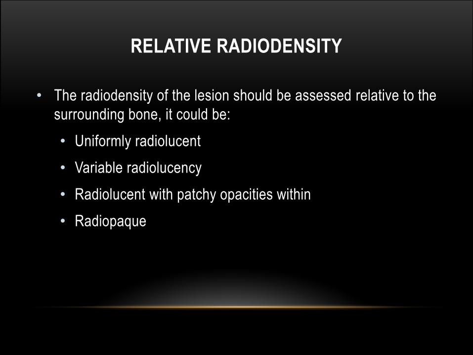
RELATIVE RADIODENSITY
• The radiodensity of the lesion should be assessed relative to the
surrounding bone, it could be:
• Uniformly radiolucent
• Variable radiolucency
• Radiolucent with patchy opacities within
• Radiopaque
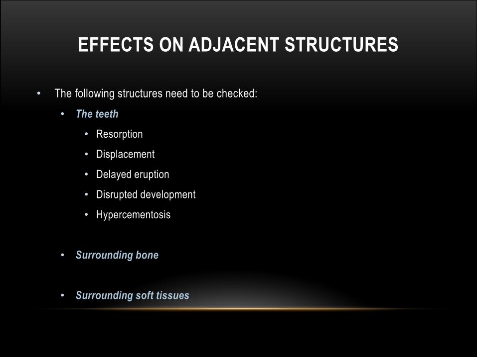
EFFECTS ON ADJACENT STRUCTURES
• The following structures need to be checked:
• The teeth
• Resorption
• Displacement
• Delayed eruption
• Disrupted development
• Hypercementosis
• Surrounding bone
• Surrounding soft tissues

Surrounding bone
• Expansion:
• Buccal
• Lingual
• In other directions
• Displacement or involvement of surrounding structures, including the:
• Inferior dental canal
• Mental foramen
• Antra
• Lower border of the mandible
• Nasal cavity
• Orbits
• Ragged destruction
• Alteration in the trabecular pattern or density
• Subperiosteal new bone formation.


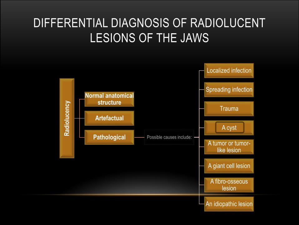
DIFFERENTIAL DIAGNOSIS OF RADIOLUCENT
LESIONS OF THE JAWS
Rad
iolu
cen
cy
Normal anatomicalstructure
Artefactual
Pathological Possible causes include:
Localized infection
Spreading infection
Trauma
A cyst
A tumor or tumor-like lesion
A giant cell lesion
A fibro-osseouslesion
An idiopathic lesion
CYSTS OF THE JAWS
Definition: A cyst is an epithelial lined, pathological cavity
having fluid, semi-fluid or gaseous contents, and surrounded
by connective tissue.
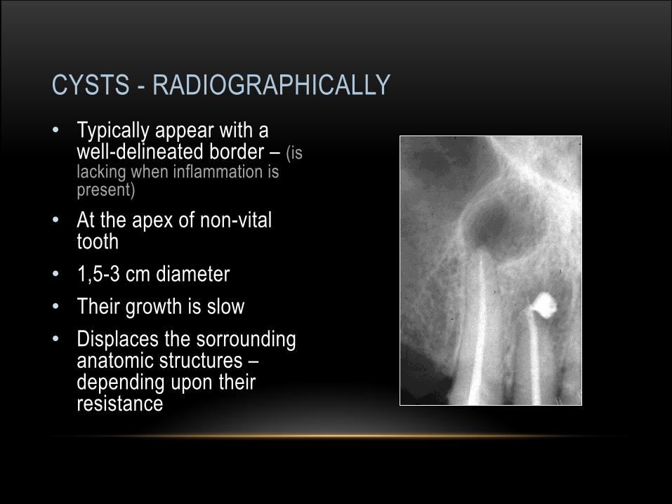
• Typically appear with a well-delineated border – (is lacking when inflammation ispresent)
• At the apex of non-vitaltooth
• 1,5-3 cm diameter
• Their growth is slow
• Displaces the sorrounding anatomic structures –depending upon theirresistance
CYSTS - RADIOGRAPHICALLY
• ETIOLOGY:
• Developmental
• Inflammatory
• Traumatic
• Neoplastic

I. ODONTOGENIC CYSTS
(with epithelial lining)
1. Radicular cysts
• Apical cyst
• Lateral cyst
2. Periodontal cysts
3. Follicular cysts
• Before formation of hard tooth substance
• Primordial cyst
• Keratocyst
• After formation of hard tooth substance
• Eruption cyst
• Coronal cyst
• Lateral cyst
• Cyst with rudimentary tooth
4. Residual cysts of all types

II. NONODONTOGENIC CYSTS
(with epithelial lining)
1. Nasopalatine cysts
2. Median (fissural) cysts
• Median alveolar cyst
• Median palatal cyst
3. Lateral (fissural) cysts
• Nasoalveolar cyst
• Globulomaxillary cyst
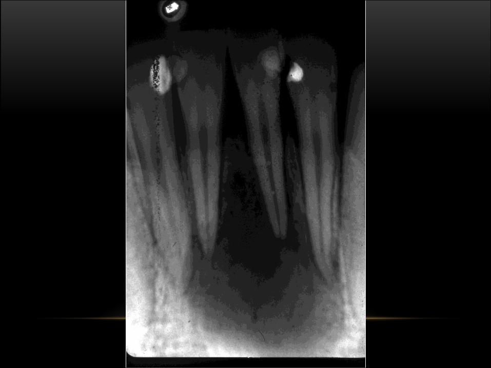
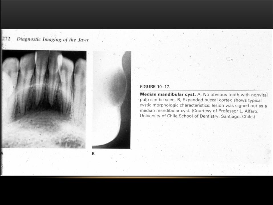
4. Median mandibular cysts
5. Residual cysts of all types

III. PSEUDOCYSTS
(without epithelial lining)
1. Solitary bone cyst
2. Aneurysmatic bone cyst
3. Latent bone cavity (Stafne)

I. Odontogenic cysts
• 1. Radicular cysts
• Apical cyst
• Lateral cyst
• 2. Periodontal cysts
• 3. Follicular cysts
• Before formation of hard tooth substance
• Primordial cyst
• Keratocyst
• After formation of hard tooth substance
• Eruption cyst
• Coronal cyst
• Lateral cyst
• Cyst with rudimentary tooth
• 4. Residual cysts of all types
II. Nonodontogenic cysts
• 1. Nasopalatine cysts
• 2. Median (fissural) cysts
• Median alveolar cyst
• Median palatal cyst
• 3. Lateral (fissural) cysts
• Nasoalveolar cyst
• Globulomaxillary cyst
• 4. Median mandibular cysts
• 5. Residual cysts of all types
III. Pseudocysts
• 1. Solitary bone cyst
• 2. Aneurysmatic bone cyst
• 3. Latent bone cavity (Stafne)
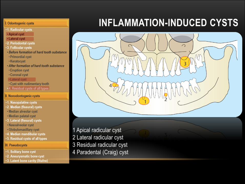
INFLAMMATION-INDUCED CYSTS
1 Apical radicular cyst
2 Lateral radicular cyst
3 Residual radicular cyst
4 Paradental (Craig) cyst
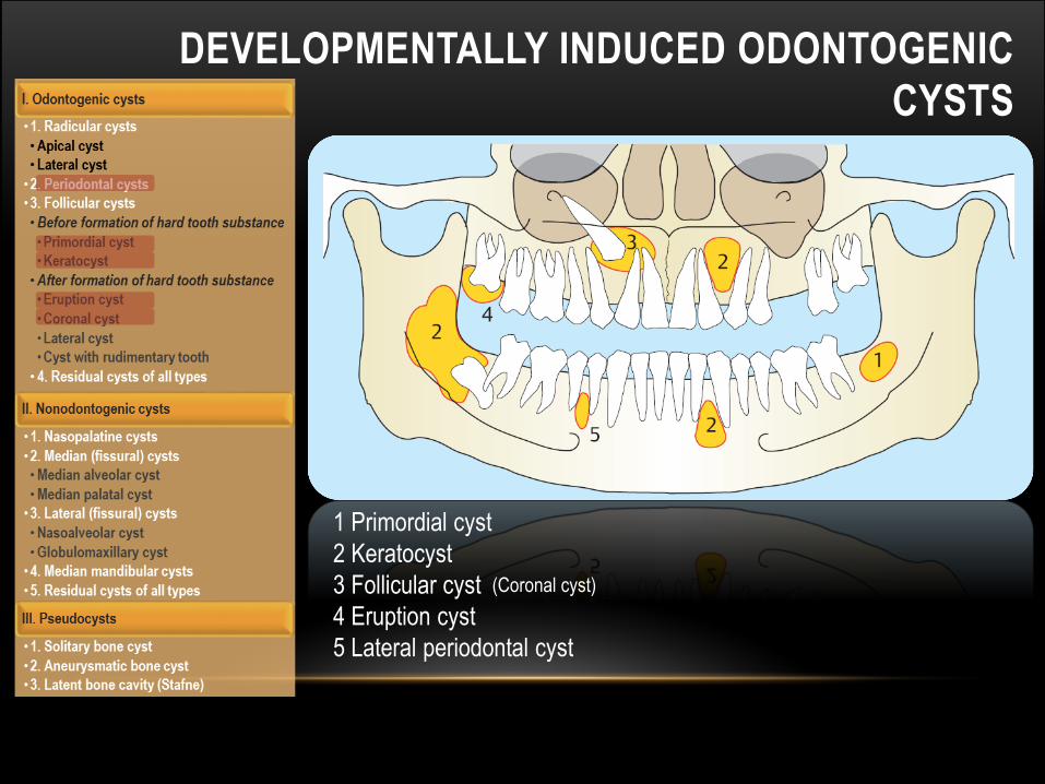
DEVELOPMENTALLY INDUCED ODONTOGENIC
CYSTS
1 Primordial cyst
2 Keratocyst
3 Follicular cyst
4 Eruption cyst
5 Lateral periodontal cyst
(Coronal cyst)

DEVELOPMENTALLY INDUCED
NONODONTOGENIC CYSTS
1 Nasopalatine cyst
2 Nasolabial (globulomaxillary) cyst
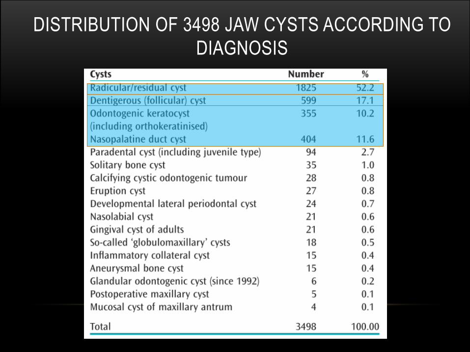
DISTRIBUTION OF 3498 JAW CYSTS ACCORDING TO
DIAGNOSIS

DISTRIBUTION OF 7121 ODONTOGENIC CYSTS ACCORDING TO
DIAGNOSIS. FROM JONES ET AL. (2006), SHEFFIELD.
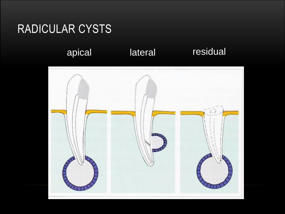
RADICULAR CYSTS
apical lateral residual
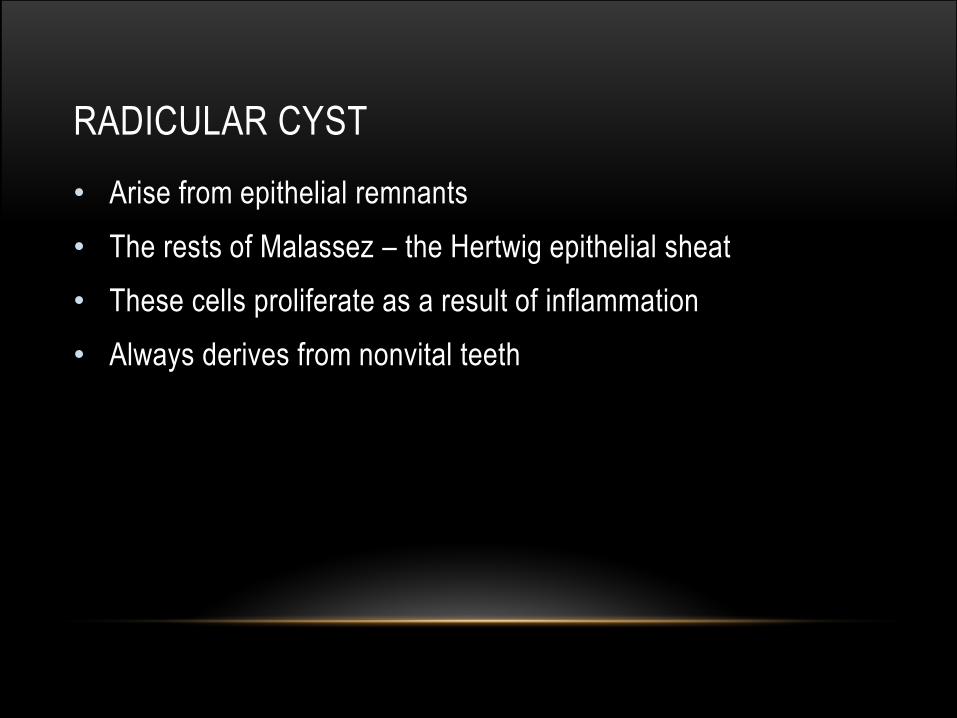
RADICULAR CYST
• Arise from epithelial remnants
• The rests of Malassez – the Hertwig epithelial sheat
• These cells proliferate as a result of inflammation
• Always derives from nonvital teeth

AGE DISTRIBUTION AGE DISTRIBUTION OF 1970
PATIENTS WITH RADICULAR CYSTS
SHEFFIELD, ENGLAND, 1990–2004 (N=1970).

SITE DISTRIBUTION OF RADICULAR CYST
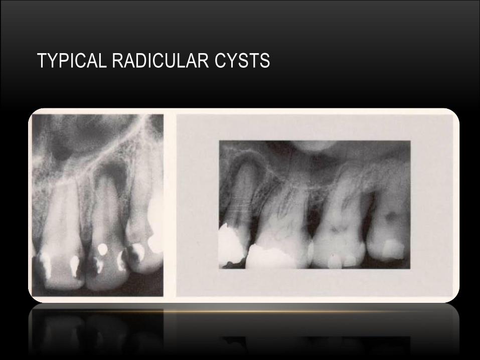
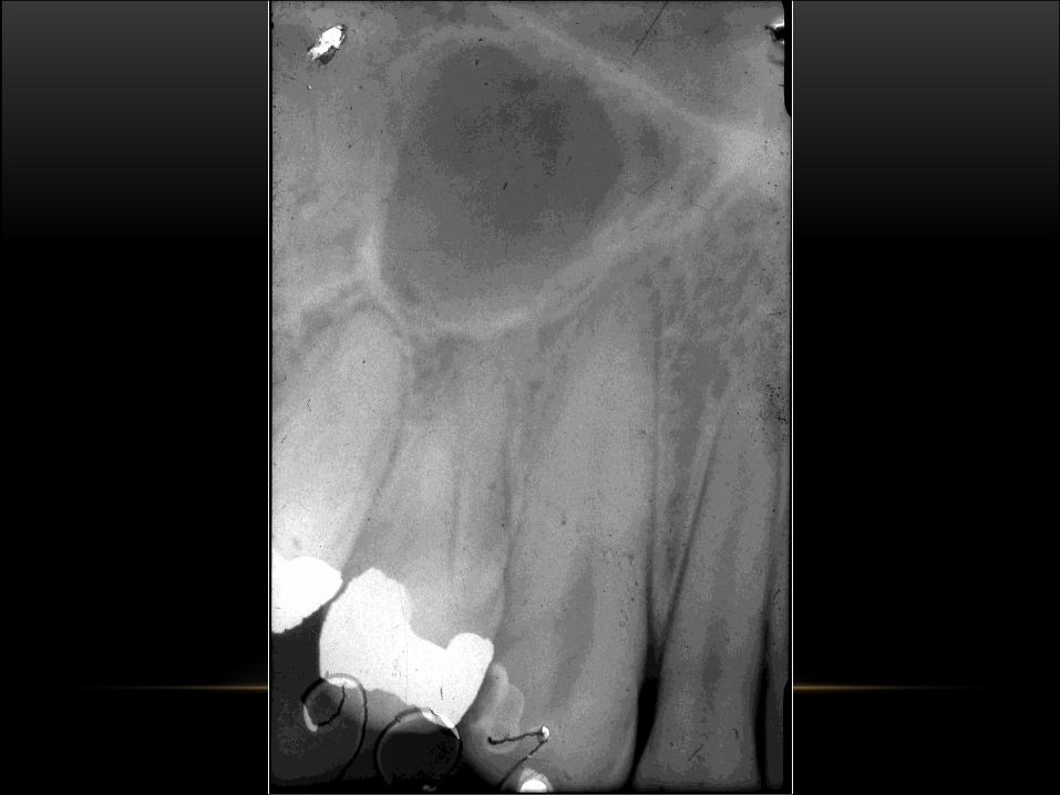
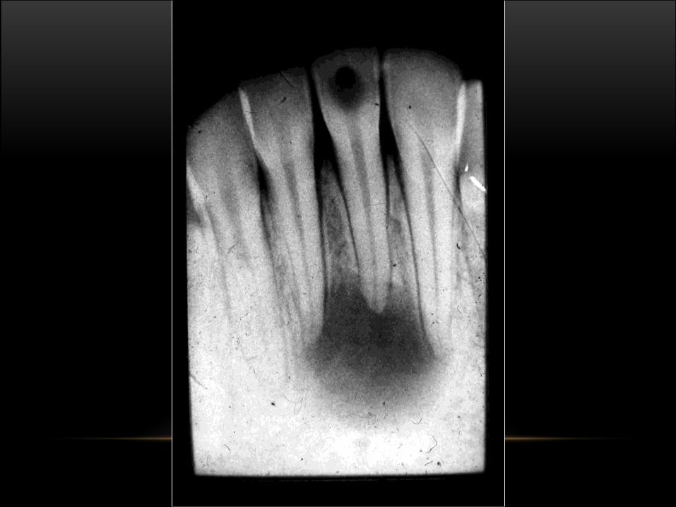
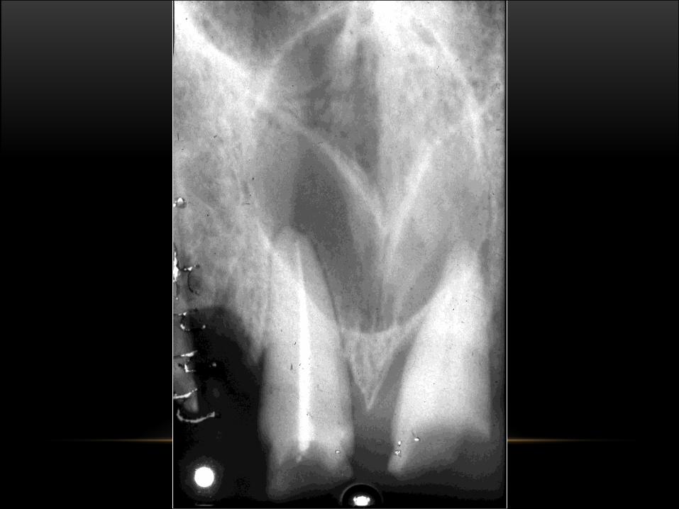
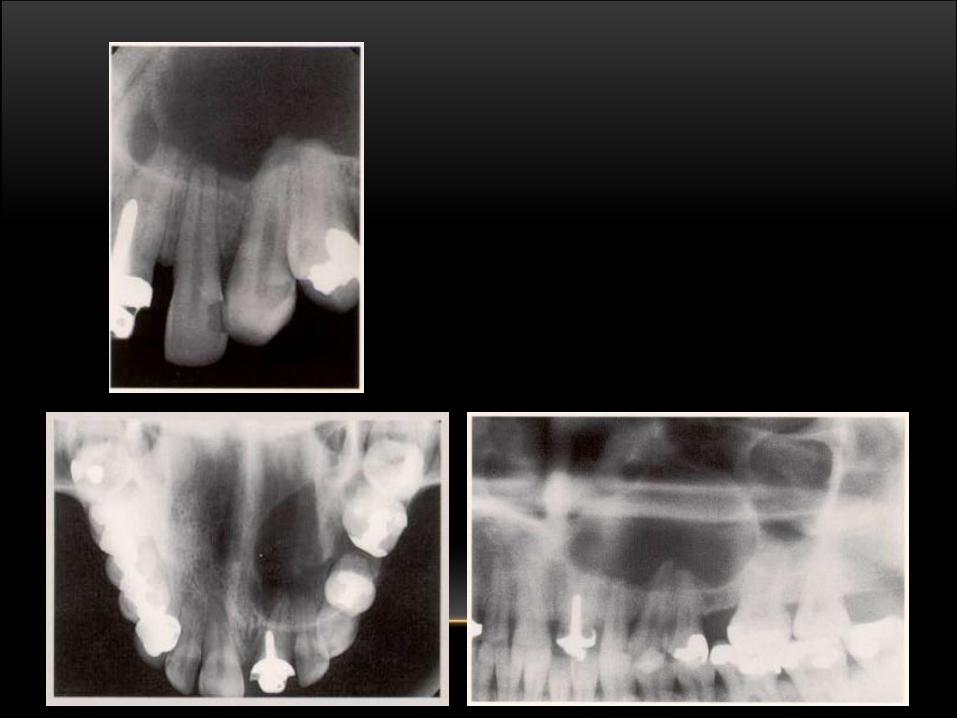
TYPICAL RADICULAR CYSTS
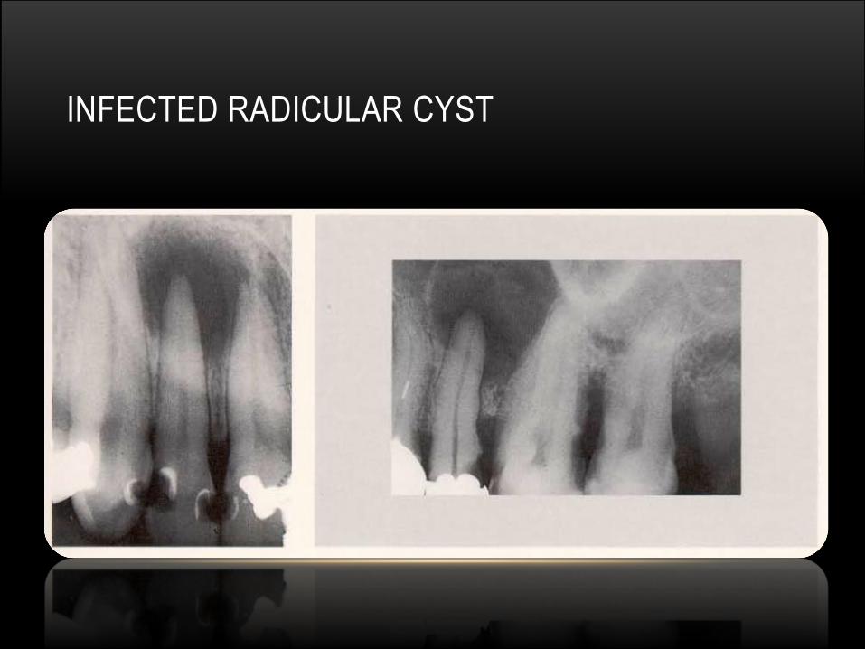
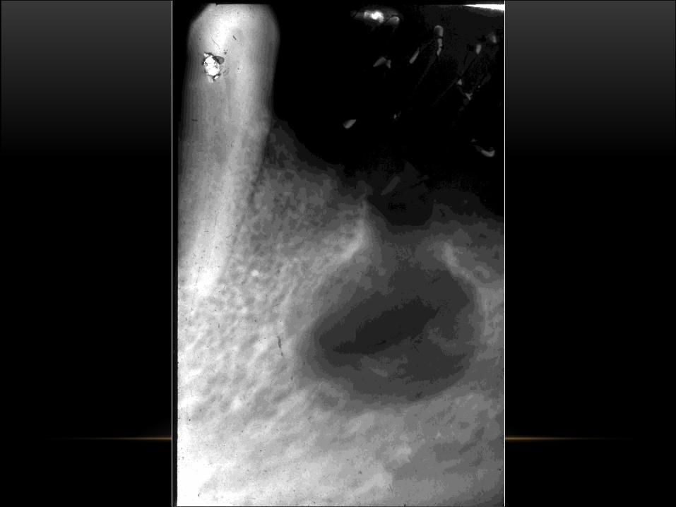
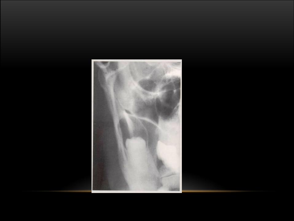
INFECTED RADICULAR CYST
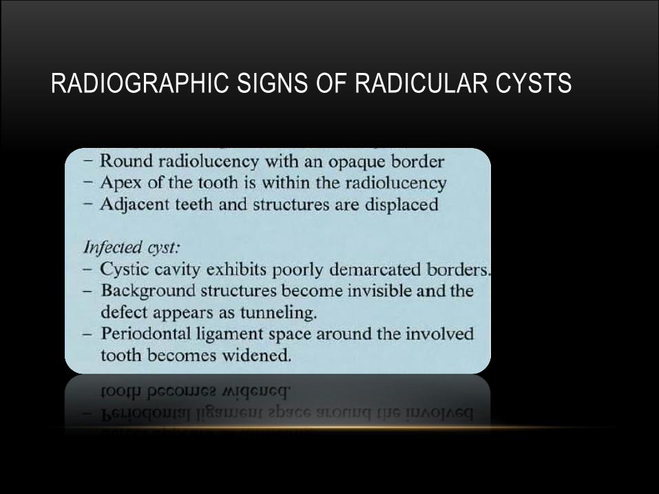
RADIOGRAPHIC SIGNS OF RADICULAR CYSTS

INFECTED RADICULAR CYST

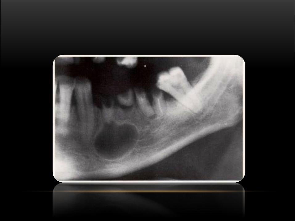
ATYPICAL MANIFESTATION OF MULTILOCULAR
RADICULAR CYST
Differential diagnosis: ameloblastoma, giant cell granuloma, keratocys
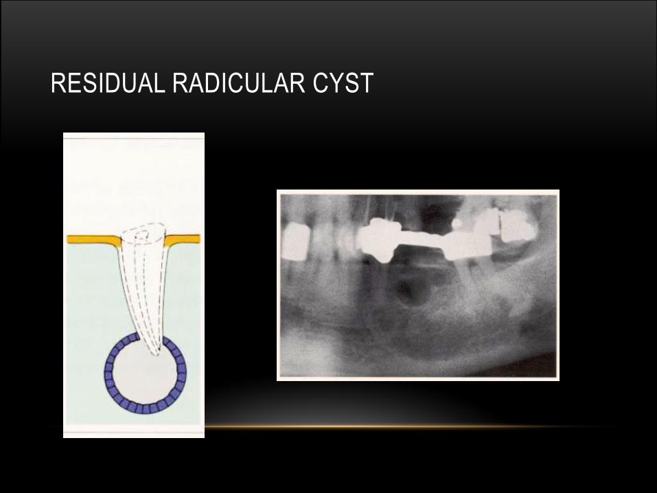
RESIDUAL RADICULAR CYST
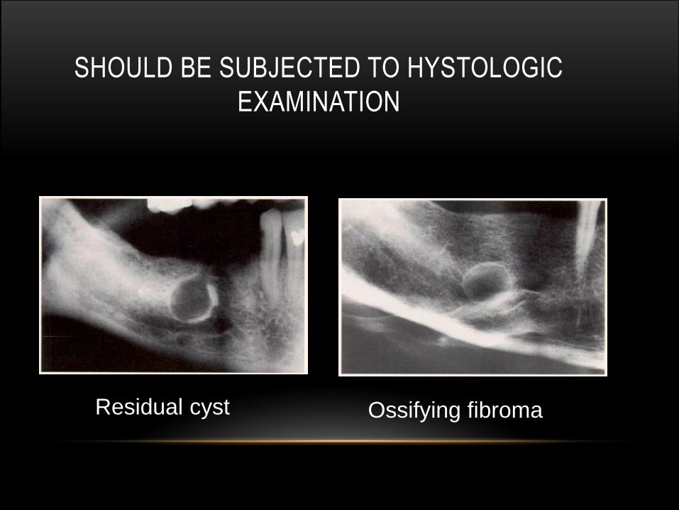
SHOULD BE SUBJECTED TO HYSTOLOGIC
EXAMINATION
Residual cyst Ossifying fibroma
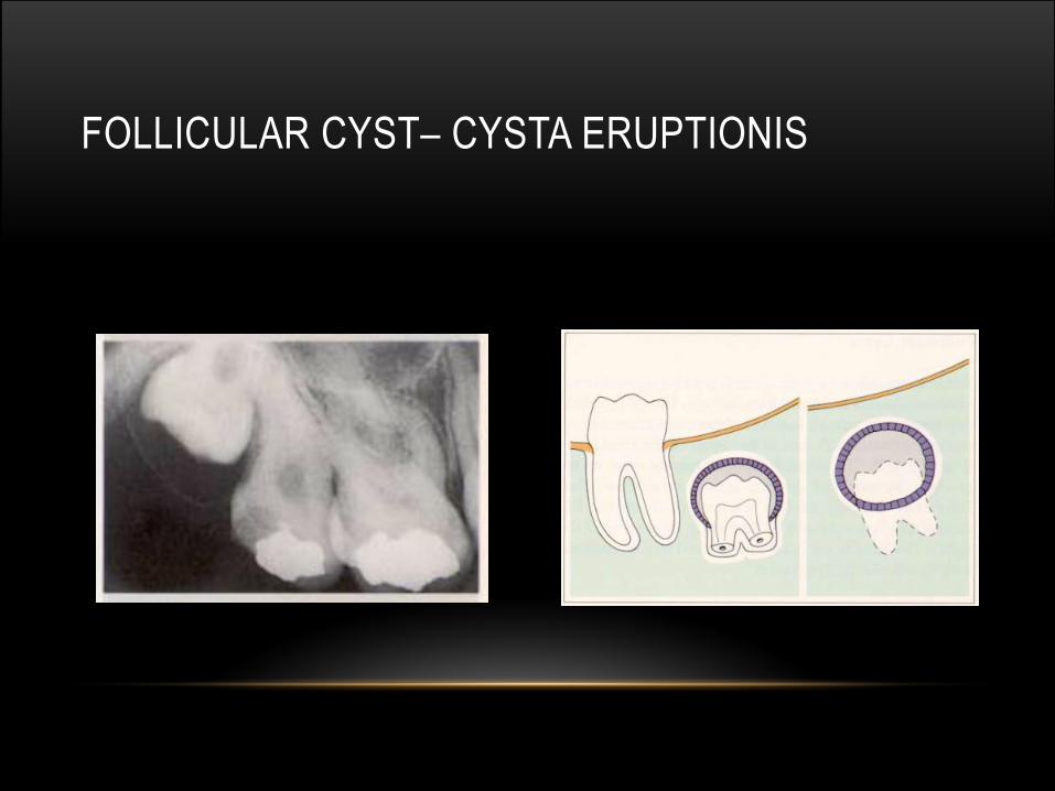
FOLLICULAR CYST– CYSTA ERUPTIONIS

PARADENTAL CYST
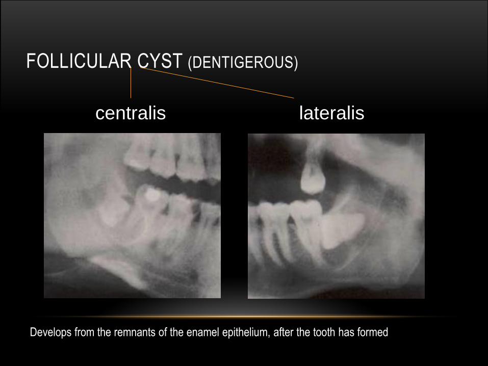
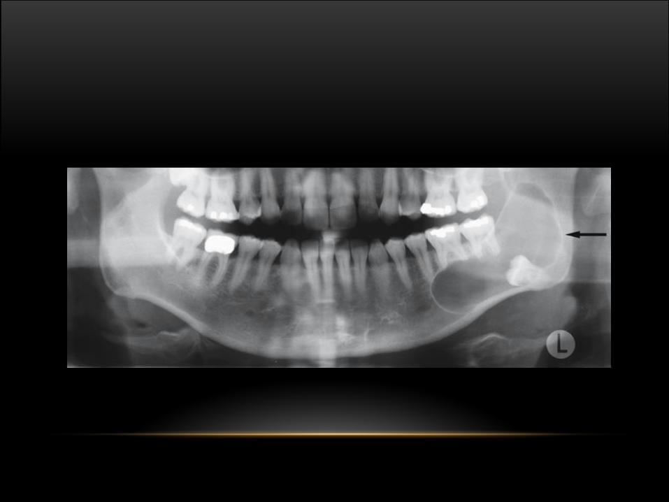
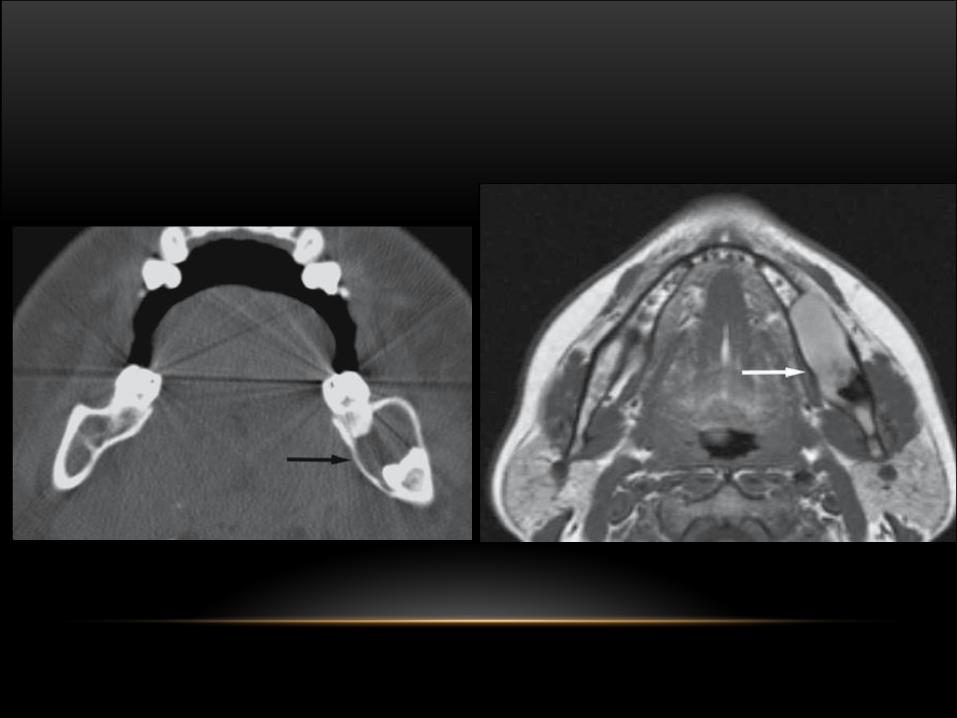
FOLLICULAR CYST (DENTIGEROUS)
centralis lateralis
Develops from the remnants of the enamel epithelium, after the tooth has formed


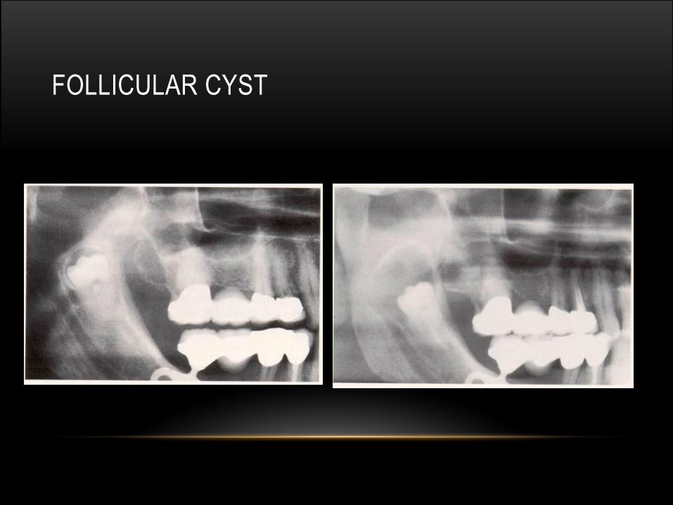
FOLLICULAR CYST
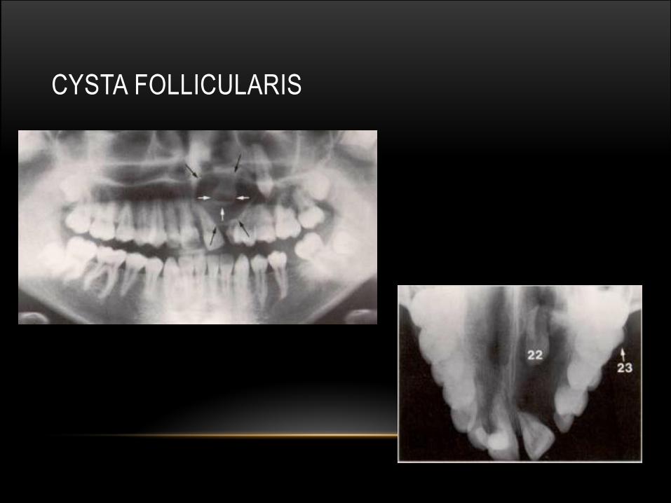
CYSTA FOLLICULARIS
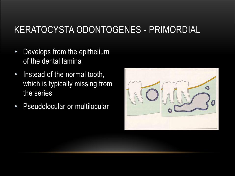
KERATOCYSTA ODONTOGENES - PRIMORDIAL
• Develops from the epithelium
of the dental lamina
• Instead of the normal tooth,
which is typically missing from
the series
• Pseudolocular or multilocular

THE MOST IMPORTANT SITES OF KERATOCYSTS
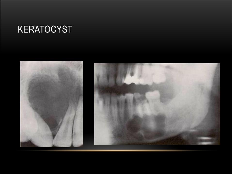
KERATOCYST

INCISIVE CANAL CYST – NASOPALATINE DUCT CYST
B: fissura mediana cysta
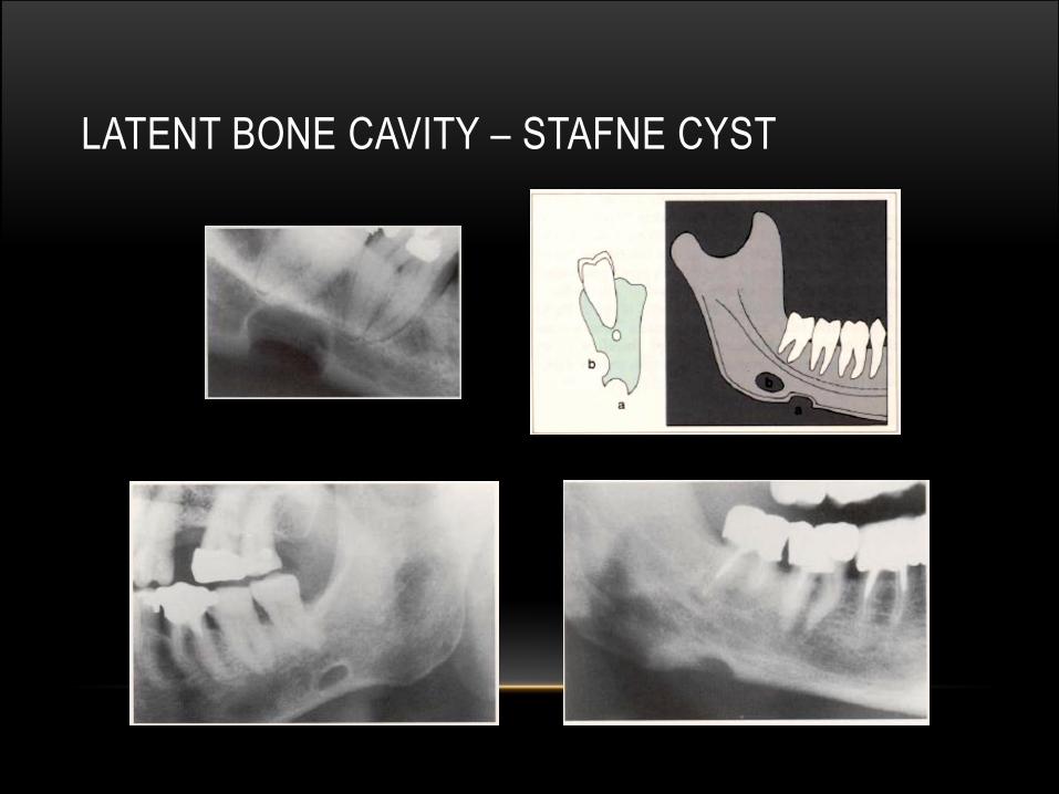
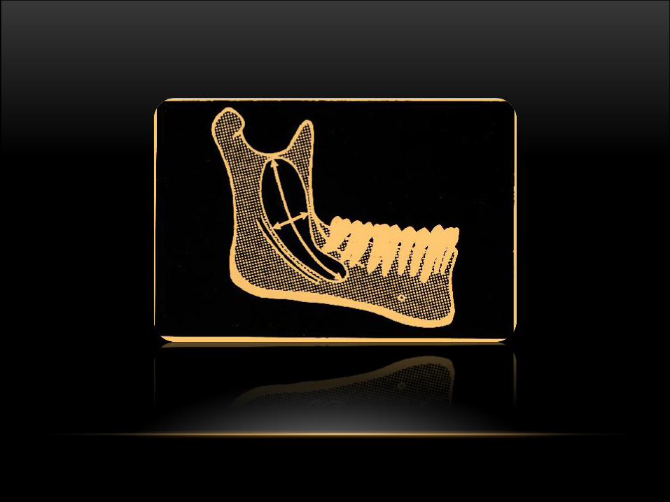
LATENT BONE CAVITY – STAFNE CYST