CYSTIC TUMORS OF THE PANCREAS - Los Angeles,...
57
CYSTIC TUMORS OF THE PANCREAS: WHAT WE KNOW AND WHAT WE DON’T Andrew L. Warshaw, M.D. W. Gerald Austen Professor of Surgery Harvard Medical School Surgeon-in-Chief Massachusetts General Hospital Longmire Surgical Society April 8, 2011 William P. Longmire Jr., M.D.
Transcript of CYSTIC TUMORS OF THE PANCREAS - Los Angeles,...
CYSTIC TUMORS OF THE PANCREAS:
WHAT WE KNOW AND WHAT WE DON’T
Andrew L. Warshaw, M.D. W. Gerald Austen Professor of Surgery
Harvard Medical School
Surgeon-in-Chief
Massachusetts General Hospital
Longmire Surgical Society
April 8, 2011
William P. Longmire Jr., M.D.

CONFLICT OF INTEREST
NOTHING
TO
DISCLOSE
(unfortunately)

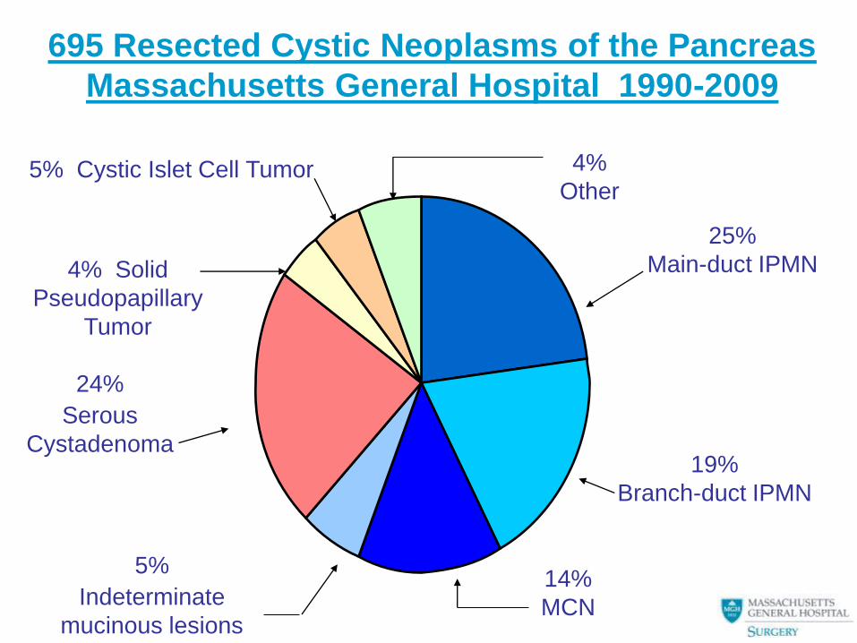
695 Resected Cystic Neoplasms of the Pancreas
Massachusetts General Hospital 1990-2009
25%
Main-duct IPMN
19%
Branch-duct IPMN
14%
MCN
5%
Indeterminate
mucinous lesions
24%
Serous
Cystadenoma
4% Solid
Pseudopapillary
Tumor
4%
Other 5% Cystic Islet Cell Tumor
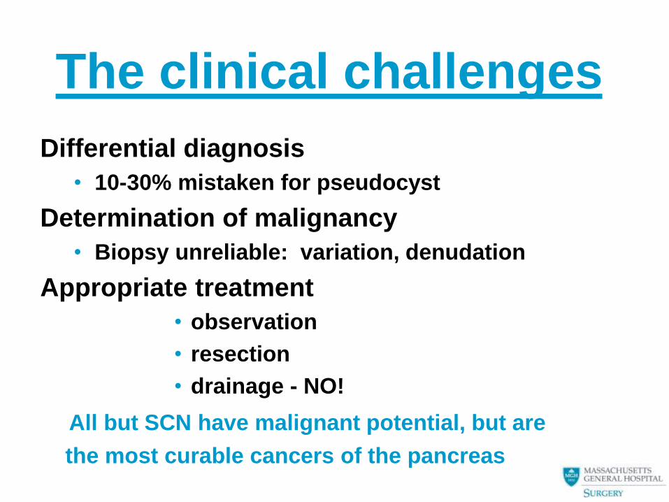
The clinical challenges
Differential diagnosis
• 10-30% mistaken for pseudocyst
Determination of malignancy
• Biopsy unreliable: variation, denudation
Appropriate treatment
• observation
• resection
• drainage - NO!
All but SCN have malignant potential, but are
the most curable cancers of the pancreas
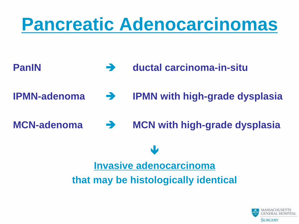
Pancreatic Adenocarcinomas
PanIN ductal carcinoma-in-situ
IPMN-adenoma IPMN with high-grade dysplasia
MCN-adenoma MCN with high-grade dysplasia
Invasive adenocarcinoma
that may be histologically identical
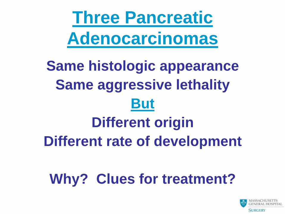
Three Pancreatic
Adenocarcinomas
Same histologic appearance
Same aggressive lethality
But
Different origin
Different rate of development
Why? Clues for treatment?
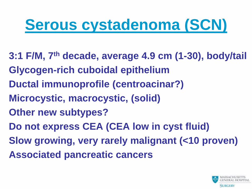
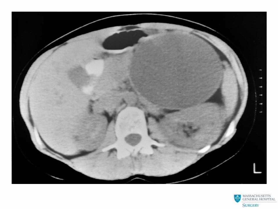
Serous cystadenoma (SCN)
3:1 F/M, 7th decade, average 4.9 cm (1-30), body/tail
Glycogen-rich cuboidal epithelium
Ductal immunoprofile (centroacinar?)
Microcystic, macrocystic, (solid)
Other new subtypes?
Do not express CEA (CEA low in cyst fluid)
Slow growing, very rarely malignant (<10 proven)
Associated pancreatic cancers
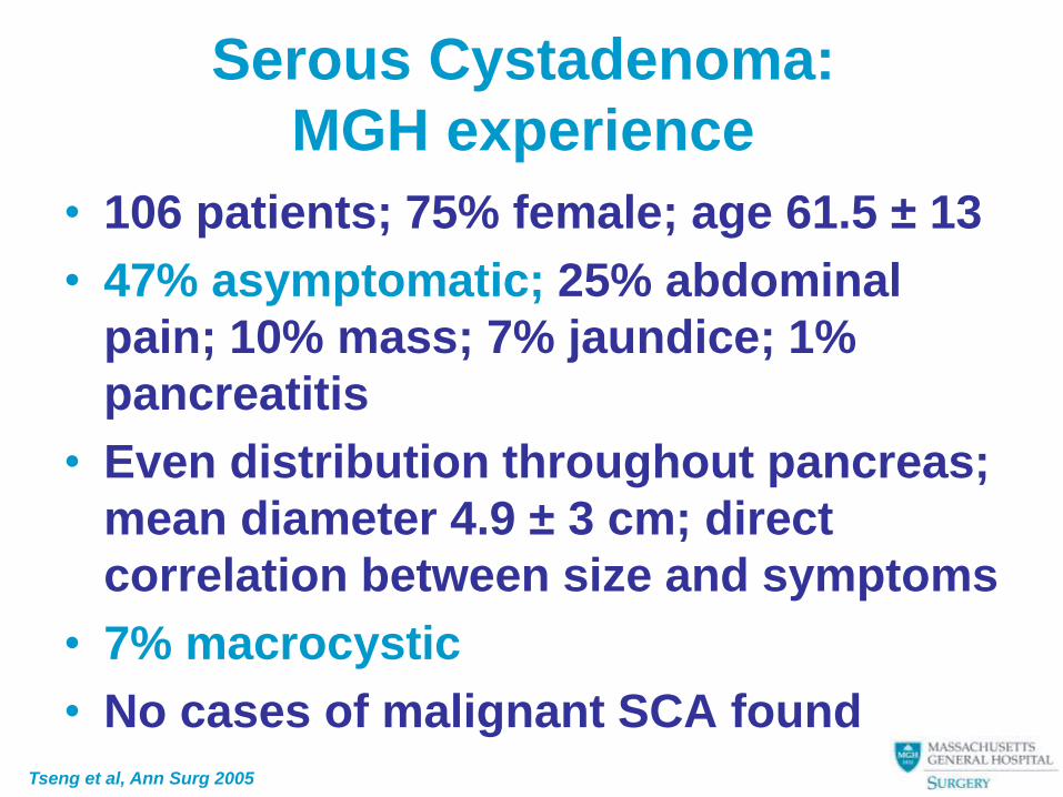
Serous Cystadenoma:
MGH experience
• 106 patients; 75% female; age 61.5 ± 13
• 47% asymptomatic; 25% abdominal
pain; 10% mass; 7% jaundice; 1%
pancreatitis
• Even distribution throughout pancreas;
mean diameter 4.9 ± 3 cm; direct
correlation between size and symptoms
• 7% macrocystic
• No cases of malignant SCA found
Tseng et al, Ann Surg 2005
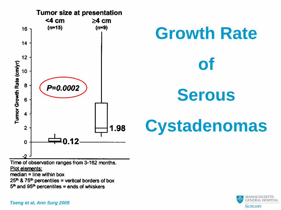
Growth Rate
of
Serous
Cystadenomas
Tseng et al, Ann Surg 2005
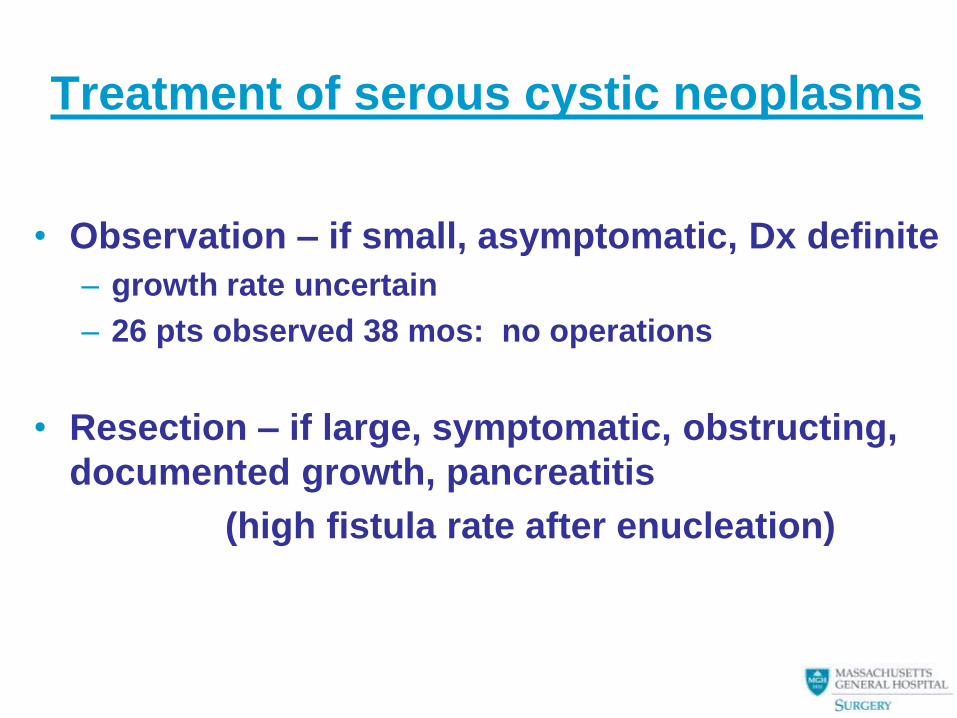
Treatment of serous cystic neoplasms
• Observation – if small, asymptomatic, Dx definite
– growth rate uncertain
– 26 pts observed 38 mos: no operations
• Resection – if large, symptomatic, obstructing,
documented growth, pancreatitis
(high fistula rate after enucleation)
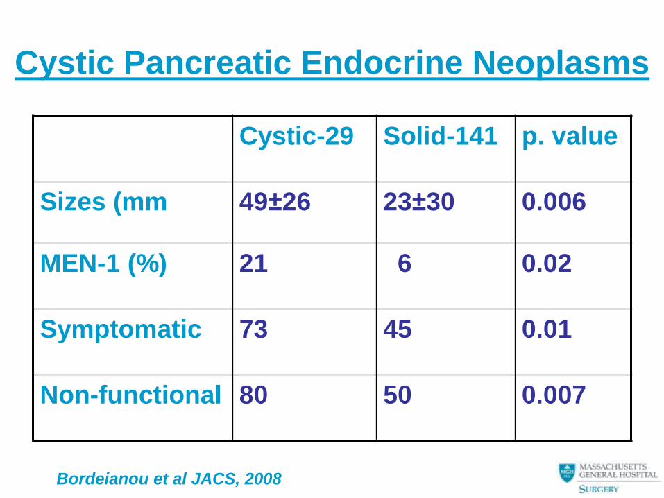
Cystic Pancreatic Endocrine Neoplasms
Cystic-29 Solid-141 p. value
Sizes (mm 49±26 23±30 0.006
MEN-1 (%) 21 6 0.02
Symptomatic 73 45 0.01
Non-functional 80 50 0.007
Bordeianou et al JACS, 2008
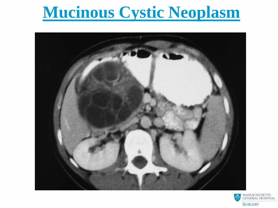
Mucinous Cystic Neoplasm
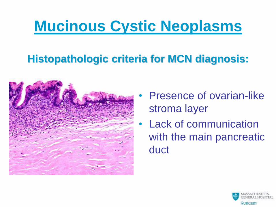
• Presence of ovarian-like
stroma layer
• Lack of communication
with the main pancreatic
duct
Mucinous Cystic Neoplasms
Histopathologic criteria for MCN diagnosis:
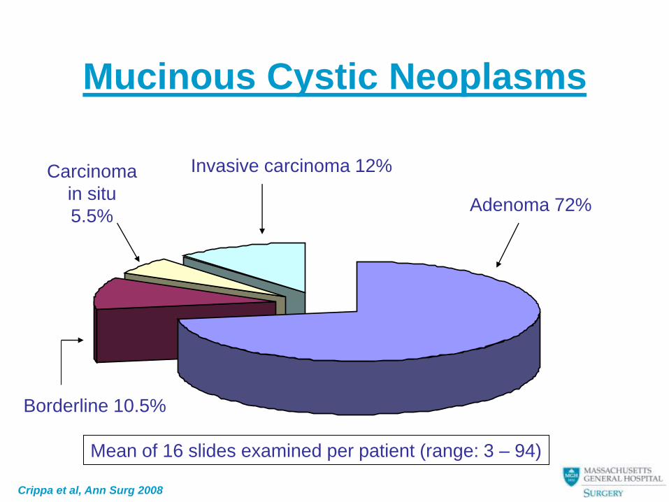
Mucinous Cystic Neoplasms
Adenoma 72%
Invasive carcinoma 12%
Borderline 10.5%
Carcinoma
in situ
5.5%
Mean of 16 slides examined per patient (range: 3 – 94)
Crippa et al, Ann Surg 2008
Mucinous Cystic Neoplasms
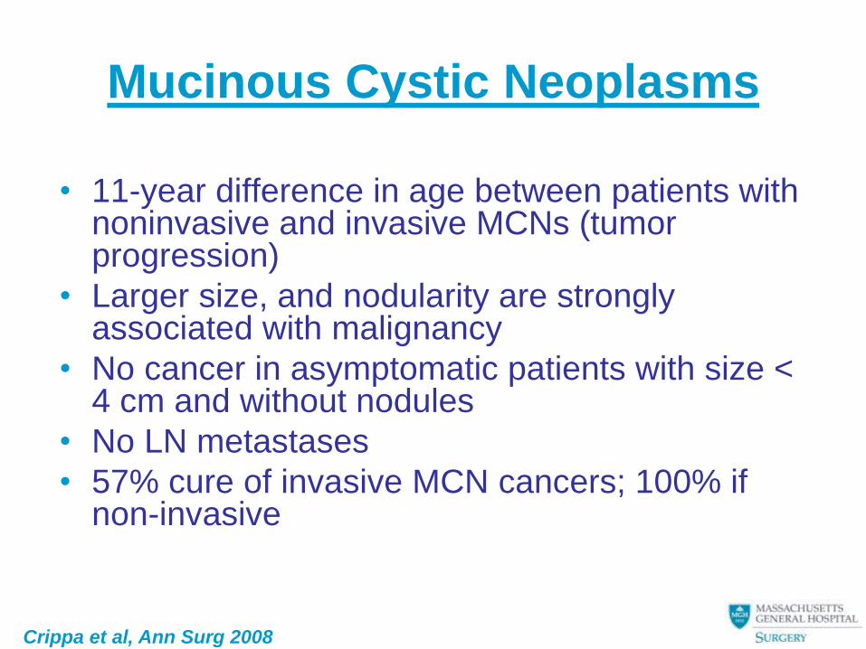
• 11-year difference in age between patients with noninvasive and invasive MCNs (tumor progression)
• Larger size, and nodularity are strongly associated with malignancy
• No cancer in asymptomatic patients with size < 4 cm and without nodules
• No LN metastases
• 57% cure of invasive MCN cancers; 100% if non-invasive
Crippa et al, Ann Surg 2008
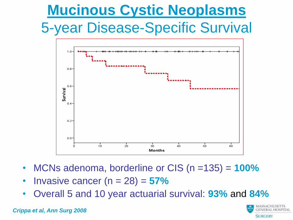
Mucinous Cystic Neoplasms
5-year Disease-Specific Survival
• MCNs adenoma, borderline or CIS (n =135) = 100%
• Invasive cancer (n = 28) = 57%
• Overall 5 and 10 year actuarial survival: 93% and 84%
Crippa et al, Ann Surg 2008
Mucinous Cystic Neoplasms
Tumor Recurrence
• No recurrence in noninvasive MCNs
• Seven patients (4.5%) comprising 37% of invasive
MCNs developed tumor recurrence; of these
– 4 patients had extracapsular infiltration
– 3 patients had diffuse intracapsular infiltration
– none had intracapsular, focally invasive carcinoma
• Recurrence site
– Peritoneum in two patients
– Liver in five
All patients with recurrence died after a mean of 6.5 months
Crippa et al, Ann Surg 2008
Mucinous Cystic Neoplasms
Recommendations
• Given the young age of patients with
MCNs, and the possibility of tumor-
progression, resection remains the
treatment of choice
• In small MCNs without nodules,
parenchyma/spleen-preserving and
minimally invasive procedures should
be performed
INTRADUCTAL PAPILLARY MUCINOUS
NEOPLASM OF THE PANCREAS
• The “new kid in the block” is now an established settler ( Has he been here all along?)
• One of the most common indications for pancreatic resection at MGH
• Clear differences in the presentation and implications of Main Duct vs Branch Duct IPMN (? Different biology)
• ? True incidence of this disease (many asymptomatic Br IPMN)
Main-Duct Intraductal Papillary Mucinous
Neoplasms (IPMN)
Men>women, head>tail, age 68
Pain/pancreatitis, exocrine insufficiency
(if chronic pancreatitis)
Increasing numbers reported
(incidence or recognition?)
Long indolent phase (probable) in progression to
invasive cancer
Serum Ca 19-9 with advanced cancer
Association with other GI tumors (colon, stomach)
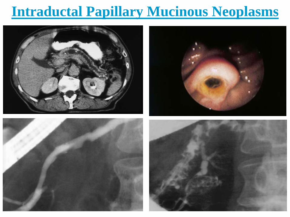
Intraductal Papillary Mucinous Neoplasms
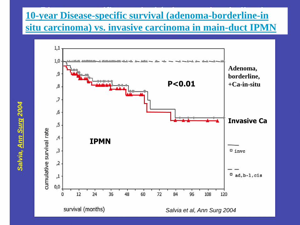
Main-duct IPMN
• 60% of resectable main-duct IPMN contain cancer, in situ or invasive
• Malignant IPMN occur in older patients and are more likely to present with jaundice or new-onset diabetes
• More than 25% of IPMN are asymptomatic (benign or malignant)
• Resection for cure is highly probable (benign including in situ cancer – 100%; invasive malignant – 60%)
• Recurrence in a pancreatic remnant is uncommon (5-7%)
• Re-resection of remnant recurrence is possible and beneficial
Adenoma,
borderline,
+Ca-in-situ
IPMN
P<0.01
Invasive Ca
10-year Disease-specific survival (adenoma-borderline-in
situ carcinoma) vs. invasive carcinoma in main-duct IPMN S
alv
ia, A
nn
Su
rg 2
00
4
Salvia et al, Ann Surg 2004
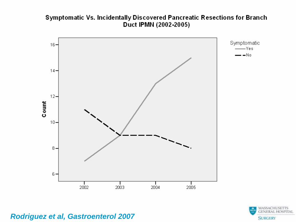
Rodriguez et al, Gastroenterol 2007
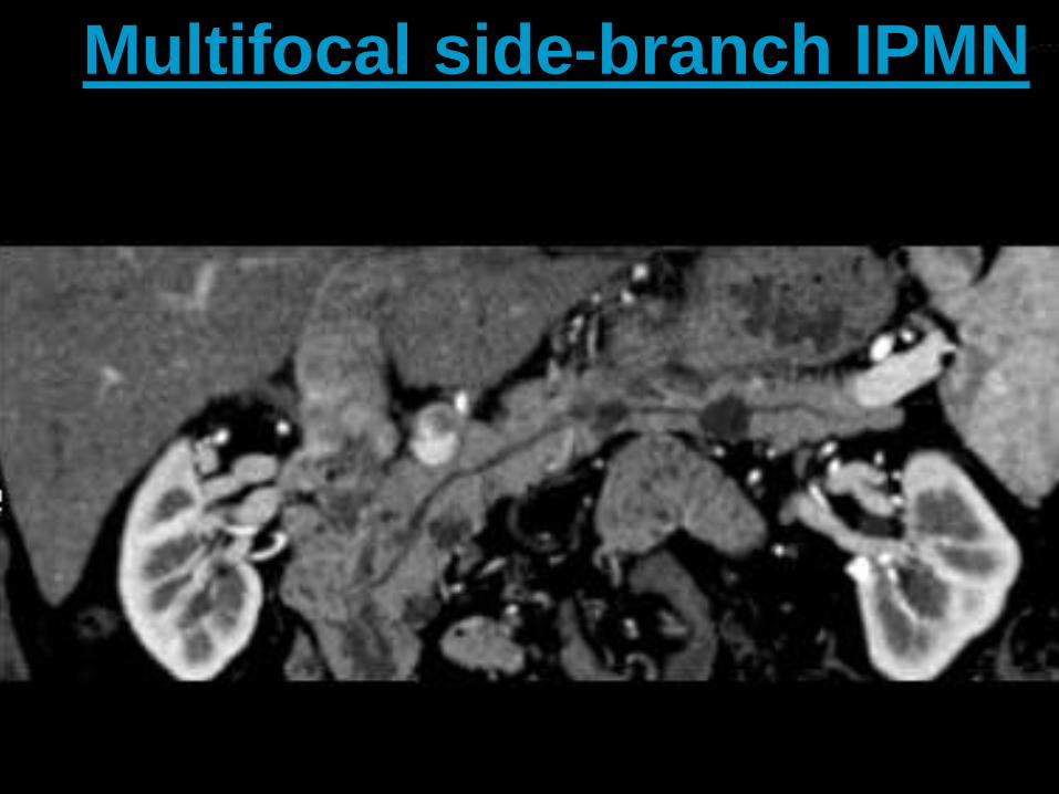
Multifocal side-branch IPMN
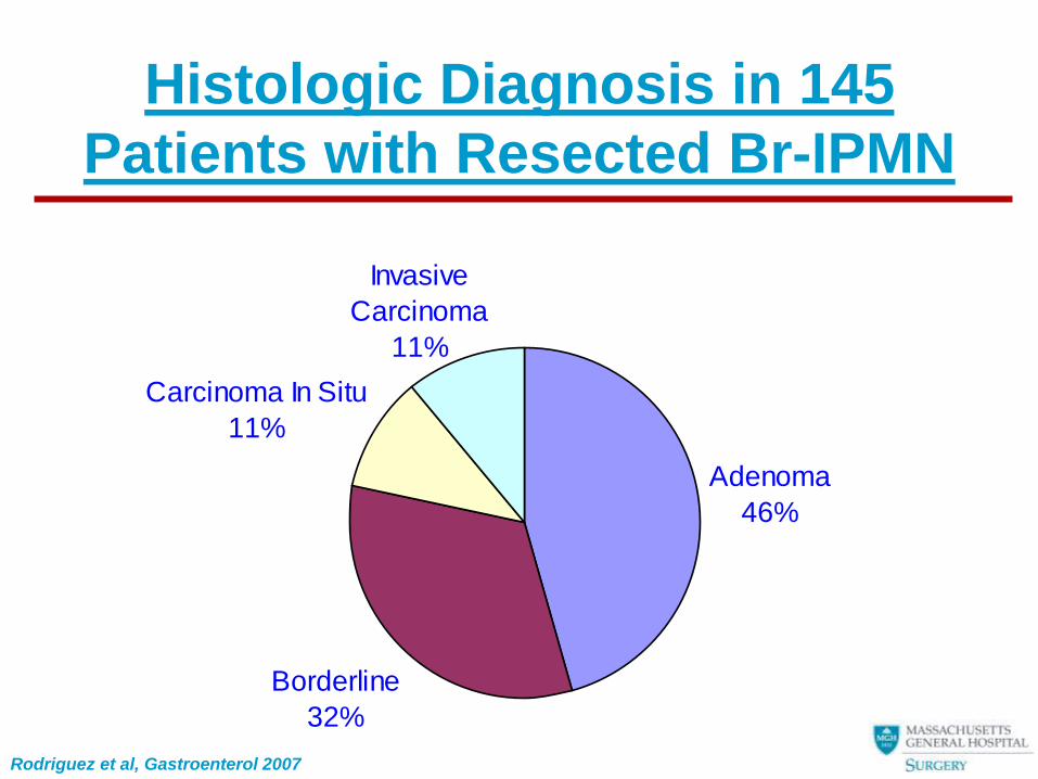
Histologic Diagnosis in 145
Patients with Resected Br-IPMN
Adenoma
46%
Borderline
32%
Carcinoma In Situ
11%
Invasive
Carcinoma
11%
Rodriguez et al, Gastroenterol 2007
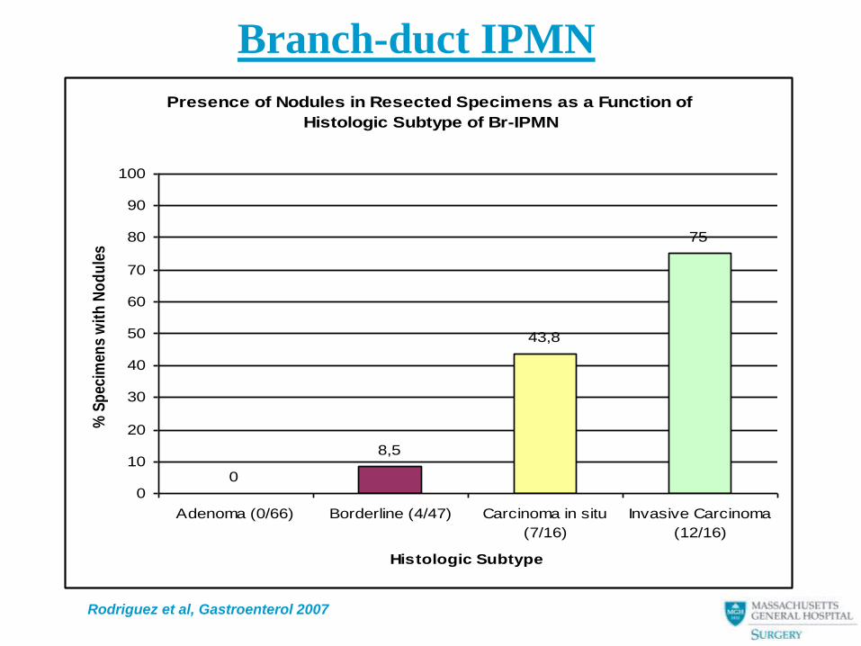
Presence of Nodules in Resected Specimens as a Function of
Histologic Subtype of Br-IPMN
0
8,5
43,8
75
0
10
20
30
40
50
60
70
80
90
100
Adenoma (0/66) Borderline (4/47) Carcinoma in situ
(7/16)
Invasive Carcinoma
(12/16)
Histologic Subtype
% S
pec
imen
s w
ith
No
du
les
Branch-duct IPMN
Rodriguez et al, Gastroenterol 2007
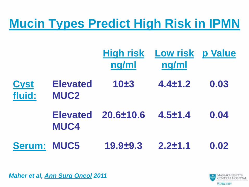
Mucin Types Predict High Risk in IPMN
High risk
ng/ml
Low risk
ng/ml
p Value
Cyst
fluid:
Elevated
MUC2
10±3 4.4±1.2 0.03
Elevated
MUC4
20.6±10.6 4.5±1.4 0.04
Serum: MUC5 19.9±9.3 2.2±1.1 0.02
Maher et al, Ann Surg Oncol 2011
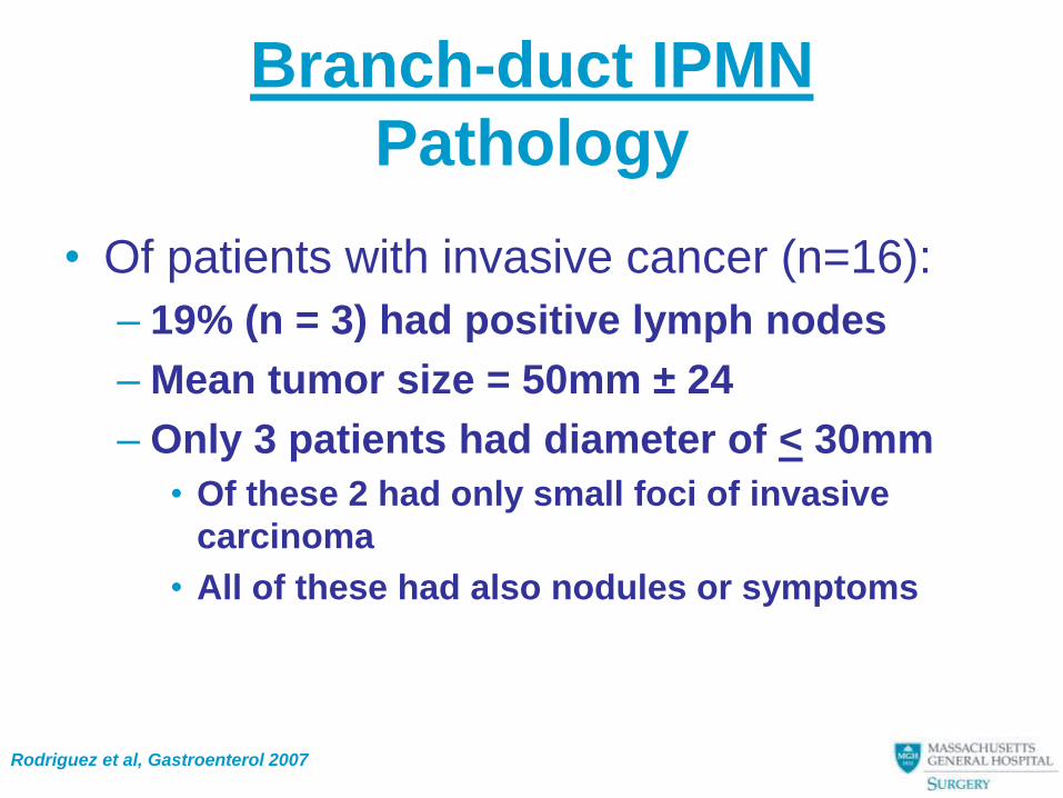
Branch-duct IPMN
Pathology
• Of patients with invasive cancer (n=16):
– 19% (n = 3) had positive lymph nodes
– Mean tumor size = 50mm ± 24
– Only 3 patients had diameter of < 30mm
• Of these 2 had only small foci of invasive
carcinoma
• All of these had also nodules or symptoms
Rodriguez et al, Gastroenterol 2007
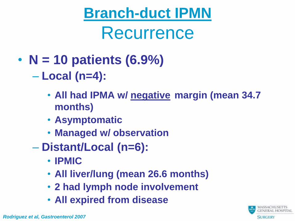
Branch-duct IPMN
Recurrence
• N = 10 patients (6.9%)
– Local (n=4):
• All had IPMA w/ negative margin (mean 34.7
months)
• Asymptomatic
• Managed w/ observation
– Distant/Local (n=6): • IPMIC
• All liver/lung (mean 26.6 months)
• 2 had lymph node involvement
• All expired from disease
Rodriguez et al, Gastroenterol 2007
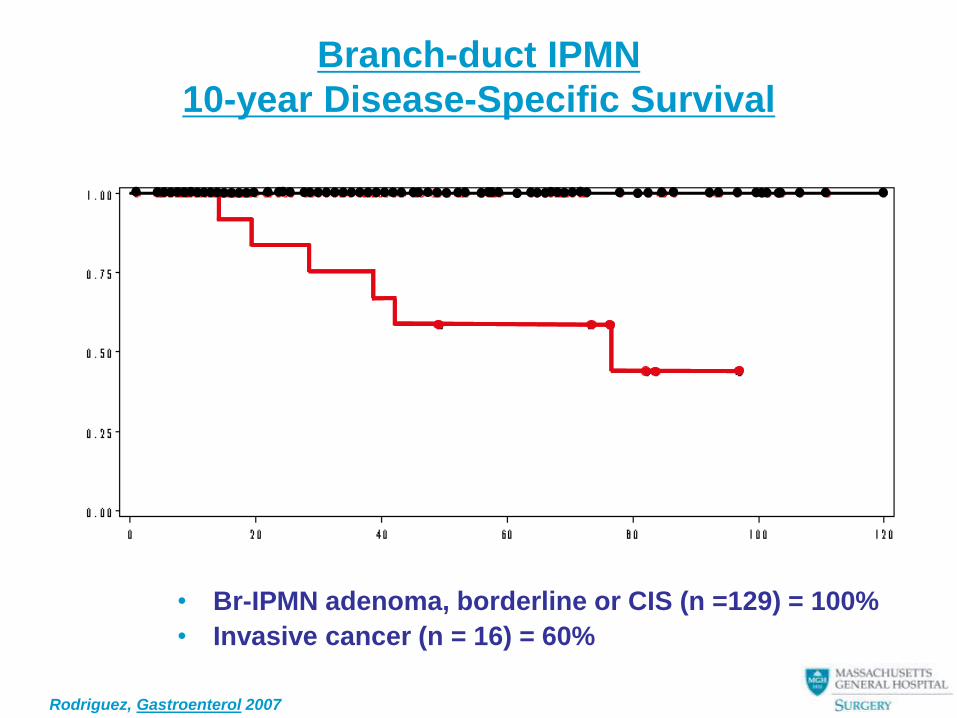
Branch-duct IPMN
10-year Disease-Specific Survival
• Br-IPMN adenoma, borderline or CIS (n =129) = 100%
• Invasive cancer (n = 16) = 60%
Rodriguez, Gastroenterol 2007
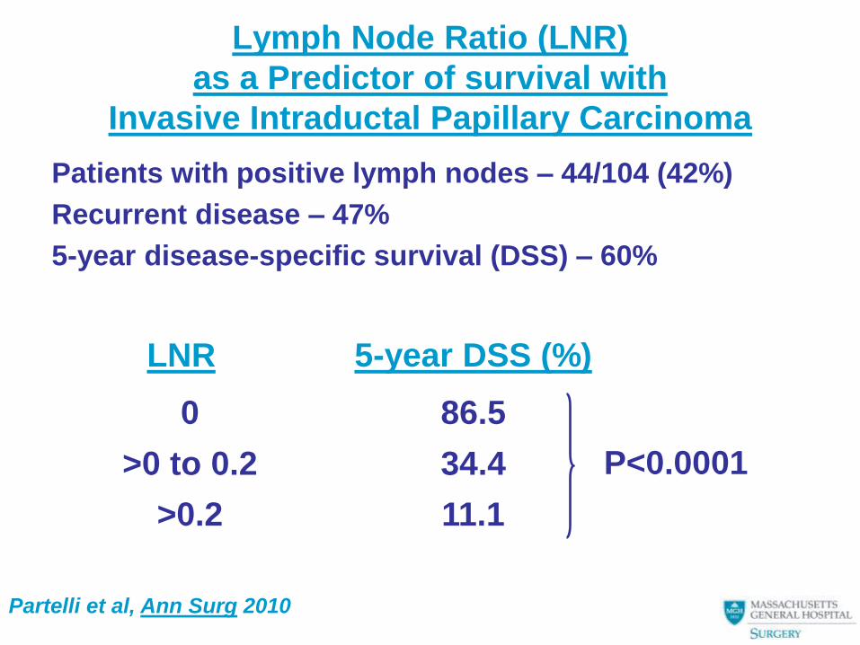
Lymph Node Ratio (LNR)
as a Predictor of survival with
Invasive Intraductal Papillary Carcinoma
Patients with positive lymph nodes – 44/104 (42%)
Recurrent disease – 47%
5-year disease-specific survival (DSS) – 60%
LNR 5-year DSS (%)
0 86.5
>0 to 0.2 34.4
>0.2 11.1
P<0.0001
Partelli et al, Ann Surg 2010
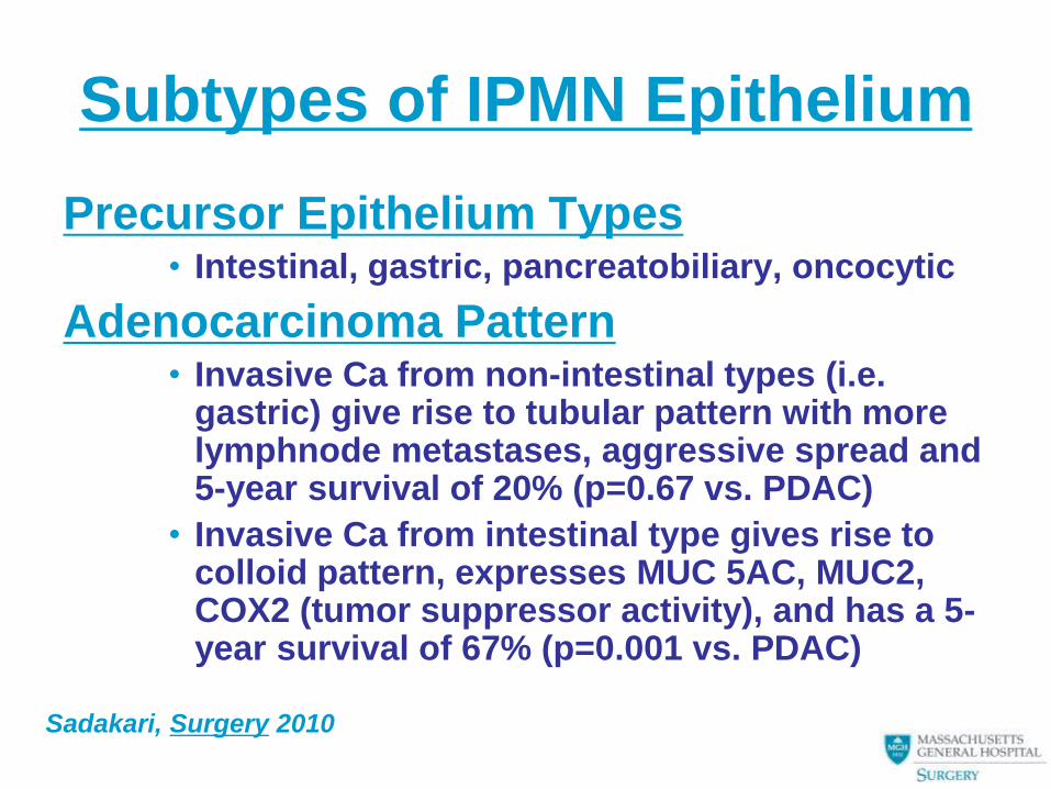
Subtypes of IPMN Epithelium
Precursor Epithelium Types • Intestinal, gastric, pancreatobiliary, oncocytic
Adenocarcinoma Pattern • Invasive Ca from non-intestinal types (i.e.
gastric) give rise to tubular pattern with more lymphnode metastases, aggressive spread and 5-year survival of 20% (p=0.67 vs. PDAC)
• Invasive Ca from intestinal type gives rise to colloid pattern, expresses MUC 5AC, MUC2, COX2 (tumor suppressor activity), and has a 5-year survival of 67% (p=0.001 vs. PDAC)
Sadakari, Surgery 2010
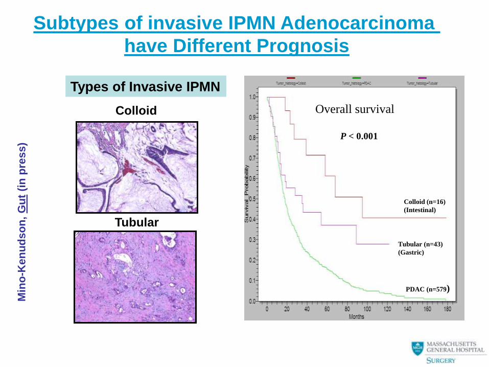
Colloid (n=16)
(Intestinal)
PDAC (n=579)
P < 0.001
Overall survival
Tubular (n=43)
(Gastric)
Types of Invasive IPMN
Colloid
Tubular
Min
o-K
en
ud
so
n,
Gu
t (i
n p
ress)
Subtypes of invasive IPMN Adenocarcinoma
have Different Prognosis
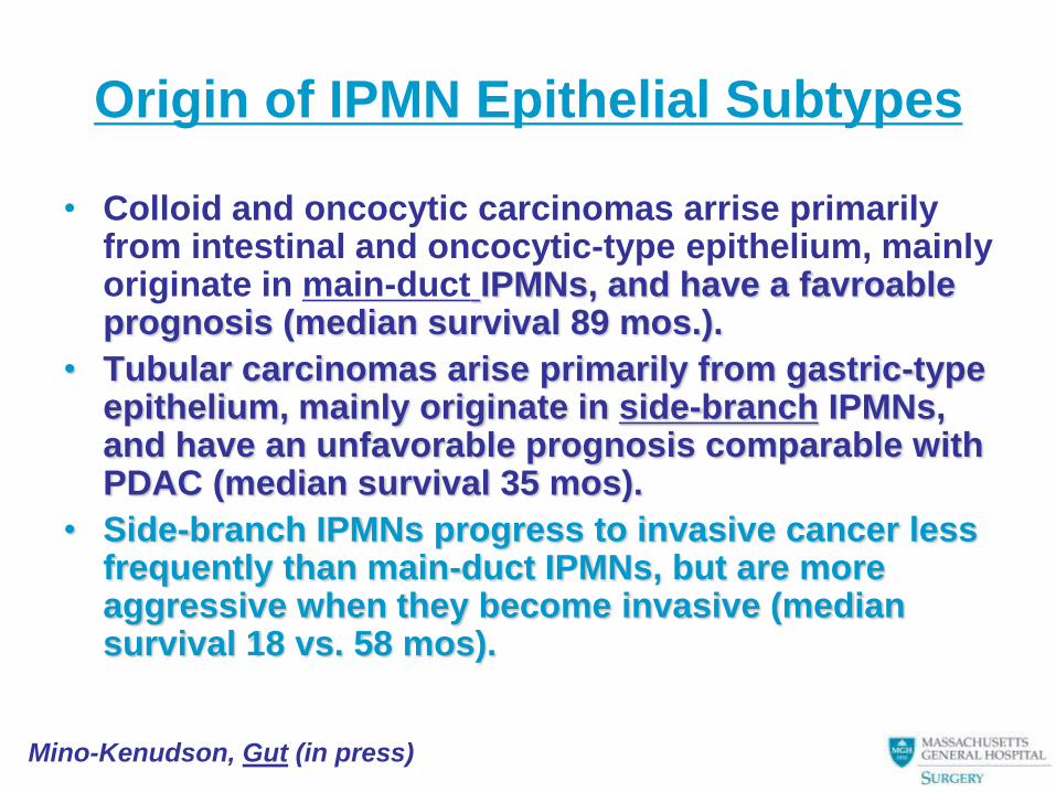
Origin of IPMN Epithelial Subtypes
• Colloid and oncocytic carcinomas arrise primarily from intestinal and oncocytic-type epithelium, mainly originate in main-duct IPMNs, and have a favroable prognosis (median survival 89 mos.).
• Tubular carcinomas arise primarily from gastric-type epithelium, mainly originate in side-branch IPMNs, and have an unfavorable prognosis comparable with PDAC (median survival 35 mos).
• Side-branch IPMNs progress to invasive cancer less frequently than main-duct IPMNs, but are more aggressive when they become invasive (median survival 18 vs. 58 mos).
Mino-Kenudson, Gut (in press)
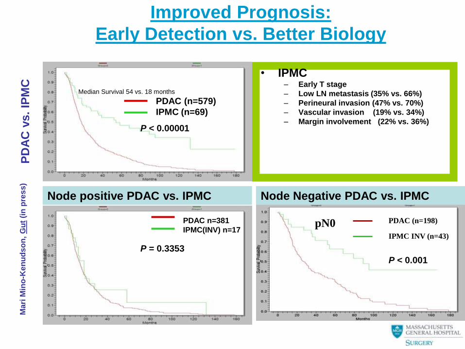
Improved Prognosis:
Early Detection vs. Better Biology
• IPMC – Early T stage
– Low LN metastasis (35% vs. 66%)
– Perineural invasion (47% vs. 70%)
– Vascular invasion (19% vs. 34%)
– Margin involvement (22% vs. 36%)
PD
AC
vs.
IPM
C
Mari Mino-Knudson et al.
PDAC (n=579)
IPMC (n=69)
P < 0.00001
Median Survival 54 vs. 18 months
Node positive PDAC vs. IPMC
PDAC n=381
IPMC(INV) n=17
P = 0.3353
IPMC INV (n=43)
PDAC (n=198)
P < 0.001
pN0
Node Negative PDAC vs. IPMC
Ma
ri M
ino
-Ken
ud
so
n,
Gu
t (i
n p
res
s)
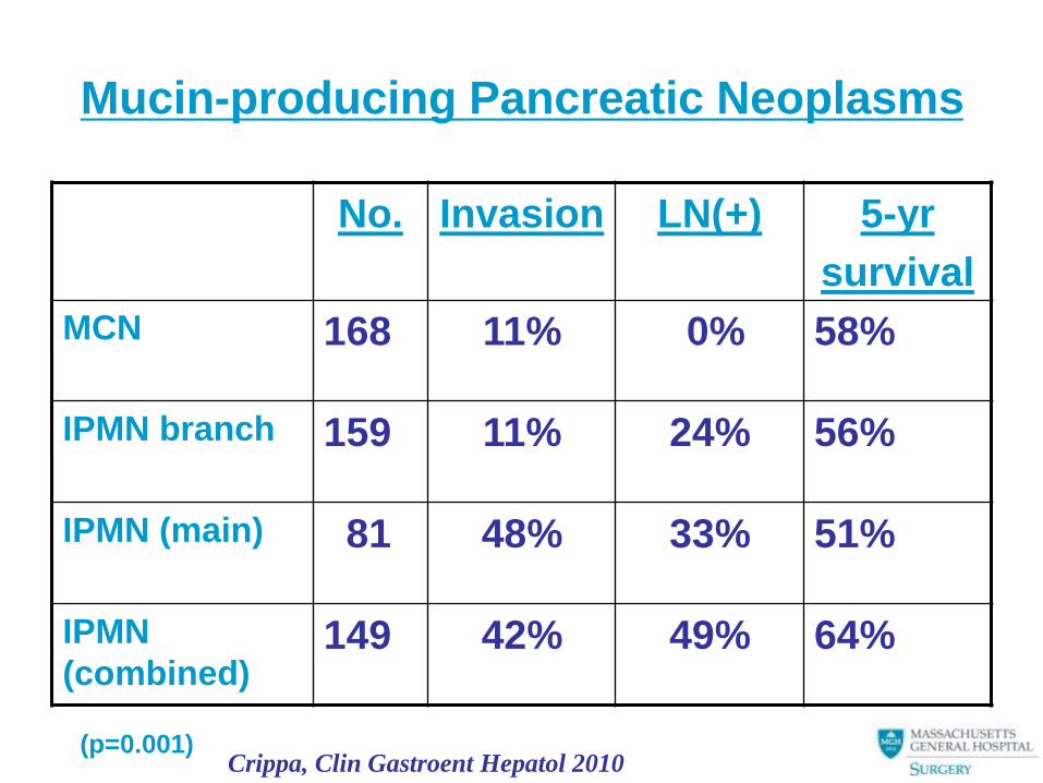
Mucin-producing Pancreatic Neoplasms
No. Invasion LN(+) 5-yr
survival
MCN 168 11% 0% 58%
IPMN branch 159 11% 24% 56%
IPMN (main) 81 48% 33% 51%
IPMN
(combined) 149 42% 49% 64%
(p=0.001) Crippa, Clin Gastroent Hepatol 2010
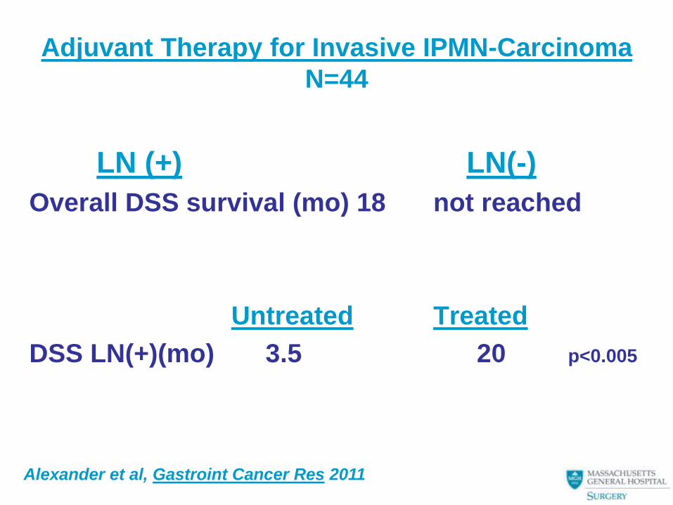
Adjuvant Therapy for Invasive IPMN-Carcinoma
N=44
LN (+) LN(-)
Overall DSS survival (mo) 18 not reached
Untreated Treated
DSS LN(+)(mo) 3.5 20 p<0.005
Alexander et al, Gastroint Cancer Res 2011
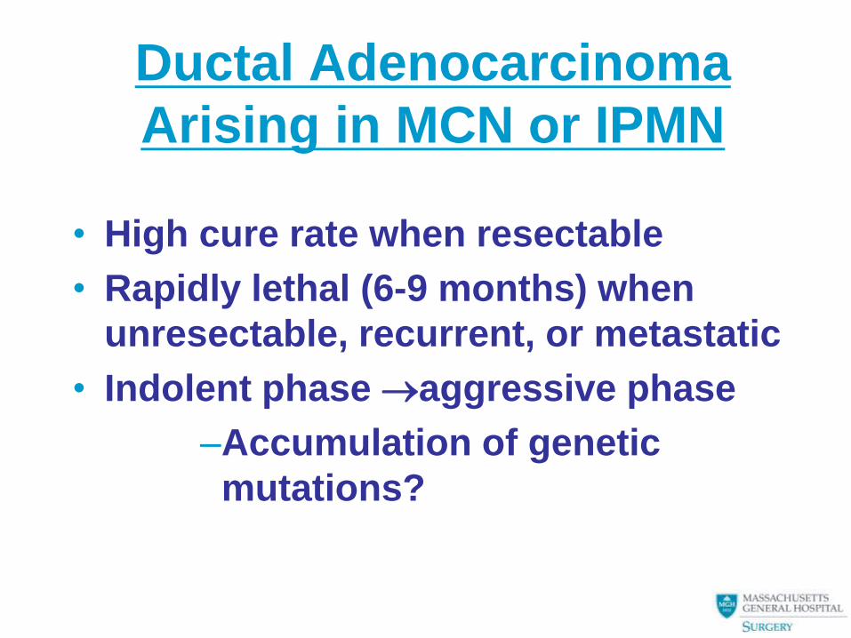
Ductal Adenocarcinoma
Arising in MCN or IPMN
• High cure rate when resectable
• Rapidly lethal (6-9 months) when
unresectable, recurrent, or metastatic
• Indolent phase aggressive phase
–Accumulation of genetic
mutations?
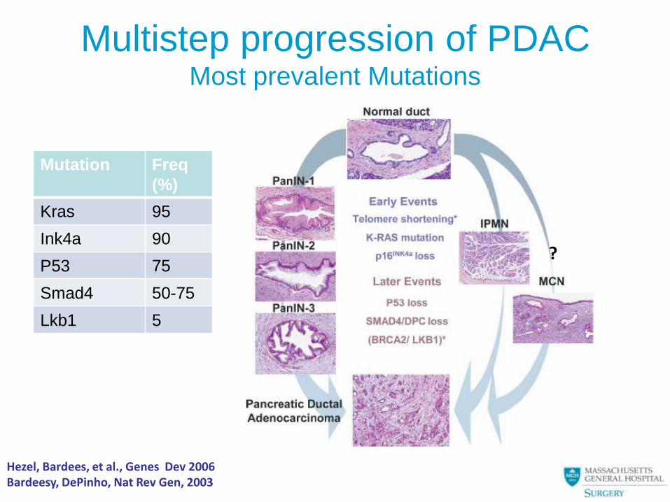
Multistep progression of PDAC Most prevalent Mutations
Mutation Freq
(%)
Kras 95
Ink4a 90
P53 75
Smad4 50-75
Lkb1 5
Hezel, Bardees, et al., Genes Dev 2006 Bardeesy, DePinho, Nat Rev Gen, 2003
?

Gene Expression Profiles
in both IPMN and PDAC
Most highly upregulated genes
(cDNA microarray analysis)
Trefoil peptide family (TFFI, TFF2, TFF3)
Lipocalin 2
Galactin 3
These genes may be involved at any early common
stage of pancreatic carcinogenesis in these alternative
routes of epithelial progression to full malignancy
Terris, Am J Pathol 2002
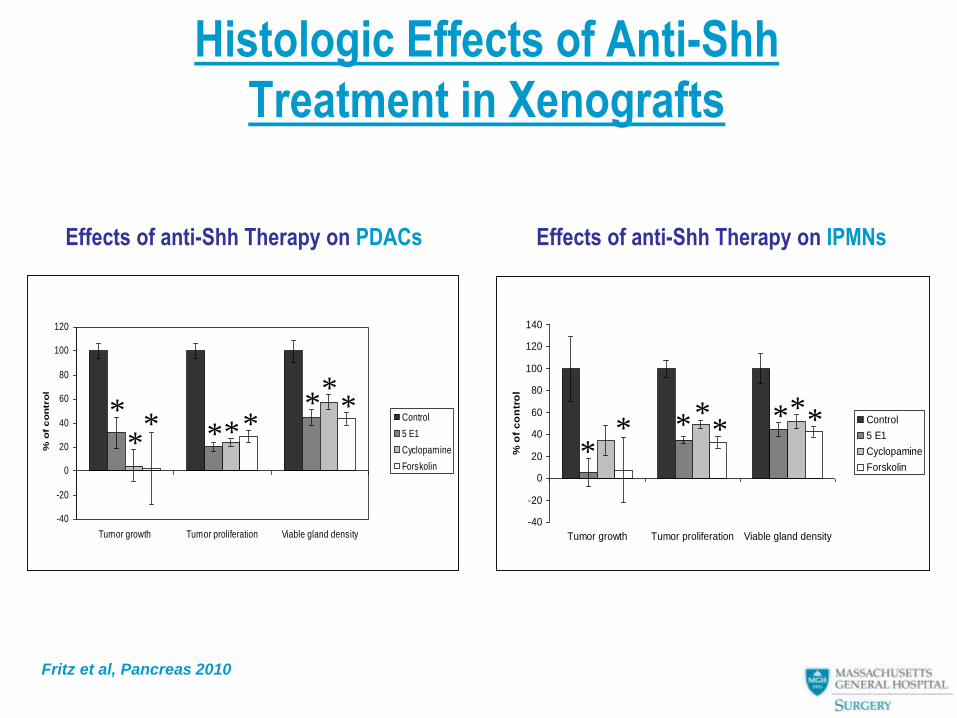
Histologic Effects of Anti-Shh
Treatment in Xenografts
-40
-20
0
20
40
60
80
100
120
140
Tumor growth Tumor proliferation Viable gland density
% o
f c
on
tro
l
Control
5 E1
Cyclopamine
Forskolin*
* * * * * * *
Effects of anti-Shh Therapy on IPMNs Effects of anti-Shh Therapy on PDACs
-40
-20
0
20
40
60
80
100
120
Tumor growth Tumor proliferation Viable gland density
% o
f c
on
tro
l
Control
5 E1
Cyclopamine
Forskolin
* * * * * *
* *
*
Fritz et al, Pancreas 2010
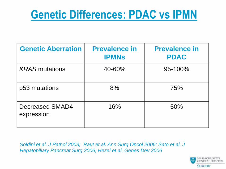
Genetic Differences: PDAC vs IPMN
Genetic Aberration Prevalence in
IPMNs
Prevalence in
PDAC
KRAS mutations
40-60% 95-100%
p53 mutations
8% 75%
Decreased SMAD4
expression
16% 50%
Soldini et al. J Pathol 2003; Raut et al. Ann Surg Oncol 2006; Sato et al. J
Hepatobiliary Pancreat Surg 2006; Hezel et al. Genes Dev 2006
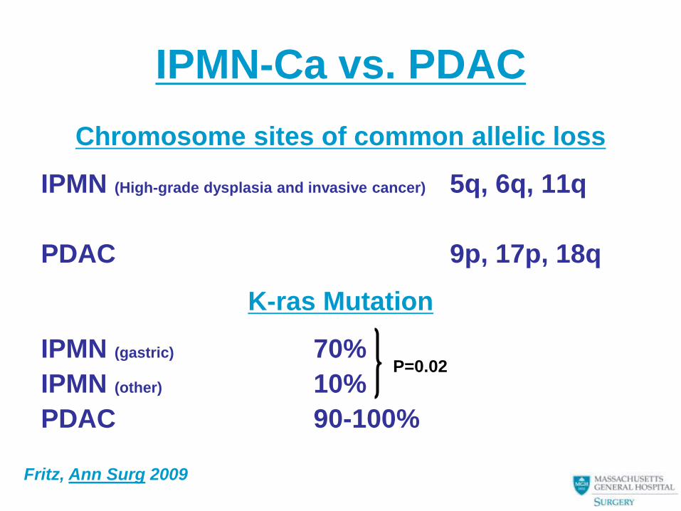
IPMN-Ca vs. PDAC
Chromosome sites of common allelic loss
IPMN (High-grade dysplasia and invasive cancer) 5q, 6q, 11q
PDAC 9p, 17p, 18q
K-ras Mutation
IPMN (gastric) 70%
IPMN (other) 10%
PDAC 90-100%
P=0.02
Fritz, Ann Surg 2009
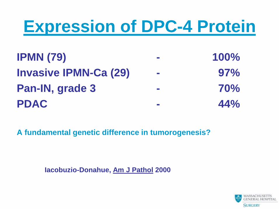
Expression of DPC-4 Protein
IPMN (79) - 100%
Invasive IPMN-Ca (29) - 97%
Pan-IN, grade 3 - 70%
PDAC - 44%
A fundamental genetic difference in tumorogenesis?
Iacobuzio-Donahue, Am J Pathol 2000
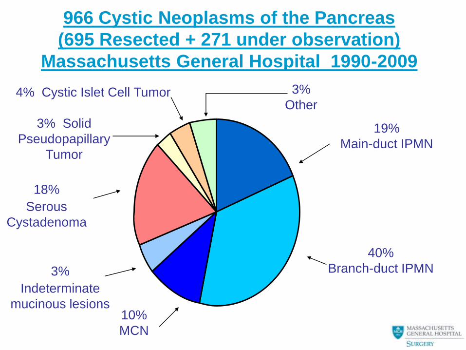
966 Cystic Neoplasms of the Pancreas
(695 Resected + 271 under observation)
Massachusetts General Hospital 1990-2009
19%
Main-duct IPMN
40%
Branch-duct IPMN
10%
MCN
3%
Indeterminate
mucinous lesions
18%
Serous
Cystadenoma
3% Solid
Pseudopapillary
Tumor
3%
Other 4% Cystic Islet Cell Tumor
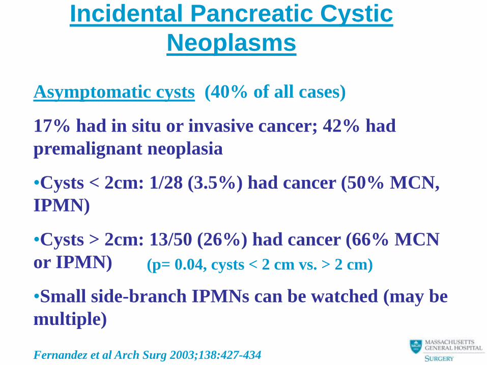
Incidental Pancreatic Cystic
Neoplasms
Asymptomatic cysts (40% of all cases)
17% had in situ or invasive cancer; 42% had
premalignant neoplasia
•Cysts < 2cm: 1/28 (3.5%) had cancer (50% MCN,
IPMN)
•Cysts > 2cm: 13/50 (26%) had cancer (66% MCN
or IPMN)
•Small side-branch IPMNs can be watched (may be
multiple)
(p= 0.04, cysts < 2 cm vs. > 2 cm)
Fernandez et al Arch Surg 2003;138:427-434
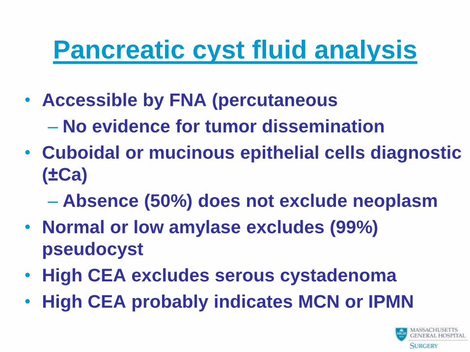
Pancreatic cyst fluid analysis
• Accessible by FNA (percutaneous
– No evidence for tumor dissemination
• Cuboidal or mucinous epithelial cells diagnostic
(±Ca)
– Absence (50%) does not exclude neoplasm
• Normal or low amylase excludes (99%)
pseudocyst
• High CEA excludes serous cystadenoma
• High CEA probably indicates MCN or IPMN
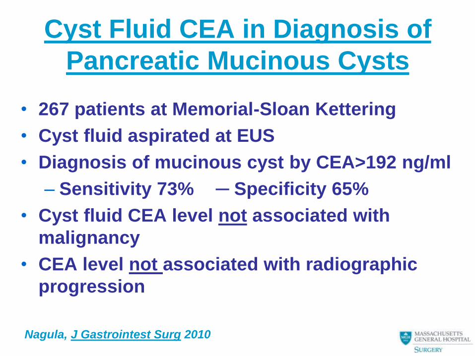
Cyst Fluid CEA in Diagnosis of
Pancreatic Mucinous Cysts
• 267 patients at Memorial-Sloan Kettering
• Cyst fluid aspirated at EUS
• Diagnosis of mucinous cyst by CEA>192 ng/ml
– Sensitivity 73% ─ Specificity 65%
• Cyst fluid CEA level not associated with
malignancy
• CEA level not associated with radiographic
progression
Nagula, J Gastrointest Surg 2010
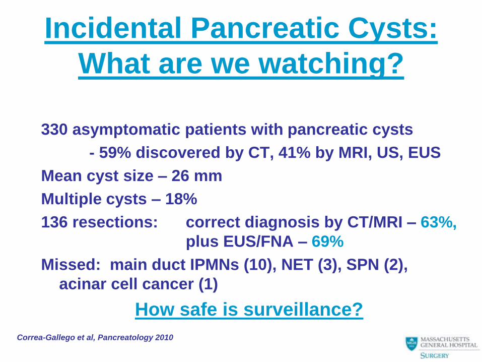
Incidental Pancreatic Cysts:
What are we watching?
330 asymptomatic patients with pancreatic cysts
- 59% discovered by CT, 41% by MRI, US, EUS
Mean cyst size – 26 mm
Multiple cysts – 18%
136 resections: correct diagnosis by CT/MRI – 63%,
plus EUS/FNA – 69%
Missed: main duct IPMNs (10), NET (3), SPN (2),
acinar cell cancer (1)
How safe is surveillance?
Correa-Gallego et al, Pancreatology 2010
Rationale for Resection of (most)
Pancreatic Cystic Neoplasms
• Uncertainty of diagnosis
• Potential for malignancy
• Symptoms
• Growth
• Cost of surveillance
• Peace of mind

Additional Reasons to Resect all
Pancreatic Cystic Neoplasms
• It helps the bottom line
• Residents need the experience
• It’s what I do
(“If you are a hammer, all the world
looks like a nail.”)
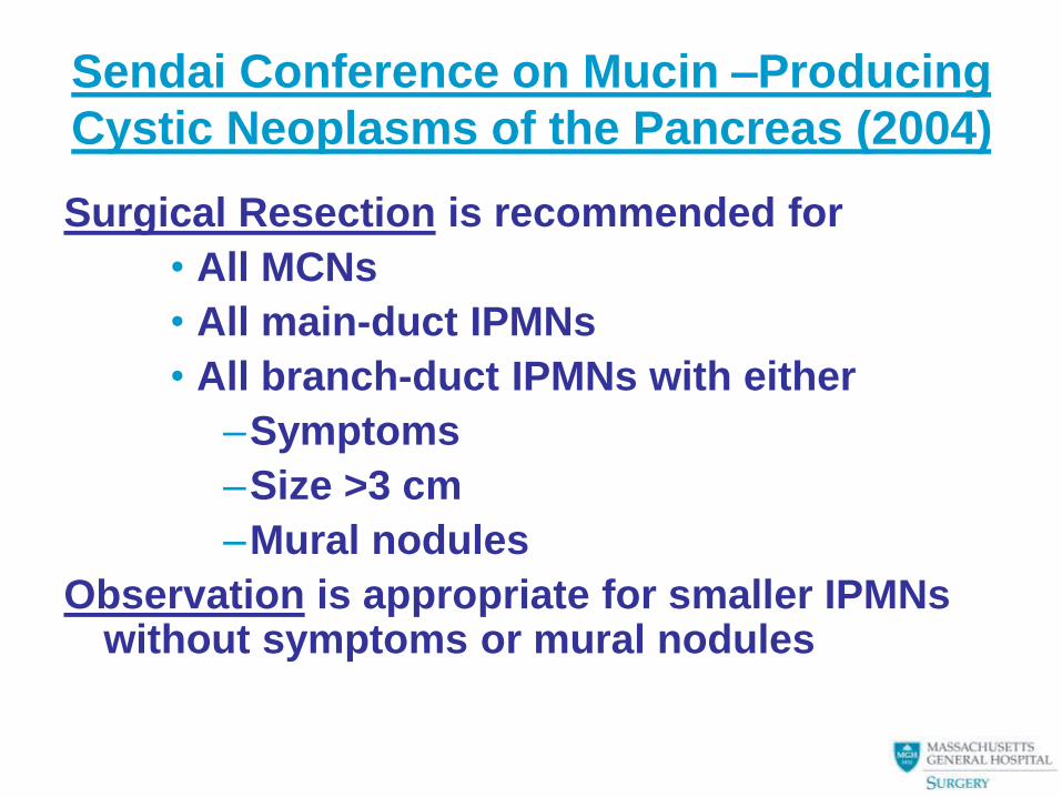
Sendai Conference on Mucin –Producing
Cystic Neoplasms of the Pancreas (2004)
Surgical Resection is recommended for
• All MCNs
• All main-duct IPMNs
• All branch-duct IPMNs with either
–Symptoms
–Size >3 cm
–Mural nodules
Observation is appropriate for smaller IPMNs without symptoms or mural nodules

Pancreatic Cancer
For patients who undergo curative
resection, their prognosis appears
to be determined by the biology of
the tumor rather than factors
involved in the resection.
Geer and Brennan, Am J Surg 1993; 165:68