Cystic Lymphangioma of the Chest Wall in a 5-Year-Old Male...
7
Case Report Cystic Lymphangioma of the Chest Wall in a 5-Year-Old Male Patient: A Rare and Atypical Localization—A Case Report and Comprehensive Review of the Literature Dimitrios Patoulias, 1 Ioannis Patoulias, 2 Christos Kaselas, 2 Maria Kalogirou, 3 Chatzopoulos Kyriakos, 4 Farmakis Konstantinos, 2 Thomas Feidantsis, 2 and Papacrivou Eleni 2 1 Department of Internal Medicine, General Hospital of Veria, Veria, Greece 2 1st Department of Pediatric Surgery, Aristotle University of essaloniki, General Hospital G. Gennimatas, essaloniki, Greece 3 General Hospital of Trikala, Trikala, Greece 4 Department of Pathology, General Hospital G. Gennimatas, essaloniki, Greece Correspondence should be addressed to Dimitrios Patoulias; [email protected] Received 22 June 2017; Accepted 23 August 2017; Published 23 October 2017 Academic Editor: Carmelo Romeo Copyright © 2017 Dimitrios Patoulias et al. is is an open access article distributed under the Creative Commons Attribution License, which permits unrestricted use, distribution, and reproduction in any medium, provided the original work is properly cited. Lymphangioma is a benign congenital malformation. e extremely rare and atypical localization of a lymphangioma in the chest wall was the real motive for the present case study. A 5-year-old boy was admitted to the Emergency Department of the 1st Depart- ment of Pediatric Surgery, Aristotle University of essaloniki, due to the presence of a mildly painful swelling in the leſt lateral chest wall, which was first noticed three months ago, aſter a blunt injury during sport. Physical examination revealed the presence of a palpable, spherical, painful, nut-sized subcutaneous lesion in the leſt lateral chest wall, respectively, with the anterior axillary line, at the height of the 6th to 7th intercostal space. Presence of ecchymosis on the overlying skin was also noticed. During palpation, we did not notice fluctuation, while transillumination was not feasible. Performance of ultrasonography, including Doppler color flow imaging, followed, depicting a subcutaneous cystic lesion, 2.1 ∗ 3.2 cm in dimensions, without extension to the thoracic cavity. Scheduled surgical excision of the lesion was decided. Histopathological examination documented the diagnosis of cystic lymphangioma. Patient is still followed up on a 6-month basis. He remains asymptomatic, aſter 2 years, without indication of relapse. 1. Introduction Lymphangioma is a benign congenital malformation charac- terized by proliferation of lymphatic vessels, resulting from the failure of communication between the primitive lym- phatic sacs and the venous system, leading to the formation of a cystic structure [1]. Rarely, it is an acquired malformation, due to inflammation, injury, or fibrosis [2]. Based upon the histopathologic features, lymphangiomas are divided into three subtypes: the cystic (macrocystic) lymphangioma, which is the most common, the capillary (supermacrocystic) lymphangioma, and the corpus (cav- ernous, microcystic) lymphangioma, which is the most rare [3]. Macrocystic lymphangioma is classified as septated and nonseptated. As for etiology, according to embryologic mech- anism, septated lymphangioma results from total obstruction of the primitive lymphatic sacs, while nonseptated lymphan- gioma from partial obstruction of them [4]. eir incidence in pediatric population rises up to 1 new case per 12000 births, while lymphangiomas represent almost 5-6% of all tumors in childhood [5]. Almost 95% of all lymphangiomas are detected in the cervical region or axillary cavity [6, 7]. Hancock et al. [8] reported that 31.4% of all lymphangiomas are found in the cervical region, 18.9% in the scalp and the face, 9.2% in the trunk, and 4.9% in the anterior chest wall and the axillary cavity. Chest wall localization of the lymphangiomas represents only 1% of all cases, with the majority of them located in the mediastinum Hindawi Case Reports in Pediatrics Volume 2017, Article ID 2083204, 6 pages https://doi.org/10.1155/2017/2083204
Transcript of Cystic Lymphangioma of the Chest Wall in a 5-Year-Old Male...

Case ReportCystic Lymphangioma of the Chest Wall in a 5-Year-Old MalePatient: A Rare and Atypical Localization—A Case Report andComprehensive Review of the Literature
Dimitrios Patoulias,1 Ioannis Patoulias,2 Christos Kaselas,2
Maria Kalogirou,3 Chatzopoulos Kyriakos,4 Farmakis Konstantinos,2
Thomas Feidantsis,2 and Papacrivou Eleni2
1Department of Internal Medicine, General Hospital of Veria, Veria, Greece21st Department of Pediatric Surgery, Aristotle University of Thessaloniki, General Hospital G. Gennimatas, Thessaloniki, Greece3General Hospital of Trikala, Trikala, Greece4Department of Pathology, General Hospital G. Gennimatas, Thessaloniki, Greece
Correspondence should be addressed to Dimitrios Patoulias; [email protected]
Received 22 June 2017; Accepted 23 August 2017; Published 23 October 2017
Academic Editor: Carmelo Romeo
Copyright © 2017 Dimitrios Patoulias et al. This is an open access article distributed under the Creative Commons AttributionLicense, which permits unrestricted use, distribution, and reproduction in any medium, provided the original work is properlycited.
Lymphangioma is a benign congenital malformation. The extremely rare and atypical localization of a lymphangioma in the chestwall was the real motive for the present case study. A 5-year-old boy was admitted to the Emergency Department of the 1st Depart-ment of Pediatric Surgery, Aristotle University of Thessaloniki, due to the presence of a mildly painful swelling in the left lateralchest wall, which was first noticed threemonths ago, after a blunt injury during sport. Physical examination revealed the presence ofa palpable, spherical, painful, nut-sized subcutaneous lesion in the left lateral chest wall, respectively, with the anterior axillary line,at the height of the 6th to 7th intercostal space. Presence of ecchymosis on the overlying skin was also noticed. During palpation,we did not notice fluctuation, while transillumination was not feasible. Performance of ultrasonography, including Doppler colorflow imaging, followed, depicting a subcutaneous cystic lesion, 2.1 ∗ 3.2 cm in dimensions, without extension to the thoraciccavity. Scheduled surgical excision of the lesion was decided. Histopathological examination documented the diagnosis of cysticlymphangioma. Patient is still followed up on a 6-month basis. He remains asymptomatic, after 2 years, without indication of relapse.
1. Introduction
Lymphangioma is a benign congenital malformation charac-terized by proliferation of lymphatic vessels, resulting fromthe failure of communication between the primitive lym-phatic sacs and the venous system, leading to the formation ofa cystic structure [1]. Rarely, it is an acquired malformation,due to inflammation, injury, or fibrosis [2].
Based upon the histopathologic features, lymphangiomasare divided into three subtypes: the cystic (macrocystic)lymphangioma, which is the most common, the capillary(supermacrocystic) lymphangioma, and the corpus (cav-ernous, microcystic) lymphangioma, which is the most rare[3]. Macrocystic lymphangioma is classified as septated and
nonseptated. As for etiology, according to embryologicmech-anism, septated lymphangioma results from total obstructionof the primitive lymphatic sacs, while nonseptated lymphan-gioma from partial obstruction of them [4].
Their incidence in pediatric population rises up to 1new case per 12000 births, while lymphangiomas representalmost 5-6% of all tumors in childhood [5]. Almost 95%of all lymphangiomas are detected in the cervical region oraxillary cavity [6, 7]. Hancock et al. [8] reported that 31.4%of all lymphangiomas are found in the cervical region, 18.9%in the scalp and the face, 9.2% in the trunk, and 4.9% inthe anterior chest wall and the axillary cavity. Chest walllocalization of the lymphangiomas represents only 1% of allcases, with the majority of them located in the mediastinum
HindawiCase Reports in PediatricsVolume 2017, Article ID 2083204, 6 pageshttps://doi.org/10.1155/2017/2083204

2 Case Reports in Pediatrics
Figure 1: Presence of a palpable, spherical, painful, nut-sizedsubcutaneous lesion in the left lateral chest wall, respectively, withthe anterior axillary line, at the height of the 6th to 7th intercostalspace. Notice the ecchymosis on the overlying skin.
[9, 10]. Omell et al. [11] were the first to describe a case ofa chest wall lymphangioma in 1973. The extremely rare andatypical localization of a lymphangioma in the chest wall wasthe real motive for the present case study, after systematic andthorough research of the relevant literature [1, 3, 7, 9, 12, 13].
2. Case Report
A 5-year-old male patient was admitted as an outpatientto the Emergency Department of the 1st Department ofPediatric Surgery, Aristotle University of Thessaloniki, dueto the presence of a mildly painful swelling in the left lateralchest wall, which was first noticed about three months ago,after a blunt injury in the context of participation in sport.Physical examination revealed the presence of a palpable,spherical, painful, nut-sized subcutaneous lesion in the leftlateral chest wall, respectively, with the anterior axillary line,at the height of the 6th to 7th intercostal space. Presence ofecchymosis on the overlying skin was also noticed (Figure 1).
Transillumination of the lesion was not feasible. Nopathological signs were detected in face and left profile chestradiograph. Performance of ultrasonographic evaluation,including Doppler color flow imaging, followed, depictinga subcutaneous cystic lesion, 2.1 ∗ 3.2 cm in dimensions,without extension to the thoracic cavity. Solid insertion tothe underlying epimysium was probable, according to theimaging findings. Lesion’s periphery was lobulated, while novascular structures, but thin septula (depicted as hyperechoicreflections), were found within it.
Scheduled surgical excision of the lesion was decided,after conduction of the typical preoperative control, whichwas normal. With the patient under general endotrachealanesthesia, we conducted a cross section above the lesion,following the direction of Langer’s lines. We then performedseparation of the adhesions between the lesion and theadjacent subcutaneous fat tissue, but also of the adhesionsbetween the posterior surface of the lesion and the underlying
fascia of the anterolateral chest muscles (Figures 2(a) and2(b)).
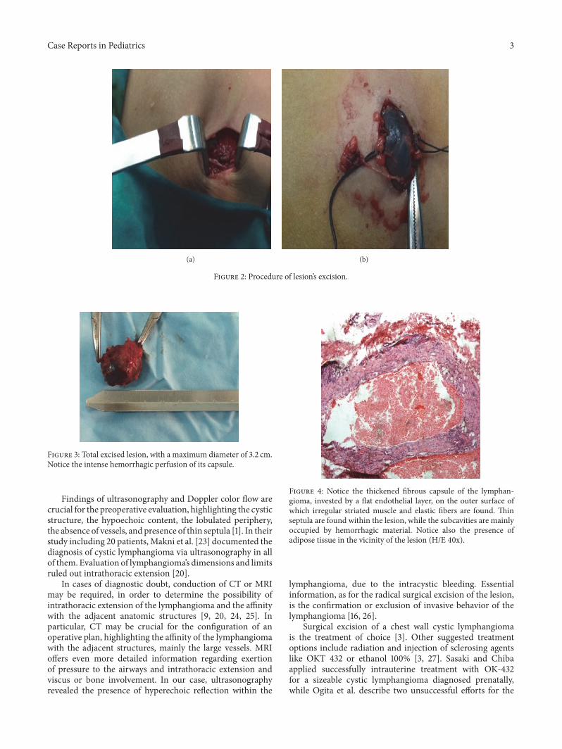
The lesion was finally excised, without incision. Wenoticed the hemorrhagic perfusion of its capsule (Figure 3).
Hemostasis and repair of the surgical wound according tothe anatomic order followed.
3. Results
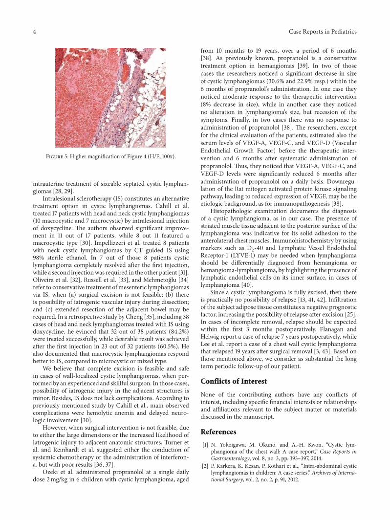
Patient was easily awakened after the completion of thesurgical procedure. Postoperative course was uneventful andhe was discharged home on the 1st postoperative day inexcellent general condition. Histopathological examinationof the excised lesion documented the diagnosis of cysticlymphangioma. It highlighted the presence of a thickenedfibrous capsule invested by a flat endothelial layer, on theouter surface of which irregular striated muscle and elasticfibers were found.Thin septula were found within the lesion,while the subcavities were mainly occupied by hemorrhagicmaterial. Adipose tissue in the vicinity of the lesion wasalso found, corresponding to the subcutaneous fat tissuesurrounding the lymphangioma (Figures 4 and 5).
Patient is still followed up on a 6-month basis. He remainsasymptomatic, after 2 years, without indication of relapse.
4. Discussion
Goldstein et al. focus on the early prenatal diagnosis of lym-phangiomas, between the 15th and 22nd week of gestation,in their study [14]. They consider that the early diagnosisof a chest wall cystic lymphangioma is not associated withpoor prognosis, as in cases of lymphangiomas of otherlocalization, due to chromosomal or anatomical anomalies,or even hydrops fetalis [14, 15]. Subsequently, based on thefact that prognosis is similar between a chest wall cysticlymphangioma and a delayed prenatally diagnosed cysticlymphangioma, it seems reasonable that development ofa chest wall cystic lymphangioma does not result fromobstruction of primitive lymphatic sacs, as their formationand conjunction with the venous system occur between 6thand 9th week of gestation [16–18].
Despite the presence of a cystic lymphangioma at birthin 50% of all cases and by the age of 2 years in 90% of allcases, diagnosis may be delayed significantly, as in our case[3, 19–22]. Mean age of diagnosis is 3 years of life. Rapidincrease in size, inflammation, or intracystic bleeding, afterinjury, resulting in local pain, or pressure phenomena toadjacent anatomic structures, usually leads to diagnosis [22].Intracystic bleeding can induce the formation of ecchymosison the overlying skin, as in our case.
Physical examination and ultrasonography substantiallycontribute to the diagnostic approach of a chest wall cysticlymphangioma [1]. Transillumination, where possible, mayalso help the diagnosis, confirming the presence of a cysticlesion and excluding that of a solid structure [1]. In our case,there was no fluctuation of the lesion, while transilluminationwas not feasible, due to the increased tension and theintracystic bleeding.
Case Reports in Pediatrics 3
(a) (b)
Figure 2: Procedure of lesion’s excision.
Figure 3: Total excised lesion, with a maximum diameter of 3.2 cm.Notice the intense hemorrhagic perfusion of its capsule.
Findings of ultrasonography and Doppler color flow arecrucial for the preoperative evaluation, highlighting the cysticstructure, the hypoechoic content, the lobulated periphery,the absence of vessels, and presence of thin septula [1]. In theirstudy including 20 patients,Makni et al. [23] documented thediagnosis of cystic lymphangioma via ultrasonography in allof them. Evaluation of lymphangioma’s dimensions and limitsruled out intrathoracic extension [20].
In cases of diagnostic doubt, conduction of CT or MRImay be required, in order to determine the possibility ofintrathoracic extension of the lymphangioma and the affinitywith the adjacent anatomic structures [9, 20, 24, 25]. Inparticular, CT may be crucial for the configuration of anoperative plan, highlighting the affinity of the lymphangiomawith the adjacent structures, mainly the large vessels. MRIoffers even more detailed information regarding exertionof pressure to the airways and intrathoracic extension andviscus or bone involvement. In our case, ultrasonographyrevealed the presence of hyperechoic reflection within the
Figure 4: Notice the thickened fibrous capsule of the lymphan-gioma, invested by a flat endothelial layer, on the outer surface ofwhich irregular striated muscle and elastic fibers are found. Thinseptula are found within the lesion, while the subcavities are mainlyoccupied by hemorrhagic material. Notice also the presence ofadipose tissue in the vicinity of the lesion (H/E 40x).
lymphangioma, due to the intracystic bleeding. Essentialinformation, as for the radical surgical excision of the lesion,is the confirmation or exclusion of invasive behavior of thelymphangioma [16, 26].
Surgical excision of a chest wall cystic lymphangiomais the treatment of choice [3]. Other suggested treatmentoptions include radiation and injection of sclerosing agentslike OKT 432 or ethanol 100% [3, 27]. Sasaki and Chibaapplied successfully intrauterine treatment with OK-432for a sizeable cystic lymphangioma diagnosed prenatally,while Ogita et al. describe two unsuccessful efforts for the
4 Case Reports in Pediatrics
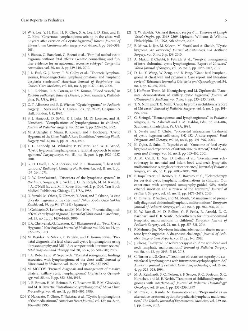
Figure 5: Higher magnification of Figure 4 (H/E, 100x).
intrauterine treatment of sizeable septated cystic lymphan-giomas [28, 29].
Intralesional sclerotherapy (IS) constitutes an alternativetreatment option in cystic lymphangiomas. Cahill et al.treated 17 patients with head and neck cystic lymphangiomas(10 macrocystic and 7 microcystic) by intralesional injectionof doxycycline. The authors observed significant improve-ment in 11 out of 17 patients, while 8 out 11 featured amacrocystic type [30]. Impellizzeri et al. treated 8 patientswith neck cystic lymphangiomas by CT guided IS using98% sterile ethanol. In 7 out of those 8 patients cysticlymphangioma completely resolved after the first injection,while a second injectionwas required in the other patient [31].Oliveira et al. [32], Russell et al. [33], and Mehmetoglu [34]refer to conservative treatment ofmesenteric lymphangiomasvia IS, when (a) surgical excision is not feasible; (b) thereis possibility of iatrogenic vascular injury during dissection;and (c) extended resection of the adjacent bowel may berequired. In a retrospective study by Cheng [35], including 38cases of head and neck lymphangiomas treated with IS usingdoxycycline, he evinced that 32 out of 38 patients (84.2%)were treated successfully, while desirable result was achievedafter the first injection in 23 out of 32 patients (60.5%). Healso documented that macrocystic lymphangiomas respondbetter to IS, compared to microcystic or mixed type.
We believe that complete excision is feasible and safein cases of wall-localized cystic lymphangiomas, when per-formed by an experienced and skillful surgeon. In those cases,possibility of iatrogenic injury in the adjacent structures isminor. Besides, IS does not lack complications. According topreviously mentioned study by Cahill et al., main observedcomplications were hemolytic anemia and delayed neuro-logic involvement [30].
However, when surgical intervention is not feasible, dueto either the large dimensions or the increased likelihood ofiatrogenic injury to adjacent anatomic structures, Turner etal. and Reinhardt et al. suggested either the conduction ofsystemic chemotherapy or the administration of interferon-a, but with poor results [36, 37].
Ozeki et al. administered propranolol at a single dailydose 2mg/kg in 6 children with cystic lymphangioma, aged
from 10 months to 19 years, over a period of 6 months[38]. As previously known, propranolol is a conservativetreatment option in hemangiomas [39]. In two of thosecases the researchers noticed a significant decrease in sizeof cystic lymphangiomas (30.6% and 22.9% resp.) within the6 months of propranolol’s administration. In one case theynoticed moderate response to the therapeutic intervention(8% decrease in size), while in another case they noticedno alteration in lymphangioma’s size, but recession of thesymptoms. Finally, in two cases there was no response toadministration of propranolol [38]. The researchers, exceptfor the clinical evaluation of the patients, estimated also theserum levels of VEGF-A, VEGF-C, and VEGF-D (VascularEndothelial Growth Factor) before the therapeutic inter-vention and 6 months after systematic administration ofpropranolol. Thus, they noticed that VEGF-A, VEGF-C, andVEGF-D levels were significantly reduced 6 months afteradministration of propranolol on a daily basis. Downregu-lation of the Rat mitogen activated protein kinase signalingpathway, leading to reduced expression of VEGF, may be theetiologic background, as for immunopathogenesis [38].
Histopathologic examination documents the diagnosisof a cystic lymphangioma, as in our case. The presence ofstriated muscle tissue adjacent to the posterior surface of thelymphangioma was indicative for its solid adhesion to theanterolateral chest muscles. Immunohistochemistry by usingmarkers such as D
2-40 and Lymphatic Vessel Endothelial
Receptor-1 (LYVE-1) may be needed when lymphangiomashould be differentially diagnosed from hemangioma orhemangioma-lymphangioma, by highlighting the presence oflymphatic endothelial cells on its inner surface, in cases oflymphangioma [40].
Since a cystic lymphangioma is fully excised, then thereis practically no possibility of relapse [13, 41, 42]. Infiltrationof the subject adipose tissue constitutes a negative prognosticfactor, increasing the possibility of relapse after excision [25].In cases of incomplete removal, relapse should be expectedwithin the first 3 months postoperatively. Flanagan andHelwig report a case of relapse 7 years postoperatively, whileLee et al. report a case of a chest wall cystic lymphangiomathat relapsed 19 years after surgical removal [3, 43]. Based onthose mentioned above, we consider as substantial the longterm periodic follow-up of our patient.
Conflicts of Interest
None of the contributing authors have any conflicts ofinterest, including specific financial interests or relationshipsand affiliations relevant to the subject matter or materialsdiscussed in the manuscript.
References
[1] N. Yokoigawa, M. Okuno, and A.-H. Kwon, “Cystic lym-phangioma of the chest wall: A case report,” Case Reports inGastroenterology, vol. 8, no. 3, pp. 393–397, 2014.
[2] P. Karkera, K. Kesan, P. Kothari et al., “Intra-abdominal cysticlymphangiomas in children: A case series,” Archives of Interna-tional Surgery, vol. 2, no. 2, p. 91, 2012.
Case Reports in Pediatrics 5
[3] W. S. Lee, Y. H. Kim, H. K. Chee, S. A. Lee, J. D. Kim, and D.C. Kim, “Cavernous lymphangioma arising in the chest wall19 years after excision of a cystic hygroma,” Korean Journal ofThoracic and Cardiovascular Surgery, vol. 44, no. 5, pp. 380–382,2011.
[4] S. Bianca, G. Bartoloni, G. Boemi et al., “Familial nuchal cystichygroma without fetal effects: Genetic counselling and fur-ther evidence for an autosomal recessive subtype,” CongenitalAnomalies, vol. 50, no. 2, pp. 139-140, 2010.
[5] J. L. Faul, G. J. Berry, T. V. Colby et al., “Thoracic lymphan-giomas, lymphangiectasis, lymphangiomatosis, and lymphaticdysplasia syndrome,” American Journal of Respiratory andCritical Care Medicine, vol. 161, no. 3, pp. 1037–1046, 2000.
[6] S. L. Robbins, R. S. Cotran, and V. Kumar, “Blood vessels,” inRobbins Pathologic Basis of Disease, p. 544, Saunders, Philadel-phia, Pa, USA, 1984.
[7] C. T. Albanese and E. S. Wiener, “Cystic hygroma,” in PediatricSurgery, L. Spitz and A. G. Coran, Eds., pp. 94-95, Chapman &Hall, London, UK, 1995.
[8] B. J. Hancock, D. St-Vil, F. I. Luks, M. Di Lorenzo, and H.Blanchard, “Complications of lymphangiomas in children,”Journal of Pediatric Surgery, vol. 27, no. 2, pp. 220–224, 1992.
[9] M. Ardenghy, Y. Miura, R. Kovach, and J. Hochberg, “CysticHygroma of theChestWall: ARareCondition,”Annals of PlasticSurgery, vol. 37, no. 2, pp. 211–213, 1996.
[10] T. L. Kennedy, M. Whitaker, P. Pellitteri, and W. E. Wood,“Cystic hygroma/lymphangioma: a rational approach to man-agement,” Laryngoscope, vol. 111, no. 11, part 1, pp. 1929–1937,2001.
[11] G. H. Omell, L. S. Anderson, and R. T. Bramson, “Chest walltumours,” Radiologic Clinics of North America, vol. 11, no. 1, pp.197–214, 1973.
[12] E. W. Fonnkaisurd, “Disorders of the lymphatic system,” inPaediatric Surgery, K. J. Welch, J. G. Randolph, M. M. Ravitch,J. A. O’Neill Jr., and M. I. Rowe, Eds., vol. 2, p. 1506, Year BookMedical Publishers, Chicago, Ill, USA, 1986.
[13] O. Suzuki, M. Ohata, K. Ohmori, Y. Sezai, and T. Okano, “A caseof cystic hygroma of the chest wall,” Nihon Kyobu Geka GakkaiZasshi, vol. 39, pp. 94–97, 1991 (Japanese).
[14] I. Goldstein, Z. Leibovitz, andM.Noi-Nizri, “Prenatal diagnosisof fetal chest lymphangioma,” Journal of Ultrasound inMedicine,vol. 25, no. 11, pp. 1437–1440, 2006.
[15] F. A. Chervenak, G. Isaacson, K. J. Blakemore et al., “Fetal CysticHygroma,”NewEngland Journal ofMedicine, vol. 309, no. 14, pp.822–825, 1983.
[16] M. Rasidaki, S. Sifakis, E. Vardaki, and E. Koumantakis, “Pre-natal diagnosis of a fetal chest wall cystic lymphangioma usingultrasonography andMRI: A case report with literature review,”Fetal Diagnosis and Therapy, vol. 20, no. 6, pp. 504–507, 2005.
[17] J. A. Robert and W. Sepulveda, “Prenatal sonographic findingsassociated with lymphangioma of the chest wall,” Journal ofUltrasound in Medicine, vol. 16, no. 9, pp. 635–637, 1997.
[18] M. MCCOY, “Prenatal diagnosis and management of massivebilateral axillary cystic lymphangioma,” Obstetrics & Gynecol-ogy, vol. 85, no. 5, pp. 853–856, 1995.
[19] L. R. Brown, H. M. Reiman, E. C. Rosenow III, P. M. Gloviczki,and M. B. Divertie, “Intrathoracic lymphangioma,”Mayo ClinicProceedings, vol. 61, no. 11, pp. 882–892, 1986.
[20] Y. Nakazato, Y. Ohno, Y. Nakataa et al., “Cystic lymphangiomaof themediastinum,”AmericanHeart Journal, vol. 129, no. 2, pp.406–409, 1995.
[21] T. W. Shields, “General thoracic surgery,” in Tumours of LymphVessel Origin, pp. 2368-2369, Lipincott Williams & Wilkins,Philadelphia, PA, USA, 5th edition, 2002.
[22] B. Mirza, L. Ijaz, M. Saleem, M. Sharif, and A. Sheikh, “Cystichygroma: An overview,” Journal of Cutaneous and AestheticSurgery, vol. 3, no. 3, p. 139, 2010.
[23] A. Makni, F. Chebbi, F. Fetirich et al., “Surgical managementof intra-abdominal cystic lymphangioma. Report of 20 cases,”World Journal of Surgery, vol. 36, no. 5, pp. 1037–1043, 2012.
[24] D. Lu, Y. Wang, W. Zeng, and B. Peng, “Giant fetal lymphan-gioma at chest wall and prognosis: Case report and literaturereview,” Taiwanese Journal of Obstetrics and Gynecology, vol. 54,no. 1, pp. 62–65, 2015.
[25] J. Hoffman-Tretin, M. Koenigsberg, and M. Ziprkowski, “Ante-natal demonstration of axillary cystic hygroma,” Journal ofUltrasound in Medicine, vol. 7, no. 4, pp. 233–235, 1988.
[26] T. N. Ninh and T. X. Ninh, “Cystic hygroma in children: a reportof 126 cases,” Journal of Pediatric Surgery, vol. 9, no. 2, pp. 191–195, 1974.
[27] G. Stringel, “Hemangiomas and lymphangiomas,” in PediatricSurgery, K. W. Ashcroft and T. M. Haldor, Eds., pp. 814–816,Saunders, Philadelphia, Pa, USA, 1993.
[28] Y. Sasaki and Y. Chiba, “Successful intrauterine treatmentof cystic hygroma colli using OK-432: A case report,” FetalDiagnosis and Therapy, vol. 18, no. 6, pp. 391–396, 2003.
[29] K. Ogita, S. Suita, T. Taguchi et al., “Outcome of fetal cystichygroma and experience of intrauterine treatment,” Fetal Diag-nosis and Therapy, vol. 16, no. 2, pp. 105–110, 2001.
[30] A. M. Cahill, E. Nijs, D. Ballah et al., “Percutaneous scle-rotherapy in neonatal and infant head and neck lymphaticmalformations: A single center experience,” Journal of PediatricSurgery, vol. 46, no. 11, pp. 2083–2095, 2011.
[31] P. Impellizzeri, C. Romeo, F. A. Borruto et al., “Sclerotherapyfor cervical cystic lymphatic malformations in children. Ourexperience with computed tomography-guided 98% sterileethanol insertion and a review of the literature,” Journal ofPediatric Surgery, vol. 45, no. 12, pp. 2473–2478, 2010.
[32] C. Oliveira, P. Sacher, and M. Meuli, “Management of prena-tally diagnosed abdominal lymphaticmalformations,”EuropeanJournal of Pediatric Surgery, vol. 20, no. 5, pp. 302–306, 2010.
[33] K. W. Russell, M. D. Rollins, G. P. Feola, R. Arnold, D. C.Barnhart, and E. R. Scaife, “Sclerotherapy for intra-abdominallymphatic malformations in children,” European Journal ofPediatric Surgery, vol. 24, no. 4, pp. 317–321, 2014.
[34] F.Mehmetoglu, “Newborn intestinal obstruction due tomesen-teric lymphangioma: A diagnostic challenge,” Journal of Pedi-atric Surgery Case Reports, vol. 17, pp. 1–5, 2017.
[35] J. Cheng, “Doxycycline sclerotherapy in children with head andneck lymphatic malformations,” Journal of Pediatric Surgery,vol. 50, no. 12, pp. 2143–2146, 2015.
[36] C. Turner and S. Gross, “Treatment of recurrent suprahyoid cer-vicofacial lymphangiomawith intravenous cyclophosphamide,”American Journal of Pediatric Hematology/Oncology, vol. 16, no.4, pp. 325–328, 1994.
[37] M. A. Reinhardt, S. C. Nelson, S. F. Sencer, B. C. Bostrom, S. C.Kurachek, andM. E. Nesbit, “Treatment of childhood lymphan-giomas with interferon-𝛼,” Journal of Pediatric Hematology/Oncology, vol. 19, no. 3, pp. 232–236, 1997.
[38] M. Ozeki, K. Kanda, N. Kawamoto et al., “Propranolol as analternative treatment option for pediatric lymphatic malforma-tion,”TheTohoku Journal of ExperimentalMedicine, vol. 229, no.1, pp. 61–66, 2013.
6 Case Reports in Pediatrics
[39] C. H. Storch and P. H. Hoeger, “Propranolol for infantile hae-mangiomas: insights into the molecular mechanisms of action,”British Journal of Dermatology, vol. 163, no. 2, pp. 269–274, 2010.
[40] A. M. Vogel and B. W. Warner, “Toward an understanding oflymphatic malformations,” Gastroenterology & Hepatology, vol.9, no. 3, pp. 195-196, 2013.
[41] J. G. Park, M.-C. Aubry, J. A. Godfrey, and D. E. Midthun,“Mediastinal lymphangioma: Mayo Clinic experience of 25cases,” Mayo Clinic Proceedings, vol. 81, no. 9, pp. 1197–1203,2006.
[42] E. Yildirim, K. Dural, T. Kaplan, and U. Sakinci, “Cystic lym-phangioma: Report of two atypical cases,” Interactive Cardiovas-cular andThoracic Surgery, vol. 3, no. 1, pp. 63–65, 2004.
[43] B. P. Flanagan and E. B. Helwig, “Cutaneous Lymphangioma,”Archives of Dermatology, vol. 113, no. 1, pp. 24–30, 1977.

Submit your manuscripts athttps://www.hindawi.com
Stem CellsInternational
Hindawi Publishing Corporationhttp://www.hindawi.com Volume 2014
Hindawi Publishing Corporationhttp://www.hindawi.com Volume 2014
MEDIATORSINFLAMMATION
of
Hindawi Publishing Corporationhttp://www.hindawi.com Volume 2014
Behavioural Neurology
EndocrinologyInternational Journal of
Hindawi Publishing Corporationhttp://www.hindawi.com Volume 2014
Hindawi Publishing Corporationhttp://www.hindawi.com Volume 2014
Disease Markers
Hindawi Publishing Corporationhttp://www.hindawi.com Volume 2014
BioMed Research International
OncologyJournal of
Hindawi Publishing Corporationhttp://www.hindawi.com Volume 2014
Hindawi Publishing Corporationhttp://www.hindawi.com Volume 2014
Oxidative Medicine and Cellular Longevity
Hindawi Publishing Corporationhttp://www.hindawi.com Volume 2014
PPAR Research
The Scientific World JournalHindawi Publishing Corporation http://www.hindawi.com Volume 2014
Immunology ResearchHindawi Publishing Corporationhttp://www.hindawi.com Volume 2014
Journal of
ObesityJournal of
Hindawi Publishing Corporationhttp://www.hindawi.com Volume 2014
Hindawi Publishing Corporationhttp://www.hindawi.com Volume 2014
Computational and Mathematical Methods in Medicine
OphthalmologyJournal of
Hindawi Publishing Corporationhttp://www.hindawi.com Volume 2014
Diabetes ResearchJournal of
Hindawi Publishing Corporationhttp://www.hindawi.com Volume 2014
Hindawi Publishing Corporationhttp://www.hindawi.com Volume 2014
Research and TreatmentAIDS
Hindawi Publishing Corporationhttp://www.hindawi.com Volume 2014
Gastroenterology Research and Practice
Hindawi Publishing Corporationhttp://www.hindawi.com Volume 2014
Parkinson’s Disease
Evidence-Based Complementary and Alternative Medicine
Volume 2014Hindawi Publishing Corporationhttp://www.hindawi.com