Cystic hygroma of the neck - europeanreview.org · lymphangioma, and CH1. CH is a misnomer for a...
6
4918 Abstract. – OBJECTIVE: Malformations of the lymphatic system are recognized as benign con- genital tumors that affect infant and children in the perinatal era. In children, these abnormali- ties usually found in the neck and the axillary re- gion, but they can present in other parts of the body such as mediastinum, pelvis, retroperito- neum as well as in solid organs (e.g., adrenal glands, pancreas, stomach). Our aim is to report our experience on cystic hygromas via two cas- es and review the literature. MATERIALS AND METHODS: Herein we pres- ent two cases of cystic hygroma, the first of female children and the second of a female adult patient respectively. Both of these patients under- went surgical excision of the masses. RESULTS: After the procedure, both patients have recovered well, and no recurrence of the lesion has been noted during the follow-up pe- riod. CONCLUSIONS: Surgical treatment remains the gold-standard treatment for these tumors, while other modalities have been used with mixed results. Key Words Cystic, Hygroma, Lymphangioma, Lymphatic, Tumor. Introduction One of the most commonly presented lymphat- ic malformation is cystic lymphangioma or cystic hygroma (CH). Lymphangiomas are divided in three groups; lymphangioma simplex, cavernous lymphangioma, and CH 1 . CH is a misnomer for a benign lesion that appears as an enlarged mass of the neck and clavicle. CH originates in embryonal life, and it is leading to a failure of communica- tion and lymph drainage into the venous system 2 . CH concerns mainly the pediatric surgeons be- cause the incidence of this particular pathology is considerably higher among children. Most common- ly it is presented in the neck, clavicle and axillary areas 3 . However, there is evidence of late manifesta- European Review for Medical and Pharmacological Sciences 2017; 21: 4918-4923 C. DAMASKOS 1,2 , N. GARMPIS 1 , M. MANOUSI 3 , A. GARMPI 4 , G.-A. MARGONIS 5 , E. SPARTALIS 2 , C. DOULA 6 , C. MICHAIL-STRANTZIA 7 , N. PATELIS 8 , D. SCHIZAS 9 , A.-T. PAPACHRISTOU 10 , N. ANDREATOS 5 , G. TSOUROUFLIS 1 , N. ZAVRAS 11 , K. MARKATOS 12 , K. KONTZOGLOU 1 , E.A. ANTONIOU 1 1 Second Department of Propedeutic Surgery, Laiko General Hospital, National and Kapodistrian University of Athens, Medical School, Athens, Greece 2 N.S. Christeas Laboratory of Experimental Surgery and Surgical Research, Medical School, National and Kapodistrian University of Athens, Athens, Greece 3 First Pediatric Surgery Department, Aglaia Kyriakou Children’s Hospital, Athens, Greece 4 Internal Medicine Department, Laiko General Hospital, University of Athens Medical School, Athens, Greece 5 Department of Surgery, The Johns Hopkins University School of Medicine, Baltimore, Maryland, USA 6 Plastic Surgery Department, Nicosia General Hospital, Cyprus 7 Department of Pathology, Aglaia Kyriakou Children’s Hospital, Athens, Greece 8 Vascular Unit, First Department of Surgery, Laiko General Hospital, Medical School, National and Kapodistrian University of Athens, Athens, Greece 9 First Department of Surgery, Laiko General Hospital, Medical School, National and Kapodistrian University of Athens, Athens, Greece 10 Medical School, National and Kapodistrian University of Athens, Athens, Greece 11 Third Department of Surgery, Attikon University Hospital, National and Kapodistrian University of Athens, Medical School, Athens, Greece 12 Biomedical Researches Foundation of the Academy of Athens, Athens Greece. Christos Damaskos and Nikolaos Garmpis equally contributed to this work Corresponding Author: Christos Damaskos, MD, MSc, PhD; e-mail: [email protected] Cystic hygroma of the neck: single center experience and literature review
Transcript of Cystic hygroma of the neck - europeanreview.org · lymphangioma, and CH1. CH is a misnomer for a...
4918
Abstract. – OBJECTIVE: Malformations of the lymphatic system are recognized as benign con-genital tumors that affect infant and children in the perinatal era. In children, these abnormali-ties usually found in the neck and the axillary re-gion, but they can present in other parts of the body such as mediastinum, pelvis, retroperito-neum as well as in solid organs (e.g., adrenal glands, pancreas, stomach). Our aim is to report our experience on cystic hygromas via two cas-es and review the literature.
MATERIALS AND METHODS: Herein we pres-ent two cases of cystic hygroma, the first of female children and the second of a female adult patient respectively. Both of these patients under-went surgical excision of the masses.
RESULTS: After the procedure, both patients have recovered well, and no recurrence of the lesion has been noted during the follow-up pe-riod.
CONCLUSIONS: Surgical treatment remains the gold-standard treatment for these tumors, while other modalities have been used with mixed results.
Key WordsCystic, Hygroma, Lymphangioma, Lymphatic, Tumor.
Introduction
One of the most commonly presented lymphat-ic malformation is cystic lymphangioma or cystic hygroma (CH). Lymphangiomas are divided in three groups; lymphangioma simplex, cavernous lymphangioma, and CH1. CH is a misnomer for a benign lesion that appears as an enlarged mass of the neck and clavicle. CH originates in embryonal life, and it is leading to a failure of communica-tion and lymph drainage into the venous system2.
CH concerns mainly the pediatric surgeons be-cause the incidence of this particular pathology is considerably higher among children. Most common-ly it is presented in the neck, clavicle and axillary areas3. However, there is evidence of late manifesta-
European Review for Medical and Pharmacological Sciences 2017; 21: 4918-4923
C. DAMASKOS1,2, N. GARMPIS1, M. MANOUSI3, A. GARMPI4, G.-A. MARGONIS5, E. SPARTALIS2, C. DOULA6, C. MICHAIL-STRANTZIA7, N. PATELIS8, D. SCHIZAS9, A.-T. PAPACHRISTOU10, N. ANDREATOS5, G. TSOUROUFLIS1, N. ZAVRAS11, K. MARKATOS12, K. KONTZOGLOU1, E.A. ANTONIOU1
1Second Department of Propedeutic Surgery, Laiko General Hospital, National and Kapodistrian University of Athens, Medical School, Athens, Greece2N.S. Christeas Laboratory of Experimental Surgery and Surgical Research, Medical School, National and Kapodistrian University of Athens, Athens, Greece3First Pediatric Surgery Department, Aglaia Kyriakou Children’s Hospital, Athens, Greece4Internal Medicine Department, Laiko General Hospital, University of Athens Medical School, Athens, Greece5Department of Surgery, The Johns Hopkins University School of Medicine, Baltimore, Maryland, USA6Plastic Surgery Department, Nicosia General Hospital, Cyprus7Department of Pathology, Aglaia Kyriakou Children’s Hospital, Athens, Greece8Vascular Unit, First Department of Surgery, Laiko General Hospital, Medical School, National and Kapodistrian University of Athens, Athens, Greece9First Department of Surgery, Laiko General Hospital, Medical School, National and Kapodistrian University of Athens, Athens, Greece10Medical School, National and Kapodistrian University of Athens, Athens, Greece11Third Department of Surgery, Attikon University Hospital, National and Kapodistrian University of Athens, Medical School, Athens, Greece12Biomedical Researches Foundation of the Academy of Athens, Athens Greece.
Christos Damaskos and Nikolaos Garmpis equally contributed to this work
Corresponding Author: Christos Damaskos, MD, MSc, PhD; e-mail: [email protected]
Cystic hygroma of the neck: single center experience and literature review
Cystic hygroma of the neck
4919
tions in early adulthood and adult patients, especial-ly as far as the other than neck and head forms are concerned. It can occur in the mediastinum, in the abdomen, in the pelvis as well as in solid abdominal organs such as the adrenal glands and the pancreas4.
The management of CHs is mainly surgical5 in order to excise the enlarging lesion, relief the patients’ symptoms and exclude other pathologies. Investigation of the protruding mass includes laboratory examina-tion, imaging studies; fine needle aspiration biopsy (FNAB) is a feasible diagnostic tool. The imaging modalities used are ultrasound (US), computed tomog-raphy (CT) and magnetic resonance imaging (MRI) scans to further appraise the imaging results. Herein, we present two cases of CHs, successfully treated by surgical excision. All patients’ data with CHs charac-teristics and procedures details are presented in Table I. A brief literature review is discussed.
Case Presentation
Case 1The first case was about a lovely girl aged one
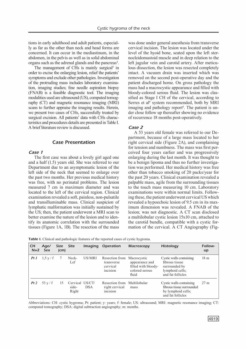
and a half (1.5) years old. She was referred to our Department due to an asymptomatic lesion of the left side of the neck that seemed to enlarge over the past two months. Her previous medical history was free, with no perinatal problems. The lesion measured 7 cm in maximum diameter and was located to the left of the cervical region. Clinical examination revealed a soft, painless, non-pulsatile and transilluminable mass. Clinical suspicion of lymphatic malformation was initially sustained by the US; then, the patient underwent a MRI scan to better examine the nature of the lesion and to iden-tify its anatomic correlation with the surrounded tissues (Figure 1A, 1B). The resection of the mass
was done under general anesthesia from transverse cervical incision. The lesion was located under the level of the hyoid bone, seated upon the left ster-nocleidomastoid muscle and in deep relation to the left jugular vein and carotid artery. After meticu-lous dissection, the lesion was resected completely intact. A vacuum drain was inserted which was removed on the second post-operative day and the patient discharged home. On gross pathology the mass had a macrocystic appearance and filled with bloody-colored serous fluid. The lesion was clas-sified as Stage I CH of the cervical, according to Serres et al6 system recommended, both by MRI imaging and pathology report6. The patient is un-der close follow up thereafter showing no evidence of recurrence 18 months post-operatively.
Case 2A 53 years old female was referred to our De-
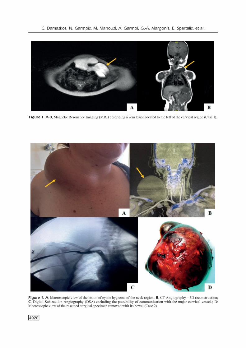
partment, because of a large mass located to her right cervical side (Figure 2A), and complaining for tension and numbness. The mass was first per-ceived four years earlier and was progressively enlarging during the last month. It was thought to be a benign lipoma and thus no further investiga-tion was performed. Her medical history was free other than tobacco smoking of 20 packs/year for the past 20 years. Clinical examination revealed a palpable mass, agile from the surrounding tissues to the touch mass measuring 10 cm. Laboratory examinations were within normal limits. Follow-ing these, the patient underwent cervical US which revealed a hypoechoic lesion of 9.5 cm in its max-imum dimension was revealed. A FNAB of the lesion; was not diagnostic. A CT scan disclosed a multilobular cystic lesion 15x10 cm, attached to the carotid bundle, compatible with a cystic for-mation of the cervical. A CT Angiography (Fig-
Table I. Clinical and pathologic features of the reported cases of cystic hygroma.
Abbreviations: CH: cystic hygroma; Pt: patient; y: years; f: female; US: ultrasound; MRI: magnetic resonance imaging; CT: computed tomography; DSA: digital subtraction angiography; m: months.
CH Age/ Size Site Imaging Operation Macroscopy Histology Follow- N=2 Sex (cm) up
Pt 1 1,5 y / f 7 Neck- US/MRI Resection from Macrocystic Cystic walls containing 18 m Lef transverse appearance and fibrous tissue cervical filled with bloody- surrounded by incision colored serous lymphoid cells; fluid and fat follicles Pt 2 53 y / f 15 Cervical US/CT/ Resection from Multilobular Cystic walls containing 27 m side- DSA right cervical mass fibrous tissue surrounded Right incision by lymphoid cells; and fat follicles
C. Damaskos, N. Garmpis, M. Manousi, A. Garmpi, G.-A. Margonis, E. Spartalis, et al.
4920
Figure 1. A-B, Magnetic Resonance Imaging (MRI) describing a 7cm lesion located to the left of the cervical region (Case 1).
Figure 1. A, Macroscopic view of the lesion of cystic hygroma of the neck region; B, CT Angiography – 3D reconstruction; C, Digital Subtraction Angiography (DSA) excluding the possibility of communication with the major cervical vessels; D: Macroscopic view of the resected surgical specimen removed with its bowel (Case 2).

Cystic hygroma of the neck
4921
ure 2B) and a Digital Subtraction Angiography (DSA) scan (Figure 2C) excluded communication with the major cervical vessels. Because of the lo-cation of the mass, and the underlying symptoms of the patient, she was planned for a surgical exci-sion of the cervical mass. She underwent athwart right cervical incision and the mass was removed with its dish intact (Figure 2D). Pathology report-ed a multilobular mass, 15x15x10 cm long that was proved to be a CH of the cervical (Figure 3). Post-operative course was uneventful and she was discharged 5 days later. She remains healthy and free of recurrence after 27 months.
Discussion
The majority (45-52%) of lymphatic malforma-tions occur in the lymphatic “rich” regions of head and neck. Nevertheless, they can also present in axil-lae, mediastinum, groin and retroperitoneum7. Worth
mentioning are the lymphatic malformations of the orbit8 due to the endangered vision as well as the so-called “giant” lymphatic malformations. The latter involve the tongue, base of the oral cavity, cervical region and mediastinum neighboring with vital struc-tures and necessitating in many instances an emer-gency tracheostomy, staged repair, tube feeding, long term speech therapy, etc.9.
Lymphatic malformations are classified as: a. macrocystic, usually under the level of mylohy-oid muscle which are located mainly within the anterior and posterior cervical triangle; b. micro-cystic, usually above mylohyoid muscle located mainly in the oral cavity, tongue, submandibular region and parotid and c. mixed type6.
Lymphatic malformations typically manifest clin-ically by age 2 years old when parents are worried and seek for a pediatric surgeon consultation. Concerns refer mainly to cosmetic reasons. Parents should be advised that these lesions do not regress spontaneous-ly but they will grow up with the child’s size.
Figure 3. A, Cystic walls containing fibrous tissue (H&E, x100); B, Fibrous tissue surrounded by lymphoid cells (H&E, x40); C, Enlarged lymphatic vessels surrounded by lymphoid tissue (H&E, x200); D, Fibrous tissue and fat cells (fat follicles) con-taining the cystic wall (H&E, x40). (Case 2).
C. Damaskos, N. Garmpis, M. Manousi, A. Garmpi, G.-A. Margonis, E. Spartalis, et al.
4922
As far as the clinical picture, it varies from life threatening (non-patent airway), serious impact on organ function (orbit – loss of vision), to as-ymptomatic. Symptoms may worsen when the le-sions are complicated with bleeding or infection10.
The baseline radiologic investigation is US of the lesion in addition to a plain chest x-ray which will rule out extension into the mediastinum. US can differentiate between cystic and solid tumors. The anatomic details offered by the MRI, a radia-tion free, child friendly tool facilitate preoperative and intraoperative decision-making and planning11. Biopsy or diagnostic needle aspiration do not apply in lymphatic malfomations diagnostic work up.
The therapeutic approach of these lesions was for years undoubtedly and without exception sur-gical. The high occurrence of recurrence as well as technical difficulties in resection due to close proximity to vital structures and poorly demarcat-ed margins of the lesions12 encouraged efforts to find and apply alternative methods of treatment. There is no consensus regarding lymphatic mal-formation treatment and no accepted guidelines exist to date13. It depends on the expertise of the Center and parents’ preference. Needless to say that there are factors which need to be considered before any intervention and individualized care is the most appropriate approach. Among the fac-tors are: symptomatic disease or not, size of the lesion and location (superficial or deep) as well as the type of lesion (macro- or micro- cystic)14.
Briefly noted beneath are the names of alterna-tive methods applied such as radiofrequemcy ab-lation (RF) ablation15, CO2 laser while a more ex-tensive reference regarding sclerotherapy follows16. However, fistula formation or infection and local recurrence have been reported, so the results of these therapies remains still to be examined17.
Percutaneous treatment of lymphatic mal-formations is an established alternative techni-que which improves itself and gains acceptance among the efficient treatment methods under spe-cific indications. The principle of percutaneous sclerotherapy is based on the chemical reaction produced by specific substances upon the en-dothelium of the lesion vessels. The inflammation process leads to thrombosis, obstruction of the lumen, scar formation and downsizing of the le-sion. The approved substances used are OK-43218 produced by Streptococcus sp., Bleomycin19 and Doxycyclin20. The latter mandates sedation due to the amount of discomfort and pain caused. In all cases the cyst liquid should be aspirated before instilling the sclerosant. One of the most serious
side effects is allergic reaction. The method of percutaneous sclerotherapy has proved to be ef-fective in macrocystic disease. Studies suggest that probably is equally effective to surgery for macrocystic lesions of the head and neck21.
Microscopically, CΗ is characterized by enlar-ged lymphatic vessels in a fibrotic and loose stro-mal background. It is characterized by the presen-ce of lymphoid tissue in various formations; even lymphoid follicles can be observed. This feature can be misleading in favor of diagnosing atypical lymphoid proliferations. Cavernous hemangioma is the entity with similar histopathological findin-gs complicates the differential diagnosis of CH. CH consists of thin walls containing fibrous tis-sue, smooth muscle and lymphoid tissue and li-ned with epithelium; guiding to the diagnosis of its particular pathology22.
In summary it can be claimed that surgical the-rapy remains to date the mainstay of treatment23. Appropriate good knowledge of the anatomy, and experience may result in successful and cosmetic results. For emergency indications moreover, opera-tion is the only indicated intervention. Nevertheless, given the disappointing rates of recurrence even in expert hands (15-53%), it is worthwhile considering the alternative techniques as an intraoperative adjun-ct24. There are still many studies to be designed and conducted in order to conclude upon the best option for lymphangioma management.
Conclusions
Lymphatic malformations are a common cau-se of benign nature masses found at the neck and clavicle area of infants and children of perinatal age. CHs are lymphatic malformations most often observed in children; few cases of adult-onset of the disease have been described in the English literature. Provided that the mass is completely excised, an excellent prognosis is of great possi-bility. In both of our cases a complete resections was achieved, with specimen wall removed in-tact; no post-operative complications nor recur-rence has been occurred. Further studies should examine the incidence of CH in adult patients to better elucidate the early diagnosis and the best treating course of the disease.
Conflict of InterestThere is no conflict of interest among the authors of this paper.

Cystic hygroma of the neck
4923
References
1) Biasotto M, Clozza E, tirElli G. Facial cystic lymph-angioma in adults. J Craniofac Surg 2012; 23: e331-334.
2) WiEGand s, Eivazi B, Barth PJ, von rautEnfEld dB, folz BJ, MandiC r, WErnEr Ja. Pathogenesis of lymphangiomas. Virchows Arch 2008; 453: 1-8.
3) BroWn lr, rEiMan hM, rosEnoW EC 3rd, GloviCzki PM, divErtiE MB. Intrathoracic lymphangioma. Mayo Clin Proc 1986; 61: 882-892.
4) kostoPoulos Gk, fEssatidis Jt, hEvas al, skordalaki as, sPyrou PG. Mediastinal cystic hygroma: report of a case with review of the literature. Eur J Cardiothorac Surg 1993; 7: 166-167.
5) orvidas lJ, kasPErBauEr Jl. Pediatric lymphangio-mas of the head and neck. Ann Otol Rhinol Laryngol 2000; 109: 411-421.
6) dE sErrEs lM, siE kC, riChardson Ma. Lymphatic malformations of the head and neck. A proposal for staging. Arch Otolaryngol Head Neck Surg 1995; 121: 577-582.
7) hoChMan M, adaMs dM, rEEvEs td. Current knowledge and management of vascular anomalies, II: malforma-tions. Arch Facial Plast Surg 2011; 13: 425-433.
8) WiEGand s, Eivazi B, BloCh lM, ziMMErMann aP, sEstErhEnn aM, sChulzE s, WErnEr Ja. Lymphatic malformations of the orbit. Clin Exp Otorhinolaryngol 2013; 6: 30-35.
9) roWlEy h, PErEz-ataydE ar, BurroWs PE, rahBar r. Management of a giant lymphatic malformation of the tongue. Arch Otolaryngol Head Neck Surg 2002; 128: 190-194.
10) hanCoCk BJ, st-vil d, luks fi, di lorEnzo M, BlanChard h. Complications of lymphangiomas in children. J Pediatr Surg 1992; 27: 220-224.
11) arnold r, Chaudry G. Diagnostic imaging of vascular anomalies. Clin Plast Surg 2011; 38: 21-29.
12) ravEh E, dE JonG al, taylor GP, fortE v. Prognostic factors in the treatment of lymphatic malforma-tions. Arch Otolaryngol Head Neck Surg 1997; 123: 1061-1065.
13) adaMs Mt, saltzMan B, PErkins Ja. Head and neck lymphatic malformation treatment: a systematic review. Otolaryngol Head Neck Surg 2012; 147: 627-639.
14) Elluru rG, Balakrishnan k, Padua hM. Lymphatic malformations: diagnosis and management. Semin Pediatr Surg 2014; 23: 178-185.
15) kiM sW, kavanaGh k, orBaCh dB, aloMari ai, MullikEn JB, rahBar r. Long-term outcome of ra-diofrequency ablation for intraoral microcystic lymphatic malformation. Arch Otolaryngol Head Neck Surg 2011; 137: 1247-1250.
16) nakazato y, ohno y, nakata y, yaMaGuChi h, hazato n, naGasaWa s, yokoyaMa M, yaMada t. Cystic lymphangioma of the mediastinum. Am Heart J 1995; 129: 406-409.
17) BossErt t, GuMMErt Jf, Mohr fW. Giant cystic lymphangioma of the mediastinum. Eur J Cardiothorac Surg 2002; 21: 340.
18) oGita s, tsuto t, nakaMura k, dEGuChi E, tokiWa k, iWai n. OK-432 therapy for lymphangioma in chil-dren: why and how does it work? J Pediatr Surg 1996; 31: 477-480.
19) yura J, hashiMoto t, tsuruGa n, shiBata k. Bleomycin treatment for cystic hygroma in children. Nihon Geka Hokan 1977; 46: 607-614.
20) BurroWs PE, Mitri rk, aloMari a, Padua hM, lord dJ, sylvia MB, fishMan sJ, MullikEn JB. Percutaneous sclerotherapy of lymphatic malformations with doxycycline. Lymphat Res Biol 2008; 6: 209-216.
21) Balakrishnan k, MEnEzEs Md, ChEn Bs, MaGit aE, PErkins Ja. Primary surgery vs primary sclerother-apy for head and neck lymphatic malformations. JAMA Otolaryngol Head Neck Surg 2014; 140: 41-45.
22) Chand EM, MCnEEly tW, frEant lJ. Pathologic quiz case: male with increasing abdominal girth. Pathologic diagnosis: multicystic intra-abdominal lymphangioma. Arch Pathol Lab Med 2000; 124: 1723-1724.
23) harsha WJ, PErkins Ja, lEWis CW, ManninG sC. Pediatric admissions and procedures for lymphat-ic malformations in the United States: 1997 and 2000. Lymphat Res Biol 2005; 3: 58-65.
24) BoardMan sJ, CoChranE la, roEBuCk d, Elliott MJ, hartlEy BE. Multimodality treatment of pediatric lymphatic malformations of the head and neck using surgery and sclerotherapy. Arch Otolaryngol Head Neck Surg 2010; 136: 270-276.