Cysteine Protease Inhibitors Block Toxoplasma gondii...
10
ANTIMICROBIAL AGENTS AND CHEMOTHERAPY, Feb. 2007, p. 679–688 Vol. 51, No. 2 0066-4804/07/$08.000 doi:10.1128/AAC.01059-06 Copyright © 2007, American Society for Microbiology. All Rights Reserved. Cysteine Protease Inhibitors Block Toxoplasma gondii Microneme Secretion and Cell Invasion Chin Fen Teo, 1 † Xing Wang Zhou, 1 ‡ Matthew Bogyo, 2 and Vern B. Carruthers 1 §* Department of Molecular Microbiology and Immunology, Johns Hopkins Bloomberg School of Public Health, 615 N. Wolfe St., Baltimore, Maryland 21205, 1 and Department of Pathology, Stanford University School of Medicine, 300 Pasteur Dr., Stanford, California 94305 2 Received 22 August 2006/Returned for modification 13 October 2006/Accepted 26 November 2006 Toxoplasma gondii enters host cells via an active, self-driven process to fulfill its need for intracellular replication and survival. Successful host cell invasion is governed by sequential release of secretory proteins from three specialized organelles, including the micronemes, which contribute adhesive proteins necessary for parasite attachment and penetration. Cumulative evidence from studies of Trypanosoma species and malaria parasites has shown that cysteine protease inhibitors represent potent anti-parasitic agents capable of curing infections in vivo. In this study, we screened a series of selective cysteine protease inhibitors for their effects on T. gondii cell invasion. Two of these compounds, morpholinourea-leucyl-homophenolalaninyl-phenyl-vinyl- sulfone and N-benzoxycarbonyl-(leucyl) 3 -phenyl-vinyl-sulfone, impaired T. gondii invasion and gliding motility at low-micromolar concentrations. Unexpectedly, these inhibitors did not affect surface proteolysis of mi- croneme products but instead impaired an earlier step by precluding the secretion of microneme-derived adhesins to the parasite surface. Our findings suggest that cysteine protease activity is required for microneme secretion and cell invasion by T. gondii. Toxoplasma gondii is a cosmopolitan protozoan that infects approximately one-third of the human population worldwide. Primary infection among pregnant women and recrudescence in immunodeficient patients are two major clinical presenta- tions that arise from T. gondii infection. The standard admin- istration for toxoplasmosis is a combination of pyrimethamine and sulfadiazine or clindamycin; however, allergic reaction to this treatment is common among patients, thereby limiting treatment in some cases (26). This problem has lingered with the ever-increasing number of individuals experiencing immu- nodeficiencies due to human immunodeficiency virus infection, organ transplantation, or cancer therapy in recent decades. The identification of novel antitoxoplasmic compounds ef- fective against crucial steps in the parasite’s life cycle may eventually improve the therapeutic options for managing toxoplasmosis. As a hallmark of an obligate intracellular parasite, T. gondii relies on an efficient and robust host cell invasion strategy to support its survival and transmission. This process is driven by the parasite’s actin-myosin motor system and is accomplished by a sequential release of secretory proteins from three spe- cialized organelles, the micronemes, rhoptries, and dense gran- ules, through its apical end (9). Unlike rhoptry and dense granule proteins, which typically associate with the parasito- phorous vacuole after invasion, microneme proteins are shed from the parasite surface into the cell media after translocating toward the posterior end during cell entry (5, 15, 25). Studies on extracellular tachyzoites have demonstrated the involve- ment of calcium-signaling events in microneme protein secre- tion (6, 8, 21, 27). Although basal secretion of microneme products into the excreted/secreted antigen (ESA) fraction harvested from culture supernatants of extracellular parasites occurs in the absence of stimulation, microneme secretion is markedly induced by treatment with secretagogues such as a calcium ionophore (e.g., A23187) or short-chain alcohols (e.g., ethanol) (6, 8). Several studies describing the identification and charac- terization of microneme proteins (MICs) have shown that the majority of MICs are subjected to posttranslational and postexocytic processing (11). One well-characterized example is the MIC2-M2AP hexameric protein complex, which is es- sential for efficient host cell invasion by T. gondii (17, 19). MIC2 is a transmembrane protein that migrates on sodium dodecyl sulfate-polyacrylamide gel electrophoresis (SDS- PAGE) as a doublet or triplet due to differential trimming of its N terminus before it is ultimately shed from the parasite surface via intramembranous cleavage (28). M2AP is a MIC2 escorter protein that is secreted in two forms, proM2AP (pM2AP) and mature M2AP (mM2AP). Whereas pM2AP is released via a nonmicronemal pathway and is not subjected to additional processing on the parasite surface (16), mM2AP is secreted from the micronemes onto the parasite surface, where it is processed into a series of truncated species (M2AP-1, M2AP-2, M2AP-3, and M2AP-4). Other MICs, including MIC3, MIC4, MIC5, MIC6, MIC10, MIC11, and AMA1, also undergo similar processing. Examination of the inhibitory profile of catalytic-type spe- * Corresponding author. Mailing address: Department of Microbi- ology and Immunology, University of Michigan School of Medicine, 1150 W. Medical Center Drive, Ann Arbor, MI 48109. Phone: (734) 763-2081. Fax: (734) 764-3562. E-mail: [email protected]. † Present address: Department of Biochemistry and Molecular Bi- ology, University of Georgia, Athens, GA 30602. ‡ Present address: Department of Biochemistry and Molecular Bi- ology, University of Maryland School of Medicine, Baltimore, MD 21201. § Present address: Department of Microbiology and Immunology, University of Michigan School of Medicine, Ann Arbor, MI 48109. Published ahead of print on 4 December 2006. 679
Transcript of Cysteine Protease Inhibitors Block Toxoplasma gondii...

ANTIMICROBIAL AGENTS AND CHEMOTHERAPY, Feb. 2007, p. 679–688 Vol. 51, No. 20066-4804/07/$08.00!0 doi:10.1128/AAC.01059-06Copyright © 2007, American Society for Microbiology. All Rights Reserved.
Cysteine Protease Inhibitors Block Toxoplasma gondii MicronemeSecretion and Cell Invasion!
Chin Fen Teo,1† Xing Wang Zhou,1‡ Matthew Bogyo,2 and Vern B. Carruthers1§*Department of Molecular Microbiology and Immunology, Johns Hopkins Bloomberg School of Public Health, 615 N. Wolfe St.,
Baltimore, Maryland 21205,1 and Department of Pathology, Stanford University School of Medicine,300 Pasteur Dr., Stanford, California 943052
Received 22 August 2006/Returned for modification 13 October 2006/Accepted 26 November 2006
Toxoplasma gondii enters host cells via an active, self-driven process to fulfill its need for intracellularreplication and survival. Successful host cell invasion is governed by sequential release of secretory proteinsfrom three specialized organelles, including the micronemes, which contribute adhesive proteins necessary forparasite attachment and penetration. Cumulative evidence from studies of Trypanosoma species and malariaparasites has shown that cysteine protease inhibitors represent potent anti-parasitic agents capable of curinginfections in vivo. In this study, we screened a series of selective cysteine protease inhibitors for their effectson T. gondii cell invasion. Two of these compounds, morpholinourea-leucyl-homophenolalaninyl-phenyl-vinyl-sulfone and N-benzoxycarbonyl-(leucyl)3-phenyl-vinyl-sulfone, impaired T. gondii invasion and gliding motilityat low-micromolar concentrations. Unexpectedly, these inhibitors did not affect surface proteolysis of mi-croneme products but instead impaired an earlier step by precluding the secretion of microneme-derivedadhesins to the parasite surface. Our findings suggest that cysteine protease activity is required for micronemesecretion and cell invasion by T. gondii.
Toxoplasma gondii is a cosmopolitan protozoan that infectsapproximately one-third of the human population worldwide.Primary infection among pregnant women and recrudescencein immunodeficient patients are two major clinical presenta-tions that arise from T. gondii infection. The standard admin-istration for toxoplasmosis is a combination of pyrimethamineand sulfadiazine or clindamycin; however, allergic reaction tothis treatment is common among patients, thereby limitingtreatment in some cases (26). This problem has lingered withthe ever-increasing number of individuals experiencing immu-nodeficiencies due to human immunodeficiency virus infection,organ transplantation, or cancer therapy in recent decades.The identification of novel antitoxoplasmic compounds ef-fective against crucial steps in the parasite’s life cycle mayeventually improve the therapeutic options for managingtoxoplasmosis.
As a hallmark of an obligate intracellular parasite, T. gondiirelies on an efficient and robust host cell invasion strategy tosupport its survival and transmission. This process is driven bythe parasite’s actin-myosin motor system and is accomplishedby a sequential release of secretory proteins from three spe-cialized organelles, the micronemes, rhoptries, and dense gran-ules, through its apical end (9). Unlike rhoptry and dense
granule proteins, which typically associate with the parasito-phorous vacuole after invasion, microneme proteins are shedfrom the parasite surface into the cell media after translocatingtoward the posterior end during cell entry (5, 15, 25). Studieson extracellular tachyzoites have demonstrated the involve-ment of calcium-signaling events in microneme protein secre-tion (6, 8, 21, 27). Although basal secretion of micronemeproducts into the excreted/secreted antigen (ESA) fractionharvested from culture supernatants of extracellular parasitesoccurs in the absence of stimulation, microneme secretion ismarkedly induced by treatment with secretagogues such as acalcium ionophore (e.g., A23187) or short-chain alcohols (e.g.,ethanol) (6, 8).
Several studies describing the identification and charac-terization of microneme proteins (MICs) have shown thatthe majority of MICs are subjected to posttranslational andpostexocytic processing (11). One well-characterized exampleis the MIC2-M2AP hexameric protein complex, which is es-sential for efficient host cell invasion by T. gondii (17, 19).MIC2 is a transmembrane protein that migrates on sodiumdodecyl sulfate-polyacrylamide gel electrophoresis (SDS-PAGE) as a doublet or triplet due to differential trimming ofits N terminus before it is ultimately shed from the parasitesurface via intramembranous cleavage (28). M2AP is a MIC2escorter protein that is secreted in two forms, proM2AP(pM2AP) and mature M2AP (mM2AP). Whereas pM2AP isreleased via a nonmicronemal pathway and is not subjected toadditional processing on the parasite surface (16), mM2AP issecreted from the micronemes onto the parasite surface, whereit is processed into a series of truncated species (M2AP-1,M2AP-2, M2AP-3, and M2AP-4). Other MICs, includingMIC3, MIC4, MIC5, MIC6, MIC10, MIC11, and AMA1, alsoundergo similar processing.
Examination of the inhibitory profile of catalytic-type spe-
* Corresponding author. Mailing address: Department of Microbi-ology and Immunology, University of Michigan School of Medicine,1150 W. Medical Center Drive, Ann Arbor, MI 48109. Phone: (734)763-2081. Fax: (734) 764-3562. E-mail: [email protected].
† Present address: Department of Biochemistry and Molecular Bi-ology, University of Georgia, Athens, GA 30602.
‡ Present address: Department of Biochemistry and Molecular Bi-ology, University of Maryland School of Medicine, Baltimore, MD21201.
§ Present address: Department of Microbiology and Immunology,University of Michigan School of Medicine, Ann Arbor, MI 48109.
! Published ahead of print on 4 December 2006.
679
cific protease inhibitors revealed that serine and cysteine pro-teases are two major classes of enzymes involved in MIC pro-cessing (7, 28). Also, the ability of two serine proteaseinhibitors, 3,4-dichloroisocoumarin (3,4-DCI) and 4-(2-amino-ethyl) benzenesulfonyl fluoride (AEBSF), to block T. gondiihost cell invasion has been reported (10). While TgROM4 andTgROM5 (rhomboid-like integral membrane serine proteases)were recently identified as key players for MICs shedding dur-ing invasion and contributors to the parasite’s sensitivity upontreatment with 3,4-DCI (4, 13), the involvement of cysteineproteases in the micronemal secretion pathway and their rolein host cell invasion remain obscure.
With the development of more selective cysteine proteaseinhibitors, recent studies using mouse models of malaria andChagas’ disease have demonstrated that cysteine protease in-hibitors can be potent anti-parasitic agents (14, 24). In light ofthese studies, it is reasonable to propose that similar effectsmight be seen for Toxoplasma if cysteine proteases participatedin host cell invasion. To test this hypothesis, we screened asmall library of cysteine protease inhibitors for their effects onT. gondii cell entry and motility. We found that two peptidylvinyl sulfone (VS) compounds, morpholinourea-leucyl-homo-phenolalaninyl-phenyl-vinyl-sulfone (LHVS) and N-benzoxy-carbonyl-(leucyl)3-phenyl-vinyl-sulfone (ZL3VS), efficiently blockparasite invasion and gliding motility by selectively impairing therelease of MIC contents.
MATERIALS AND METHODS
Materials. E64 {N-[N-(L-3-trans-carboxyoxirane-2-carbonyl)-L-leucyl]-agma-tine}, E64d [(2S,3S)-trans-epoxysuccinyl-L-leucylamido-3-methylbutane ethyl es-ter], and 3,4-DCI (3,4-dichloroisocoumarin) were purchased from Roche (Indi-anapolis, IN), and K777 was a generous gift from James McKerrow (Universityof California, San Francisco, CA). The papain family cysteine protease diazo-methyl ketone inhibitor Z-YA-CHN2 was purchased from Enzyme SystemsProducts (Livermore, CA). All other cysteine protease inhibitors were synthe-sized in the laboratory of M. Bogyo and have been published elsewhere. Specif-ically, all the VS compounds other than LHVS were designed to target theproteasome but have also been shown to react with papain family cathepsins (1,2, 23). The acyloxymethyl ketones (AOMKs) were designed to target both CA-and CD-clan cysteine proteases (20), and LHVS, LHVS-PhOH, MB-074, andJPM-OEt all target papain family cysteine proteases (3). All reagents for tissueculture were obtained from Biowhittaker Inc. (Walkersville, MD) or Gibco/BRL(Gaithersburg, MD). Fluorochrome-conjugated secondary antibodies were pur-chased from Molecular Probes (Eugene, OR). Unless otherwise stated, all otherchemicals were obtained from Sigma (St. Louis, MO).
Parasites and host cells. Human foreskin fibroblasts (HFF) were grown inD10 complete medium (Dulbecco’s modified Eagle’s medium [DMEM] contain-ing 10% fetal bovine serum, 2 mM glutamine, 10 mM HEPES, and 50 "g/mlpenicillin-streptomycin). T. gondii tachyzoites, RH or 2F1 strains, were propa-gated in HFF under the above conditions.
Invasion assays. For the #-galactosidase (#-gal) invasion assay, 2F1 parasiteswere resuspended in invasion medium (IM; DMEM containing 1% fetal bovineserum and 10 mM HEPES) at 4 $ 106/ml. One-hundred microliters of parasiteswas incubated in the presence of 10 "M or 50 "M of each protease inhibitor ordimethyl sulfoxide (DMSO) at room temperature for 15 min before loading ontoHFF in 96-well plates. The plates were then incubated for 30 min at 37°C, with5% CO2. Excess parasites were removed from monolayers by washing with coldphosphate-buffered saline (PBS) (containing 1 mM CaCl2 and 1 mM MgCl2) sixtimes (2 min on the shaker per wash). After the infected HFF were lysed with 100"l ice-cold lysis buffer (100 mM HEPES, pH 8.0, 1 mM MgSO4, 0.1% TritonX-100, 5 mM dithiothreitol), 50 "l of the lysates was aliquoted and mixed with200 "l of assay buffer (100 mM phosphate buffer, pH 7.3, 102 mM #-mercapto-ethanol, and 9 mM MgCl2) and 40 "l of 6.25 mM CPRG (chlorophenol red-#-D-galactopyranoside; Roche, Indianapolis, IN). After incubating the reactionmixtures at 37°C for 45 min, the enzymatic activity of #-galactosidase was mea-sured at an absorbance of 550 nm using a kinetic plate reader (Molecular
Devices) and Softmax pro3 alias software. Data was compiled from two inde-pendent experiments, each with duplicate samples.
For the red/green invasion assay, strain 2F1 parasites were resuspended in IMat 5 $ 107/ml. Two-hundred microliters of parasites was incubated with variousconcentrations of protease inhibitors as indicated or DMSO at room tempera-ture for 15 min before adding to HFF monolayers in 8-well chamber slides. Afterincubation for 15 min at 37°C in 5% CO2, the chamber was removed and the slidewas rinsed with PBS prior to fixation with 4% formaldehyde–0.02% glutaralde-hyde in PBS (20 min at room temperature). After three washes with PBS, theslides were blocked with 10% fetal bovine serum (FBS) in PBS and washed oncewith antigen dilution buffer (ADB; 1% fetal bovine serum and 1% normal goatserum in PBS). Attached parasites were stained with rabbit %-SAG1 (P30)polyclonal antibody (1:1,000), whereas the invaded parasites were stained withmonoclonal antibody (MAb) 9E11 %-SAG1 after permeabilizing with 0.1% Tri-ton X-100. Secondary antibodies used were goat anti-rabbit Alexa Fluoro 594(1:1,000) and goat anti-mouse Alexa Fluoro 488 (1:500). 4&,6&-Diamidino-2-phenylindole (DAPI; 5 "g/ml) was added in the secondary solution for nucleistaining. The slides were washed three times with ADB after each staining step.Finally, the slides were mounted in Mowiol and visualized by phase contrast andepifluorescence using a Nikon Eclipse E800 equipped with an RT spot sliderCCD camera. Data were compiled from three independent experiments, eachfrom eight random fields/well under $600 total magnification.
Preparation of excretory/secretory antigens. To perform the screening andquantification of MIC secretion experiments, 2F1 parasites were resuspended inDGH medium (DMEM with 2 mM glutamine and 10 mM HEPES) ('3 $ 107
to 6 $ 107 parasites with a total volume of 100 "l per reaction) and pretreatedwith 50 "M protease inhibitors or DMSO at room temperature for 15 min. Forinduced secretion, the parasites were incubated in a 37°C water bath for 2 min inthe presence of 1% (vol/vol) ethanol. For basal secretion, the parasites wereincubated in a 37°C water bath for 20 min without ethanol treatment. Theparasites were then removed from the water bath and left on ice to quenchmicroneme protein secretion. After centrifuging twice (1,000 $ g, 3 min, 4°C),the supernatant (ESA) was mixed with 5$ sample buffer containing 2% mer-captoethanol and boiled. The samples were stored at (20°C before use inSDS-PAGE.
Large-scale ESA (basal secretion, without ethanol treatment) used in two-dimensional differential in-gel electrophoresis (2D-DIGE) experiments was pre-pared using the procedure described above, with '4 $ 109 parasites in 15 mlDGH medium.
SDS-PAGE and immunoblotting. The ESA fractions were resolved by SDS–12.5% PAGE or SDS–15% PAGE (for MIC5, MIC10, and MIC11) prior tosemidry electrotransfer to nitrocellulose membranes for immunoblotting. Theprimary antibodies used included MAb 40-1a S/N (#-gal; 1:100), MAb 6D10ascites fluid (MIC2; 1:15,000), R%M2AP (M2AP; 1:10,000), TgCL7 (AMA1;1:30,000), R%MIC5 (MIC5; 1:7,500), R%P18 (MIC10; 1:7,500), R%MIC11(MIC11; 1:7,500), and Tg17-43 (GRA1; 1:60,000). Secondary antibodies wereeither goat anti-mouse- or goat anti-rabbit-conjugated horseradish peroxidaseused at 1:5,000 dilution. After adding Supersignal substrate (Pierce, Rockford,IL), direct chemiluminescence was captured with the Fujifilm LAS-1000 CCDcamera before conventional exposure onto films. Signal intensities were quanti-fied using Fujifilm Image Gauge Software.
2D-DIGE. Fifty micrograms of lyophilized DMSO-treated or 50 "M LHVS-treated large-scale ESA was dissolved in 10 "l of labeling buffer {30 mM Tris, 8M urea, 2% 3-[(3-cholamidopropyl)-dimethylammonio]-1-propanesulfonate, pH8.5} and reacted with 400 pmol of either Cy3 or Cy5, respectively. Reactionswere carried out on ice for 30 min in the dark, and the labeling reactions werequenched by adding 1 "l of 10 mM lysine. The samples were mixed together inequal parts and resolved by two-dimensional electrophoresis as described previ-ously (28).
Gliding motility assay. Eight-well chamber slides (Nalge-Nunc Intl., Roches-ter, NY) were coated with either 50% FBS in PBS (37°C for 1 h) or 0.1%poly-L-lysine (room temperature) and washed with PBS before use. Freshlyharvested RH parasites were resuspended in 5 ml HHE medium (Hank’s bal-anced salt solution supplemented with 10 mM HEPES and 1 mM EGTA).Parasites (0.5 ml) were then treated with 50 "M protease inhibitors or DMSO atroom temperature for 15 min before applying onto chamber slides. The parasiteswere allowed to glide on the coated slides at 37°C for 15 min. The slides werethen rinsed with PBS and fixed in 4% formaldehyde–0.02% glutaraldehyde inPBS at room temperature for 20 min. After blocking with 10% FBS in PBS for30 min, trails left by gliding parasites were detected by MAb 9E11 %-SAG1antibody (1:1,000) followed by goat anti-mouse Alexa Fluoro 594 (1:1,000). Theslides were washed three times with ADB after each staining step. After theslides were mounted in Mowiol, the images were visualized and captured under
680 TEO ET AL. ANTIMICROB. AGENTS CHEMOTHER.

epifluorescence (with $600 total magnification) using a Nikon Eclipse E800equipped with an RT spot slider CCD camera.
RESULTS
Two peptidyl vinyl sulfones, LHVS and ZL3VS, effectivelyinhibit Toxoplasma invasion and microneme protein release.We compiled a set of 22 cysteine protease inhibitors that weresynthesized initially to target either the proteasome (all vinylsulfones [VS], except LHVS and LHVS-PhOH), papain familycysteine proteases (LHVS, LHVS-PhOH, MB-074, K777, andJPM-OEt), or general CA- and CD-clan cysteine proteases (allAOMK compounds) and three commercially available generalcysteine protease inhibitors (E64, its membrane-permeableanalog E64d, and Z-YA-CHN2) (Table 1). The VS compoundsthat were designed against the proteasome are also likely totarget papain family cysteine proteases, as they contain thesame reactive vinyl sulfone group as LHVS, and several (i.e.,ZL3VS and YL3-VS) also contain the critical P2 leucine resi-due found in LHVS. These compounds were used for prelim-inary screening of effects on T. gondii invasion as well as mi-croneme protein secretion and processing. 3,4-DCI, a serineprotease inhibitor previously shown to impair Toxoplasma in-vasion, was included as a positive control, whereas the solventvehicle (DMSO) was used as a negative control. The inhibitoryeffect on invasion was evaluated using the #-galactosidase-
FIG. 1. Cysteine protease inhibitor screen for Toxoplasma gondii invasion. (A) A representative assay plate showing color development from#-gal activity as an indicator of parasite attachment and invasion. Wells showing minimal attachment/invasion are yellow, whereas those with higherattachment/invasion are orange to red. Parasites were treated with 50 "M of each inhibitor prior to being added to HFF monolayers. (B) Quan-tification of attachment/invasion. The data are mean values ) standard errors of the means of two independent experiments, each with duplicatesamples, and are expressed as a percentage of the solvent control (DMSO). The peptidyl vinyl sulfone inhibitors LHVS and ZL3VS were the mosteffective inhibitors tested. DMSO and 3,4-DCI were included as negative and positive controls, respectively.
TABLE 1. Protease inhibitor characteristics
Compoundname Target Class Source or
reference
LHVS-PhOH Papain family Vinyl sulfone 3YL3-VS Proteasome Vinyl sulfone 2ZL3-VS Proteasome Vinyl sulfone 1NLVS Proteasome Vinyl sulfone 1LHVS Papain family Vinyl sulfone 3Ac-PRKN-VS Proteasome Vinyl sulfone 23Ac-PRLN-VS Proteasome Vinyl sulfone 23Ac-PKLN-VS Proteasome Vinyl sulfone 23Ac-PKRN-VS Proteasome Vinyl sulfone 23Ac-PKKN-VS Proteasome Vinyl sulfone 23Ac-YRKN-VS Proteasome Vinyl sulfone 23Ac-YRLN-VS Proteasome Vinyl sulfone 23Ac-YKKN-VS Proteasome Vinyl sulfone 23Ac-YKLN-VS Proteasome Vinyl sulfone 23Ac-YKRN-VS Proteasome Vinyl sulfone 23G-AOMK General cysteine Acyloxymethyl ketone 20L-AOMK General cysteine Acyloxymethyl ketone 20N-AOMK General cysteine Acyloxymethyl ketone 20R-AOMK General cysteine Acyloxymethyl ketone 20JPM-OEt Papain family Epoxysuccinyl 3Z-YA-CHN2 Papain family Diazomethyl ketone Enzyme Systems
ProductsMB-074 Papain family Epoxysuccinyl 3K777 Papain family Vinyl sulfone J. McKerrowE-64 Papain family Epoxysuccinyl RocheE-64d Papain family Epoxysuccinyl Roche3,4-DCI Serine protease Isocoumarin Roche
VOL. 51, 2007 CYSTEINE PROTEASE INHIBITORS AND TOXOPLASMA INVASION 681

expressing parasite 2F1 (8) and a modified procedure based onMcFadden et al. (22). Compounds were screened at 10 "Mand 50 "M. Whereas none of the compounds inhibited inva-sion at 10 "M (data not shown), several compounds signifi-cantly impaired invasion when administered at 50 "M (Fig. 1).The two most effective compounds were LHVS (morpho-linourea-leucyl-homophenolalaninyl-phenyl-vinyl-sulfone), withapproximately 70% inhibition compared to the DMSO control,and ZL3VS [N-benzoxycarbonyl-(leucyl)3-phenyl-vinyl-sulfone],which also inhibited parasite invasion by approximately 50%.These vinyl sulfone-based compounds were more effective thanthe epoxide-based general cysteine protease inhibitors E-64 andE-64d but were similar in effectiveness to 3,4-DCI.
Proteins released from the micronemal pathway are sub-jected to extensive proteolytic processing both before and afterexocytosis (11). The correlation between microneme secretionand a successful host cell invasion is well established (5, 15). Todetermine whether inhibition of invasion was linked to a defi-ciency in the micronemal secretion pathway, both basal andinduced secretion of MIC2 and M2AP in ESA fractions wasassessed by immunoblotting. The level of #-galactosidase de-tected in the ESA was monitored for inadvertent parasite lysisduring manipulation. Consistent with the results from invasionscreening, LHVS and ZL3VS were also the most effectiveinhibitors of microneme secretion among the collection ofcompounds tested (Fig. 2). Moreover, we observed a differen-tial inhibition between the basal and induced secretions in
which a better inhibitory effect was repeatedly seen duringbasal secretion. Interestingly, these compounds appeared toreduce the total amount of MIC2 and M2AP without markedlyaffecting the profile of the processed species (note that pM2APis not a processed species). This observation suggests that bothcompounds impair invasion by blocking microneme proteinsecretion, not surface processing. Also, secretion of pM2APwas unaffected, or perhaps slightly increased, as a result oftreatment, implying that the inhibition is selective for the mi-cronemal pathway.
Because LHVS and ZL3VS were two of the most effectiveinhibitors emerging from the screens, we focused on thesecompounds for further investigation. Two chemically relatedcompounds, LH (morpholinourea-leucyl-homophenolalanine)and NLVS [4-hydroxy-5-iodo-3-nitrophenylacetyl-(leucyl)3-phenyl-vinyl-sulfone], were synthesized (Fig. 3). LH is a trun-cated version of LHVS synthesized without the thioreactivevinyl sulfone moiety. NLVS is structurally similar to ZL3VS butwith a distinct N-terminal capping moiety, and it was includedas a negative control because it showed minimal activity in theinvasion assay (Fig. 1).
Inhibition occurs primarily at the attachment step duringinvasion. As part of its active entry strategy, T. gondii utilizesmicroneme proteins to initiate attachment to the host cell via itsapical end. We used a sequential staining method (red/greenassay) and fluorescence microscopy to distinguish T. gondiitachyzoites that were attached to host cells from those parasites
FIG. 2. Effect of cysteine protease inhibitors on microneme protein secretion and processing. Protein levels of MIC2 and M2AP in ESA fractionswere examined by immunoblotting after (A) basal secretion or (B) induced secretion, as described in Materials and Methods. #-Gal levels were monitoredas an indicator of general toxicity and inadvertent parasite lysis. LHVS and ZL3VS partially blocked the release of MIC2 and the processed forms ofM2AP into the ESA, an effect that was more prominent for basal secretion.
682 TEO ET AL. ANTIMICROB. AGENTS CHEMOTHER.

that had actively invaded the monolayers. BAPTA-AM (B-AM),a calcium chelator that can inhibit microneme protein secre-tion, thereby precluding attachment, was used as a positivecontrol along with 3,4-DCI. Cytochalasin D (CytD), an actinpolymerization inhibitor that blocks parasite penetration with-out affecting attachment, was also included.
As shown in Fig. 4A, LHVS and ZL3VS effectively blockedboth attachment and penetration during invasion, leading to anoverall decrease of parasite association with host cells. On thecontrary, parasites treated with LH or NLVS exhibited a sim-ilar invasion capacity compared to that of the solvent control.Also, the red/green assay appears to be more sensitive than the#-galactosidase assay since, for example, LHVS showed noinhibitory activity at 10 "M in the #-gal invasion assay yetexhibited '50% inhibition at the same concentration in thered/green assay. This may be due to the more extensive washesperformed in the red/green assay. As established by previousstudies (17), an overall decrease in both attached and invadedparasites strongly indicates an effect on parasite attachment,since attachment is an essential prerequisite for penetration.Nonetheless, we cannot rule out a secondary effect on pene-tration that is masked by the attachment deficit. Collectively,the above findings indicate that both LHVS and ZL3VS impairtachyzoite attachment by blocking the release of at least twokey invasion proteins, MIC2 and M2AP, from the micronemes.
To determine the dose-response relationship for invasion,the parasites were treated with various concentrations (0.1 to100 "M) of compounds, and the inhibition was evaluated by
the red/green assay. The total number of associated parasitescounted (regardless of attached or invaded parasites) per hostcell nuclei was normalized to that of the solvent control (being100%). LHVS inhibited T. gondii invasion with a 50% inhibi-tory concentration (IC50) of 10 "M, whereas the IC50 ofZL3VS and 3,4-DCI was approximately 12.5 "M (Fig. 4B). Agradual decrease in the number of successfully attached/in-vaded parasites was also observed in LH and NLVS treatmentsas higher concentrations were tested, but the decrease did notreach statistical significance, even at 100 "M. Similar trendswere also observed using the #-gal invasion assay (data notshown).
Inhibition of LHVS and ZL3VS selectively impairs mi-croneme protein secretion. To further investigate whether the
FIG. 3. Structures of LHVS, ZL3VS, LH, and NLVS.FIG. 4. LHVS and ZL3VS impair T. gondii invasion by blocking
parasite attachment and penetration. (A) Attached (red) and invaded(green) parasites in an invasion assay were distinguished by a sequen-tial staining method as described in Materials and Methods. Data areexpressed as the number of tachyzoites per host cell nucleus, visualizedby DAPI staining. Both attached and invaded parasites from LHVSand ZL3VS treatments were significantly decreased (*) compared tothat of the DMSO solvent control, whereas LH and NLVS treatmentdid not substantially inhibit parasite invasion. 3,4-DCI, B-AM, andCytD were included as positive control treatments that impair parasiteinvasion. Data are mean values ) standard errors of the means ofthree independent experiments. (B) To determine the dose-responseeffect of LHVS treatment on T. gondii invasion, parasites associated(regardless attached or invaded) per host nuclei were enumerated andnormalized as a percentage of solvent control (DMSO)-treated para-sites. LHVS inhibited T. gondii invasion with an IC50 of 10 "M. Asimilar trend was observed using the #-gal invasion assay (data notshown). Data are mean values ) standard errors of the means of threeindependent experiments. Log [PI], log10 concentration of proteaseinhibitor.
VOL. 51, 2007 CYSTEINE PROTEASE INHIBITORS AND TOXOPLASMA INVASION 683

invasion blockage was due specifically to an effect on mi-croneme protein release, especially from basal secretion, weperformed a differential gel electrophoresis (DIGE) experi-ment. DMSO- and LHVS-treated large-scale ESA fractionswere collected under basal secretion conditions, and proteinstherein were covalently modified with Cy3 (pseudocoloredgreen) and Cy5 (pseudocolored red), respectively, mixed to-gether, and resolved by two-dimensional electrophoresis. Inthis scheme, proteins that show no change in abundance areyellow, whereas those that are lower in abundance are green.As shown in Fig. 5A, all of the green spots that were success-
fully identified were derived from micronemes (MIC2, MIC4,MIC5, MIC10, MIC11, M2AP, and SUB1), while the levels ofproteins released from other sites (GRA1, a dense granuleprotein; Cyp18, an endoplasmic reticulum protein; and SAG1,a glycosylphosphatidylinositol-linked surface protein) did notchange appreciably, as indicated by their yellow appearance.
To confirm the inhibitory activity of compounds in ESAfractions, we examined individual microneme proteins, includ-ing AMA1, MIC5, and MIC10, by immunoblotting. As shownin Fig. 5B and C, secretion of these microneme proteins wasreduced after treating with LHVS or ZL3VS, while samples
FIG. 5. LHVS selectively impairs microneme protein secretion. (A) For DIGE analysis, proteins from DMSO or 50 "M LHVS-treatedlarge-scale ESA were modified with Cy3 (pseudocolored green) and Cy5 (pseudocolored red) fluorescent dyes, respectively. Yellow spots indicateno change in abundance, whereas green spots correspond to proteins diminished by LHVS treatment. Spots were identified by comparing themobility of proteins on two-dimensional gel electrophoresis to previously resolved DIGE data (28, 29). The inhibitory effect of LHVS and ZL3VSon secretion of AMA1, MIC5, and MIC10 was verified by immunoblotting ESA from (B) basal secretion or (C) induced secretion. Little or noeffect was seen for GRA1 secretion.
684 TEO ET AL. ANTIMICROB. AGENTS CHEMOTHER.

treated with LH or NLVS showed levels similar to that ofDMSO treatment. Levels of GRA1 were unchanged or slightlyupregulated after treatment with LHVS or ZL3VS.
The dose-response relationship was evaluated based on therelative amount of MIC2 in ESA fractions. The results shownin Fig. 6 revealed that the IC50 of LHVS and ZL3VS in block-ing microneme protein secretion was '10 to 25 "M, i.e., in thesame potency range as that for impairing parasite attachment.
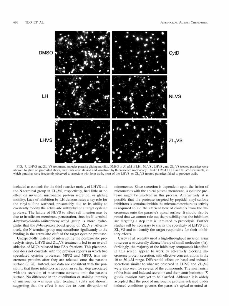
Treatments of LHVS and ZL3VS diminish parasite glidingmotility. Gliding motility is thought to be a critical factor indriving parasite invasion. MIC2 has previously been shown toserve as a bridge between the internal actin-myosin motorsystem and external substrates (receptors, surfaces, etc.),thereby serving as a mediator in T. gondii movement (18).From the above experiments, we have established that LHVSand ZL3VS blocked the release of microneme contents thatwas associated with parasite invasion. Next, we examinedwhether their effects were also related to parasite motility byperforming a gliding assay on plastic chamber slides. The trails,containing SAG1 and produced by parasite movements, werevisualized by immunofluorescence staining.
LH or NLVS had little or no effect on gliding, as indicatedby the abundant presence of trails, including some of consid-erable length, compared to DMSO solvent control (Fig. 7).Although few of the LHVS- and ZL3VS-treated parasites werestill able to deposit relatively long and normal trails, the overalltrail deposition was greatly diminished in a manner similar toCytD treatment. Collectively, these observations suggest thatboth LHVS and ZL3VS effectively disrupt parasite gliding mo-tility, presumably as a consequence of their effects on mi-croneme secretion.
LHVS and ZL3VS do not affect MIC protein shedding.Based on the immunoblotting data from the above experi-
ments, we concluded that the peptidyl vinyl sulfones did notinhibit the surface-trimming process. However, our results donot readily distinguish whether the effect was due to an effecton surface shedding of MIC products. Although surfaceshedding occurs through intramembrane proteolysis byserine proteases of the rhomboid family (4, 12), a role forother types of proteases in shedding has not been ruled out.To determine if LHVS and ZL3VS affect MIC shedding, welooked for accumulation of MIC2 on the surface of inhibi-tor-treated tachyzoites.
As a known sheddase inhibitor, 3,4-DCI treatment of extra-cellular parasites resulted in a significantly stronger surfacestaining due to the retention of MIC2 under basal (Fig. 8A) orinduced (Fig. 8B) secretion conditions. By contrast, no accu-mulation of MIC2 was seen after treatment with LHVS,ZL3VS, or any of the other control compounds. This resultstrongly indicates that LHVS and ZL3VS do not target theshedding step. Therefore, we conclude that these compoundsin some way block the secretion of MIC proteins from themicronemes.
DISCUSSION
We report herein that two vinyl sulfone cysteine proteaseinhibitors, LHVS and ZL3VS, effectively block T. gondii mi-croneme protein secretion, gliding motility, and cell invasion.The anti-invasion activity of LHVS was found to slightly ex-ceed that of the serine protease inhibitor 3,4-DCI, whereasZL3VS was as effective as 3,4-DCI. No significant inhibitionwas observed when general cysteine protease inhibitors, suchas E-64 and E-64d, were tested, suggesting that peptidyl vinyl-based compounds may be more potent than these epoxide-based compounds. In addition, LH and NLVS, which were
FIG. 6. Dose-response of LHVS and ZL3VS in blocking microneme protein secretion. The potency of LHVS and ZL3VS was examined byimmunoblotting MIC2 and M2AP in ESA fractions from (A) basal secretion and (B) induced secretion. Collectively, LHVS inhibited micronemeprotein secretion with an IC50 of '25 "M in both basal and induced secretions, while ZL3VS showed a higher IC50 at a concentration of '50 "M.
VOL. 51, 2007 CYSTEINE PROTEASE INHIBITORS AND TOXOPLASMA INVASION 685
included as controls for the thiol-reactive moiety of LHVS andthe N-terminal group in ZL3VS, respectively, had little or noeffect on invasion, microneme protein secretion, or glidingmotility. Lack of inhibition by LH demonstrates a key role forthe vinyl-sulfone warhead, presumably due to its ability tocovalently modify the active-site sulfhydryl of a target cysteineprotease. The failure of NLVS to affect cell invasion may bedue to insufficient membrane penetration, since its N-terminal4-hydroxy-5-iodo-3-nitrophenylacetyl group is more hydro-philic that the N-benzoxycarbonyl group on ZL3VS. Alterna-tively, the N-terminal group may contribute significantly to thebinding in the active-site cleft of the target cysteine protease.
Unexpectedly, instead of interrupting the postexocytic pro-teolysis steps, LHVS and ZL3VS treatments led to an overallablation of MICs released into ESA fractions. This phenome-non does not correlate with the previous reports in which twospeculated cysteine proteases, MPP2 and MPP3, trim mi-croneme proteins after they are released onto the parasitesurface (7, 28). Instead, our data are consistent with the pos-sibility that these inhibitors act upon an earlier step associatedwith the secretion of microneme contents onto the parasitesurface. No difference in the distribution or staining intensityof micronemes was seen after treatment (data not shown),suggesting that the effect is not due to overt disruption of
micronemes. Since secretion is dependent upon the fusion ofmicronemes with the apical plasma membrane, a cysteine pro-tease might be involved in this process. Alternatively, it ispossible that the protease targeted by peptidyl vinyl sulfoneinhibitors is contained within the micronemes where its activityis required to aid the efficient flow of contents from the mi-cronemes onto the parasite’s apical surface. It should also benoted that we cannot rule out the possibility that the inhibitorsare targeting a step that is unrelated to proteolysis. Furtherstudies will be necessary to clarify the specificity of LHVS andZL3VS and to identify the target responsible for their inhibi-tory effects.
Carey et al. recently used a high-throughput invasion assayto screen a structurally diverse library of small molecules (4a).Strikingly, the majority of the inhibitory compounds identifiedin this screen appear to work by selectively blocking mi-croneme protein secretion, with effective concentrations in the10 to 50 "M range. Differential effects on basal and inducedsecretions similar to what we observed in LHVS and ZL3VSwere also seen for several of the compounds. The mechanismof the basal and induced secretion and their contributions to T.gondii invasion have yet to be clarified. Although it is widelyaccepted that the pool of microneme proteins released underinduced conditions governs the parasite’s apical-oriented at-
FIG. 7. LHVS and ZL3VS treatment impedes parasite gliding motility. DMSO or 50 "M of LH-, NLVS-, LHVS-, and ZL3VS-treated parasites wereallowed to glide on precoated slides, and trails were stained and visualized by fluorescence microscopy. Unlike DMSO, LH, and NLVS treatments, inwhich parasites were frequently observed to associate with long trails, most of the LHVS- or ZL3VS-treated parasites failed to produce trails.
686 TEO ET AL. ANTIMICROB. AGENTS CHEMOTHER.

tachment during invasion, Lovett et al. (21) has elegantly,albeit unexpectedly, demonstrated that the intracellular cal-cium in the parasite is sharply down-regulated during the ini-tial stage of host cell invasion, precisely the opposite of whatwas expected. Combined with our results herein, this impliesthat basal secretion may serve a more prominent role in cellinvasion than previously thought. Although both pathways arecalcium dependent, based on their sensitivity to the calciumagonist BAPTA-AM, basal secretion likely has a lower con-centration threshold for calcium than induced secretion.Therefore, the drop in intracellular calcium during apical at-tachment presumably limits secretion to a basal rate that mayprovide an optimal supply of surface ligands to direct penetra-tion into the target cell.
For those microneme proteins, such as M2AP and MIC5,which require en route proteolysis to remove their propeptidesduring transportation to microneme compartments, the levelsof their proforms were found to be more abundant in basalsecretion than those of induced secretion. There are two pos-sible explanations for this observation. First, since pM2AP andpMIC5 are released in a calcium independent manner (16)(S. D. Brydges and V. B. Carruthers, unpublished), these pro-proteins are likely discharged into the ESA via the dense gran-ules (DG), which represents the default secretory pathway inT. gondii. DG secretion (based on detection of GRA1) appears
to be unaffected or slightly elevated by inhibitor treatment,therefore the increased levels of pM2AP and pMIC5 may bedue to activation of the DG pathway. Furthermore, treatmentsthat suppress microneme secretion have been observed to ac-tivate DG secretion (6, 8), thus, our results are consistent withthis inverse regulatory relationship between the two pathways.
A second factor potentially contributing to elevated releaseof pM2AP and pMIC5 is that the proteolytic maturation ofthese precursor proteins may be inhibited by both LHVS andZL3VS treatments. An ongoing study by our group character-izing a candidate micronemal maturase, TgCPL (cathepsinproteinase L), has found that LHVS can selectively bind thisparasite-derived cathepsin L-like protease both in vivo and invitro (F. Parussini, C. I. Phillips, M. Bogyo, and V. B. Carruth-ers, unpublished data). Preliminary localization of TgCPLsuggests that this protease is largely confined to a post-Golgicompartment that is distinct from the micronemes. UnlabeledLHVS can compete for labeling of this compartment by afluorescent conjugate of LHVS (bodipy-LHVS) in extracellu-lar parasites (but not intracellular parasites), suggesting thatLHVS is at least somewhat membrane permeable (C.-F. Teo,F. Parussini, and V. B. Carruthers, unpublished data). Sincepresumably the bulk of the microneme material is processedand trafficked during parasite replication, it remains unclearhow a brief blockage of propeptide processing in extracellular
FIG. 8. LHVS and ZL3VS do not affect MIC protein shedding. Distribution of surface MIC was detected after treating with 50 "M of LH,NLVS, LHVS, and ZL3VS. DMSO was a solvent control, whereas 3,4-DCI was an inhibitor specific to the shedding process. Shown is (A) basalsecretion or (B) induced secretion.
VOL. 51, 2007 CYSTEINE PROTEASE INHIBITORS AND TOXOPLASMA INVASION 687

parasites would impair microneme secretion. It is possible thatsome TgCPL is trafficked to the micronemes, where it couldplay a role in facilitating the release of microneme contentsupon fusion with the parasite apical membrane. Additionalstudies will be required to clarify the role of TgCPL in mi-croneme secretion, and further work will also be necessary todetermine whether LHVS acts upon alternative targets thatare responsible for the observed effects.
Cysteine protease inhibitors, especially peptidyl vinyl sul-fones, have gained interest in pharmaceutical research duringthe past decades. Importantly, peptidyl vinyl sulfones havebeen demonstrated to be potent anti-parasitic agents inTrypanosoma cruzi and Plasmodium species (14, 24). In bothcases, the target enzymes have been localized to digestive com-partments: the lysosomes/reservasomes of T. cruzi (cruzain)and the food vacuole of P. falciparum (falcipain-2a, falcipain-2b, and falcipain-3). Interestingly, digestive compartmentshave not been described in Toxoplasma. Therefore, with thediscovery of both LHVS and ZL3VS as effective cysteine pro-tease inhibitors, future work may not only unveil chemother-apeutic candidates for treating Toxoplasma infections but mayalso provide a useful tool in identifying the target enzyme(s)and unravel how and where it fulfills its key role in micronemesecretion, gliding motility, and cell invasion.
ACKNOWLEDGMENTS
We thank Claudia Bordon and Bjorn Kafsack for help with experi-ments, Marie-France Cesbron-Delauw and Lloyd Kasper for providingantibodies, and James McKerrow for providing K777.
This study was supported by a Stanley Medical Research Institutegrant (to V.B.C.) and U.S. National Institutes of Health NationalTechnology Center for Networks and Pathways grant U54 RR020843(to M.B.).
REFERENCES
1. Bogyo, M., J. S. McMaster, M. Gaczynska, D. Tortorella, A. L. Goldberg,and H. Ploegh. 1997. Covalent modification of the active site threonine ofproteasomal beta subunits and the Escherichia coli homolog HslV by a newclass of inhibitors. Proc. Natl. Acad. Sci. USA 94:6629–6634.
2. Bogyo, M., S. Shin, J. S. McMaster, and H. L. Ploegh. 1998. Substratebinding and sequence preference of the proteasome revealed by active-site-directed affinity probes. Chem. Biol. 5:307–320.
3. Bogyo, M., S. Verhelst, V. Bellingard-Dubouchaud, S. Toba, and D.Greenbaum. 2000. Selective targeting of lysosomal cysteine proteaseswith radiolabeled electrophilic substrate analogs. Chem. Biol. 7:27–38.
4. Brossier, F., T. J. Jewett, L. D. Sibley, and S. Urban. 2005. A spatiallylocalized rhomboid protease cleaves cell surface adhesins essential for inva-sion by Toxoplasma. Proc. Natl. Acad. Sci. USA 102:4146–4151.
4a.Carey, K. L., N. J. Westwood, T. J. Mitcheson, and G. E. Ward. 2004. Asmall-molecule approach to studying invasive mechanisms of Toxoplasmagondii. Proc. Natl. Acad. Sci. USA 101:7433–7438.
5. Carruthers, V. B., O. K. Giddings, and L. D. Sibley. 1999. Secretion ofmicronemal proteins is associated with Toxoplasma invasion of host cells.Cell Microbiol. 1:225–235.
6. Carruthers, V. B., S. N. J. Moreno, and L. D. Sibley. 1999. Ethanol andacetaldehyde elevate intracellular calcium and stimulate microneme dis-charge in Toxoplasma gondii. Biochem. J. 342:379–386.
7. Carruthers, V. B., G. D. Sherman, and L. D. Sibley. 2000. The Toxoplasmaadhesive protein MIC2 is proteolytically processed at multiple sites by twoparasite-derived proteases. J. Biol. Chem. 275:14346–14353.
8. Carruthers, V. B., and L. D. Sibley. 1999. Mobilization of intracellularcalcium stimulates microneme discharge in Toxoplasma gondii. Mol. Micro-biol. 31:421–428.
9. Carruthers, V. B., and L. D. Sibley. 1997. Sequential protein secretion fromthree distinct organelles of Toxoplasma gondii accompanies invasion of hu-man fibroblasts. Eur. J. Cell Biol. 73:114–123.
10. Conseil, V., M. Soete, and J. F. Dubremetz. 1999. Serine protease inhibitorsblock invasion of host cells by Toxoplasma gondii. Antimicrob. Agents Che-mother. 43:1358–1361.
11. Dowse, T., and D. Soldati. 2004. Host cell invasion by the apicomplexans: thesignificance of microneme protein proteolysis. Curr. Opin. Microbiol. 7:388–396.
12. Dowse, T. J., J. C. Pascall, K. D. Brown, and D. Soldati. 2005. Apicomplexanrhomboids have a potential role in microneme protein cleavage during hostcell invasion. Int. J. Parasitol. 35:747–756.
13. Dowse, T. J., and D. Soldati. 2005. Rhomboid-like proteins in Apicomplexa:phylogeny and nomenclature. Trends Parasitol. 21:254–258.
14. Engel, J. C., P. S. Doyle, I. Hsieh, and J. H. McKerrow. 1998. Cysteineprotease inhibitors cure an experimental Trypanosoma cruzi infection. J. Exp.Med. 188:725–734.
15. Garcia-Reguet, N., M. Lebrun, M.-N. Fourmaux, O. Mercereau-Puijalon, T.Mann, C. J. M. Beckers, B. Samyn, J. Van Beeumen, D. Bout, and J.-F.Dubremetz. 2000. The microneme protein MIC3 of Toxoplasma gondii is asecretory adhesin that binds to both the surface of the host cells and thesurface of the parasite. Cell Microbiol. 2:353–364.
16. Harper, J. M., M. H. Huynh, I. Coppens, F. Parussini, S. Moreno, and V. B.Carruthers. 2006. A cleavable propeptide influences Toxoplasma Infectionby facilitating the trafficking and secretion of the TgMIC2-M2AP invasioncomplex. Mol. Biol. Cell 17:4551–4563.
17. Huynh, M. H., K. E. Rabenau, J. M. Harper, W. L. Beatty, L. D. Sibley, andV. B. Carruthers. 2003. Rapid invasion of host cells by Toxoplasma requiressecretion of the MIC2-M2AP adhesive protein complex. EMBO J. 22:2082–2090.
18. Jewett, T. J., and L. D. Sibley. 2003. Aldolase forms a bridge between cellsurface adhesins and the actin cytoskeleton in apicomplexan parasites. Mol.Cell 11:885–894.
19. Jewett, T. J., and L. D. Sibley. 2003. The Toxoplasma proteins MIC2 andM2AP form a hexameric complex necessary for intracellular survival. J. Biol.Chem. 279:9362–9369.
20. Kato, D., S. H. Verhelst, K. B. Sexton, and M. Bogyo. 2005. A general solidphase method for the preparation of diverse azapeptide probes directedagainst cysteine proteases. Org. Lett. 7:5649–5652.
21. Lovett, J. L., N. Marchesini, S. N. Moreno, and L. D. Sibley. 2002. Toxo-plasma gondii microneme secretion involves intracellular Ca(2!) releasefrom inositol 1,4,5-triphosphate (IP[3])/ryanodine-sensitive stores. J. Biol.Chem. 277:25870–25876.
22. McFadden, D. C., F. Seeber, and J. C. Boothroyd. 1997. Use of Toxoplasmagondii expressing beta-galactosidase for colorimetric assessment of drug ac-tivity in vitro. Antimicrob. Agents Chemother. 41:1849–1853.
23. Nazif, T., and M. Bogyo. 2001. Global analysis of proteasomal substratespecificity using positional-scanning libraries of covalent inhibitors. Proc.Natl. Acad. Sci. USA 98:2967–2972.
24. Olson, J. E., G. K. Lee, A. Semenov, and P. J. Rosenthal. 1999. Antimalarialeffects in mice of orally administered peptidyl cysteine protease inhibitors.Bioorg. Med. Chem. 7:633–638.
25. Opitz, C., M. Di Cristina, M. Reiss, T. Ruppert, A. Crisanti, and D. Soldati.2002. Intramembrane cleavage of microneme proteins at the surface of theapicomplexan parasite Toxoplasma gondii. EMBO J. 21:1577–1585.
26. van der Ven, A. J., P. P. Koopmans, T. B. Vree, and J. W. van der Meer. 1991.Adverse reactions to co-trimoxazole in HIV infection. Lancet 338:431–433.
27. Vieira, M. C., and S. N. Moreno. 2000. Mobilization of intracellular calciumupon attachment of Toxoplasma gondii tachyzoites to human fibroblasts isrequired for invasion. Mol. Biochem. Parasitol. 106:157–162.
28. Zhou, X. W., M. J. Blackman, S. A. Howell, and V. B. Carruthers. 2004.Proteomic analysis of cleavage events reveals a dynamic two-step mechanismfor proteolysis of a key parasite adhesive complex. Mol. Cell Proteomics3:565–576.
29. Zhou, X. W., B. F. Kafsack, R. N. Cole, P. Beckett, R. F. Shen, and V. B.Carruthers. 2005. The opportunistic pathogen Toxoplasma gondii deploys adiverse legion of invasion and survival proteins. J. Biol. Chem. 280:34233–34244.
688 TEO ET AL. ANTIMICROB. AGENTS CHEMOTHER.