
CYCLIC CHANGES IN EXCITABILITY OF THE OPTIC … · CYCLIC CHANGES IN EXCITABILITY OF THE OPTIC...
12
CYCLIC CHANGES IN EXCITABILITY OF THE OPTIC PATH- WAY OF THE RABBIT GEO. H. BISHOP From the Department of Ophthalmology, Oscar Johnson Institute, Washington Uni- versit y, Saint Louis Received for publication September 2, 1932 It has been shown (Bartley and Bishop, 1933a) t,hat electric stimuli applied to the stump of the optic nerve of the rabbit result in electrical phenomena that may be recorded from the contralateral optic cortex, as a complicated sequence of cortical action potentials. The general pro- cedure followed in the work cited has been employed here, except for details to be noted below. Stimuli repeated at various intervals, from one hundred sigma up to three seconds, in general do not result in identical responsesto each stimulus. The question whether the variat,ion in re- sponse of the cortex is a random one, or whether some orderly cycle of events gives the appearance of random variation only because of some uncontrolled factor in our procedure, is of fundamental importance, not only in ordering further experimental work, but in intlerpretling the activity of the cortex itself. Experiments show that the variations observed in responses to repeated constant stimuli are due to the fact that such stimuli, except at certain critical frequencies, fall at random times in a cyclic variation of cortical excitability. Whether the actual level at which such excitability is vary- ing is cortical or sub-cortical we do not know; it is at some level along the optic pathway. We have good evidence that the cortex itself varies not only in threshold, but in the continuous and apparently spontaneous activity that occurs without specific stimulation of the optic nerve. On the other hand we have some reason to believe that this will not account for all the phenomena observed. For instance, after a maximal response to optic nerve steimulatlion,the cortex does not appear to be quiescent for the period of refractoriness to a second stimulus. Further, when strych- nine is applied t’o a circumscribed region of the cortex (Bartley, 1933) distant points show a rhythmic activity spreading from the strychninized area into normal regions, at a frequency higher than could be obtained by repetitive stimulation of t,he opt,ic nerve, and with a constant and not a varying amplitude of response. Finally, the spontaneous cortical activity shows no major fluctuation with a periodicity corresponding to that de- 213 by 10.220.33.1 on April 13, 2017 http://ajplegacy.physiology.org/ Downloaded from
Transcript of CYCLIC CHANGES IN EXCITABILITY OF THE OPTIC … · CYCLIC CHANGES IN EXCITABILITY OF THE OPTIC...

CYCLIC CHANGES IN EXCITABILITY OF THE OPTIC PATH- WAY OF THE RABBIT
GEO. H. BISHOP
From the Department of Ophthalmology, Oscar Johnson Institute, Washington Uni- versit y, Saint Louis
Received for publication September 2, 1932
It has been shown (Bartley and Bishop, 1933a) t,hat electric stimuli applied to the stump of the optic nerve of the rabbit result in electrical phenomena that may be recorded from the contralateral optic cortex, as a complicated sequence of cortical action potentials. The general pro- cedure followed in the work cited has been employed here, except for details to be noted below. Stimuli repeated at various intervals, from one hundred sigma up to three seconds, in general do not result in identical responses to each stimulus. The question whether the variat,ion in re- sponse of the cortex is a random one, or whether some orderly cycle of events gives the appearance of random variation only because of some uncontrolled factor in our procedure, is of fundamental importance, not only in ordering further experimental work, but in intlerpretling the activity of the cortex itself.
Experiments show that the variations observed in responses to repeated constant stimuli are due to the fact that such stimuli, except at certain critical frequencies, fall at random times in a cyclic variation of cortical excitability. Whether the actual level at which such excitability is vary- ing is cortical or sub-cortical we do not know; it is at some level along the optic pathway. We have good evidence that the cortex itself varies not only in threshold, but in the continuous and apparently spontaneous activity that occurs without specific stimulation of the optic nerve. On the other hand we have some reason to believe that this will not account for all the phenomena observed. For instance, after a maximal response to optic nerve steimulatlion, the cortex does not appear to be quiescent for the period of refractoriness to a second stimulus. Further, when strych- nine is applied t’o a circumscribed region of the cortex (Bartley, 1933) distant points show a rhythmic activity spreading from the strychninized area into normal regions, at a frequency higher than could be obtained by repetitive stimulation of t,he opt,ic nerve, and with a constant and not a varying amplitude of response. Finally, the spontaneous cortical activity shows no major fluctuation with a periodicity corresponding to that de-
213
by 10.220.33.1 on April 13, 2017
http://ajplegacy.physiology.org/D
ownloaded from

214 GEO. H. BISHOP
t.ected by repet)itive stimulation. Apparently the rhythmic variation in the excitability of the pathway is in part at least subcortical, and does not correspond simply to a rhythmic variation in the spontaneous activity of the cortex itself.
Following each effective stimulus to the nerve, two manifestations of activity are observed in the cortex. First, a series of (typically) five mono- phasic potentials occur at about one-fifth second intervals, the first of the series arising within from five to fifty sigma after the stimulus, depending on the strength of t#he latter. Second, a single diphasic potential is super- posed upon the first monophasic wave of the former series, obscuring its
60
L
4
20
15 110 210 TiMI? de 3 IO 510
Fig. 1. Upper: Diagram of form of potential of first component of the optic corti- cal record, indicating the low monophasic wave A at threshold, occupying positions 1, 2, and by inference 6 at successive increases of stimulus strength, and the diphasic wave B which falls earlier, and at successive increases of stimulus appears as 3, ,$ and 6, its second phase obscuring the first wave to appear, A 6.
Lower: Plot of time-strength values for the two elements of the first potential complex. The dotted where B obscures it.
line is the inferred curve of A, extrapolated, at a strength
form when above threshold value. It is therefore impossible to observe what is happening to the first member of the monophasic series; but the subsequent responses can be followed, and all members of this series appear to vary in amplitude together. Further, while the thresholds of these two types of waves are different, when a shock well above threshold is delivered, the amplitudes of the two sets of responses vary in a parallel manner with repeated constant stimuli, if not too frequent. This is further evidence that the magnitude of the response is determined not only by the number of fibers stimulated in the optic nerve, but also by the condition of excitability of some critical level of a pathway common to the two impulses. To be
by 10.220.33.1 on April 13, 2017
http://ajplegacy.physiology.org/D
ownloaded from
EXCITABILITY OF THE OPTIC PATHWAY 215
sure, when stimuli follow each other too frequently, only the single diphasic response may occur, the series of monophasic responses being at least too low to be clearly distinguishable, a circumstance suggesting a rather funda- mental difference in the significance of these responses in vision; but in most of what follows the intervals between stimuli are relatively long. The ampli- tude of the larger diphasic potential may therefore be taken as one index of the typical cortical response, (fig. 1) and the details of these responses will be ignored for the present, the whole being discussed in terms of the single diphasic potential.
One major difficulty in this work must be taken carefully into account. Some of the most striking details of the series of events to be described have not been detectable in every attempt to study them. They might thus be taken to be fortuitous occurrences, rather than essential phenomena in the functioning of the visual mechanism. However, in satisfactory preparations the results are so specific and so clear-cut as to leave no doubt in the minds of the observers that they are valid and fundamental evidences of a definite type of functioning. Furthermore, other aspects of this chain of events are constant and reproducible in all rabbits, and the former and the latter types of activity are obviously closely related. We therefore presume that something fundamental which occurs in all experiments is plainly manifested only in some of them because of the complexity of the situation, and venture the opinion that if adequate controls were possible over a nervous mechanism so elaborate as the optic nerve-thalamus- cortex complex, presumably also capable of being influenced by the rest of the nervous system, our experiments would be less equivocal. The best we can say for them at present, however, is that they are not contradictory, and even this at times has somewhat taxed our ingenuity.
In every preparation the following events can be demonstrated. As the stimulus to the optic nerve is increased from below threshold, stimulation at each strength being repeated at three second intervals for at least ten times, a value is reached such that only an occasional response from the cortex is elicited, and never more than a just threshold response. This will be termed the threshold stimulus, as being potentially threshold; it must activate optic nerve fibers that are potentially capable of sending impulses to the cortex, but only occasionally do so. As the strength of stimulus is increased, and more fibers are consequently stimulated each time, cortical responses follow more of the stimuli than previously, and the largest of the responses are greater in amplitude, responses of various amounts being obtained. With further increase, some of these responses become maximal, that is, as large as can be obtained from a stimulus of any greater strength; other responses grade from this value down to nothing. This will be termed a just maximal stimulus even though not all responses to it are maximal. With still stronger stimuli, each is followed by a response, every response
by 10.220.33.1 on April 13, 2017
http://ajplegacy.physiology.org/D
ownloaded from

216 GEO. H. BISHOP
being finally maximal. This will be termed a repetitive maximal stimulus. That the variation in response is not to be assigned to variation in the number of fibers activated in the nerve by a constant stimulus may be checked directly by recording the action potential from the optic nerve just beyond the chiasma, and is indicated indirectly by the fact that at certain even shorter intervals the response of the cortex does not vary at all.
If the frequency of stimulation is now increased, say to once per second, a second factor enters. At any given strength, the first response occurs as usual, but subesquent responses are uniformly lower than they would have been at a slower rate of stimulation, even though the stimulus is a repetitive maximal one. After three or four successively lower responses to such a maximal stimulus the rest occur at a new and constant level. This level depends on the frequency of stimulation, with qualifications that will appear presently. For less than maximal stimulation, the responses to a constant stimulus vary, with this new repetitive maximal level as an upper limit. The appearance is similar to relative refractoriness in nerve fibers, where the higher the frequency, the lower the response in any given fiber; but at these frequencies, the locus of such refractoriness certainly cannot be the optic nerve fibers themselves.
Starting at the other extreme, a second stimulus of any strength following a first maximal stimulus is ineffective after an interval of less than fifteen sigma, the two being applied every three seconds. As the interval is increased, a small second response of the diphasic component appears and grows larger, if the stimulus is maximal. If both stimuli are weaker than this, but equal in strength, when the response to the first happens to be large, that following the second will be smaller, but if the first is small or lacking, the second will be large. Two such responses therefore see-saw up and down continuously. This indicates that when, for some unknown reason, a stimulus has been ineffective, it has not left the pathway refractory to a subsequent impulse; that is, the apparently random variations of excitability described above are independent of the impulses sent up the optic nerve by which this variability is made evident.
These events suggest that, insofar as the pathway is concerned which is activated by fibers of the optic nerve having low threshold (activated by submaximal stimuli), there is a spontaneously rhythmic variation in excita- bility; that the time in this cycle at which the stimulus falls determines whether or not a response shall take place in the cortex; and that if the first of two stimuli falls in a refractory part of the cycle, it does not alter the cycle in phase, and a second stimulus, falling necessarily later in this cycle, must always be effective if the first is ineffective, because by this time these particular elements will have become excitable again. The fact, however, that a stronger stimulus will give a maximal response at any
by 10.220.33.1 on April 13, 2017
http://ajplegacy.physiology.org/D
ownloaded from

EXCITABILITY OF THE OPTIC PATHWAY 217
random time it may be delivered, suggests that if enough fibers respond, the refractoriness of the pathway may be overcome, whatever phase the cycle may be in at the moment. This is not strictly true, as will be shown below; it applies to any first stimulus, but not to properly timed successive ones. An alternate or even a parallel possibility to this last is that different elements are refractory at different times, the fibers of low threshold con- necting with elements going through the cycle more or less as a group, those of higher threshold being in a slightly different phase from the first, etc., so that a few elements will be refractory at whatever time the impulses arrive, but on the average, a constant number of elements will be excita#ble, and therefore a constant maximum response may be obtained. This is a permissible assumption so far as any data we have will indicate. If a submaximal stimulus causes a submaximal response, a maximal stimulus closely following it will always cause a second response, even at a time when a repetition of the first stimulus is ineffective. That is, more of the elements, not activated by the first stimulus, will respond if more optic nerve fibers are activated, even though the elements activated by the first impulse cannot respond again. Further, that all the elements capable of responding at one point of the cortex never can be made to respond to a single stimulus applied to the optic nerve, is shown by experiments with strychnine (Bartley, 1933, l.c.). After having applied strychnine to a small sharply localized area of the cortical surface, repetitive stimulation at one per second will induce responses not only from the strychninized area, but from points at some distance, which are higher in amplitude than any responses ever obtained from the untreated cortex. The evidence (q.v.) indicates that in this case the strychninized area acts as a pace-maker for the adjacent regions of the cortex, which under these circumstances only respond when stimulated by way of the strychninized area; a paradoxical situation, the analysis of which is presented elsewhere. Thus all the ele- ments may respond, instead of only these elements which normally would be non-refractory at the moment of stimulation.
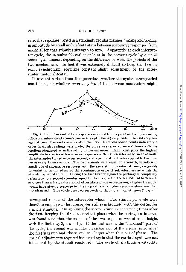
By modification of the procedure of the foregoing experiments, a more specific evidence of rhythmic excitability could be obtained. The optic nerve was first stimulated at the rate of one per second with a submaximal shock, successive responses varying in amplitude as usual (fig. 2). Now the speed of the circuit breaker was altered slowly, until a frequency of stimulation could be found that permitted responses of constant amplitude. That is, the stimulus was brought into constant phase relationship with the cyclic excitability of the optic pathway. If these responses were large, a slight lag of the stimulation cycle caused them to become smaller, even to extinction. In this situation a stronger stimulus would give a response. When the interrupter was timed at a very slightly lower rate than exactly coincided with the rhythm of the optic pathway, or at a slightly higher
by 10.220.33.1 on April 13, 2017
http://ajplegacy.physiology.org/D
ownloaded from
218 GEO. H. BISHOP
rate, the responses varied in a strikingly regular manner, waxing and waning in amplitude by small and definite steps between successive responses, from maximal for that stimulus strength to zero. Apparently at each interrup- ter cycle, the stimulus fell earlier or later in the nervous cycle by a small amount, an amount two mechanisms.
depending on the differen In fact it was extremely
.ce .
dl between the periods of the
fficult to keep the two in exact synchronism, requiring constant slight adjustment of the inter- rupter motor rheostat.
It was not certain from this procedure whether the cycles corresponded one to one, or whether several cycles of the nervous mechanism might
Fig. 2. Plot of second of two responses recorded from a point on the optic cortex, following submaximal stimulation of the optic nerve; amplitude of second response against time of second stimulus after the first. Numbers beside points indicate the order in which readings were made; the curve was repeated several times with the readings staggered as indicated by numerical order. Each point plots the highest amplitude in a series of ten or more responses with a given interval between stimuli; the interrupter turned once per second, and a pair of stimuli were applied to the optic nerve every three seconds. The two stimuli were equal in strength, variation in amplitude of successive responses with the same stimulus interval being assignable to variation in the phase of the spontaneous cycle of refractoriness at which the stimuli happened to fall. During the first twenty sigma the pathway is completely refractory to a second stimulus equal to the first, but if the second had been made stronger than a first, activation of other fibers in the nerve having a higher threshold would have given a response in this interval, and a higher response elsewhere than was observed. This whole curve corresponds to the interval syz of figure 3 b, q.v.
correspond to one of the interrupter wheel. Two sCmuli Per cycle w ‘ere therefore employed, the interrupter still syn .chronized with the cortex for a single stimulus. By applying the second stimulus at varying times after the first, keeping the first in constant phase with the cortex, an interval was found such that the second of the two responses was of equal height with the first (fig. 3, a and b). If the first was in the “maximal” part of the cycle, the second was smaller on either side of the critical interval; if the first was minimal, the second was larger when thus out of phase. The critical adjustments required indicated again that the cortical cycle was not influenced by the stimuli employed. The cycle of rhythmic excitability
by 10.220.33.1 on April 13, 2017
http://ajplegacy.physiology.org/D
ownloaded from

EXCITABILITY OF THE OPTIC PATHWAY 219
in the optic pathway so detected was somewhat more than one-fifth second, and varied in different preparations from one-fifth to one-third. Not all animals gave such quantitatively clear-cut results, but in all attempts the most constant series of responses was obtained at a critical frequency of stimulation, with submaximal stimuli.
a.
c.
Fig. 3. Diagram to indicate the cyclic fluctuations in excitability of the optic pathway to submaximal and maximal stimulation of the optic nerve. a, cyclic varia- tion in refractoriness for fibers of lowest threshold, or for the pathways they lead to. Pathway accessible at 0, 3, etc., refractory half way between these points. It is inferred that pathways of higher threshold are out of phase with those of lower thres- hold, as indicated by dotted curves, such that to a stronger stimulus a response may be obtained even when those paths responding to a weaker stimulus are refractory.
b. Diagram of responses to second of two equal submaximal stimuli when the first falls in the non-refractory part of the cycle. For fifteen to twenty sigmas the path- way is left completely refractory by the first response; it is partly refractory after that (from z to y and even from y to z), due to the cyclic variation going on inde- pendently of the stimulation (represented by the curve of fig. 2), and for this reason the maximum at y is not as high as the first response. Beyond z the response rises again to reach a second maximum practically as high as the first response, one cycle after the first stimulus, in the phase of non-refractoriness, and so repeats. The cycle of refractoriness is not altered by the stimulation.
c. Responses as in b, but both stimuli supermaximal. Apparently a sufhciently strong first stimulus activating a sufficient number of fibers can render all the path- ways refractory; and when this occurs the pathways are refractory again one nervous cycle later, but after one and a half cycles the second stimulus may cause a second maximal response. Such a strong stimulus has thus thrown all pathways into the same phase, and thereafter the phase relationships impressed upon the system by the stimulus are maintained for some time, contrary to the case of weaker stimulation, (3, b,) where the phase relationships cannot be shown to shift from their spontaneous rhythm.
THE AMERICAN JOURNAL OF PHYSIOLOGY, VOL. 103, NO. 1
by 10.220.33.1 on April 13, 2017
http://ajplegacy.physiology.org/D
ownloaded from

220 GEO. H. BISHOP
In one particularly clear-cut experiment, the interrupter was speeded up until definitely out of step with the cortex, and since the interval between the two stimuli in each cycle was changed in consequence, this was again adjusted to its previous value of exactly one nervous cycle. Now a single stimulus of submaximal value gave varying responses, but when both stimuli were applied at each interrupter revolution, both responses were of the same amplitude, whatever that value happened to be. Again when nearly in time, the responses, both first and second in this case, increased and decreased slowly and rhythmically as the cycles of interrupter and cortex shifted in and out of phase. The response is determined therefore not primarily by the frequency of stimulation through causing refractori- ness or fatigue, but by the phase of the nervous cycle at which the stimulus is applied. This fact is emphasized in view of what follows.
If stimuli, of submaximal value in the preceding trials, are increased to fully maximal values, this series of events is materially altered. It will be recalled that such maximal stimuli call forth in general maximal responses; even when applied at one per second frequency the amplitude of response, although lower than with slower stimulation, usually becomes constant, or varies but slightly. When a second stimulus is applied at a shorter interval than one second after a first, this is not true. The former experi- ments with two stimuli per cycle were repeated with strong stimuli. In one experiment, at the interval corresponding to one nervous cycle as tested by submaximal stimuli, a second maximat stimulus gave no cortical response at all (fig. 3, c). This interval was now prolonged, until at one and one- half cycles as determined previously, the second response became nearly as large as the first, and at greater intervals became again smaller. This occurred for any one pair of stimuli, applied after a period of rest, and without regard to the phase relationship of interrupter and nervous mecha- nism. In other words, it appears as if a maximal stimulus excited all or at least a constant number of elements of the pathway regardless of the phase they were in, and in so doing, forced them all into the same phase, rendering them refractory thereby, so that at the end of one complete cycle they were all again refractory, but after one and one-half cycles, all were again irritable.
Phenomena similar to these, and obviously having the same basis, could be produced by various manipulations of the two shocks, and some of them were too complicated for analysis at the present stage of our infor- mation, and not all could be repeated on different preparations. They are referred to here as giving further and striking evidence not only of definite rhythmicity in the optic pathway, but as indicating that by strong stimuli this rhythm can be changed in phase by the stimulation of the optic nerve. For instance, two maximal shocks were spaced at an interval corresponding to one and one-half nervous cycles, both giving maximal responses as above.
by 10.220.33.1 on April 13, 2017
http://ajplegacy.physiology.org/D
ownloaded from

EXCITABILITY OF THE OPTIC PATHWAY 221
The interrupter was run at three and one-half nervous cycles per revolution, so that at successive revolutions a given stimulus would be alternately in and out of phase with the optic mechanism. Now during the first inter- rupter cycle after the stimulating circuits were closed, both responses were maximal; at the second cycle, both were just visible, and equal; and this alternation repeated indefinitely, and with successive trials. Further, changing the speed of the interrupter, faster or slower, did not throw the responses out of phase, over a range that with sub-maximal stimulation always throws the two mechanisms throughly out of synchronism.
In other words, not only could the nervous mechanism be driven, but the rhythm so set up maintained the phase imposed upon it for at least the greater part of a second, that is, during several cycles of the nervous activity during which stimuli were not given it. Each effective stimulus apparently set a pace for the rest during one interrupter cycle at least, and another stimulus arrived before the two mechanisms were allowed to get out of phase enough to interfere with the regularity of the results. In other experiments, instead of presence and absence of responses, regular varia- tions in amplitude of lesser degree were obtained.
DISCUSSION. Several major conditions are presented here which seem to us to form a scaffolding within which an explanation of the functioning of the visual pathway must somehow be constructed. No pretense is made that even the scaffolding is complete. First, while the retina and the cortex are presumably connected in a one-to-one point relationship, that is, unit areas of the retina are projected on the cortex by means of potentially simple pathways, these conductile elements are not entirely independent of each other. Activation of one group of elements may affect the time of arrival of impulses at the cortex over another group, even without the mediation of retinal interconnections. Secondly, there is some level at which a rhythmic variation in irritability takes place, apparently spon- taneously, but this rhythm can be driven if enough elements are activated at once; again evidence that one element of the pathway can affect the activity or excitability of others. Third, there are connections available, from one element of the cortex to others, accessible under the influence of strychnine even at a distance from the strychninized area, and presumably normally occupied in the “spontaneous” activity shown by the cortex. Whether there may be such connections by way of the thalamus we have ourselves obtained no evidence, but we have evidence from direct stimula- tion of the cortex (Bartley and Bishop, 1933b) that they are available at the cortical level. Added to these facts, the further data that two more or less independent pathways exist from optic nerve to cortex, as indicated by two distinct potential components (Bartley and Bishop, 1933a), and t,hat one of these at least can be activated only at such a rate of stimulation t’hat it could not reasonably account for the immediate facts of vision, and
by 10.220.33.1 on April 13, 2017
http://ajplegacy.physiology.org/D
ownloaded from

222 GEO. H. BISHOP
we have the major limitations that seem to be imposed by this work so far on the elaboration of an analysis of visual functioning. Even this takes no account whatever of the complications of the retina.
It is obviously futile at present therefore to attempt to “explain” any significant aspect of vision in terms of the records of cell activity that we have obtained so far, and we disavow any such purpose in the remarks which follow. However, it is proper to enquire what limits the facts of vision impose upon the interpretation of our records of nervous phenomena, as well as the other way around. One way of doing both is to construct a hypothetical system that might work if it really existed, one consistent with known facts, but with the gaps that must inevitably appear filled in by figments of the imagination. Such a working hypothesis then suggests further experimental tests, and is perhaps the more stimulating for being constructed so largely out of pure moonshine.
A primary requirement of such a system is that there should be possible, not only a punctate representation of retinal elements on the cortex, but that this representation should be sensibly continuous in time. That is, it is desirable that each unit area of the retina (permissibly including a number of sense endings) be able to activate the cortex at least as frequently as the rate at which separate light stimuli can be differentiated. We do not know what fusion frequency a rabbit has, if any, but may assume it is of the same order of frequency as in man, or possibly lower.
The simple diphasic potential wave recorded from the cortex can follow the second of two stimuli to the optic nerve when the interval is not over fifteen sigma, but the second response is low, and a third stimulus after the same interval is ineffective. Spaced at a reasonable fusion frequency (10 to 20 per second), each would be effective when it did not fall in a refractory point of the cycle of the optic pathway. This potential might thus signalize the nrst response by the cortex in the simplest form of sensory perception. We have found no evidence of cortical activity that specifi- cally follows as a continuation of this first step; but the reverberations through the optic or other cortical regions of such an impulse, presumably attenuating in intensity as it disperses in space, might easily be lost among the spontaneous discharges so continually in evidence. We need not even assume, however, that it goes any further; perhaps the modification by such an impulse of the cortical activity continuously taking place is in itself the visual act. Presumably the retinal discharges may be higher in frequency than the cortex can follow; the summation of such discharges at lower levels with a change in frequency of the response has been but briefly enquired into, but we have some evidence that a second stimulus to the nerve which fails to give a second separate response from the cortex may increase the amplitude of a first response. The limit of fusion frequency may be im- posed by sub-cortical or cortical synapses, at the same level as the refractory
by 10.220.33.1 on April 13, 2017
http://ajplegacy.physiology.org/D
ownloaded from

EXCITABILITY OF THE OPTIC PATHWAY 223
phenomena we have studied, and the variation of fusion frequency with intensity of stimulation has an obvious analogue in the summation phe- nomena we have met with. An increme in number of fibers stimulated lessens the shortest interval between responses, and an increase in the frequency of stimulation seems to increase the number of cortical elements responding.
The other response obtainable from the cortex, a series of monophasic waves, cannot be made to repeat at a frequency that corresponds to the more obvious events of visual experience. However, visual events are not all obvious; in particular there is demanded some machinery by which the activity of the thalamus as a visual level in itself may be correlated with the activity of the cortex. As the more complicated processes require more time for their accomplishment, so they could be handled by mecha- nisms more flexible and more complex, but less quick-acting. At least this succession of responses has some of the characteristics that seem appropriate to such complex activity.
We have been able to record specific responses from the cortex only by causing many elements to respond at the same time, and thus tosummate their results. Inferences from the results of this simple procedure to the normal case when impulses arise from the retina, will be precarious ones. It is not necessary to suppose that even from a unit area of the retina, only a unit pathway leads to the cortex. There may be several such pathways in parallel, and the cyclic alteration of excitability that we observe may affect them in rotation, leaving some pathway always accessible. In particular, it is too striking to be fortuitous that all of the optic nerve fibers of lowest threshold lead to elements that are approximately in phase with each other, but are out of phase with elements whose fibers have higher thresholds. Such grouping of fibers according to thresholds is reminiscent of the conditions in peripheral nerves (Heinbecker, Bishop and O’Leary, 1933), where fibers serving a common function are usually thus set off from fibers serving other functions. The optic nerve itself is being investigated from this point of view.
The only assumption required to allow continuous representation in the cortex of unit retinal areas is that each such pair of loci, in retina and cortex, be connected by several fibers having different thresholds to electrical stimuli; this threshold difference, whatever other significance it may have, connoting according to our findings a difference in phase of cyclic excitability of the pathways. An impulse from the retinal sense organs might then avail itself of whatever pathway was open at the time and, by means of cortical cross-connections, affect the same cortical locus over any pathway of a group.
In general, it is not necessary to infer that each individual impulse travel- ling up a fiber from the retina arrives as a unit impulse at the cortex, and
by 10.220.33.1 on April 13, 2017
http://ajplegacy.physiology.org/D
ownloaded from

224 GEO. H. BISHOP
registers there as such. activity, the
Rather we would look upon the cortex as being in the whole network of neu- physiological activity of constant
rones bearing some direct relationship to the “present state” of the animal’s complex behavior which is sometimes referred to as his “mental state.” Impulses coming in from the periphery, by one route more or less directly, by a second which reflected or interpreted the general state of activity of the th .alamus as cond itioned or modified by - such impulses, would then take effect by modifying the cortical activity continuously going on, and the change in the whole picture of such continuous activity, rather than the specific set of impulses sent in, would correspond to visual perception. Such activity would then tend to revert, gradually and possibly with rhythmic disturbances still persisting, to its previous state of activity. Failure to return completely would correspond in a sense to memory; rhythmic disturbances persisting after the exciting impulses ceased would have an analogy at least in such phenomena as after-images.
This imaginary picture already far outdistances our experimental find- ings, but that is the privilege of hypotheses in general. We may at least rest assured that such a picture does not err on the side of complexity, however improbable as fact.
The writer wishes to acknowledge the assistance of Dr. S. Howard Bart- ley in this work.
SUMMARY
Successive equal stimuli applied to the optic nerve of the rabbit in general evoke from the contralateral optic cortex responses of variable magnitude, unless the stimuli are very strong, or unless they are timed at certain critical intervals.
The shortest critical interval of one-third to one-fifth second presumably corresponds to a cyclic spontaneous variation in the accessibility of the optic pathway, of the character of partial refractoriness, but independent of the stimuli applied. Strong stimuli (activating many fibers of the optic nerve), seem to be able to activate such pathways even when partially refractory, as if by summation at any one element of the effects of the impulses arriving over many optic nerve fibers. This and other similar evidence suggests that the to a corresponding point
projection pathwav from one point s of the cortex is multiple, and su
of the retina bject to tlhe
influence of impulses over other similar pathways. Various experimental and hypothetical considerations are advanced in
an attempt to construct fat ts with vision.
a workable scheme that would reconcile these
BIBLIOGRAPHY
BARTLEY, S. H. 1933. This Journal, in press. BARTLEY, S. H. AND G. H. BISHOP. 1933a. This Journal, in press.
1933b. This Journal, in press. HEINBECKER, P.,G.H. BISHOPAND J. O'LEARY. 1933. Arch. Neurol. and Psych&t.,
in press.
by 10.220.33.1 on April 13, 2017
http://ajplegacy.physiology.org/D
ownloaded from