CXCL16-Mediated Cell Recruitment to Rheumatoid Arthritis
14
ARTHRITIS & RHEUMATISM Vol. 54, No. 3, March 2006, pp 765–778 DOI 10.1002/art.21662 © 2006, American College of Rheumatology CXCL16-Mediated Cell Recruitment to Rheumatoid Arthritis Synovial Tissue and Murine Lymph Nodes Is Dependent Upon the MAPK Pathway Jeffrey H. Ruth, 1 Christian S. Haas, 1 Christy C. Park, 2 M. Asif Amin, 1 Rita J. Martinez, 1 G. Kenneth Haines, III, 2 Shiva Shahrara, 2 Phillip L. Campbell, 1 and Alisa E. Koch 3 Objective. Rheumatoid arthritis (RA) is charac- terized by profound mononuclear cell (MNC) recruit- ment into synovial tissue (ST), thought to be due in part to tumor necrosis factor (TNF), a therapeutic target for RA. Although chemokines may also be involved, the mechanisms remain unclear. We undertook this study to examine the participation of CXCL16, a novel che- mokine, in recruitment of MNCs to RA ST in vivo and to determine the signal transduction pathways mediating this process. Methods. Using a human RA ST–SCID mouse chimera, immunohistochemistry, enzyme-linked immu- nosorbent assay, real-time reverse transcription– polymerase chain reaction, flow cytometry, and in vitro chemotaxis assays, we defined the expression and func- tion of CXCL16 and its receptor, CXCR6, as well as the signal transduction pathways utilized by them for MNC homing in vitro and in vivo. Results. CXCL16 was markedly elevated in RA synovial fluid (SF) samples, being as high as 145 ng/ml. Intense macrophage and lining cell staining for CXCL16 in RA ST correlated with increased CXCL16 messenger RNA levels in RA ST compared with those in osteoarthritis and normal ST. By fluorescence-activated cell sorting analysis, one-half of RA SF monocytes and one-third of memory lymphocytes expressed CXCR6. In vivo recruitment of human MNCs to RA ST implanted in SCID mice occurred in response to intragraft injec- tion of human CXCL16, a response similar to that induced by TNF. Lipofection of MNCs with antisense oligodeoxynucleotides for ERK-1/2 resulted in a 50% decline in recruitment to engrafted RA ST and a 5-fold decline in recruitment to regional lymph nodes. Inter- estingly, RA ST fibroblasts did not produce CXCL16 in response to TNF in vitro, suggesting that CXCL16 protein may function in large part independently of TNF. Conclusion. Taken together, these results point to a unique role for CXCL16 as a premier MNC recruiter in RA and suggest additional therapeutic possibilities, targeting CXCL16, its receptor, or its signaling pathways. One of the earliest events in rheumatoid arthritis (RA) is the ingress of leukocytes into inflamed synovial tissue (ST). A number of cell-derived factors facilitate this process, including macrophage inflammatory pro- tein 3 (MIP-3), granulocyte–macrophage colony- stimulating factor, monocyte chemoattractant protein 1 (MCP-1), MIP-1, epithelial neutrophil–activating pep- tide 78 (ENA-78), fractalkine, and others (1–6). Thera- Supported by the American Heart Association (grant 0425758Z), the Gallagher Professorship for Arthritis Research, the William D. Robinson and Frederick Huetwell Endowment, and funds from the Veterans Administration Research Service. Dr. Ruth’s work was supported by the NIH (grant AR-049907), the Great Lakes Regional Center for AIDS Research, and the Michigan Chapter of the Arthritis Foundation. Dr. Koch’s work was supported by the NIH (grants AI-40987, HL-58695, and AR-48267). 1 Jeffrey H. Ruth, PhD, Christian S. Haas, MD, M. Asif Amin, MD, Rita J. Martinez, BS, Phillip L. Campbell, BS: University of Michigan Medical School, Ann Arbor, and Northwestern University Feinberg School of Medicine, Chicago, Illinois; 2 Christy C. Park, MD, G. Kenneth Haines III, MD, Shiva Shahrara, PhD: Northwestern University Feinberg School of Medicine, Chicago, Illinois; 3 Alisa E. Koch, MD: University of Michigan Medical School, Ann Arbor, Northwestern University Feinberg School of Medicine, Chicago, Illi- nois, Veterans Administration Chicago Health Care Medical Center, Chicago, Illinois, and Ann Arbor Veterans Administration, Ann Arbor, Michigan. Address correspondence and reprint requests to Alisa E. Koch, MD, University of Michigan Medical School, Department of Medicine, Rheumatology Division, 1150 West Medical Center Drive, Ann Arbor, MI 48109-0680. E-mail: [email protected]. Submitted for publication August 22, 2005; accepted in revised form December 1, 2005. 765
Transcript of CXCL16-Mediated Cell Recruitment to Rheumatoid Arthritis

ARTHRITIS & RHEUMATISMVol. 54, No. 3, March 2006, pp 765–778DOI 10.1002/art.21662© 2006, American College of Rheumatology
CXCL16-Mediated Cell Recruitment toRheumatoid Arthritis Synovial Tissue andMurine Lymph Nodes Is Dependent Upon
the MAPK Pathway
Jeffrey H. Ruth,1 Christian S. Haas,1 Christy C. Park,2 M. Asif Amin,1 Rita J. Martinez,1
G. Kenneth Haines, III,2 Shiva Shahrara,2 Phillip L. Campbell,1 and Alisa E. Koch3
Objective. Rheumatoid arthritis (RA) is charac-terized by profound mononuclear cell (MNC) recruit-ment into synovial tissue (ST), thought to be due in partto tumor necrosis factor � (TNF�), a therapeutic targetfor RA. Although chemokines may also be involved, themechanisms remain unclear. We undertook this studyto examine the participation of CXCL16, a novel che-mokine, in recruitment of MNCs to RA ST in vivo and todetermine the signal transduction pathways mediatingthis process.
Methods. Using a human RA ST–SCID mousechimera, immunohistochemistry, enzyme-linked immu-nosorbent assay, real-time reverse transcription–polymerase chain reaction, flow cytometry, and in vitrochemotaxis assays, we defined the expression and func-
tion of CXCL16 and its receptor, CXCR6, as well as thesignal transduction pathways utilized by them for MNChoming in vitro and in vivo.
Results. CXCL16 was markedly elevated in RAsynovial fluid (SF) samples, being as high as 145 ng/ml.Intense macrophage and lining cell staining forCXCL16 in RA ST correlated with increased CXCL16messenger RNA levels in RA ST compared with those inosteoarthritis and normal ST. By fluorescence-activatedcell sorting analysis, one-half of RA SF monocytes andone-third of memory lymphocytes expressed CXCR6. Invivo recruitment of human MNCs to RA ST implantedin SCID mice occurred in response to intragraft injec-tion of human CXCL16, a response similar to thatinduced by TNF�. Lipofection of MNCs with antisenseoligodeoxynucleotides for ERK-1/2 resulted in a 50%decline in recruitment to engrafted RA ST and a 5-folddecline in recruitment to regional lymph nodes. Inter-estingly, RA ST fibroblasts did not produce CXCL16 inresponse to TNF� in vitro, suggesting that CXCL16protein may function in large part independently ofTNF�.
Conclusion. Taken together, these results point toa unique role for CXCL16 as a premier MNC recruiterin RA and suggest additional therapeutic possibilities,targeting CXCL16, its receptor, or its signaling pathways.
One of the earliest events in rheumatoid arthritis(RA) is the ingress of leukocytes into inflamed synovialtissue (ST). A number of cell-derived factors facilitatethis process, including macrophage inflammatory pro-tein 3� (MIP-3�), granulocyte–macrophage colony-stimulating factor, monocyte chemoattractant protein 1(MCP-1), MIP-1�, epithelial neutrophil–activating pep-tide 78 (ENA-78), fractalkine, and others (1–6). Thera-
Supported by the American Heart Association (grant0425758Z), the Gallagher Professorship for Arthritis Research, theWilliam D. Robinson and Frederick Huetwell Endowment, and fundsfrom the Veterans Administration Research Service. Dr. Ruth’s workwas supported by the NIH (grant AR-049907), the Great LakesRegional Center for AIDS Research, and the Michigan Chapter of theArthritis Foundation. Dr. Koch’s work was supported by the NIH(grants AI-40987, HL-58695, and AR-48267).
1Jeffrey H. Ruth, PhD, Christian S. Haas, MD, M. Asif Amin,MD, Rita J. Martinez, BS, Phillip L. Campbell, BS: University ofMichigan Medical School, Ann Arbor, and Northwestern UniversityFeinberg School of Medicine, Chicago, Illinois; 2Christy C. Park, MD,G. Kenneth Haines III, MD, Shiva Shahrara, PhD: NorthwesternUniversity Feinberg School of Medicine, Chicago, Illinois; 3Alisa E.Koch, MD: University of Michigan Medical School, Ann Arbor,Northwestern University Feinberg School of Medicine, Chicago, Illi-nois, Veterans Administration Chicago Health Care Medical Center,Chicago, Illinois, and Ann Arbor Veterans Administration, AnnArbor, Michigan.
Address correspondence and reprint requests to Alisa E.Koch, MD, University of Michigan Medical School, Department ofMedicine, Rheumatology Division, 1150 West Medical Center Drive,Ann Arbor, MI 48109-0680. E-mail: [email protected].
Submitted for publication August 22, 2005; accepted inrevised form December 1, 2005.
765

pies designed to block the activity or inhibit the produc-tion of these mediators and their correspondingreceptors are currently being developed. Some chemo-kines function in a variety of ways, including initiatingangiogenesis (6–8), binding of human immunodeficiencyvirus (HIV) surface proteins (9), and directly regulatingimmune responses to antigen (10). However, one of theprimary functions of chemokines is their contribution toleukocyte homing (11).
Chemokines are redundant by nature, but theycan be subdivided into inducible chemokines producedin response to inflammation and noninducible chemo-kines. The first group defines chemokines that recruitleukocytes, dendritic cells, and activated T cells to sitesof inflammation. The second group defines the nonin-flammatory, constitutive chemokines expressed in bonemarrow, thymus, and secondary lymphoid organs. Che-mokines in the latter group are produced for normalphysiologic leukocyte trafficking (12). However, regula-tion of leukocyte recruitment is complex and involvesboth secretion and cell surface presentation of chemo-kines, as well as their receptors, during leukocyte differ-entiation and activation (13). Thus, chemokines areproduced in response to a variety of stimuli. For in-stance, in RA ST fibroblasts, interleukin-1� (IL-1�) andtumor necrosis factor � (TNF�) are well-known stimuli(14–18).
Chemokines are further subdivided into so-called“CXC” (�) or “CC” (�) chemokines. These designationsare derived from the location of 2 adjacent amino-terminus cysteine residues. We and others have shownthat many CC chemokines and their receptors, such asCCR5, a receptor for the CC chemokines MIP-1� andRANTES, are up-regulated in the RA joint (19–21). TheCXC chemokines are also active mediators of inflamma-tion in the RA joint. Examples of this class of chemo-kines that are important in RA include IL-8 andENA-78 (6,14,22,23).
Much like fractalkine, CXCL16 has a chemokinedomain without the proangiogenic “ELR” (glutamate-leucine-arginine) motif (4,24), and it is flanked by atypical mucin structure that is rich in serine, threonine,and proline. Both fractalkine and CXCL16 contain ahydrophobic transmembrane domain and a short cyto-plasmic tail (24). Other similarities exist betweenCXCL16 and fractalkine, including the expression of atransmembrane domain suspended by a heavily glycosy-lated mucin stalk and the fact that both proteins exist aseither membrane-associated or secreted forms. Thesechemokines contain a small cytoplasmic domain with a“YXPV” motif that is a potential tyrosine phosphoryla-
tion and SH2-binding site, and is preferentially ex-pressed by type 1 lymphocytes. Finally, CXCL16 has aunique receptor–ligand interaction, much like fracta-lkine (24–26). These characteristics are important, con-sidering the proinflammatory functions of fractalkine inRA and in rat adjuvant-induced arthritis (AIA) (3,4).Thus, the similar physical properties of fractalkine andCXCL16 are likely to result in similar proinflammatoryactivity.
CXCR6, the only known receptor for CXCL16, isalso known as Bonzo, TYMSTR, and STRL33, and is anewly characterized chemokine receptor initially de-scribed as an HIV coreceptor expressed by a subset ofmemory/T effector cells, natural killer cells, and plasmacells (26–30). The present study directly addresses theparticipation of both CXCR6 and its ligand CXCL16 inleukocyte migration in RA. CXCR6-expressing leuko-cytes may also use this receptor to migrate intolymphocyte-rich regions such as lymph nodes (LNs). Byexpressing a receptor known to actively bind HIV,CXCR6� lymphocytes may aid in the spread of viralinfections (24). We show here that CXCL16 contributesto chronic inflammation, since it is highly expressed inRA synovial fluid (SF), is a potent chemoattractant formononuclear cells (MNCs) in vitro, and is chemotacticfor peripheral blood mononuclear cells (PBMCs) to RAST and LNs in vivo.
PATIENTS AND METHODS
Patient samples. SF samples were obtained duringarthrocentesis from patients with RA, osteoarthritis (OA), andother diseases including juvenile rheumatoid arthritis (JRA),psoriatic arthritis (PsA), polyarthritis, spondylarthropathy, in-flammatory myopathy, and gout. Peripheral blood (PB) serawere also obtained from patients with arthritides (RA, OA,JRA, PsA, polyarthritis, and gout), and PB samples wereobtained from healthy normal volunteers. STs were obtainedfrom RA patients undergoing total joint replacement who metthe American College of Rheumatology (formerly, the Amer-ican Rheumatism Association) 1987 revised criteria for theclassification of RA (31). Normal STs were obtained fromfresh autopsies or from amputations. All specimens wereobtained with Institutional Review Board approval.
SCID mice. SCID/NCr mice were purchased from theNational Cancer Institute (Bethesda, MD). SCID mice weremaintained in a pathogen-free animal facility and given foodand water ad libitum.
Depletion of rheumatoid factor (RF) from sera andSF. To avoid any possible confounding effects of RF on assays,RF was immunodepleted from sera and SF samples usinganti-IgM antibodies coupled to agarose beads (Sigma, St.Louis, MO) as previously described (3). Removal of RF (IgM)was determined by randomly choosing 5 RA SF samples andmeasuring RF levels before and after immunodepletion using
766 RUTH ET AL

an RF enzyme-linked immunosorbent assay (ELISA) kit(RDI, Flanders, NJ). Before immunodepletion, RF levelsranged from 5 IU/ml to 300 IU/ml. After immunodepletion, allsamples had RF levels below the detection limit of the assay(0.031 IU/ml; data not shown). Samples immunodepleted ofRF were used in the ELISA and chemotaxis studies.
Sandwich ELISA. CXCL16 levels were measured bycoating 96-well polystyrene plates with rabbit anti-humanCXCL16 (PeproTech, Rocky Hill, NJ), followed by a blockingstep as described previously (3). All samples were added intriplicate. Biotinylated rabbit anti-human antibody (Pepro-Tech) was used to detect CXCL16 using a streptavidin–peroxidase method (PharMingen, San Diego, CA), with atetramethylbenzidine substrate (Sigma). Assay sensitivity wasroutinely 125 pg/ml, and rabbit anti-human CXCL16 antibodydemonstrated �5% cross-reactivity with other recombinanthuman chemokines (data not shown).
Fluorescence-activated cell sorting (FACS) analysis.PB samples were obtained from normal volunteers and RApatients. SF cells were obtained from the joints of RA patientsundergoing arthrocentesis. CXCR6 expression on CD14�monocytes was evaluated by FACS analysis as previouslydescribed (11). Briefly, cells were incubated with fluoresceinisothiocyanate (FITC)–labeled mouse anti-human CD14(PharMingen) and mouse anti-human CXCR6 antibodies(CXCR6 IgG; kindly provided by Millennium Pharmaceuti-cals, Cambridge, MA). CXCR6 receptor expression was de-tected with R-phycoerythrin (R-PE)–labeled goat anti-mouseantibodies (Jackson ImmunoResearch, West Grove, PA). Forsome studies, CXCR6 receptor expression on lymphocytesubsets was evaluated by incubating cells with anti-CXCR6antibodies or with energy-coupled dye–labeled anti-CD4 oranti-CD8 antibodies (Beckman Coulter, Miami, FL) with theaddition of FITC-labeled (anti-CD45RA or anti-CD45RO)and Cy-Chrome–labeled (anti-CD3) antibodies (PharMingen)as previously described (5,32).
Immunohistochemistry. We performed immunohisto-logic staining on cryosections from RA, OA, and normal STsusing immunoperoxidase and avidin–biotin technique as de-scribed previously (3). Antibodies, including control antibod-ies, were used at 10 �g/ml. Polyclonal rabbit anti-humanCXCL16 antibodies (PeproTech) or monoclonal mouse anti-CXCR6 antibodies were used as specific antibodies, andtissues were counterstained with Harris’ hematoxylin. STs werealso stained with the appropriate IgG control antibodies forcomparison (CXCL16 control antibody was rabbit IgG and wasobtained from R&D Systems [Minneapolis, MN]; CXCR6control antibody was mouse IgG2� and was obtained fromBeckman Coulter). For some immunohistologic experiments,STs were stained with mouse anti-human CXCR6 from adifferent vendor to check for staining consistency (mouseanti-human CXCR6 was obtained from R&D Systems; controlmouse IgG2�� antibody was obtained from BeckmanCoulter). Various ST cell types were evaluated for staining,including macrophages, lymphocytes, fibroblasts, and endothe-lial cells. Immunostaining was evaluated in a blinded mannerand graded by a pathologist as described previously (33).
For 2-color immunofluorescence histology, serial STsections (8 �m) were cut, allowed to dry for 2 hours at roomtemperature, and subsequently fixed in acetone for 10 minutesat 4°C. Following a wash with phosphate buffered saline (PBS),
sections were blocked with 3% fetal bovine serum (FBS)/PBSfor 30 minutes at 37°C. Either mouse anti-human CXCR6antibodies or goat anti-human CXCL16 antibodies (final con-centration 10 �g/ml; both from R&D Systems) were added asprimary antibodies and incubated for 2 hours at room temper-ature. Mouse or goat IgG served as controls. After washingtwice with PBS, Alexa 488–labeled donkey anti-mouse oranti-goat antibody was added and incubated at room temper-ature for 1 hour. PE-tagged rat anti-human CD14 antibody(diluted 1:200 in PBS; Ancell, Bayport, MN) was used to detectCD14� macrophages. After washing with PBS, nuclei werecounterstained with 4�,6-diamidino-2-phenylindole (MolecularProbes, Eugene, OR) and coverslipped. Serial sections wereexamined with a BX51 Fluorescence Microscope System usingDP Manager imaging software (Olympus America, Melville,NY). Photographs were merged, and CXCL16 (red)– and/orCXCR6 (red)–expressing CD14� macrophages (green) wereidentified by yellow fluorescence microscopy.
Real-time reverse transcription–polymerase chain re-action (RT-PCR) (TaqMan). Total RNA was extracted fromjoints by the procedure of Chomczynski and Sacchi (34). Thefinal RNA was digested with DNase and with an RNaseinhibitor added. Reverse transcription was performed simulta-neously using Superscript II reverse transcriptase (Invitrogen,Carlsbad, CA) followed by the PCR reactions and preparationof serial dilutions of the purified PCR products (35). PCRprimers and TaqMan fluorogenic probes were designed usingthe Primer Express program, version 1.01 (Applied Biosys-tems, Foster City, CA). For human CXCL16, the followingprimers and probe sequences were designed and used: 5�primer, AAGCCATTGAGACACCAGCTG; 3� primer, AC-CTCGCTCTGACTCCCAGA; and the probe, ACGT-CACGCGCCGGAGCA. TaqMan probes carry a 5�-FAMreporter dye and a 3�-TAMRA quencher dye (Mega Bases,Evanston, IL). The quantity of complementary DNA (cDNA)of the gene of interest is directly related to the fluorescencedetection of FAM. The amount of cDNA was calculated usinga comparative cycle threshold method and the standard curvemethod according to Perkin Elmer ABI Prism 7700 UserBulletin no. 2, 1997 (36). The estimated amount of the gene ofinterest was normalized to the amount of GAPDH.
Isolation of human MNCs and chemotaxis. PB (�100ml) was collected from normal healthy adult volunteers toobtain MNCs, and chemotaxis was performed as previouslyoutlined (3) using 48-well chemotaxis chambers (Neuroprobe,Cabin John, MD) with a 5-mm polyvinylpyrrolidone-free poly-carbonate filter (Poretics, Livermore, CA). Results were ex-pressed as the number of MNCs migrating per high-powerfield (hpf). For CXCL16 neutralization studies, SF sampleswere preincubated either with goat anti-human CXCL16 IgGantibody or with an equivalent amount of a correspondingcontrol antibody (nonspecific goat IgG; Beckman Coulter) for1 hour at 37°C. Neutralized SF samples were assayed forchemotactic activity for normal PBMCs. All chemotaxis assaysincluded Hanks’ balanced salt solution (HBSS) as the negativecontrol and fMLP as the positive control.
RA ST fibroblast isolation and culture. ST fibroblastsisolated from patients with RA were cultured in 24-well plates(Becton Dickinson, Franklin Lakes, NJ) at 100,000/ml for 24 or48 hours at 37°C in a 5% CO2 atmosphere. RA ST fibroblastswere grown in RPMI medium (Invitrogen, Carlsbad, CA)
EXPRESSION AND FUNCTION OF CXCL16 IN A HUMAN RA ST–SCID MOUSE CHIMERA 767

supplemented with 10% FBS/1% penicillin–streptomycin. Forsome cultures, fibroblasts were grown in media containingeither TNF� (30 ng/ml; Pfizer, Kalamazoo, MI) or IL-1� (30ng/ml; Pfizer). Supernatants were assayed for CXCL16 andcontrol chemokines.
Cell culture, cell lysis, immunoblotting, and immuno-precipitation. Cell lysis and immunoblotting were performedas described previously (37,38). Briefly, freshly isolated humanMNCs were incubated in 6-well plates for at least 12 hours(2 � 106/well) in serum-free RPMI 1640 prior to stimulationwith 10 nM CXCL16 for various time periods. Immunoprecipi-tation was performed according to the protocol of Roche(Basel, Switzerland). Protein A–agarose or protein G–agarose(Roche) was washed twice with 1 ml of lysis buffer. MNC lysate(400 �g) was added to the washed protein A–agarose orprotein G–agarose for 1 hour at 4°C. After centrifugation,supernatant was incubated with 7 �l of antiphosphotyrosineantibody (Cell Signaling Technology, Beverly, MA) and 50 �lof protein A–agarose or protein G–agarose overnight at 4°C.Suspensions were washed twice with lysis buffer. Thereafter,pellets were resuspended in 2� Laemmli sample buffer andboiled for 3 minutes. Supernatants were used in Western blotanalysis.
Sodium dodecyl sulfate–polyacrylamide gel electro-phoresis (SDS-PAGE) and Western blotting. Protein lysate(15–20 �g) was run on SDS-PAGE gels and transblotted tonitrocellulose membranes using a semidry transblotting appa-ratus (Bio-Rad, Hercules, CA). Nitrocellulose membraneswere blocked with 5% nonfat milk in Tris buffered saline–Tween 20 (TBST) for 60 minutes at room temperature. Blotswere incubated overnight at 4°C with optimally diluted specificprimary antibody in TBST containing 5% nonfat milk. Phos-phorylation state–specific antibodies to ERK-1/2 (Cell Signal-ing Technology), Src (BioSource International, Camarillo,CA), and Raf-1 (BioSource International) were used as pri-mary antibodies. Blots were washed 3 times and incubated inhorseradish peroxidase–conjugated antibody (1:10,000 dilu-tions) for 1 hour at room temperature. Protein bands weredetected using enhanced chemiluminescence (GE Healthcare,Piscataway, NJ) in accordance with the manufacturer’s instruc-tions. Blots were scanned and analyzed for band intensitiesusing UN-SCAN-ITJ version 5.1 software (Silk Scientific,Orem, UT).
Generating human ST–SCID mouse chimeras. Togenerate chimeras, we obtained human ST from RA patientsundergoing joint replacement. SCID/NCr mice (6–8 weeksold), having a B lymphocyte and T lymphocyte defect, wereanesthetized, and graft beds were prepared subcutaneously inthe backs (behind the left ear) of mice. One RA ST specimen(�1 � 1 � 0.05 cm) was implanted per animal, and the woundwas gently sutured. Grafts were allowed to “take” and wereused 4–6 weeks after transplantation. Only those animalswithout gross evidence of inflammation or rejection were used.
After engraftment, freshly isolated human MNCs weredye-tagged with PKH26 fluorescent dye (Sigma, St. Louis,MO). Cytospins were prepared to determine successful label-ing of cells using a fluorescence microscope with a 550-nmfilter. Labeled cells (5 � 106) were injected intravenously (IV)via the tail vein, and 48 hours later, animals were killed andgrafts and LNs were removed and snap-frozen in liquid
nitrogen. Cryosections (10-�m thick) were examined in ablinded manner for cell homing using a fluorescence micro-scope at 550 nm. Migrating fluorescent MNCs were quantifiedin grafts and LNs by counting the total number of cells per hpfin at least 3 sections per tissue. To examine homing of MNCsin response to chemotactic stimuli, we injected 1 �g recombi-nant human CXCL16 or 200 ng TNF� directly into implantedgrafts prior to IV injection of labeled cells. This is done bygently palpating the mouse around the scruff of the neck forengrafted RA ST and carefully injecting the needle directlyinto the grafts. The concentrations of intragraft-injected cyto-kines were those previously shown to be effective in vivo (39).Control chimeras received labeled cells but no stimulus.
Transient transfection of human MNCs. Isolated hu-man PBMCs were plated overnight in 6-well plates withserum-free RPMI 1640 medium and subsequently transfectedusing Lipofectin reagent (Invitrogen). Oligodeoxynucleotide(ODN) DNA (10 �M) and Lipofectin (5 �l) were incubatedseparately in 100 �l of serum-free medium for 30 minutes.Solutions were mixed gently, and 880 �l of medium wasadded. A DNA–Lipofectin mixture was added to the preincu-bated MNCs with an additional incubation of at least 5 hoursbefore use for the in vitro and in vivo signaling studies. Fortransient transfection of human MNCs, the following senseand antisense ODNs were used with subsequent CXCL16stimulation for in vivo migration assays: ERK-1/2 sense, 5�-ATGGCGGCGGCGGCGGC-3� and ERK-1/2 antisense, 5�-GCCGCCGCCGCCGCCAT-3�. Transfection of ODNspeaked at 5 hours, with an efficiency routinely �80%.
Once transfected, MNCs were labeled with PKH26dye, washed, and injected IV into RA ST–engrafted SCIDmice at a concentration of 5 � 106/100 �l/mouse. We per-formed viability counts on cells transfected with sense andantisense ODNs for several signaling molecules, includingERK-1/2 (cell viability 82% sense, 92% antisense), Src (cellviability 98% sense, 97% antisense), and phosphatidylinositol3-kinase (PI 3-kinase) (cell viability 97% sense, 100% anti-sense). Cell viabilities were evaluated with at least 100 cellscounted per group. As described above, some mice receivedsimultaneous intragraft injections of CXCL16 (1 �g/100 �l permouse) when dye-tagged cells were administered IV.
Statistical analysis. Statistical analysis was done byStudent’s t-test. P values less than 0.05 were consideredsignificant.
RESULTS
Up-regulation of CXCR6 expression in CD14�monocytes and memory lymphocytes in RA SF. Normalor RA PB monocytes expressed virtually no CXCR6,while 50% of RA SF monocytes expressed this receptor(Figure 1A). This suggests that monocytes in RA SFmight be a target for CXCL16. While only a smallminority of normal or RA PB CD3� lymphocytesexpressed CXCR6, a mean of 33% of RA SF CD3�lymphocytes expressed this receptor, as determined by4-color flow cytometry (Figure 1B). Similar results wereobtained for CD3�,CD8�,CD45RO�,CXCR6� lym-
768 RUTH ET AL

Figure 1. Flow cytometric analysis identifying the percentage of CD14� monocytes expressing CXCR6 (red cells in dot plots) in normal (NL)peripheral blood (PB), PB from patients with rheumatoid arthritis (RA), and synovial fluid (SF) from RA patients. A, Representative dot plot ofCD14� monocyte CXCR6 expression with corresponding graph of results. CD14� monocytes did not significantly express CXCR6 in normal or RAPB. However, �50% of CD14� monocytes from RA SF expressed CXCR6. B, Four-color fluorescence-activated cell sorting analysis on memorylymphocytes (with corresponding graph of results) identifying elevated percentages of memory CD3�,CD4�,CD45RO�,CXCR6� lymphocytes inRA SF compared with normal and RA PB. Similar results were obtained for memory CD3�,CD8�,CD45RO�,CXCR6� lymphocytes (data notshown). C, CXCL16 concentrations in RA SF were significantly elevated compared with those in normal PB, RA PB, SF from patients withosteoarthritis (OA), and other SF samples. D, Normal, OA, and RA synovial tissue (ST) CXCL16 mRNA expression was also evaluated by real-timereverse transcription–polymerase chain reaction. RA ST had elevated expression of CXCL16 mRNA, indicating that factors in the RA joint supportCXCL16 expression at the level of transcription. E, Fibroblasts are one source of CXCL16 in RA. RA fibroblasts produced CXCL16 after culturefor 24 or 48 hours and did not respond significantly to exogenous cytokine (tumor necrosis factor � [TNF�] or interleukin-1� [IL-1�]) stimulationat either time point. Values are the mean and SEM (n � number of patient samples). Color figure can be viewed in the online issue, which is availableat http://www.arthritisrheum.org.
EXPRESSION AND FUNCTION OF CXCL16 IN A HUMAN RA ST–SCID MOUSE CHIMERA 769

phocytes (data not shown). This indicates that CXCR6receptor expression is on both monocytes and T lympho-cytes in the RA joint.
Elevated CXCL16 concentration in RA. Wefound that the CXCL16 concentration in RA SF wassignificantly elevated compared with that in OA and
Figure 2. A and C, Immunostaining of RA ST for CXCL16 and CXCR6, respectively. B and D, Staining with the IgG control antibody. Macrophagesstained positive for both CXCL16 and CXCR6 in RA ST (arrows in A and C). E and F, Phycoerythrin-tagged rat anti-human CD14 antibody wasadded to detect CD14� macrophages. Photographs were merged, and CXCL16-expressing (red in E) and/or CXCR6-expressing (red in F) CD14�macrophages (green) were identified by yellow fluorescence microscopy (arrows). RA ST CD14� macrophages expressed both CXCL16 andCXCR6. Table 1 summarizes the various cell types staining positive for CXCL16 and CXCR6 in RA, OA, and normal ST. �-CXCL16 � antibodyto CXCL16; �-CXCR6 � antibody to CXCR6 (see Figure 1 for other definitions). (Original magnification � 200 in A–D; � 400 in E and F.)
770 RUTH ET AL
other SF samples (P � 0.05) and 8 times greater than thatin sera from normal healthy people (Figure 1C). Theseresults point toward a significant function of CXCL16 inthe inflammatory environment of the RA joint.
We also measured CXCL16 messenger RNA(mRNA) expression in RA ST by real-time RT-PCR.Normal ST as well as ST obtained from patients with OAand RA was snap-frozen and prepared for mRNAanalysis. Samples were reverse-transcribed into cDNAand examined for CXCL16 mRNA by real-time RT-PCR. RA ST contained elevated levels of CXCL16mRNA compared with normal and OA ST, confirmingthat CXCL16 was up-regulated in RA joint inflamma-tion (Figure 1D). We identified one source of CXCL16expression by culturing RA fibroblasts for 48 hours withand without cytokine stimulation. Figure 1E shows thatunstimulated RA fibroblasts produced significantamounts of CXCL16 (up to 0.5 ng/ml at 24 hours and 1ng/ml at 48 hours), which were not significantly up-regulated with the addition of TNF� or IL-1�. This iscontrary to our previous findings with fractalkine andMIP-3�, since we showed that RA ST fibroblasts couldbe stimulated to produce significantly elevated levels ofthese chemokines when stimulated with TNF� (frac-talkine and MIP-3�) or IL-1� (MIP-3�) (3,5).
Expression of CXCL16 and CXCR6 in RA ST byimmunohistology. As shown in Figure 2, CXCL16 andCXCR6 were immunolocalized in ST obtained frompatients with active RA. Figure 2A shows representativeimmunostaining for CXCL16 in macrophages, while thiswas not observed in RA ST stained with the IgG controlantibody (Figure 2B). Figure 2C shows RA ST stainedwith antibody to CXCR6. CXCL16 and CXCR6 werealso immunolocalized primarily on macrophages in RAST, while RA ST stained with control IgG antibody didnot show significant reactivity (Figure 2D).
For 2-color immunofluorescence histology, serialST sections were cut and stained either with mouse
anti-human CXCR6 antibody or with goat anti-humanCXCL16. Alexa 488–labeled donkey anti-mouse or don-key anti-goat antibody was also added as described inPatients and Methods. PE-tagged rat anti-human CD14antibody was added to detect CD14� macrophages.Photographs were merged, and CXCL16 (red)– and/orCXCR6 (red)–expressing CD14� macrophages (green)were identified by yellow fluorescence microscopy (Fig-ures 2E and F, respectively). As shown, RA ST CD14�macrophages expressed both CXCL16 and CXCR6.
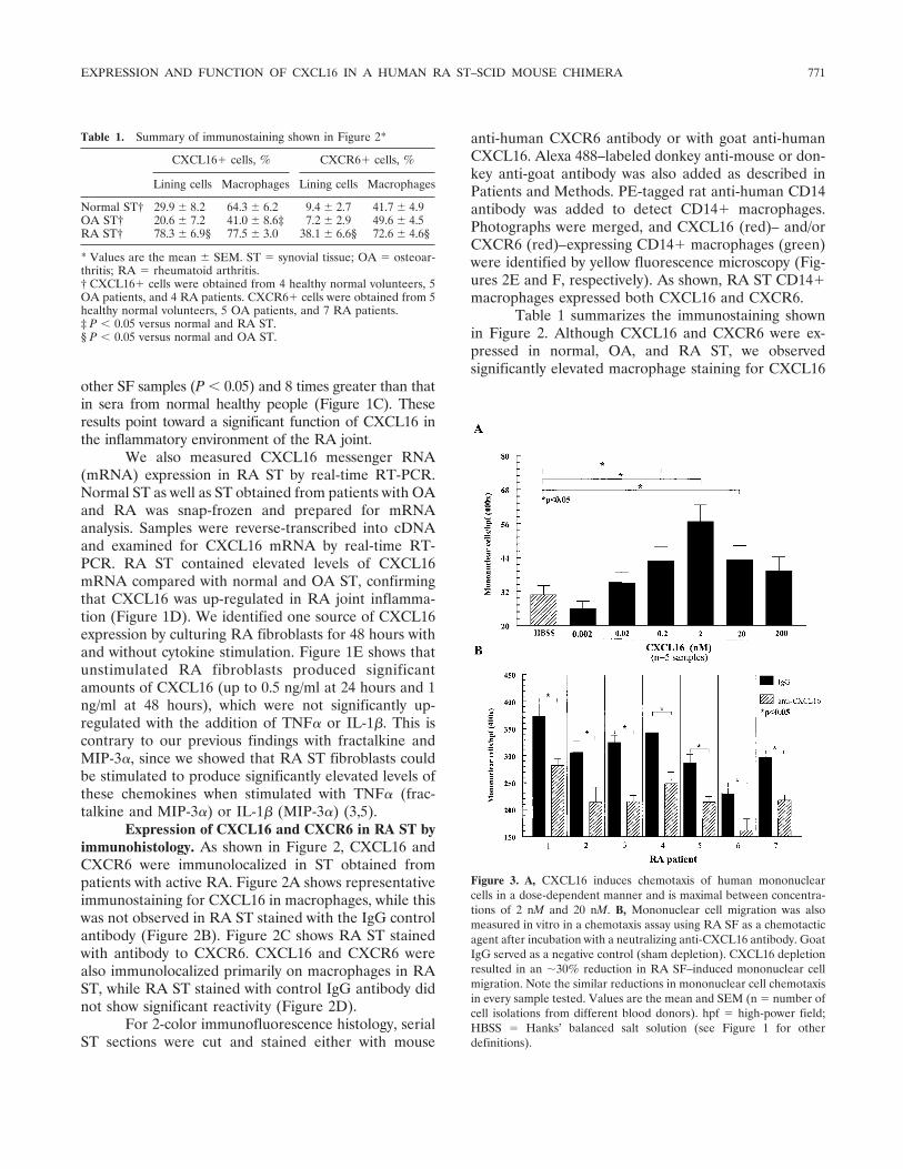
Table 1 summarizes the immunostaining shownin Figure 2. Although CXCL16 and CXCR6 were ex-pressed in normal, OA, and RA ST, we observedsignificantly elevated macrophage staining for CXCL16
Figure 3. A, CXCL16 induces chemotaxis of human mononuclearcells in a dose-dependent manner and is maximal between concentra-tions of 2 nM and 20 nM. B, Mononuclear cell migration was alsomeasured in vitro in a chemotaxis assay using RA SF as a chemotacticagent after incubation with a neutralizing anti-CXCL16 antibody. GoatIgG served as a negative control (sham depletion). CXCL16 depletionresulted in an �30% reduction in RA SF–induced mononuclear cellmigration. Note the similar reductions in mononuclear cell chemotaxisin every sample tested. Values are the mean and SEM (n � number ofcell isolations from different blood donors). hpf � high-power field;HBSS � Hanks’ balanced salt solution (see Figure 1 for otherdefinitions).
Table 1. Summary of immunostaining shown in Figure 2*
CXCL16� cells, % CXCR6� cells, %
Lining cells Macrophages Lining cells Macrophages
Normal ST† 29.9 � 8.2 64.3 � 6.2 9.4 � 2.7 41.7 � 4.9OA ST† 20.6 � 7.2 41.0 � 8.6‡ 7.2 � 2.9 49.6 � 4.5RA ST† 78.3 � 6.9§ 77.5 � 3.0 38.1 � 6.6§ 72.6 � 4.6§
* Values are the mean � SEM. ST � synovial tissue; OA � osteoar-thritis; RA � rheumatoid arthritis.† CXCL16� cells were obtained from 4 healthy normal volunteers, 5OA patients, and 4 RA patients. CXCR6� cells were obtained from 5healthy normal volunteers, 5 OA patients, and 7 RA patients.‡ P � 0.05 versus normal and RA ST.§ P � 0.05 versus normal and OA ST.
EXPRESSION AND FUNCTION OF CXCL16 IN A HUMAN RA ST–SCID MOUSE CHIMERA 771

in RA ST compared with OA ST, and we observedelevated macrophage staining for CXCR6 in RA STcompared with both normal and OA ST. RA ST liningcells, containing macrophages and fibroblasts, expressedsignificantly more CXCL16 and CXCR6 than did nor-mal or OA ST.
Induction of MNC migration by CXCL16. Nor-mal human PBMCs were assayed for their chemotacticresponse to CXCL16 in a modified Boyden chamber todetermine a proinflammatory role for CXCL16. HBSSand fMLP served as negative and positive controls,respectively. We found a dose-dependent migration of
Figure 4. CXCL16 activates Raf and ERK-1/2 in a time-dependent manner. Monocytes (2.5 � 106) were stimulated with 20 nM recombinant humanCXCL16. Cell lysates were probed for p-Raf and p–ERK-1/2 by Western blotting, showing a marked increase in phosphorylation after 5 and 15minutes, respectively. A, Representative blots for both p-Raf and p–ERK-1/2. B, Graphs corresponding to blots in A. Values are the mean and SEM(n � number of blood donors). C, Chemotaxis for human monocytes was also measured in the presence of signaling pathway inhibitors (inh). Valuesare the mean and SEM (n � 4–7 donors per group). hpf � high-power field; HBSS � Hanks’ balanced salt solution; PP2 � a Src inhibitor; PD �PD98059 (a MAPK inhibitor); SB � SB203508 (a p38 MAPK inhibitor).
772 RUTH ET AL

human MNCs to CXCL16, with a peak between 2 nM(20 ng/ml) and 20 nM (200 ng/ml) (Figure 3A). Theselevels were consistent with those found in RA SF(mean � SEM 44.4 � 4.4 ng/ml). To determine theparticipation of CXCL16 in MNC migration in RA, RASF samples containing average levels of CXCL16 (45ng/ml) were incubated with anti-human CXCL16 anti-body. SF samples were then tested in an in vitromigration assay and compared with sham-depleted con-trols. RA SF samples showed a 28% decrease (P � 0.05)in MNC migratory activity after incubation with neutral-izing antibody to CXCL16 (Figure 3B).
Activation of Raf and ERK-1/2 in monocytes byCXCL16. To determine the signaling pathways mediat-ing CXCL16 function, isolated normal human mono-cytes were cultured overnight and stimulated withCXCL16 for various time periods. Cell lysates wereprepared and probed with anti–phospho–ERK-1/2 andanti–phospho-Raf by Western blotting. The blots werestripped and reprobed with �-actin to confirm equalloading of the samples. Monocyte stimulation withCXCL16 induced a marked increase in an early phos-phorylation of Raf at 5 minutes and of ERK-1/2 at 15minutes (Figure 4A). The graphs of 3 experiments withRaf and 5 experiments with ERK-1/2 are also shown(Figure 4B). These results were consistent with MAPKsignaling, since Raf is upstream of ERK-1/2, and theyindicated that CXCL16 may induce some of its proin-flammatory properties through this pathway.
To further analyze the relevance of the MAPKpathway for monocyte recruitment, migration assayswere performed with 20 nM CXCL16 in the presence ofvarious signaling pathway inhibitors at a final concentra-tion of 10 �M (40). As shown in Figure 4C, monocytemigration was significantly blocked by PP2 (an inhibitorof Src), by PD98059 (a MAPK inhibitor), and bySB203508 (a p38 MAPK inhibitor), suggesting partici-pation of Src and MAPK pathways for monocyte che-motaxis toward CXCL16 in vitro.
Recruitment by CXCL16 of MNCs in vivo toengrafted human ST in a SCID mouse chimera. To testMNC migration in vivo, we used a human ST–SCIDmouse chimera. After 4–6 weeks, animals engrafted withhuman RA ST were killed. Well-engrafted tissues withno signs of rejection were observed and were used at thistime point (Figure 5A). To determine homing of normalhuman MNCs, freshly isolated cells were dye-taggedwith PKH26, and 5 � 106 cells/100 �l/mouse wereinjected IV via the tail vein 48 hours before mice werekilled. Cryosections (10 �m) of the RA ST grafts wereexamined using a fluorescence microscope with a
550-nm filter. Injection of CXCL16 or TNF� (positivecontrol) into the engrafted RA ST prior to adoptive celltransfer resulted in a significant increase of MNC mi-gration (39) (Figure 5B). To ensure that cell recruitmentwas induced by CXCL16 and not by contaminants, onlyadditive-free and sterile-filtered CXCL16 was used. Inaddition, the soluble CXCL16 used in all experimentscontained �0.1 ng/�g (1 endotoxin unit/�g) endotoxin.These results indicate that exogenously administeredCXCL16 is chemotactic for human MNCs in vivo.
Figure 5. A, A SCID mouse grafted with RA ST. Left, An area wherethe RA ST graft was not inserted. Right, Side of the mouse where theRA ST graft was inserted, clearly showing a viable, growing RA STgraft (arrow). The photograph was taken 5 weeks after engraftment. B,PKH26 red fluorescent dye–tagged human PB mononuclear cells (5 �106) were injected intravenously into SCID mice engrafted for 6–8weeks with human RA ST. Before administering cells, ST grafts wereinjected with TNF� (200 ng/graft) or CXCL16 (1,000 ng/graft) or weresham injected (no stimulus). After 48 hours, grafts were harvested andtissue sections were examined by immunofluorescence microscopy at550 nm. Top, PKH26 dye–tagged mononuclear cells (original magni-fication � 100). Bottom, Numbers of dye-tagged cells migrating toengrafted RA ST in response to the various stimuli. Cell migration wasquantified by dividing the number of cells per high-power field (hpf).Values are the mean and SEM (n � number of tissue sectionscounted). See Figure 1 for other definitions. Color figure can be viewed inthe online issue, which is available at http://www.arthritisrheum.org.
EXPRESSION AND FUNCTION OF CXCL16 IN A HUMAN RA ST–SCID MOUSE CHIMERA 773

MNC chemotaxis to exogenously administeredintragraft CXCL16 is reduced by 50% to RA ST, and5-fold to murine LNs, in cells transfected with antisenseODNs for ERK-1/2. We further examined the participa-tion of CXCL16 in the recruitment of MNCs in vivo byinhibiting the signaling pathways it utilizes. To do this,we transfected MNCs with sense or antisense ODNs forERK-1/2, and then we labeled the transfected cells withPKH26 fluorescent dye. Before dye-tagging the cells, weperformed viability counts in order to make certain that
cells did not experience an increased rate of cell death.Transfected dye-tagged cells were injected into tail veinsof SCID mice bearing RA ST receiving simultaneousintragraft injections of CXCL16. Engrafted RA ST andmurine inguinal LNs were removed 48 hours later.Transfection with antisense ODNs resulted in significantreductions of MNC migration compared with sense-treated controls (Figures 6A and B). The attenuatedmigration to LNs was especially impressive consideringthe elevated mean � SEM concentrations of soluble
Figure 6. Intravenous administration of fluorescent dye–tagged hu-man mononuclear cells to SCID mice engrafted with human RA ST.A, Fluorescent human mononuclear cells lipofected with sense (S)oligodeoxynucleotides (ODNs) to ERK-1/2 migrated readily to en-grafted human RA ST in response to an intragraft injection of humanCXCL16, whereas those cells lipofected with antisense (AS) ODNs toERK-1/2 showed reduced migratory activity. B, Fluorescent humanmononuclear cells lipofected with sense ODNs to ERK-1/2 migratedreadily to murine lymph nodes (LNs) in response to an intragraftinjection of human CXCL16, whereas those cells lipofected withantisense ODNs to ERK-1/2 showed reduced migratory activity.Values in A and B are the mean and SEM (n � number of tissuesections counted from 3 mice per group). C, Mononuclear cellstransfected with antisense ODNs to ERK-1/2, but not nontransfected(NT) or sense-transfected mononuclear cells, showed significant inhi-bition of p–ERK-1/2 expression. D, A significant percentage of mono-nuclear cell inhibition of ERK-1/2 expression using antisense ODNs toERK-1/2 is shown compared with sense-transfected mononuclear cells.Values are the mean and SEM. The experiment was repeated 3 times.�-actin served as the loading control. hpf � high-power field (seeFigure 1 for other definitions). (Original magnification � 100.) Colorfigure can be viewed in the online issue, which is available athttp://www.arthritisrheum.org.
774 RUTH ET AL

human CXCL16 in the serum of mice receiving intra-graft injections of CXCL16 (CXCL16-injected mice1.91 � 0.23 ng/ml, n � 7 mice; noninjected mice 0.84 �0.21 ng/ml, n � 6 mice) (P � 0.05) (data not shown).
To ensure that ERK-1/2 expression was inhibitedby antisense ODNs, we performed Western blot analysisfor ERK-1/2 expression in MNCs as described in Pa-tients and Methods. As shown in Figure 6C, MNCstransfected with antisense ODNs to ERK-1/2, but notnontransfected or sense-transfected MNCs, showed sig-nificant inhibition of ERK-1/2 expression. These data,graphed in Figure 6D, show a significant percentage ofMNC inhibition of ERK-1/2 expression using antisenseODNs to ERK-1/2 compared with sense-transfectedMNCs. These results show that targeting CXCL16-mediated signaling pathways influences MNC migrationto RA ST and regional LNs in vivo.
DISCUSSION
Substantial progress in characterizing chemo-kines and chemokine receptor function in vitro has ledto better understanding of cell migration (3,5,11,32), butthe limitations of the in vitro environment make itdifficult to interpret how chemokines and correspondingreceptors function in vivo. Limitations of in vitro migra-tion assays include evaluating the role of a particularchemokine as it relates to other chemokines and chemo-kine receptors, and ranking the importance of chemo-kines and chemokine receptors in a particular disease.Indeed, many investigators have used a similar SCIDmouse chimera to study the effects of gene therapy andRA ST fibroblast infiltration in vivo (41–43). Therefore,to evaluate the proinflammatory function of chemokinesin RA, we developed an in vivo cell migration assay todetermine the relative importance of chemokines andtheir respective cellular receptors in a human RA ST–SCID mouse chimera.
Manipulation of chemokines has shown promisefor RA therapy. We previously showed that ENA-78, aCXC chemokine, is up-regulated in RA SF as well as ina rodent model of arthritis (22,44). Immunodepletion ofhuman ENA-78 resulted in decreased ability to che-moattract polymorphonuclear cells in vitro (22). Fur-thermore, anti–ENA-78 given systemically amelioratedsigns and symptoms of rat AIA (44). Inhibition of CCchemokines also reduced joint inflammation. For exam-ple, in vivo inhibition of the CC chemokine MIP-1� byIL-10 delayed the onset and reduced the severity ofcollagen-induced arthritis (CIA) in mice (45). A neutral-izing antibody to MCP-1, another CC chemokine, sig-
nificantly reduced ankle swelling in a rat model of RA(46). Chemokine antagonists have similarly shownpromise. For instance, a 67–amino acid sequence ofMCP-1 that specifically antagonizes MCP-1 abrogatedarthritis in MRL-lpr autoimmune mice, and an antago-nist of RANTES ameliorated CIA in mice (47–49). IL-1receptor antagonist protein, a naturally produced spe-cific antagonist of IL-1�, has consistently shown potentantiinflammatory properties in animal models of inflam-mation (50–52) and is used in the treatment of RA(53–58). CXCL16 is a novel chemokine that has notbeen fully characterized in chronic disease, and it isespecially of interest because its only known receptor todate has been shown to be highly expressed in chronicTh1 diseases such as RA (28).
Initial reports on CXCL16 in mice and humansshowed predominant gene expression in spleen, LNs,and Peyer’s patches, with little or no expression in otherorgans (24). Immunohistologic analysis revealed thatmacrophages in RA ST stained intensely for bothCXCL16 and CXCR6. This staining is consistent with arecent report that CXCL16 is produced by macrophages(59). This was also confirmed by real-time RT-PCRanalysis showing elevated CXCL16 mRNA in RA STcompared with OA and normal ST. CXCL16 was alsostrongly expressed on a subset of PB leukocytes, includ-ing CD14� monocytes and macrophages, from whichfunctional CXCL16 has been shown to be shed (25).These findings are completely consistent with FACSresults indicating that �40% of CD14� monocytes and33% of memory CD4� and CD8� lymphocytes expressCXCR6 in RA SF. The FACS results suggest that bothmonocytes and lymphocytes may use the CXCR6 recep-tor to migrate from the normal PB to the joints inresponse to CXCL16 expression.
In combination with expression of its receptorBonzo/CXCR6 on T cell subsets, these results suggest aunique role for CXCL16 in chronic inflammation. Re-cent studies showing expression of both CXCL16 and itsreceptor on T lymphocytes, akin to IL-2 and its receptor,point to a possible mechanism of autocrine stimulation(60). Our LN data support this role for CXCL16 as apossible T lymphocyte regulatory factor. We observedelevated serum levels of CXCL16 when CXCL16 wasinjected intragraft into SCID mice (data not shown),explaining in part the impressive recruitment of MNCsto murine LNs.
RA patient data show that CXCL16 is highlyexpressed in RA SF, is a potent chemoattractant forMNCs in vitro, and is chemotactic for PBMCs to RA STin vivo. These findings suggest that inhibition of CXCR6
EXPRESSION AND FUNCTION OF CXCL16 IN A HUMAN RA ST–SCID MOUSE CHIMERA 775

expression or CXCL16 signaling pathways may substan-tially halt migration of inflammatory cells to RA ST. Ourhypothesis was formulated on the basis of strong prelim-inary data suggesting that CXCL16 is abundant inchronic inflammation and may be associated with type 1inflammatory responses (28). We have reason to believethat although CXCL16 may play a dominant role in RA,it may very well be important in other chronic diseasessuch as OA, since we observed elevated mRNA expres-sion of CXCL16 in OA compared with normal ST, andelevated soluble CXCL16 in OA SF compared withnormal serum. Chandrasekar et al (61) demonstratedthat CXCL16 signals via Gi, PI 3-kinase, Akt, I�Bkinase, and NF-�B in aortic smooth muscle cells, point-ing to specific signaling pathways after CXCL16–CXCR6 binding. We found that stimulation of mono-cytes with CXCL16 induced dose-dependent increasesin phosphorylation of the signaling molecules Raf andERK-1/2. This correlates with in vitro MNC migrationassays. For example, in the presence of ERK-1/2 signal-ing pathway inhibitors, CXCL16-induced MNC migra-tion was significantly blocked. Interestingly, FACS ana-lysis showed that normal PB CD14� monocytes hadlittle or no expression of CXCR6.
These findings may indicate that other yet-unknown receptors may also bind and respond toCXCL16. Indeed, our observation of robust in vitrorecruitment of PB monocytes toward CXCL16 may bethe result of monocytes using an alternative receptor forCXCL16. Also, it is possible that in the inflammatorymilieu of the RA SF, SF monocytes predominantly usethe CXCR6 receptor while inactivated PB monocytesuse another receptor. Our data cannot completely ruleout this possibility. We recently observed a similarphenomenon with MIP-3�, finding that PB monocytesdo not express the only known receptor for MIP-3�,namely, CCR6, but that they readily migrate dose de-pendently toward MIP-3� in vitro (5).
We tested the capacity of CXCL16 to recruitMNCs in vivo using the human RA ST–SCID mousechimera. This model has been used successfully by othergroups to show that engrafted RA ST not only keepsmany of the morphologic and pathologic characteristicsof RA ST (62), but also can be used as an appropriatemodel of RA (63). We found that tail vein–injected cellsmigrate to engrafted RA ST and murine LNs in re-sponse to intragraft injections of human CXCL16. Weexamined possible signaling mechanisms underlying thisresponse. We found that preincubation of MNCs withantisense ODNs against ERK-1/2 resulted in a pro-nounced decline in cell recruitment to engrafted RA ST
and inguinal LNs in CXCL16-injected SCID mice. Al-though other signaling pathways may certainly be in-volved, we report here that inhibition of a chemokine-induced signaling cascade can temper the inflammatoryinfiltrate in vivo.
These findings have twofold importance. First,we observed an increase in MNC migration in responseto exogenously added chemokine. Second, the primaryfunction of this chemokine could be reversed in vivo byinhibiting its signaling cascade. CXCL16 may have che-motactic potency apart from other chemokines shown tobe expressed in RA (1–3,5,6,14,16,21–23,44,64). This issupported by the finding that CXCL16 may functionindependently of TNF� and IL-1�, at least in vitro. Thiswas initially unexpected, considering the potent proin-flammatory activity of these cytokines in RA (8). Wefound that cultured RA ST fibroblasts constitutivelyproduce large amounts of CXCL16 (�1 ng/ml), explain-ing in part the highly elevated levels of CXCL16 in RASF. Previous reports show that TNF� further stimulatesRA synovial fibroblasts to release a variety of chemo-kines, suggesting that cell recruitment is largely due toTNF�, and implicating TNF� as an ultimate down-stream eliciting agent (3,5).
Interestingly, we found that incubation of RA STfibroblasts with either TNF� or IL-1� failed to furtherincrease CXCL16 expression, contrary to previous find-ings with fractalkine and MIP-3� (3,5). This is especiallyimportant when considering the amounts of CXCL16found in patient samples, reaching levels as high as 145ng/ml in some cases. It suggests that large amounts ofCXCL16 may be produced in RA independently ofestablished proinflammatory cytokines. This finding isconsistent with results of a recent study showing that RAfibroblasts express CXCL16 directly when stimulated byway of the Toll-like receptor 2 ligand peptidoglycan (65).
In conclusion, we evaluated the expression andfunction of CXCL16 and its receptor CXCR6 in RA. Wefound both the ligand and receptor to be very highlyexpressed in RA, and we observed that CXCL16 func-tions to aggressively recruit MNCs in vivo via the Rafand ERK-1/2 kinase pathways. These results point toCXCL16 as a novel, potent MNC recruiter in RAinflammation.
ACKNOWLEDGMENTS
The authors would like to thank Dr. Steven M. Wolin-sky for advice on this project. His thoughtful comments helpedto make these studies possible. The authors would also like tothank Drs. Anthony J. Coyle and James B. Rottman of
776 RUTH ET AL

Millennium Pharmaceuticals for supplying the mouse anti-human CXCR6 IgG antibody used in the FACS studies.
REFERENCES
1. Koch AE, Kunkel SL, Harlow LA, Johnson B, Evanoff HL, HainesGK, et al. Enhanced production of monocyte chemoattractantprotein-1 in rheumatoid arthritis. J Clin Invest 1992;90:772–9.
2. Koch AE, Kunkel SL, Harlow LA, Mazarakis DD, Haines GK,Burdick MD, et al. Macrophage inflammatory protein-1�: a novelchemotactic cytokine for macrophages in rheumatoid arthritis.J Clin Invest 1994;93:921–8.
3. Ruth JH, Volin MV, Haines GK III, Woodruff DC, Katschke KJJr, Woods JM, et al. Fractalkine, a novel chemokine in rheumatoidarthritis and in rat adjuvant-induced arthritis. Arthritis Rheum2001;44:1568–81.
4. Volin MV, Woods JM, Amin MA, Connors MA, Harlow LA,Koch AE. Fractalkine: a novel angiogenic chemokine in rheuma-toid arthritis. Am J Pathol 2001;159:1521–30.
5. Ruth JH, Shahrara S, Park CC, Morel JC, Kumar P, Qin S, et al.Role of macrophage inflammatory protein-3� and its ligand CCR6in rheumatoid arthritis. Lab Invest 2003;83:579–88.
6. Szekanecz Z, Strieter RM, Kunkel SL, Koch AE. Chemokines inrheumatoid arthritis. Springer Semin Immunopathol 1998;20:115–32.
7. Szekanecz Z, Koch AE. Chemokines and angiogenesis. Curr OpinRheumatol 2001;13:202–8.
8. Szekanecz Z, Koch AE, Kunkel SL, Strieter RM. Cytokines inrheumatoid arthritis: potential targets for pharmacological inter-vention. Drugs Aging 1998;12:377–90.
9. Alkhatib G, Combadiere C, Broder CC, Feng Y, Kennedy PE,Murphy PM, et al. CC CKR5: a RANTES, MIP-1�, MIP-1�receptor as a fusion cofactor for macrophage-tropic HIV-1. Sci-ence 1996;272:1955–8.
10. Ruth JH, Lukacs NW, Warmington KS, Polak TJ, Burdick M,Kunkel SL, et al. Expression and participation of eotaxin duringmycobacterial (type 1) and schistosomal (type 2) antigen-elicitedgranuloma formation. J Immunol 1998;161:4276–82.
11. Katschke KJ Jr, Rottman JB, Ruth JH, Qin S, Wu L, LaRosa G,et al. Differential expression of chemokine receptors on peripheralblood, synovial fluid, and synovial tissue monocytes/macrophagesin rheumatoid arthritis. Arthritis Rheum 2001;44:1022–32.
12. Murdoch C, Finn A. Chemokine receptors and their role ininflammation and infectious diseases. Blood 2000;95:3032–43.
13. Heydtmann M, Adams DH. Understanding selective trafficking oflymphocyte subsets. Gut 2002;50:150–2.
14. Koch AE, Kunkel SL, Burrows JC, Evanoff HL, Haines GK, PopeRM, et al. Synovial tissue macrophage as a source of the chemo-tactic cytokine IL-8. J Immunol 1991;147:2187–95.
15. Koch AE, Harlow LA, Haines GK, Amento EP, Unemori EN,Wong WL, et al. Vascular endothelial growth factor: a cytokinemodulating endothelial function in rheumatoid arthritis. J Immu-nol 1994;152:4149–56.
16. Koch AE, Kunkel SL, Shah MR, Hosaka S, Halloran MM, HainesGK, et al. Growth-related gene product �: a chemotactic cytokinefor neutrophils in rheumatoid arthritis. J Immunol 1995;155:3660–6.
17. Koch AE, Halloran MM, Haskell CJ, Shah MR, Polverini PJ.Angiogenesis mediated by soluble forms of E-selectin and vascularcell adhesion molecule-1. Nature 1995;376:517–9.
18. Loetscher P, Dewald B, Baggiolini M, Seitz M. Monocyte che-moattractant protein 1 and interleukin 8 production by rheuma-toid synoviocytes: effects of anti-rheumatic drugs. Cytokine 1994;6:162–70.
19. Qin S, Rottman JB, Myers P, Kassam N, Weinblatt M, LoetscherM, et al. The chemokine receptors CXCR3 and CCR5 mark
subsets of T cells associated with certain inflammatory reactions.J Clin Invest 1998;101:746–54.
20. Loetscher P, Uguccioni M, Bordoli L, Baggiolini M, Moser B,Chizzolini C, et al. CCR5 is characteristic of Th1 lymphocytes.Nature 1998;391:344–5.
21. Volin MV, Shah MR, Tokuhira M, Haines GK, Woods JM, KochAE. RANTES expression and contribution to monocyte chemo-taxis in arthritis. Clin Immunol Immunopathol 1998;89:44–53.
22. Koch AE, Kunkel SL, Harlow LA, Mazarakis DD, Haines GK,Burdick MD, et al. Epithelial neutrophil activating peptide-78: anovel chemotactic cytokine for neutrophils in arthritis. J ClinInvest 1994;94:1012–8.
23. Koch AE, Polverini PJ, Kunkel SL, Harlow LA, DiPietro LA,Elner VM, et al. Interleukin-8 as a macrophage-derived mediatorof angiogenesis. Science 1992;258:1798–801.
24. Matloubian M, David A, Engel S, Ryan JE, Cyster JG. Atransmembrane CXC chemokine is a ligand for HIV-coreceptorbonzo. Nat Immunol 2000;1:298–304.
25. Wilbanks A, Zondlo SC, Murphy K, Mak S, Soler D, Langdon P,et al. Expression cloning of the STRL33/BONZO/TYMSTRligandreveals elements of CC, CXC, and CX3C chemokines. J Immunol2001;166:5145–54.
26. Nakayama T, Hieshima K, Izawa D, Tatsumi Y, Kanamaru A,Yoshie O. Cutting edge: profile of chemokine receptor expressionon human plasma cells accounts for their efficient recruitment totarget tissues. J Immunol 2003;170:1136–40.
27. Kim CH, Johnston B, Butcher EC. Trafficking machinery of NKTcells: shared and differential chemokine receptor expressionamong V � 24(�)V � 11(�) NKT cell subsets with distinctcytokine-producing capacity. Blood 2002;100:11–6.
28. Kim CH, Kunkel EJ, Boisvert J, Johnston B, Campbell JJ,Genovese MC, et al. Bonzo/CXCR6 expression defines type1-polarized T-cell subsets with extralymphoid tissue homing po-tential. J Clin Invest 2001;107:595–601.
29. Kim CH, Rott L, Kunkel EJ, Genovese MC, Andrew DP, Wu L,et al. Rules of chemokine receptor association with T cell polar-ization in vivo. J Clin Invest 2001;108:1331–9.
30. Unutmaz D, Xiang W, Sunshine MJ, Campbell J, Butcher E,Littman DR. The primate lentiviral receptor Bonzo/STRL33 iscoordinately regulated with CCR5 and its expression pattern isconserved between human and mouse. J Immunol 2000;165:3284–92.
31. Arnett FC, Edworthy SM, Bloch DA, McShane DJ, Fries JF,Cooper NS, et al. The American Rheumatism Association 1987revised criteria for the classification of rheumatoid arthritis.Arthritis Rheum 1988;31:315–24.
32. Ruth JH, Rottman JB, Katschke KJ Jr, Qin S, Wu L, LaRosa G,et al. Selective lymphocyte chemokine receptor expression in therheumatoid joint. Arthritis Rheum 2001;44:2750–60.
33. Volin MV, Szekanecz Z, Halloran MM, Woods JM, Magua J,Damergis JA Jr, et al. PECAM-1 and leukosialin (CD43) expres-sion correlate with heightened inflammation in rat adjuvant-induced arthritis. Exp Mol Pathol 1999;66:211–9.
34. Chomczynski P, Sacchi N. Single-step method of RNA isolation byacid guanidinium thiocyanate-phenol-chloroform extraction. AnalBiochem 1987;162:156–9.
35. Endesfelder S, Krahn A, Kreuzer KA, Lass U, Schmidt CA,Jahrmarkt C, et al. Elevated p21 mRNA level in skeletal muscle ofDMD patients and mdx mice indicates either an exhausted satel-lite cell pool or a higher p21 expression in dystrophin-deficientcells per se. J Mol Med 2000;78:569–74.
36. Favy DA, Lafarge S, Rio P, Vissac C, Bignon YJ, Bernard-GallonD. Real-time PCR quantification of full-length and exon 11 splicedBRCA1 transcripts in human breast cancer cell lines. BiochemBiophys Res Commun 2000;274:73–8.
37. Kumar P, Amin MA, Harlow LA, Polverini PJ, Koch AE. Src and
EXPRESSION AND FUNCTION OF CXCL16 IN A HUMAN RA ST–SCID MOUSE CHIMERA 777

phosphatidylinositol 3-kinase mediate soluble E-selectin-inducedangiogenesis. Blood 2003;101:3960–8.
38. Zhu K, Amin MA, Kim MJ, Katschke KJ Jr, Park CC, Koch AE.A novel function for a glucose analog of blood group H antigen asa mediator of leukocyte-endothelial adhesion via intracellularadhesion molecule 1. J Biol Chem 2003;278:21869–77.
39. Blades MC, Ingegnoli F, Wheller SK, Manzo A, Wahid S, PanayiGS, et al. Stromal cell–derived factor 1 (CXCL12) induces mono-cyte migration into human synovium transplanted onto SCIDmice. Arthritis Rheum 2002;46:824–36.
40. Morel JC, Park CC, Zhu K, Kumar P, Ruth JH, Koch AE. Signaltransduction pathways involved in rheumatoid arthritis synovialfibroblast interleukin-18-induced vascular cell adhesion mole-cule-1 expression. J Biol Chem 2002;277:34679–91.
41. Jungel A, Distler JH, Kurowska-Stolarska M, Seemayer CA, SeiblR, Forster A, et al. Expression of interleukin-21 receptor, but notinterleukin-21, in synovial fibroblasts and synovial macrophages ofpatients with rheumatoid arthritis. Arthritis Rheum 2004;50:1468–76.
42. Muller-Ladner U, Gay S. The SCID mouse: a novel experimentalmodel for gene therapy in human rheumatoid arthritis. DrugsToday (Barc) 1999;35:379–88.
43. Neidhart M, Seemayer CA, Hummel KM, Michel BA, Gay RE,Gay S. Functional characterization of adherent synovial fluid cellsin rheumatoid arthritis: destructive potential in vitro and in vivo.Arthritis Rheum 2003;48:1873–80.
44. Halloran MM, Woods JM, Strieter RM, Szekanecz Z, Volin MV,Hosaka S, et al. The role of an epithelial neutrophil-activatingpeptide-78-like protein in rat adjuvant-induced arthritis. J Immu-nol 1999;162:7492–500.
45. Kasama T, Strieter RM, Lukacs NW, Lincoln PM, Burdick MD,Kunkel SL. Interleukin-10 expression and chemokine regulationduring the evolution of murine type II collagen-induced arthritis.J Clin Invest 1995;95:2868–76.
46. Ogata H, Takeya M, Yoshimura T, Takagi K, Takahashi K. Therole of monocyte chemoattractant protein-1 (MCP-1) in thepathogenesis of collagen-induced arthritis in rats. J Pathol 1997;182:106–14.
47. Gong JH, Ratkay LG, Waterfield JD, Clark-Lewis I. An antago-nist of monocyte chemoattractant protein 1 (MCP-1) inhibitsarthritis in the MRL-lpr mouse model. J Exp Med 1997;186:131–7.
48. Wooley PH, Schaefer C, Whalen JD, Dutcher JA, Counts DF. Apeptide sequence from platelet factor 4 (CT-112) is effective in thetreatment of type II collagen induced arthritis in mice. J Rheu-matol 1997;24:890–8.
49. Plater-Zyberk C, Hoogewerf AJ, Proudfoot AE, Power CA, WellsTN. Effect of a CC chemokine receptor antagonist on collageninduced arthritis in DBA/1 mice. Immunol Lett 1997;57:117–20.
50. Ruth JH, Bienkowski M, Warmington KS, Lincoln PM, KunkelSL, Chensue SW. IL-1 receptor antagonist (IL-1ra) expression,function, and cytokine-mediated regulation during mycobacterialand schistosomal antigen-elicited granuloma formation. J Immu-nol 1996;156:2503–9.
51. Gabay C, Marinova-Mutafchieva L, Williams RO, Gigley JP,
Butler DM, Feldmann M, et al. Increased production of intracell-ular interleukin-1 receptor antagonist type I in the synovium ofmice with collagen-induced arthritis: a possible role in the resolu-tion of arthritis. Arthritis Rheum 2001;44:451–62.
52. Palmer G, Talabot-Ayer D, Szalay-Quinodoz L, Maret M, ArendWP, Gabay C. Mice transgenic for intracellular interleukin-1receptor antagonist type 1 are protected from collagen-inducedarthritis. Eur J Immunol 2003;33:434–40.
53. Arend WP. The balance between IL-1 and IL-1Ra in disease.Cytokine Growth Factor Rev 2002;13:323–40.
54. Arend WP, Malyak M, Guthridge CJ, Gabay C. Interleukin-1receptor antagonist: role in biology. Annu Rev Immunol 1998;16:27–55.
55. Firestein GS, Boyle DL, Yu C, Paine MM, Whisenand TD,Zvaifler NJ, et al. Synovial interleukin-1 receptor antagonist andinterleukin-1 balance in rheumatoid arthritis. Arthritis Rheum1994;37:644–52.
56. Fleishmann RM. Safety of anakinra, a recombinant interleukin-1receptor antagonist (r-metHuIL-1ra), in patients with rheumatoidarthritis and comparison to anti-TNF� agents. Clin Exp Rheuma-tol 2002;20 Suppl 27:S35–41.
57. Kay J, Calabrese L. The role of interleukin-1 in the pathogenesisof rheumatoid arthritis. Rheumatology (Oxford) 2004;43 Suppl3:III2–9.
58. Olsen NJ, Stein CM. New drugs for rheumatoid arthritis. N EnglJ Med 2004;350:2167–79.
59. Van der Voort R, van Lieshout AW, Toonen LW, Sloetjes AW,van den Berg WB, Figdor CG, et al. Elevated CXCL16 expressionby synovial macrophages recruits memory T cells into rheumatoidjoints. Arthritis Rheum 2005;52:1381–91.
60. Shashkin P, Simpson D, Mishin V, Chesnutt B, Ley K. Expressionof CXCL16 in human T cells. Arterioscler Thromb Vasc Biol2003;23:148–9.
61. Chandrasekar B, Bysani S, Mummidi S. CXCL16 signals via Gi,PI3 kinase, Akt, I�B kinase and nuclear factor-�B, and inducescell-cell adhesion and aortic smooth muscle cell proliferation.J Biol Chem 2004;279:3188–96.
62. Davis LS, Sackler M, Brezinschek RI, Lightfoot E, Bailey JL,Oppenheimer-Marks N, et al. Inflammation, immune reactivity,and angiogenesis in a severe combined immunodeficiency modelof rheumatoid arthritis. Am J Pathol 2002;160:357–67.
63. Geiler T, Kriegsmann J, Keyszer GM, Gay RE, Gay S. A newmodel for rheumatoid arthritis generated by engraftment ofrheumatoid synovial tissue and normal human cartilage into SCIDmice. Arthritis Rheum 1994;37:1664–71.
64. Amin MA, Volpert OV, Woods JM, Kumar P, Harlow LA, KochAE. Migration inhibitory factor mediates angiogenesis via mito-gen-activated protein kinase and phosphatidylinositol kinase. CircRes 2003;93:321–9.
65. Pierer M, Rethage J, Seibl R, Lauener R, Brentano F, Wagner U,et al. Chemokine secretion of rheumatoid arthritis synovial fibro-blasts stimulated by Toll-like receptor 2 ligands. J Immunol2004;172:1256–65.
778 RUTH ET AL